HAL Id: dumas-01745906
https://dumas.ccsd.cnrs.fr/dumas-01745906
Submitted on 5 Jun 2018HAL is a multi-disciplinary open access archive for the deposit and dissemination of sci-entific research documents, whether they are pub-lished or not. The documents may come from teaching and research institutions in France or abroad, or from public or private research centers.
L’archive ouverte pluridisciplinaire HAL, est destinée au dépôt et à la diffusion de documents scientifiques de niveau recherche, publiés ou non, émanant des établissements d’enseignement et de recherche français ou étrangers, des laboratoires publics ou privés.
Bretagne ayant une sclérose en plaque sous fingolimod
Cécile Malrain
To cite this version:
Cécile Malrain. Évolution clinique et radiologique des patients de Bretagne ayant une sclérose en plaque sous fingolimod. Sciences du Vivant [q-bio]. 2016. �dumas-01745906�
THÈSE D'EXERCICE / UNIVERSITÉ DE RENNES 1
FACULTÉ DE MÉDECINE
sous le sceau de l’Université Européenne de Bretagne
THÈSE EN VUE DU
DIPLÔME D'ÉTAT DE DOCTEUR EN MÉDECINE
présentée par
Cécile
Malrain
né(e) le 19 Novembre 1986 à Versailles
Evolution clinique et
radiologique des
patients de Bretagne
ayant une sclérose
en plaque sous
fingolimod
Thèse soutenue à Rennes le 1er avril 2016
devant le jury composé de :
Marc VERIN
Professeur – CHU de Rennes / président
Jean-Christophe FERRE
Professeur – CHU de Rennes / Juge
David LAPLAUD
Professeur – CHU de Nantes / Juge
Véronique DEBURGHGRAEVE
Praticien attaché – CHU de Rennes / Juge
Emmanuelle LEPAGE
Praticien hospitalier – CHU de Rennes / Membre
invité
Anne KERBRAT
Chef de Clinique assistante - CHU de Rennes /
Membre invité
Marc MERIENNE
Praticien hospitaliser – Centre hospitalier de Saint Malo / Membre invité
Gilles EDAN
Professeurs des Universités – Praticiens Hospitaliers 2015-2016
ANNE-GALIBERT Marie Dominique Biochimie et biologie moléculaire
BELAUD-ROTUREAU Marc-Antoine Histologie; embryologie et cytogénétique
BELLISSANT Eric Pharmacologie fondamentale; pharmacologie clinique; addictologie
BELLOU Abdelouahab Thérapeutique; médecine d'urgence; addictologie BELOEIL Hélène Anesthésiologie-réanimation; médecine d'urgence BENDAVID Claude Biochimie et biologie moléculaire
BENSALAH Karim Urologie BEUCHEE Alain Pédiatrie
BONAN Isabelle Médecine physique et de réadaptation
BONNET Fabrice Endocrinologie, diabète et maladies métaboliques; gynécologie médicale
BOUDJEMA Karim Chirurgie générale
BOUGET Jacques Thérapeutique; médecine d'urgence; addictologie BOURGUET Patrick
Professeur des Universités en surnombre
Biophysique et médecine nucléaire
BRASSIER Gilles Neurochirurgie
BRETAGNE Jean-François Gastroentérologie; hépatologie; addictologie BRISSOT Pierre
Professeur des Universités en surnombre
Gastroentérologie; hépatologie; addictologie
CARRE François Physiologie CATROS Véronique Biologie cellulaire CHALES Gérard
Professeur des Universités émérite Rhumatologie
CORBINEAU Hervé Chirurgie thoracique et cardiovasculaire
CUGGIA Marc Biostatistiques, informatique médicale et technologies de communication
DARNAULT Pierre Anatomie DAUBERT Jean-Claude
Professeur des Universités émérite Cardiologie
DAVID Véronique Biochimie et biologie moléculaire DAYAN Jacques
Professeur des Universités associé Pédopsychiatrie; addictologie DE CREVOISIER Renaud Cancérologie; radiothérapie
DECAUX Olivier Médecine interne; gériatrie et biologie du vieillissement; addictologie
DELAVAL Philippe Pneumologie; addictologie DESRUES Benoît Pneumologie; addictologie DEUGNIER Yves
Professeur des Universités en surnombre
Gastroentérologie; hépatologie; addictologie
DONAL Erwan Cardiologie
DRAPIER Dominique Psychiatrie d'adultes; addictologie DUPUY Alain Dermato-vénéréologie
ECOFFEY Claude Anesthésiologie-réanimation; médecine d'urgence
EDAN Gilles Neurologie
FERRE Jean Christophe Radiologie et imagerie Médecine FEST Thierry Hématologie; transfusion
FLECHER Erwan Chirurgie thoracique et cardiovasculaire FREMOND Benjamin Chirurgie infantile
GANDEMER Virginie Pédiatrie
GANDON Yves Radiologie et imagerie Médecine GANGNEUX Jean-Pierre Parasitologie et mycologie
GARIN Etienne Biophysique et médecine nucléaire GAUVRIT Jean-Yves Radiologie et imagerie Médecine GODEY Benoit Oto-rhino-laryngologie
GUGGENBUHL Pascal Rhumatologie GUIGUEN Claude
Professeur des Universités émérite Parasitologie et mycologie GUILLÉ François Urologie
GUYADER Dominique Gastroentérologie; hépatologie; addictologie HOUOT Roch Hématologie; transfusion
HUGÉ Sandrine
Professeur des Universités associé Médecine générale HUSSON Jean-Louis
Professeur des Universités en surnombre
Chirurgie orthopédique et traumatologique
JEGO Patrick Médecine interne; gériatrie et biologie du vieillissement; addictologie
JEGOUX Franck Oto-rhino-laryngologie JOUNEAU Stéphane Pneumologie; addictologie
KAYAL Samer Bactériologie-virologie; hygiène hospitalière KERBRAT Pierre Cancérologie; radiothérapie
LAMY DE LA CHAPELLE Thierry Hématologie; transfusion
LAVIOLLE Bruno Pharmacologie fondamentale; pharmacologie clinique; addictologie
LAVOUE Vincent Gynécologie-obstétrique; gynécologie médicale LE BRETON Hervé Cardiologie
LE GUEUT Maryannick Médecine légale et droit de la santé LE TULZO Yves Réanimation; médecine d'urgence LECLERCQ Christophe Cardiologie
LEGUERRIER Alain Chirurgie thoracique et cardiovasculaire LEJEUNE Florence Biophysique et médecine nucléaire
LEVEQUE Jean Gynécologie-obstétrique; gynécologie médicale LIEVRE Astrid Gastroentérologie; hépatologie; addictologie MABO Philippe Cardiologie
MALLEDANT Yannick Anesthésiologie-réanimation; médecine d'urgence MEUNIER Bernard Chirurgie digestive
MICHELET Christian Maladies infectieuses; maladies tropicales MOIRAND Romain Gastroentérologie; hépatologie; addictologie
MORANDI Xavier Anatomie
MORTEMOUSQUE Bruno Ophtalmologie
MOSSER Jean Biochimie et biologie moléculaire MOULINOUX Jacques Biologie cellulaire
MOURIAUX Frédéric Ophtalmologie
ODENT Sylvie Génétique
OGER Emmanuel Pharmacologie fondamentale; pharmacologie clinique; addictologie
PERDRIGER Aleth Rhumatologie PLADYS Patrick Pédiatrie
POULAIN Patrice Gynécologie-obstétrique; gynécologie médicale RAVEL Célia Histologie; embryologie et cytogénétique RIFFAUD Laurent Neurochirurgie
RIOUX-LECLERCQ Nathalie Anatomie et cytologie pathologiques ROBERT-GANGNEUX Florence Parasitologie et mycologie
SAINT-JALMES Hervé Biophysique et médecine nucléaire
SEGUIN Philippe Anesthésiologie-réanimation; médecine d'urgence SEMANA Gilbert Immunologie
SIPROUDHIS Laurent Gastroentérologie; hépatologie; addictologie
SOMME Dominique Médecine interne; gériatrie et biologie du vieillisement; addictologie
SULPICE Laurent Chirurgie générale
TARTE Karin Immunologie
TATTEVIN Pierre Maladies infectieuses; maladies tropicales THIBAULT Ronan Nutrition
THIBAULT Vincent Bactériologie-virologie; hygiène hospitalière THOMAZEAU Hervé Chirurgie orthopédique et traumatologique TORDJMAN Sylvie Pédopsychiatrie; addictologie
VERGER Christian
Professeur des Universités émérite Médecine et santé au travail
VERHOYE Jean-Philippe Chirurgie thoracique et cardiovasculaire
VERIN Marc Neurologie
VIEL Jean-François Epidémiologie, économie de la santé et prévention VIGNEAU Cécile Néphrologie
VIOLAS Philippe Chirurgie infantile
WATIER Eric Chirurgie plastique, reconstructrice et esthétique; brûlologie WODEY Eric Anesthésiologie-réanimation; médecine d'urgence
Maitres de conférences des universités – Praticiens Hospitaliers 2015-2016
AME-THOMAS Patricia Immunologie
AMIOT Laurence Hématologie; transfusion
BARDOU-JACQUET Edouard Gastroentérologie; hépatologie; addictologie BEGUE Jean-Marc Physiologie
BOUSSEMART Lise Dermato-vénéréologie CABILLIC Florian Biologie cellulaire
CAUBET Alain Médecine et santé au travail DAMERON Olivier Informatique
DE TAYRAC Marie Biochimie et biologie moléculaire DEGEILH Brigitte Parasitologie et mycologie DUBOURG Christèle Biochimie et biologie moléculaire
DUGAY Frédéric Histologie; embryologie et cytogénétique EDELINE Julien Cancérologie; radiothérapie
GALLAND Françoise Endocrinologie, diabète et maladies métaboliques; gynécologie médicale GARLANTEZEC Ronan Epidémiologie, économie de la santé et prévention
GUILLET Benoit Hématologie; transfusion HAEGELEN Claire Anatomie
JAILLARD Sylvie Histologie; embryologie et cytogénétique
LAVENU Audrey Sciences physico-chimiques et technologies pharmaceutiques LE GALL François Anatomie et cytologie pathologiques
LE RUMEUR Elisabeth Physiologie
MAHÉ Guillaume Chirurgie vasculaire; médecine vasculaire MARTINS Raphaël Cardiologie
MASSART Catherine Biochimie et biologie moléculaire MATHIEU-SANQUER Romain Urologie
MENARD Cédric Immunologie MENER Eric Médecine générale MILON Joëlle Anatomie
MOREAU Caroline Biochimie et biologie moléculaire MOUSSOUNI Fouzia Informatique
MYHIE Didier Médecine générale PANGAULT Céline Hématologie; transfusion RENAUT Pierric Médecine générale
RIOU Françoise Epidémiologie, économie de la santé et prévention ROBERT Gabriel Psychiatrie d'adultes; addictologie
ROPARS Mickaël Anatomie SAULEAU Paul Physiologie
TADIÉ Jean-Marc Réamination; médecine d'urgence TATTEVIN-FABLET Françoise Médecine générale
TURLIN Bruno Anatomie et cytologie pathologiques
VERDIER Marie-Clémence Pharmacologie fondamentale; pharmacologie clinique; addictologie VINCENT Pascal Bactériologie-virologie; hygiène hospitalière
Remerciements
A monsieur le Professeur Marc VERIN, vous me faites l’honneur de présider ce travail et je vous en remercie. J’ai beaucoup appris à vos côtés, merci de m’avoir épaulée pendant ces années d’internat. Veuillez trouver l’expression de mon plus grand respect tant pour vos qualités professionnelles qu’humaines.
A monsieur le Professeur Jean-Christophe FERRE : vous me faîtes l’honneur de juger ce travail et je vous en remercie.
A monsieur le Professeur David LAPLAUD de l’université de Nantes : vous me faîtes l’honneur de juger ce travail et je vous en remercie.
Au Dr Véronique DEBURGHGRAEVE : vous me faîtes l’honneur de juger ce travail et je vous en remercie.
Au Dr Anne KERBRAT : je te remercie d’avoir accepté de faire partie des membres de mon jury, j’en suis très honorée. J’ai beaucoup apprécié travailler avec toi pour tes qualités médicales et humaines.
Au Dr Emmanuelle LEPAGE : je vous remercie de me faire l’honneur de juger mon travail et de m’avoir guidée pendant ces années d’internat.
Au Dr Marc MERIENNE : vous me faîtes l’honneur de juger ce travail et je vous en remercie. Travailler à vos côtés est un plaisir, merci pour votre disponibilité et votre goût de partager vos connaissances.
A monsieur le Professeur Gilles EDAN : vous m’avez fait l’honneur de diriger mon travail et je vous en remercie. Veuillez trouver ici l’expression de ma profonde reconnaissance pour vos qualités humaines et professionnelles.
A Emmanuelle LERAY : merci infiniment pour ton aide et ta disponibilité.
A l’ensemble des services de neurologie et neurologues m’ayant permis de compléter mon recueil.
Au Dr Anne SALMON, travailler avec toi a été un réel plaisir, merci pour tes qualités humaines et médicales, ta façon d’aborder la médecine m’a beaucoup apporté.
Au Dr Thomas RONZIERE, merci de m’avoir fait partager tes connaissances en vasculaire, j’ai beaucoup apprécié travailler à tes côtés.
A l’ensemble de l’équipe médicale du service de neurologie du CHU de Rennes : au Dr LASSALLE Véronica pour sa bonne humeur, au Dr DRAPIER Sophie, au Dr Paul SAULEAU, au Dr PINEL Jean-François, au Dr PIHAN Morgane, au Dr NICA Anca, au Dr BELLIARD Serge, au Dr CAHAGNE Vincent.
Au Dr Hélène HUSSON, au Dr Marie-Christine MINOT-MYHIE, au Dr Hélène OLIVET, merci pour vos conseils.
A mes co-internes devenus docteurs avec qui j’ai eu beaucoup de plaisir à travailler : au Dr Lucie COURAULT-BODI, au Dr Margaux GENEVRAY, au Dr Stéphane VANNIER, au Dr Sophie LEMERCIER-LANGNER, au Dr Frédérique LEH, au Dr Anne MAURUS, au Dr Claire RICORDEAU, au Dr Julien BONNENFANT.
Un remerciement particulier à mes co-internes : Audrey RIOU et Valentine MARGOT pour leur soutien, les goûters et les rires (heureusement qu’on s’entendait bien !)
Merci beaucoup à mes co-internes pour ce semestre en médecine interne qui restera un très bon souvenir : au Dr Clémence SAILLARD, à Camille LEPART, à Alice BALLERIE, à
Benjamin LEFEVRE.
Merci à mes co-internes : Etienne, Benjamin, Charlotte, Marine, Benoît, Jules, Christian, Ronan, Mélo.
Merci aux équipes para-médicales : Hélène, Gaëlle, Sylvie, Laurence, Marie, Elodie, Nicole, Evelyne…
Merci à Véronique TOURILLON pour sa bonne humeur et son efficacité, travailler avec toi a été un plaisir !
Merci au service de neurologie de Vannes pour m’avoir accueillie pour mes premiers pas d’interne, particulièrement au Dr Françoise BRUNET-BOURGIN et au Dr Philippe
KASSIOTIS.
Merci au service de réanimation de Lorient.
Merci au service de médecine interne du CHU de Rennes, notamment au Dr Thomas LE GALLOU, j’ai beaucoup appris à tes côtés.
Merci au service de neurologie de l’hôpital de Saint Malo, merci au Dr Gregory TAURIN de m’avoir accueillie.
A ma copine de toujours le Dr Audrey-Céline WEMEL. Merci également à ses parents Pierre et Leïla pour m’avoir soutenue dans des moments difficiles.
Aux zouzous, je vous suis reconnaissante pour votre amitié et votre soutien, merci pour tous les bons moments passés ensemble et ceux à venir : Anne-Chouette, Kristel, Audrey, Marine, Tamara, Thalie, Virginie, Erwan, Karim, Claire.
Aux amies du premier semestre à Vannes : Marie-Laure, Solinne, merci pour votre présence et votre soutien.
A mes grands-parents et ma famille.
A mes parents, merci de m’avoir aidée et soutenue pendant toutes ces années, merci pour votre amour.
Table des matières
PAGE DE TITRE ...1
LISTE DES PROFESSEURS DES UNIVERSITES-PRATICIENS HOSPITALIERS 2015-2016 ...2
LISTE DES MAITRES DE CONFERENCES DES UNIVERSITES-PRATICIENS HOSPITALIERS 2015-2016 ...5
REMERCIEMENTS ...6
TABLE DES MATIERES...9
INTRODUCTION ... 10
Multiple sclerosis ... 10
Fingolimod ... 11
Efficacy ... 12
Study endpoint ... 13
MATERIEL AND METHODS ... 14
Study design ... 14
Statistical analyses ... 15
RESULTS ... 15
Baseline characteristics ... 15
Efficacy ... 18
Adverse effects and reasons to interruption ... 23
Sub groups analyses ... 26
DISCUSSION ... 30
Efficacy ... 31
Safety ... 32
Sub groups ... 33
INTRODUCTION
Multiple sclerosis
Multiple sclerosis (MS) is a chronic inflammatory autoimmune disease of the central nervous system (CNS) mainly affecting young adults.
The national prevalence in France is 94.7 per 100 000 patients and the incidence is 7.5 per 100 000 [1]. MS is a common cause of handicap with also social consequences [2].
The etiology of MS is still unknown, but some environmental, genetic [3] and immunological factors have been identified.
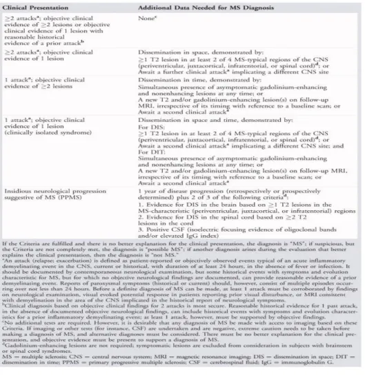
The diagnosis is based on the principles of dissemination in space and dissemination in time of CNS lesions, including clinical and radiological criteria, defined according to the Mac Donald classification [4] (Table 1), which allows an early diagnosis.
Lesions observed in the CNS of MS patients are characterized by multifocal areas of myelin sheath destruction, oligodendrocyte death, axonal and neuronal damages, and activation of glial cells. The migration of activated T lymphocytes from the periphery into the CNS is a crucial step in the formation of MS lesions, in the CNS, T lymphocytes need to be reactivated.
Fingolimod
Fingolimod (FTY720) is a sphingosine-1-phosphate–receptor modulator that prevents lymphocyte egressing from lymph nodes, by reducing recirculation of auto aggressive lymphocytes to the CNS. Fingolimod can cross the blood brain barrier due to its lipophilic properties, and it is phosphorylated within the CNS. Through the interaction with sphingosine-1-phosphate receptors on neural cells, fingolimod may have neuroprotective or reparative effects.
Fingolimod is the first oral treatment approved for relapsing remitting (RR) MS, introduced from 2011 in France. The efficacy of fingolimod was demonstrated in two phase III clinical trials:
- In the FREEDOMS trial [5], it reduced annualized relapse rate (ARR) by 54% and risk of disability progression by 30% compared to placebo. Percentage brain volume change was significantly lower in fingolimod group.
- In the TRANSFORMS trial [6], compared to IFNB-1a once weekly, fingolimod reduced ARR by 50%, reduced new or enlarged hyperintense lesions on T2-weighted images and gadolinium (Gd) enhancing lesions on T1-T2-weighted images, but it did not reduce the risk of disability progression. IFNB-1a is a well-known treatment, which reduces relapses incidence, disability and lesions accumulation on MRI [7] for patients with a RRMS.
Fingolimod is indicated as single disease modifying therapy in highly active RRMS for the following adult patients’ groups:
- patients presenting high disease activity despite treatment with at least one disease modifying therapy: these patients may be defined as those who have failed to respond to a full and adequate course (normally at least one year of treatment) of at least one disease modifying therapy. Patients should have had
at least one relapse in the previous year while on therapy, and have at least 9 T2-hyperintense lesions in cranial MRI or at least 1 Gd enhancing lesion. A “non-responder” could also be defined as a patient with an unchanged or increased relapse rate or ongoing severe relapses, as compared to the previous year.
- or patients with rapidly evolving severe relapsing remitting multiple sclerosis defined by 2 or more disabling relapses in one year, and with 1 or more Gd enhancing lesions on brain MRI or a significant increase in T2 lesion load as compared to a previous recent MRI.
The most common side effects with fingolimod are flu infections, sinusitis, headache, cough, diarrhea, back pain, raised liver enzyme levels, and lymphopenia. The most serious side effects are infections (multifocal leuocencephalopathy has been described), macular oedema and transient atrioventricular block at the start of treatment [8].
Women should avoid becoming pregnant while taking fingolimod and for at least two months after treatment withdrawal.
Efficacy
The efficacy of treatment in MS use following features: - no new or enlarging T2-weighted lesions;
- no new gadolinium-enhancing lesions; - no relapses;
- no confirmed worsening of Expanded Disability Status Scale scores (EDSS). Table 2
Table 2. Expanded disability status scale (EDSS)
MRI criteria have been recently as biomarkers since they are considered to be more sensitive than relapses or disability progression for measuring disease activity.
New Gd-enhancing lesions, even when completely asymptomatic, are associated with significant demyelination and axonal loss and up to 55% of them may become chronic T1 hypointense lesions (“black holes”) indicative of permanent tissue loss [9]. This accumulation of permanent tissue loss, as seen on MRI, has ultimately proven prognostic value for disability progression [10-11-12].Thus, the occurrence of MRI activity in this setting would justify a treatment switch to prevent future disability.
Study end point
The aim of this study was to review our clinical experience with fingolimod looking at different outcomes including efficacy (relapse, EDSS, MRI) and safety in real world.
MATERIAL AND METHODS :
Study design/Study procedures
Our study is an observational restrospective multicentric review of a prospectively followed cohort.
We included all patients registered in the data base EDMUS affected with RRMS, and who received fingolimod before 31.12.2013.
EDMUS is a multicentric data base in Brittany including several hospitals: university hospital of Rennes, university hospital of Brest, La Cavale blanche, Quimper, Saint Brieuc, Saint Malo, Lannion, Pontivy.
The fills had been consulted to collect EDSS, relapse, MRI evolution and adverse effects until 31.12.2014.
EDSS before treatment was defined as the EDSS at the introduction or during the year before fingolimod introduction if patients were free of relapses between the EDSS and fingolimod introduction.
Disability progression was defined as an increase of ≥1 point in EDSS score (or ≥ 0.5 points if EDSS baseline was ≥5).
A relapse was defined as the occurrence of new or worsening of previously stable neurological symptoms suggestive of demyelination and supported by objective findings on physical examination, lasting for at least 24 h in the absence of infection or fever.
Since patients underwent MRI at different timings, MRI between 2 and 4 months was considered as the MRI at 3 months, MRI between 5 and 7 months: the MRI at 6 months; the MRI between 9 and 15 months: the MRI at 12 months; the MRI between 15 and 21 months: the MRI at 18 months; and the MRI between 21 and 27 months: the MRI at 24 months.
The same process was used for the EDSS evaluation: EDSS between 2 and 4 months was considered as the EDSS at 3 months, EDSS between 5 and 7 months was the EDSS at 6 months, EDSS between 9 and 15 months is EDSS at one year, EDSS
between 15 and 21 months is EDSS at 18 months, and EDSS between 21 and 27 months is EDSS at 24 months.
Statistical analysis
Continuous variables were reported as means, standard deviations while discrete data were reported in contingency tables as absolute and relative frequencies.
Comparisons between matched groups with categorial variables were made using Mc Nemar test.
Comparisons between unmatched groups with categorial variables were made using Fischer exact test.
Comparisons between independent groups for continuous variables were made with Kruskal-Wallis test.
Statistical significance was considered at a p-value of <0.05.
The time taken for a relapse to occur was used to perform survival analysis. Survival rates were calculated by means of the Kaplan–Meier method.
RESULTS
Baseline characteristics
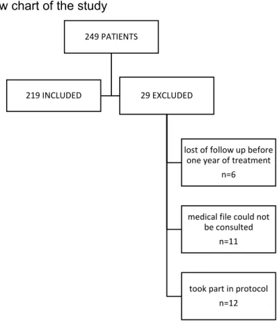
A total of 248 patients were identified from the data base EDMUS. Out of 248 patients, 29 patients were excluded due to lost of follow up during the year after treatment initiation (n=6), inclusion in clinical trials (n=12), missing data because the medical file could not be consulted (n=11). (Figure 1).
Figure 1. Follow chart of the study
Out of the 219 patients included in the study, they had a mean age of 38 +/- 9.4 years and a mean disease duration of 9.6 +/- 6.0 years.
The mean duration of treatment with fingolimod was 1.7 +/- 0.9 years.
Fingolimod was used as first line therapy in 3.2% (n=7) and as second line therapy in 96.8% (n=212). Previous treatment and reason for switching from other treatment are detailed in the table 3.
The annual rate relapse (ARR) was 1 in the previous year, the number of relapses is detailed in the table 5.
32.4% patients (n=71) were relapse free in the previous year.
The mean baseline EDSS was 2.5 +/- 1.8, the number of EDSS by the number of patients is detailed in the table 4.
58.5% patients (n=128) had no Gd enhanced on cerebral MRI, MRI characteristics before fingolimod are detailed in the table 3.
249 PATIENTS
219 INCLUDED 29 EXCLUDED
lost of follow up before one year of treatment
n=6
medical file could not be consulted
n=11
took part in protocol n=12
Table 3. Baseline characteristics
Baseline characteristic (n=219)
Age, mean +/- SD 38,0 +/- 9,4 years
Sexe male, n= 58 ( 26,5 %)
female, n=161 (73,5%) sex ratio (F/M) 2,8 MS one set, mean +/- SD 28,4 +/- 8,5 years Disease duration, mean +/- SD 9,6 +/- 6,0 years
Previous treatment
Previous treatment teriflunomide, n=1 (0.5%) interferon, n= 69 (31,5%) mycophenolate mofetil, n=2 (0.9%) glatiramere acetate, n=75 (35.4%) mitoxantrone, n=13 (6.1%) cyclophosphamide, n=2 (0.9%) pixantrone, n=1 (0.5%) dimethyle fumarate, n=1 (0.5%) natalizumab, n=47 (22.6%) none, n=7 (3,2%)
Main reason for switching from other DMT to fingolimod Lack of efficacy, n=115 (52,5%) Poor tolerance, n=40 (18,3%) Programed stop, n=58 (26,5%) JCV seroconversion, n=41 (18,7%) Wish of pregnancy, n=5 (2,3%) Personal wish, n=5 (2,3%) Duration of previous treatment, mean (+/-SD) 2,1 +/- 2.0 years
Duration between the previous treatment and fingolimod mean+/-SD 0,5 +/- 1,0 years
Relapses
Number of relapses in the previous year, mean +/-SD (min-max) 1.0 +/- 0.9 (0-5) Relapse free patients the year before fingolimod 71 (32,4%) Annual rate relapse (ARR) in the previous year 1
Duration between the last relapse and fingolimod beginning, mean +/- SD 5,1 +/- 4,6 months
EDSS
Baseline EDSS score, mean ± SD (min-max) (n=160, 75,47%) 2,5 +/- 1,8 (0-7,5)
MRI
Time between cerebral MRI and fingolimod begining, mean +/- SD 0,3 +/- 0,3 years Time between medullar MRI and fingolimod begining, mean +/- SD 1,1 +/- 2,1 years Cerebral MRI before fingolimod, n= 211 (96,34%) Gd+, n= 83 (37,9%)
Gd-, n= 128 (58,5%) Medullar MRI before fingolimod, n= 86 (39,27%) Gd+, n= 12 (5,5%)
Gd-, n= 70 (32.0%) Patient with out Gd enhanced on cerebral and medullar MRI, n= 81 n=48 (59,3%)
Fingolimod
Duration of fingolimod therapy, mean +/- SD (min-max) 1,7 +/- 0,9 years Duration of following since fingolimod begining, mean+/-SD 2,2 +/- 0,7 years
Table 4. EDSS in the previous year.
EDSS score in the previous year Patients , n=160 0 - 2,5 n= 99 (61,9%) 3 - 4,5 n= 43 (26,9%) 5 - 6,0 n= 8 (5,0%) ≥6,5 n= 10 (6,3%)
Table 5. Number of relapses in the previous year
Number of relapses
in the previous year Patients
0 n=71 (32,4%) 1 n=71 (32,4%) 2 n=96 (43,8%) 3 n=40 (18,2%) 4 n=8 (3,6%) 5 n=3 (1,4%) Efficacy Relapse
The ARR decreased significantly from 1 the year before fingolimod to 0.28 with fingolimod representing a relative risk reduction of 72% in the ARR, IC95%: [0.22-0.36]. Figure 2.
Figure 2. ARR the year before fingolimod and during fingolimod
If we exclude patients who were treated by natalizumab before, we have 172 patients, the ARR decreased significantly from 1,17 the year before fingolimod to 0.25 with fingolimod, representing a relative risk reduction of 78% in the ARR, IC95%: [0.1; 0.28]. Figure 3. 1 0,28 0 0,2 0,4 0,6 0,8 1 1,2
ARR
Before fingolimod During fingolimod IC 95% [0,22 ; 0,36]
Relative risk reduction of 72%
Figure 3. ARR the year before fingolimod and during fingolimod (patients treated by natalizumab before excluded)
We have a significant increase of the proportion of patients remaining relapse free post-treatment at 69.41% compared to 32.42% the year before fingolimod initiation (p < 0.0001). Figure 4.
Figure 4. Relapse free before and after fingolimod
1,17 0,25 0 0,2 0,4 0,6 0,8 1 1,2 1,4
Before fingolimod Under fingolimod
ARR
patients treated by
natalizumab before excluded
Relative risk reduction of 78% IC95% [0.1; 0.28] 32,42% 69,41% 0,00% 10,00% 20,00% 30,00% 40,00% 50,00% 60,00% 70,00% 80,00%
Relapse free patients
Before fingolimod During fingolimod p < 0,0001
If we exclude patients who were treated by natalizumab before, we have a significant difference between the 22.7% relapse free the year before fingolimod and the 72.1% who are relapse free with fingolimod (p<0.0001). Figure 5.
Figure 5. Relapse before and during fingolimod, patients who were treated by natalizumab before excluded.
The number of relapse with fingolimod therapy is detailed in the table 6.
Table 6. Number of relapses. Number of relapse Number of patients
0 n=152 (69,4%) 1 n=39 (17,8%) 2 n=21 (9,6%) 3 n=5 (2,3%) 4 n=1 (0,5%) 7 n=1 (0,5%)
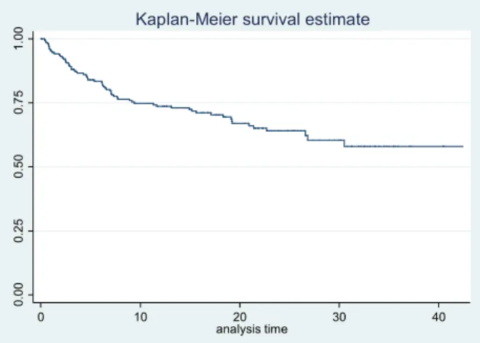
The mean time from initiation of fingolimod therapy to onset relapse was 0.7 +/- 0.6 years. The duration between the first relapse and fingolimod initiation is represented on the figure 6. 22,70% 72,10% 0,00% 10,00% 20,00% 30,00% 40,00% 50,00% 60,00% 70,00% 80,00%
Before fingolimod During fingolimod
Patients relapse free,
patients treated by natalizumab
before excluded
Figure 6. Kaplan-Meier survival estimate for relapse.
The mean time from initiation of fingolimod and other relapse is described in table 7.
Table 7. Mean time for relapse.
Relapse Time from initiation of fingolimod therapy, mean +/-SD years
1 0,7 +/- 0,6 2 1,3 +/- 0,8 3 1,7 +/- 0,7 4 1,3 +/- 0,5 5 1,2 6 1,7 7 1,9 EDSS, disability
The proportion of patients remaining free of disability progression was 85% at 3 months, 82.4% at 6 months, 86.8% at 12 months, 82.8% at 18 months, 84.4% at 24 months. Table 8.
Table 8. EDSS evolution.
Follow up Number of patients Improvement Stability Worsening
M3 53 7 (13,2%) 38 (71,7%) 8 (15,1%) M6 34 6 (17,7%) 22 (64,7%) 6 (17,7%) M12 83 15 (18,1%) 57 (68,7%) 11 (13,3%) M18 58 6(10,3%) 42 (72,4%) 10 (17,2%) M24 32 6 (18,8%) 21 (65,6%) 5 (15,6%) 0.00 0.25 0.50 0.75 1.00 0 10 20 30 40 analysis time
There is no significant difference on the EDSS during the follow up at 3, 6, 12, 18, 24 months compared to the baseline EDSS score. Table 9.
Table 9. EDSS during fingolimod therapy compared to EDSS baseline.
Following Number of patients M0 Follow up p
M3 53 2,1 +/- 1,9 2,1 +/- 1,8 0,93 M6 34 1,9 +/- 1,6 2,1 +/- 1,5 0,42 M12 83 2,0 +/- 1,6 2,0 +/- 1,7 0,64 M18 58 2,6 +/- 2,0 2,5 +/-2,1 0,73 M24 32 2,5 +/- 1,9 2,5 +/- 2,1 0,86 MRI outcome
Patients have in mean 1.2 +/- 1.1 MRI during the treatment with fingolimod, 0.7 MRI per year.
60.9% (n=14) patients are free from MRI activity at M6, 71.2% (n=52) at M12, and 81.5% (n=22) at M24.
We have a significant decrease for Gd enhancement at M12 and M24 compared to Gd enhancement at M0.
Table 10. MRI outcome
Number of patients with a follow up MRI and a MRI before fingolimod No new lesion compared to M0 MRI Follow up Gd- M0 Gd- p Gd- (follow up-M0) No new lesion and Gd- M6 23 15 (65,2%) 17 (73,9%) 12 (52,2%) 0,182 14 (60,9%) M12 73 53 (72,6%) 60 (82,2%) 47 (64,4%) 0,03 52 (71,2%) M24 27 21 (77,8%) 23 (85,2%) 15 (53,6%) 0,01 22 (81,5%) Disease activity
Out of 24 patients in whom relapse rate and MRI outcomes were measured at M6, 11 patients (45.8%) had no disease activity.
Out of 75 patients in whom relapse rate and MRI outcomes were measured at M12, 35 patients (46.7%) had no disease activity.
Out of 27 patients in whom relapse rate and MRI outcomes were measured at M24, 15 patients (55.6%) had no disease activity.
Table 11. Patients relapse free and MRI activity free Relapse free and MRI activity free Number of patients
M6, n=24 11 (45,8%)
M12, n=75 35 (46,7%)
M24, n=27 15 (55,6%)
Adverse effects and reasons of interruption
Fingolimod therapy was discontinued in 30.14% of patients (n=66) due to suboptimal response in 6.8% patients (n=15), poor clinical tolerance in 14.5% patients (n=31), biologic trouble in 4.7% patients (n=10) and whish of pregnancy in 5% patients (n=11). Table 12.
Table 12. Reason to stop the treatment. Reason to stop the treatment n= 66
poor clinical tolerance 31 (47%)
pain 7 (10,6%) asthenia 7 (10,6%) diarrhea 5 (7,6%) depression 5 (7,6%) infectieux 4 (6%) ORL 1 (1,5%) sepsis 1 (1,5%) cystitis 1 (1,5%) Dermatologic trouble 4 (6%) Discomfort 3 (4,5%) cardiologic trouble 3 (4,5%) head heach 2 (3%) insomnia 1 (1,5%) thrombocytopenic purpura 1 (1,5%) cancer 2 (3%) others 4 (6%) biologic trouble 10 (15,2%) lymphopenia 4 (6%) hepatic trouble 6 (9%) whish of pregnancy 11 (16,7%) inefficacity 15 (22,7%)
48% of patients (n=104) met adverse events with fingolimod therapy.
Lymphopenia defined as lymphocyte count inferior to 200 × 10^6/L, occurred in 3.2% patients (n=7) of patients, requiring discontinuation in 4 of them (one of them had still lymphopenia with fingolimod every two days) and decrease of the dose in other 3 patients.
13.7% patients (n=30) had infections, 1.8% of them (n=4) stopped the treatment for this reason.
Three patients presented a cancer during the treatment, 2 of them stopped the treatment for this reason, one had an epidermoide carcinoma, and the other had a melanoma. The two cancer described in the patient who continue the treatment are two cutaneous neoplasia in the same patiente: one melanoma, and one basocellular carcinoma.
Two patients started a pregnancy before two months of fingolimod discontinuation: one of them had a precoce miscarriage, and the other had a normal pregnancy.
3.2% of patients (n=7) had cardiologic troubles, 3 of them had to stop the treatment, two of them the day of introduction because of one atrio-ventricular block, and the other for severe bradycardia requiring isuprel, the third stopped the treatment one day after the introduction because of a persistant bradycardia.
The other patient with cardiologic trouble who follow the treatment had palpitations, one of them had an autentified cardiac arythmia.
The patiente with thrombocytopenic purpura had already this pathology before the treatment, but she had a relapse of her thrombopenia with the fingolimod.
The patiente who had a stroke had a retina ischemia.
None of the patients developed macular edema or multifocal leukoencephalopathy. 2.7% patients (n=6) developed a progressive form under fingolimod.
Table 13. Adverse events Adverse events Patients, n= 219 Reason to stop fingolimod Adverse event continuing fingolimod poor clinical tolerance 87 (40,7%) 31 (14,15%) 80 (36,5%)
infectieux 30 (13,7%) 4 (1,8%) 26 (11,9%)
ORL 5 (2,3%) 1 (0,5%) 4 (1,8%)
pyelonephritis 3 (1,4%) 3 (1,4%)
abscess 2 (1%) 2 (1%)
bronchitis 2 (1%) 2 (1%)
clostridium difficile colitis 1 (0,5%) 2 (1%)
herpes virus infection 7 (3,2%) 7 (3,2%)
prostatis 1 (0,5%) 1 (0,5%)
sepsis 2 (1%) 1 (0,5%) 1 (0,5%)
vaginal yeast infection 2 (1%) 2 (1%)
cystitis 2 (1%) 1 (0,5%) 1 (0,5%) zona 2 (1%) 2 (1%) pain 19 (8,7%) 7 (3,2%) 12 (5,5%) asthenia 17 (7,8%) 7 (3,2%) 10 (4,6%) depression 17 (7,8%) 5 (2,3%) 12 (5,5%) head heach 10 (4,6%) 2 (1%) 8 (3,7%) dermatologic trouble 10 (4,6%) 4 (1,8%) 6 (2,7%) diarrhea 8 (3,7%) 5 (2,3%) 3 (1,4%) cardiologic trouble 7 (3,2%) 3 (1,4%) 4 (1,8%) discomfort 7 (3,2%) 3 (1,4%) 4 (1,8%) cancer 4 (1,8%) 2 (1%) 2 (1%) nausea 2 (1%) 2 (1%) cough 2 (1%) 2 (1%) insomnia 2 (1%) 1 (0,5%) 1 (0,5%) thrombocytopenic purpura 1 (0,5%) 1 (0,5%) stroke 1 (0,5%) 1 (0,5%) phlebitis 1 (0,5%) 1 (0,5%) others 10 (5%) 3 (1,4%) 7 (3,2%) biologic trouble 30 (13,7%) 10 (4,6%) 20 (9,1%) lymphopenia 7 (3,2%) 4 (1,8%) 3 (1,4%) hepatic trouble 20 (9,1%) 6 (2,7%) 14 (6,4%) cytolysis 6 (2,7%) 1 (0,05%) cholestasis 13 (5,9%) thrombopenia 1 (0,5%) 1(0,5%) neutropenia 1 (0,5%) 1 (0,5%) hypereosino 1 (0,5%) 1 (0,5%) pregnancy 2 (1%) 2 (1%)
Sub-groups analyses
Switch from natalizumab to fingolimod
21.9% of patients (n=47) had natalizumab as previous treatment, their characteristic before fingolimod are detailed in the table 14.
Table 14. Baseline characteristics for patients who stopped natalizumab for fingolimod
Previous treatment by natalizumab n= 47
Reason of switch
JCV conversion n=41 (85,4%)
Inefficacity n=1 (2,0%)
Intolerance n=3(6,0%)
Convenance n=2 (4,0%)
Duration of natalizumab, mean +/- SD 2,0 +/- 1,2 years
Wash out period, mean +/- SD 0,3 +/- 0,4 years
Number of patients with a wash out period inferior to 3 months 23 (48.9%) Number of relapse the year before fingolimod, mean +/- SD 0,4 +/- 0,6
Relapse free the previous year 33 (68,8%)
AAR the year before fingolimod 0,4
ARR with natalizumab 0,2
EDSS before fingolimod, mean +/- SD 2,7 +/- 2,0
Duration of fingolimod, mean+/- SD 1,7 +/- 1,0 years
There is a significant difference between the ARR with natalizumab at 0.2 and the ARR with fingolimod at 0.38, IC 95% [0.30; 0.94]. Figure 7.
Figure 7. ARR with natalizumab and with fingolimod
And we have a significant difference between the ARR with natalizumab at 0.2 and the ARR after natalizumab stop and during fingolimod at 0.4, IC 95% [1.13; 3.41].
The mean time for the first relapse after natalizumab stop is 11.1 months. 5 patients (10.6%) presented a relapse during the wash out period, they all had a wash out period superior to 3 months. Only one patient had a relapse before 3 months, and he presented his relapse before one month, so before the rebound period with natalizumab. The others patients who had relapsed during the wash out period presented their relapse after 3 months.
23 patients (48.9%) had a wash out period inferior to 3 months, those patients had no relapse during the wash out period.
We have no significant difference between the ARR after natalizumab stop for patients with a wash out period inferior to 3 months (0.32) and the ARR after natalizumab for patients with a wash out period superior to 3 months (0.42), IC 95% [0.39; 1.55].
0,2 0,38 0 0,05 0,1 0,15 0,2 0,25 0,3 0,35 0,4 natalizumab fingolimod
ARR with natalizumab and with fingolimod
There is no significant difference for the EDSS during the follow up with fingolimod compared to EDSS baseline with natalizumab. Table 15.
Table 15. EDSS during fingolimod therapy compared with EDSS at M0
Follow up Number of patients M0 Follow up p
M3 10 2,2 +/- 2,2 2,5 +/- 2,1 0,33
M6 9 1,9 +/-1,2 2,3 +/- 1,7 0,46
M12 17 2,0 +/- 1,1 2,1 +/- 1,4 0,53
M18 14 2,9 +/- 2,1 3,1 +/- 2,3 0,27
M24 7 2,0 +/- 2,8 2,1 +/- 2,7 0,88
Patients who stopped fingolimod for inefficacity
6.9% patients (n=15) stopped fingolimod because of inefficacity, they had fingolimod during 1.4 +/- 1.0 year.
33.3% patients (n=5) were relapse free before fingolimod, and 13.3% patients (n=2) were relapse free with fingolimod, there is no significant difference (p=0.37).
The ARR before fingolimod was 0.93, and the ARR with fingolimod is 1.07, there is no significant difference IC95% [0.08 ; 14.15].
The first relapse appeared at 7.1 +/- 8.3 months.
There is no significant difference between EDSS before fingolimod and EDSS during the treatment for people who stopped fingolimod for inefficacy. Table 16.
Table 16. EDSS evolution compared to EDSS baseline for patients who stopped fingolimod for inefficacy.
Number of patients EDSS M0 Follow EDSS IC95% EDSS M0-following EDSS
M3 2 2,6 +/- 0,3 2,3 +/- 0,3 0
M6 6 1,8 +/-0,2 2,3 +/- 0,2 [-1,26 ; 0,42]
M12 5 2,4 +/- 0,3 2,4 +/- 0,3 [-0,44 ; 0,44]
M18 3 2,8 +/- 0,6 3,1 +/- 0,6 [-1,77 ; 1,1]
There is no significant difference between patients who stopped the treatment for inefficacity and others patients on gender, age, age of MS one set, disease duration, gadolinium enhancement before fingolimod, EDSS baseline, relapse free before fingolimod, ARR before and with fingolimod, table 17.
Table 17. Characteristics of patients who stopped fingolimod for inefficacity compared to the others patients.
Stop for inefficacy, n=15 Others patients, n=204 Significance Sexe, female 14 (93,3%) 147 ( 72,1%) p=0,125 Age 34,5 +/- 9,9 38,3 +/- 9,3 p=0,137 MS onset 25,6 +/- 10,2 28,6 +/- 8,4 p=0,188 Disease duration 8,9 +/- 6,1 9,7 +/- 4,9 p=0,649
Cerebral Gd+ before fingolimod 7 (46,7%) 76 (38,8%) p=0,590
Medullar Gd+ before fingolimod 1 (10,0%) 11 (15,3%) p=1
Baseline EDSS 2,7 +/- 1,3 2,5 +/- 1,9 p=0,7
Relapse free before fingolimod 5 (33,3%) 66 (32,4%) p=1
ARR before fingolimod 0,9 1 IC95% [0,06 ; 18,1]
ARR with fingolimod 1,1 0,2 IC95% [0,01 ; 6,88]
Patients who stopped fingolimod for wish of pregnancy
11 patients (16.7%) stopped fingolimod for wish of pregnancy.
They had fingolimod in mean 1.2 +/- 0.7 years, and they stopped the treatment in mean 1.1 +/- 0.4 years.
72.7% of this patient where relapse free with fingolimod, they had a mean EDSS of 1.0 +/- 0.8.
5 of them had a temporary stop of fingolimod, the 6 others had not taken fingolimod at the end of the study.
We have a significant increase of the ARR from 0.40 to 1.26, IC95% [0.05-0.64] after fingolimod stop for patient who stop the treatment for wish of pregnancy. Figure 8. The mean time to the first relapse is 3.4 +/- 2.8 months.
Only two of them had a relay treatment when fingolimod is stopped: one had copaxone and had no relapse after fingolimod stop. The other had an interferon and had two relapses with this treatment, she had only one relapse with fingolimod. A third one had alemtazumab, 3 months after fingolimod stop because of a serious relapse with an MRI with a lot of Gd enhancement.
Figure 8. ARR with and after fingolimod for patients who had stopped fingolimod for wish of pregnancy.
DISCUSSION :
Our cohort reflects the “real world condition” of fingolimod use during in mean 1.7 +/- 0.9 years in 219 patients. This is important because patients are not exactly the same as in randomized controlled trials and in real clinical practice: patients are of different ages, different disease duration, different disease activity, varying comorbidities, patients can be less invested in their treatment: doses may be omitted.
The main limitation of this cohort study is the retrospective nature of our review, and the lack of EDSS and MRI.
We report here our clinical experience with the use of fingolimod in daily practice in Brittany. 0,24 1,26 0 0,2 0,4 0,6 0,8 1 1,2 1,4
ARR with fingolimod ARR after fingolimod stop
ARR with and after fingolimod for patients who
stopped fingolimod for wish of pregnancy
Efficacy
Compared with other studies
The ARR decreased by 72% on fingolimod therapy in our study, which was slightly lower than the relative risk reduction of 88% and 89% observed in the clinical trials FREEDOMS [5] and TRANSFORMS [6], respectively. Other post marketing studies reported a quite similar relative risk reduction for ARR such as: Latin America [13], Lebanese [14] with a ARR reduction of 69% and 75%, respectively.
Some studies published a lower relative risk reduction for ARR as a Portuguese study (51.92%) [15], or a US study reporting a reduced risk of ARR of 59% for patients switching from IFNB to fingolimod [16]. A possible explanation for these differences is the variability of ARR at baseline which was highest in the study who reported the highest relative risk reduction for ARR: 1.5 in FREEDOMS [5] and TRANSFORMS [6] , moderate in our cohort (1) and in the Lebanese cohort (1.16) [14], and lowest in the study who find a lower relative risk reduction for ARR (0.46 in the US study) [16].
The proportion of patients relapse free in our cohort is 69.41% comparable with the original trial FREEDOMS [5]: 70.4%, and some observational studies: Lebanese (77.3%) [14], Baldi Italian study (69.30%) [17]. Other studies have a higher rate of patients relapse free: 82.6% in TRANSFORMS [6], 88.10% in Totaro Italia study [18], 86.3% in a study from Koweit [19], 87.3% in Hersh study [20]. But the proportion relapse free before fingolimod was the same in our study (32%), and in the study from Koweit.
The proportion of patients free of disability progression in our cohort was 84.37% at 24 months comparable to 82.3% in FREEDOMS trial [5], 80% in Lebanese study [14]. The proportion of patients free of disability progression was lower in Totaro Italian study at 70% [18] and higher in TRANSFORMS trial [6] at 94.1%.
Results are discordant in different study on the significant improvement of EDSS. We have no significant change in the EDSS with fingolimod as it was described in TRANSFORMS [6] and FREEDOM [5], and in other study: Portuguese one [15], and Italian studies [17-18]. Some study have shown an improvement on the EDSS: the
Lebanese study [14], Mexican study [21], study from Koweit [19]. This does not seem to be due to the EDSS difference before fingolimod or treatment naive.
The proportion of patients free from any radiological activity (New T2 or Gd+ lesions) was 84.8% in our cohort and other observational studies (81.%) [22] compared to 50.5 % in FREEDOMS trial [5] and 54.8 % in TRANSFORMS trial [6], 66.3% in Lebanese study [14], 54.9% in Baldi Italian study [17], this is higher but the percentage of patients free form Gd enhancement is similar before treatment around 60% in the different study.
We have not a significance difference at M6 for Gd enhancement, this could be explain, because fingolimod is not yet to its full efficacy.
For disease activity we did not include EDSS evolution, because of data missing.
In real world, patients treatment naïve are very lower 3.2% in our study, 6% in
Portuguese study [15], 3.5% in Hersh study [20], 12% in Lebanese study [14], 21% in a study from Koweit [19], than in clinical trials 57% in FREEDOMS [5] and 45.8% in TRANSFORMS [6].
Compared with others treatments
It seems fingolimod is less effective than natalizumab to prevent relapses, disability progression and new T2 and Gd-enhancing lesions at 1 and 2 years [23 - 24]. Fingolimod therapy results in a higher probability of NEDA than dimethyl fumarate and teriflunomide therapy when phase 3 trial data are indirectly compared and differences between trials are adjusted for [25].
Safety
30.14% patients discontinued treatment permanently, this is higher than in all other studies 7.5% in FREEDOMS [5], 5.6% in TRANSFORMS [6], 1.6% in Lebanese study [14], 10.6% in Portuguese study [15], 9.10% in Totaro Italian study [18], 24.8% in Hersh study [20], maybe patients are less tolerant to clinical adverse effects, because the proportion of patients who stopped for inefficacy or biologic intolerance are the same as in the other study, and we do not have more adverse events.
48% met adverse event in our study, this is quite heterogenous between different studies from 94% in FREEDOMS [5] to 24% in Koweit study [19], sometimes it is difficult to know if the adverse event is really due to fingolimod.
None of our patients developed macular oedema, or multifocal leukoencephalopathy. In our study two patients had to stop the treatment the day of the introduction because of serious bradycardia, this should encourage us to be careful with cardiologic follow. Infections occurred in 13.7% of our patients and were mostly mild to moderate. The rate of infections was much higher in both the FREEDOMS [5] and TRANSFORMS [6] trials, but this is probably related to the strict adverse event reporting in clinical trial study.
These data will be assess by the French national data base VIRGIL.
Due to its safety and efficacy fingolimod had an extension of indication in second line use to ‘patients with active disease defined by clinical or imaging features despite treatment with at least one disease modifying therapy’.
Sub groups
Switch natalizumab to fingolimod
One French study find 27% patients relapsed during the wash out period [26], in our study only 10.6% relapsed during the wash out period, maybe due to the wash out period which is a little shorter in our study (14 weeks versus 17 weeks). This same French study has shown that a wash out period shorter than 3 months was associated with a lower risk of relapse but in our study we have no significant difference between the ARR after natalizumab stop for patients with wash out period inferior to 3 months compared to those with a wash out period superior to 3 months, but we have no relapse during the wash out period for patients with a wash out period shorter than 3 months. In our study, we have a significant increase of the ARR with fingolimod compared with natalizumab, as shown in an other study [27], maybe fingolimod is not the ideal relay treatment after natalizumab stop.
Patients who stop the treatment for wish of pregnancy
We have a significant increase of the ARR after fingolimod stop, this could encourage us to discuss a relay treatment after fingolimod stop for wish of pregnancy.
CONCLUSION
In conclusion, our cohort and other post marketing studies confirm the efficacy and safety of fingolimod in the treatment of RRMS in a real world setting, this has been assessed by its extension of indication.
[1]. Agnes Fromont et al., “Geographic Variations of Multiple Sclerosis in France,” Brain 133, no. 7 (July 1, 2010): 1889–99, doi:10.1093/brain/awq134.
[2]. A. Créange and P. Labauge, “[Social handicap at the onset of the multiple sclerosis],” Revue
Neurologique 165 Suppl 4 (March 2009): S167–72, doi:10.1016/S0035-3787(09)72130-7.
[3]. Stephen Sawcer, Robin J M Franklin, and Maria Ban, “Multiple Sclerosis Genetics,” The Lancet
Neurology 13, no. 7 (July 2014): 700–709, doi:10.1016/S1474-4422(14)70041-9.
[4]. Ron Milo and Ariel Miller, “Revised Diagnostic Criteria of Multiple Sclerosis,” Autoimmunity Reviews, Diagnostic criteria in Autoimmune diseases9th International Congress on Autoimmunity, 13, no. 4–5 (April 2014): 518–24, doi:10.1016/j.autrev.2014.01.012.
[5]. Ludwig Kappos et al., “A Placebo-Controlled Trial of Oral Fingolimod in Relapsing Multiple Sclerosis,”
New England Journal of Medicine 362, no. 5 (February 4, 2010): 387–401, doi:10.1056/NEJMoa0909494.
[6]. Jeffrey A. Cohen et al., “Oral Fingolimod or Intramuscular Interferon for Relapsing Multiple Sclerosis,”
New England Journal of Medicine 362, no. 5 (February 4, 2010): 402–15, doi:10.1056/NEJMoa0907839.
[7]. L. D. Jacobs et al., “Intramuscular Interferon Beta-1a for Disease Progression in Relapsing Multiple Sclerosis. The Multiple Sclerosis Collaborative Research Group (MSCRG),” Annals of Neurology 39, no. 3 (March 1996): 285–94, doi:10.1002/ana.410390304.
[8]. Peter A Calabresi et al., “Safety and Efficacy of Fingolimod in Patients with Relapsing-Remitting Multiple Sclerosis (FREEDOMS II): A Double-Blind, Randomised, Placebo-Controlled, Phase 3 Trial,” The Lancet
Neurology 13, no. 6 (June 2014): 545–56, doi:10.1016/S1474-4422(14)70049-3.
[9]. Francesca Bagnato et al., “Evolution of T1 Black Holes in Patients with Multiple Sclerosis Imaged Monthly for 4 Years,” Brain 126, no. 8 (August 1, 2003): 1782–89, doi:10.1093/brain/awg182.
[10]. D. S. Goodin and D. Bates, “Treatment of Early Multiple Sclerosis: The Value of Treatment Initiation after a First Clinical Episode,” Multiple Sclerosis (Houndmills, Basingstoke, England) 15, no. 10 (October 2009): 1175–82, doi:10.1177/1352458509107007.
[11]. J. Río et al., “Relationship between MRI Lesion Activity and Response to IFN-β in Relapsing–remitting Multiple Sclerosis Patients,” Multiple Sclerosis 14, no. 4 (May 1, 2008): 479–84,
doi:10.1177/1352458507085555.
[12]. Ruth Dobson et al., “Assessing Treatment Response to Interferon-β: Is There a Role for MRI?,”
Neurology 82, no. 3 (January 21, 2014): 248–54, doi:10.1212/WNL.0000000000000036.
[13]. Jorge Correale et al., “Use of Fingolimod in the Management of Relapsing–Remitting Multiple Sclerosis: Experience from Latin America,” Advances in Therapy 32, no. 7 (2015): 612–25, doi:10.1007/s12325-015-0226-0.
[14]. Bassem I. Yamout et al., “Safety and Efficacy of Reduced Fingolimod Dosage Treatment,” Journal of
Neuroimmunology 285 (August 15, 2015): 13–15, doi:10.1016/j.jneuroim.2015.05.012.
[15]. I. Correia et al., “The Effectiveness of Fingolimod in a Portuguese Real-World Population,” Multiple
Sclerosis and Related Disorders 6 (March 2016): 41–48, doi:10.1016/j.msard.2016.01.003.
[16]. Niklas Bergvall et al., “Relapse Rates in Patients with Multiple Sclerosis Switching from Interferon to Fingolimod or Glatiramer Acetate: A US Claims Database Study,” PLoS ONE 9, no. 2 (February 6, 2014), doi:10.1371/journal.pone.0088472.
[17]. Eleonora Baldi et al., “Previous Treatment Influences Fingolimod Efficacy in Relapsing–remitting Multiple Sclerosis: Results from an Observational Study,” Current Medical Research and Opinion 30, no. 9 (September 1, 2014): 1849–55, doi:10.1185/03007995.2014.921144.
[18]. Rocco Totaro et al., “Fingolimod Treatment in Relapsing-Remitting Multiple Sclerosis Patients: A Prospective Observational Multicenter Postmarketing Study,” Multiple Sclerosis International 2015 (2015), doi:10.1155/2015/763418.
[19]. Jasem Al-Hashel et al., “Real-World Use of Fingolimod in Patients with Relapsing Remitting Multiple Sclerosis: A Retrospective Study Using the National Multiple Sclerosis Registry in Kuwait,” CNS Drugs 28, no. 9 (September 2014): 817–24, doi:10.1007/s40263-014-0185-z.
[20]. Carrie M. Hersh et al., “Experience with Fingolimod in Clinical Practice,” The International Journal of
Neuroscience 125, no. 9 (2015): 678–85, doi:10.3109/00207454.2014.969839.
[21]. I. Treviño-Frenk et al., “The Real-Life Experience with Fingolimod in Mexico: A Multicenter Post-Marketing Study,” Journal of the Neurological Sciences, Abstracts from the World Congress of Neurology (WCN 2015), 357, Supplement 1 (October 15, 2015): e301, doi:10.1016/j.jns.2015.08.1061.
[22]. Ibid.
[23]. Laetitia Barbin et al., “Comparative Efficacy of Fingolimod vs Natalizumab A French Multicenter Observational Study,” Neurology, January 29, 2016, 10.1212/WNL.0000000000002395,
doi:10.1212/WNL.0000000000002395.
[24]. Tomas Kalincik et al., “Switch to Natalizumab versus Fingolimod in Active Relapsing-Remitting Multiple Sclerosis,” Annals of Neurology 77, no. 3 (March 2015): 425–35, doi:10.1002/ana.24339.
[25]. Richard Nixon et al., “No Evidence of Disease Activity: Indirect Comparisons of Oral Therapies for the Treatment of Relapsing–Remitting Multiple Sclerosis,” Advances in Therapy 31, no. 11 (2014): 1134–54, doi:10.1007/s12325-014-0167-z.
[26]. Cohen M et al., “Switching from Natalizumab to Fingolimod in Multiple Sclerosis: A French Prospective Study,” JAMA Neurology 71, no. 4 (April 1, 2014): 436–41, doi:10.1001/jamaneurol.2013.6240.
[27]. Vilija G. Jokubaitis et al., “Fingolimod after Natalizumab and the Risk of Short-Term Relapse,”
U.F.R DE MEDECINE DE RENNES N°
MALRAIN Cécile- Evolution clinique et radiologique des patients de Bretagne ayant une sclérose en plaques sous fingolimod
37 feuilles, 8 figures, 17 tableaux. 30 cm. Thèse : médecine. Rennes 1. 2016. N°
Résumé
Introduction : Le fingolimod est un traitement de fond des formes très actives de sclérose en plaque rémittente-récurrente (SEP RR) commercialisé en France depuis 2011. C’est est un immunosuppresseur modulateur du récepteur de la sphingosine-1-phosphate permettant de bloquer les lymphocytes en périphérie empêchant leur passage dans le système nerveux central. Le but de notre étude est de voir l’évolution clinique et radiologique des patients de Bretagne ayant une SEP RR sous fingolimod afin d’avoir un reflet de son efficacité et de sa sécurité d’emploi en pratique courante.
Matériel et méthodes : Nous avons inclus tous les patients de Bretagne de la base EDMUS ayant une SEP RR ayant reçu du fingolimod jusqu’au 31.12.2013. Le recueil de données s’est arrêté au 31.12.2014. L’ensemble des dossiers ont été relus afin de collecter les poussées, les EDSS, les IRM, les effets indésirables et les motifs d’arrêt sous fingolimod.
Résultats: Nous avons inclus 219 patients. Sous fingolimod, le taux annuel de poussée (TAP) a diminué de façon significative de 72% de 1 à 0.28, IC95%: [0.22-0.36]. Sous fingolimod, il y avait une augmentation significative à 69.41% du taux de patients ne présentant pas de poussée (p<0.0001). Le délai moyen de survenue de la première poussée était de 0.65 +/- 0.63 mois. Nous n’avons pas constaté de différence significative sur les EDSS de suivi comparés à l’EDSS à l’instauration du traitement. Parmi les patients 84.37% ne présentaient pas de progression du handicap à 2 ans. 60.87% patients n’avaient pas d’activité IRM à six mois, 71.23% à un an, et 81.48% à deux ans. Il y a une diminution significative du taux de patients avec des prises de contraste à M12 (p=0.03) et à M24 (p=0.01), comparé à l’instauration du traitement. 60.9% des patients n’ont pas d’activité IRM ni poussée à M6, 71.2% à M12 et 81.5% à M24.
Le fingolimod a été arrêté chez 30.14% des patients : 6.8% ont cessé pour inefficacité, 14.5% pour mauvaise tolérance clinique, 4.7% pour intolérance biologique (lymphopénie ou trouble du bilan hépatique), 5% pour désir de grossesse. Des effets indésirables ont été rapportés chez 48% des patients : 13.7% ont présenté des
évènements infectieux, 3.2% ont présenté des évènements cardiaques. Aucun patient n’a présenté d’œdème maculaire ni de leucoencéphalopathie multiple progressive.
Pour les patients précédemment traités par natalizumab, il y a une augmentation significative du TAP sous fingolimod (0.38) comparé au TAP sous natalizumab (0.2) IC 95% [0.3 ; 0.94]. Il n’y a pas de différence significative sur l’EDSS de suivi. Il n’y a pas de différence significative sur le TAP après l’arrêt du natalizumab pour les patients ayant une durée de wash out inférieure à 3 mois et ceux ayant une durée de wash out supérieure à 3 mois.
Les patients ayant arrêtés le fingolimod pour inefficacité avaient des caractéristiques initiales similaires aux autres patients (âge, durée d’évolution de la maladie, âge de la première poussée, EDSS, TAP, pourcentage de patients sans poussée) avant la mise sous fingolimod.
Il y a une augmentation significative du TAP de 0.4 à 1.26 IC95% [0.05-0.64], après l’arrêt du fingolimod chez les patientes l’ayant arrêté pour désir de grossesse.
Conclusion : Notre étude ainsi que les autres études suite à la mise sur le marché du fingolimod confirment son efficacité et sa sécurité d’emploi, ce qui va dans le sens de son extension d’indication.
Mots clés : sclérose en plaque, fingolimod
Mots clés anglais MeSH : multiple sclerosis fingolimod