Development of an enzyme immobilization platform
based on microencapsulation for paper-based biosensors
par Yufen Zhang Département de Chimie Faculté des arts et de sciences
Thèse présentée à la Faculté des études supérieures et postdoctorales en vue de l’obtention du grade de Philosophiae Doctor (Ph.D)
en Chimie
November, 2011
Faculté des études supérieures et postdoctorales
Cette thèse intitulée :
Development of an enzyme immobilization platform based on microencapsulation for paper-based biosensors
présentée par : Yufen Zhang
a été évaluée par un jury composé des personnes suivantes :
Prof. Karen C. Waldron, président-rapporteur Prof. Dominic Rochefort, directeur de recherche
Prof. Julian X. Zhu, membre du jury Prof. Gil Garnier, examinateur externe
Résumé
Un papier bioactif est obtenu par la modification d’un papier en y immobilisant une ou plusieurs biomolécules. La recherche et le développement de papiers bioactifs est en plein essor car le papier est un substrat peu dispendieux qui est déjà d’usage très répandu à travers le monde. Bien que les papiers bioactifs n’aient pas connus de succès commercial depuis la mise en marche de bandelettes mesurant le taux de glucose dans les années cinquante, de nombreux groupes de recherche travaillent à immobiliser des biomolécules sur le papier pour obtenir un papier bioactif qui est abordable et possède une bonne durée de vie. Contrairement à la glucose oxidase, l’enzyme utilisée sur ces bandelettes, la majorité des biomolécules sont très fragiles et perdent leur activité très rapidement lorsqu’immobilisées sur des papiers. Le développement de nouveaux papiers bioactifs pouvant détecter des substances d’intérêt ou même désactiver des pathogènes dépend donc de découverte de nouvelles techniques d’immobilisation des biomolécules permettant de maintenir leur activité tout en étant applicable dans la chaîne de production actuelle des papiers fins.
Le but de cette thèse est de développer une technique d’immobilisation efficace et versatile, permettant de protéger l’activité de biomolécules incorporées sur des papiers. La microencapsulation a été choisie comme technique d’immobilisation car elle permet d’enfermer de grandes quantités de biomolécules à l’intérieur d’une sphère poreuse permettant leur protection. Pour cette étude, le polymère poly(éthylènediimine) a été choisi
afin de générer la paroi des microcapsules. Les enzymes laccase et glucose oxidase, dont les propriétés sont bien établies, seront utilisées comme biomolécules test. Dans un premier temps, deux procédures d’encapsulation ont été développées puis étudiées. La méthode par émulsion produit des microcapsules de plus petits diamètres que la méthode par encapsulation utilisant un encapsulateur, bien que cette dernière offre une meilleure efficacité d’encapsulation. Par la suite, l’effet de la procédure d’encapsulation sur l’activité enzymatique et la stabilité thermique des enzymes a été étudié à cause de l’importance du maintien de l’activité sur le développement d’une plateforme d’immobilisation. L’effet de la nature du polymère utilisé pour la fabrication des capsules sur la conformation de l’enzyme a été étudié pour la première fois.
Finalement, l’applicabilité des microcapsules de poly(éthylèneimine) dans la confection de papiers bioactifs a été démontré par le biais de trois prototypes. Un papier réagissant au glucose a été obtenu en immobilisant des microcapsules contenant l’enzyme glucose oxidase. Un papier sensible à l’enzyme neuraminidase pour la détection de la vaginose bactérienne avec une plus grande stabilité durant l’entreposage a été fait en encapsulant les réactifs colorimétriques dans des capsules de poly(éthylèneimine). L’utilisation de microcapsules pour l’immobilisation d’anticorps a également été étudiée.
Les avancées au niveau de la plateforme d’immobilisation de biomolécules par microencapsulation qui ont été réalisées lors de cette thèse permettront de mieux comprendre l’effet des réactifs impliqués dans la procédure de microencapsulation sur la
stabilité, l’activité et la conformation des biomolécules. Les résultats obtenus démontrent que la plateforme d’immobilisation développée peut être appliquée pour la confection de nouveaux papiers bioactifs.
Mots-clés :
Microencapsulation, microcapsules de poly (éthylèneimine), encapsulateur, buse vibrations, papiers bioactifs, conformation des enzymes, fluorescence, microscopie confocale à balayage laser (CLSM), biocapteur colorimétrique, microcapsules d'anticorps sensibilisés.
Abstract
Biosensing paper attracts increasing attention due to its benefits of being simple, visible, portable and useful for detecting various contaminants, pathogens and toxins. While there has been no bioactive paper commercialized since glucose paper strips developed in the fifties, many research groups are working to immobilize biomolecules on paper to achieve a bioactive paper that is affordable and has good shelf life. The goal of this research is to develop some highly useful bioactive paper that could, for example, measure blood glucose, or immediately detect and simultaneously deactivate pathogens such as neuraminidase and E.coli. Previously, bioactive paper was produced either through physically absorbing biorecognition elements or printing bio-ink onto paper substrate. Our methodology for fabrication of bioactive paper strips is compatible with existing paper making process and includes three procedures: the fabrication of microcapsules, enzyme or antibody microencapsulation, immobilization of enzymes or antibody-entrapped microcapsules into paper pulp.
The first step, in fabricating of bioactive paper strips is to produce biocompatible and inexpensive microcapsules with suitable parameters. To do so, two types of microencapsulation methods were compared; the emulsion method and the vibration nozzle method accomplished with an encapsulator. The parameters for producing optimal microcapsules with both methods were studied. Factors that affect their diameter, wall thickness, shell pore size, encapsulation efficiency and membrane compositions were also discussed. By comparison, microcapsules prepared with poly(ethyleneimine) (PEI) by the
emulsion method exhibit properties that were more suitable for enzyme encapsulation and paper making process, whereas the microcapsules prepared by the vibration nozzle method were too big to be immobilized within paper pulp, and had lower encapsulation efficiency, enzymatic activity and productivity. Thus the emulsion method was chosen for subsequent experiments such as enzyme and antibody microencapsulation and bacterial vaginosis (BV) paper preparation. Microcapsules made by the emulsion method were semi-permeable in that the diffusion of substrate and product molecules were allowed freely across the membranes but the encapsulated enzymes would be retained inside.
Glucose oxidase from Aspergillus niger (GOx) and laccase from Trametes versicolor (TvL) microcapsules showed high encapsulation efficiency, but the encapsulation process caused a severe decrease in the specific activities of both enzymes. Results from circular dichroism (CD) studies, fluorescence properties, enzymatic activities of free enzymes and Michaelis-Menten behavior demonstrated that the Vmax decrease for
GOx was due to the restriction of diffusion across microcapsule membranes with pore size less than 5 nm. The microencapsulation process improved the thermal stability of GOx but decreased that of laccase.
Bioactive papers were fabricated either by incorporating microcapsules containing different enzymes or empty microcapsules soaked in substrate and enhancer solution into the paper pulp during the sheet making process. Both the GOx and the BV paper strips underwent a color change in the presence of glucose and potassium iodide, and sialidase from Clostridium perfringens respectively. Some preliminary studies on antibody
sensitized microcapsules, in which antibody was either encapsulated within the PEI microcapsules or conjugated to its membranes, were also performed.
Our objective was to establish an enzyme immobilization platform based on microencapsulation techniques for paper based biosensors. Even though our current studies only focused on the microencapsulation of two enzymes, TvL and GOx, as well as the bioactive paper preparation, a similar approach can be applied to other enzymes. We believe that this immobilization method can potentially be employed for bioactive paper preparation on an industrial scale.
Keywords
Microencapsulation, microcapsules, poly(ethyleneimine), encapsulator, vibration nozzle, bioactive paper, enzymes conformation, fluorescence, confocal laser scanning microscope (CLSM), colorimetric biosensor, antibody sensitized microcapsules.
Contribution of co-author
The present work has been supported financially by the Natural Sciences and Engineering Research Council of Canada (NSERC) through SENTINEL--- A Canadian bioactive paper network. The research has been supervised by Dr. Rochefort, Université de Montréal (Montréal, Canada). Dr. Rochefort is the co-author for the articles that have been published and will be co-author for the manuscripts that have been submitted or in preparation for publication.
Parts of this work have been published or submitted
1. Yufen Zhang, Dominic Rochefort, Comparison of emulsion and vibration nozzle methods for microencapsulation of laccase and glucose oxidase by interfacial reticulation of poly(ethyleneimine), Journal of Microencapsulation, 2010, 27(8), 703-713. (Chapter 4)
2. Yufen Zhang, Dominic Rochefort, Activity, conformation and thermal stability of laccase and glucose oxidase in poly(ethyleneimine) microcapsules for immobilization in paper, Process Biochemistry, 2011, 46(4), 993-1000. (Chapter 5)
3. Yufen Zhang, Dominic Rochefort, Characterization and applications of microcapsules obtained by interfacial polycondensation, Journal of Microencapsulation, 2012, 29(7), 636-649. (Chapter 3)
4. Yufen Zhang, Dominic Rochefort, Fast and Effective Bioactive Paper for Self-Diagnosis of Bacterial Vaginosis, Lab on a Chip, in preparation. (Chapter 6)
List of Abbreviations, initials and nomenclature
Ab AntibodyABTS 2,2’-azinobis(3-ethylbenzthiazoline-6-sulfonate) AFM Atomic force microscope
BCA Bicinchoninic acid
BCIN 5-Bromo-4-chloro-3-indolyl-α-D-N-acetylneuraminic acid BET Brunauer-Emmett-Teller
BSA Bovine serum albumin BV Bacterial vaginosis CD Circular dichroism
CDEMA 2-chloro-N-(2,6-diethylphenyl)-N-(methoxy methyl)acetamide CFU Colony forming units
CLSM Confocal laser scanning microscope CV Coefficient of variation
DBS Dibutyl sebacate DEG Diethylene glycol DETA Diethylene triamine DHEA Dehydroepiandrosterone
DSC Differential scanning calorimetry EA Elemental analysis
EDA Ethylenediamine
EGDE Ethyleneglycol diglycidylether
ELISA Enzyme-Linked immunosorbent assay FITC Fluorescein isothiocyanate
GOx Glucose oxidase from Aspergillus Niger HAD 1,6 Hexamethylenediamine
HMDA 1,6-hexamethylenediamine HMW High molecular weight LMW Low molecular weight HPVA Hydrolyzed polyvinyl alcohol HRP Horseradish peroxidase IPDI Isophorone diisocyanate L-B plot Lineweaver-Burk plot MCR Monocrotophos
MDI Diphenylmethane-4,4'-diisocyanate MF Melamine-formaldehyde resin NBT Nitroblue tetrazolium
NC Negative control
NMR Nuclear magnetic resonance OES Oligoethoxysiloxane OM Optical microscope OPD o-phenylenediamine
PBS Phosphate-buffered saline
PBST phosphate-buffered saline with Tween 20 PCM Phase change material
PEI Poly(ethyleneimine) PPD p-phenylenediamine PTFE Polytetrafluoroethylene PU Polyurethane
PVA Poly(vinyl alcohol) SC Sebacoyl chloride
SEM Scanning electron microscope SMA Styrene–maleic anhydride TBS Tris-buffered saline TC Trimesoyl chloride TCMP (Trichloromethyl)pyridine TDC Terephthaloyl dichloride TDI Toluene-2,4-diisocyanate TDITC Terephthaloyl diisothiocyanate TEM Transmission electron microscope TEOHS Tetradecaethoxyhexasiloxane TGA Thermogravimetric analysis TMB 3, 3’, 5, 5’ –Tetramethylbenzidine TMP Trimethylolpropane
TPC Acid dichloride (or Terephthalic acid) TvL Laccase from Trametes Versicolor UF Urea-formaldehyde
Table of Contents
Résumé ... iii
Abstract ... vi
Contribution of co-author ... ix
List of Abbreviations, initials and nomenclature ... x
Table of Contents ... xiv
List of Tables ... xix
List of Figures ... xx
Acknowledgements ... xxiv
Chapter 1 Introduction ... 1
1.1 General introduction ... 1
1.2 Rationale and objectives of the study ... 7
1.3 Structure of thesis ... 8
1.4 References ... 9
Chapter 2 Instrumental and experimental methodologies ... 16
2.1 Oxygen electrode ... 16
2.3 Particle size analyzer ... 20
2.4 Emulsion microencapsulation method ... 23
2.5 Encapsulator microencapsulation method ... 27
2.6 Scheme of enzyme microcapsules ... 29
2.7 Preparation of paper strips ... 33
2.8 References ... 35
Chapter 3 Characterization and applications of microcapsules obtained by interfacial polycondensation ... 39
3.1 Abstract ... 39
3.2 Introduction ... 40
3.3 Classification, mechanism and characterization ... 41
3.4 Applications ... 55
3.5 Microencapsulation for paper modification in our group ... 66
3.6 Conclusion ... 68
3.7 References ... 70
Chapter 4 Comparison of emulsion and vibration nozzle methods for microencapsulation of laccase and glucose oxidase by interfacial reticulation of poly(ethyleneimine) ... 93
4.1 Abstract ... 93
4.3 Materials and methods ... 96
4.3.1. Materials ... 96
4.3.2. Preparation methods of PEI microcapsules... 97
4.3.3. Characterization of microcapsules ... 98
4.4 Results and Discussion ... 101
4.4.1 Microencapsulation with an encapsulation device ... 101
4.4.2 Characterisation of microcapsules ... 105
4.4.3 Enzyme microcapsules ... 117
4.5 Conclusions ... 119
4.6 References ... 120
Chapter 5 Activity, conformation and thermal stability of laccase and glucose oxidase in poly(ethyleneimine) microcapsules for immobilization in paper ... 126
5.1 Abstract ... 126
5.2 Introduction ... 127
5.3 Materials and methods ... 130
5.4 Results ... 134
5.4.1 Enzymatic activity of microencapsulated enzymes ... 134
5.4.2 Preliminary results on bioactive paper ... 143
5.5 Discussions ... 145
5.6 Conclusions ... 150
Chapter 6 Fast and effective bioactive paper for self-diagnosis of bacterial vaginosis . 159
6.1 Abstract ... 159
6.2 Introduction ... 160
6.3 Materials and methods ... 163
6.3.1 Preparation of microcapsules ... 163
6.3.2 Fabrication of bioactive paper ... 164
6.3.3 Loading efficiency of BCIN ... 166
6.4 Results and discussion ... 167
6.4.1 Microcapsules in bioactive paper ... 167
6.4.2 Different kinds of paper ... 167
6.4.3 The same paper with different amounts of sialidase ... 175
6.4.4 Loading efficiency of BCIN ... 177
6.5 Conclusions ... 178
6.6 References ... 179
Chapter 7 Antibody sensitized microcapsules for the detection and deactivation of pathogens ... 184
7.1 Introduction ... 184
7.2 Materials and methods ... 186
7.3 Results and discussions ... 191
7.3.1 Activity of conjugated GOx microcapsules ... 191
7.3.2 CLSM study of antibody-microcapsule conjugation ... 192
7.3.4 Killing assay ... 194
7.3.5 Microencapsulation of Immunoglobulin G from human plasma ... 196
7.4 References ... 199
Chapter 8 Conclusions and future work ... 203
8.1 Conclusions ... 203
8.2 Future work ... 205
Annexes ... I
Annex 1. The procedure of antibody conjugation to microcapsules. ... I
Annex 2. Confocal laser scanning microscope (CLSM) for the detection of conjugation.
... II
Annex 3. Conventional ELISA for the detection of conjugation. ... III
List of Tables
Chapter 3
Table 3-1 The core and shell phases and applications of microcapsules by interfacial polycondensation. ... 44
Chapter 4
Table 4-1 Optimal reagent ratios and instrumental parameters for preparation of PEI microcapsules with the encapsulation device. ... 105 Table 4-2 Results from the elemental analysis of the microcapsule membranes composition
prepared by the emulsion method and the encapsulation device. ... 112
Chapter 5
Table 5-1 Decrease in laccase activity observed after the microencapsulation procedure.. ... 134 Table 5-2 Monitoring of TvL’s and GOx’s activities by the oxygen consumption method. ... 139
Chapter 6
Table 6-1 Materials used for the preparation of four kinds of paper ... 165 Table 6-2. Paper 1 components per square centimeter of paper. ... 175
Chapter 7
Table 7-1 Killing capacity of GOx microcapsules with various dilutions ... 195 Table 7-2 Killing capacity of GOx microcapsules and GOx microcapsules-Ab ... 196
List of Figures
Chapter 2
Figure 2-1 Setup of the Perspex oxygen electrode from Rank Brothers ... 17 Figure 2-2 Schematic illustration of the reaction of encapsulated TvL (a) and GOx (b) in
oxygen cell. ... 18 Figure 2-3 Typical oxygen consumption curve measured with the oxygen electrode.. ... 18 Figure 2-4 The configuration scheme of Leica TCS SP5 ... 21 Figure 2-5 3-D image of the cross-sections of microcapsules prepared by the emulsion
method.. ... 22 Figure 2-6 Idealized laser diffraction setup (angle of scatter VS. particle size) . ... 23 Figure 2-7 Schematic illustration of the emulsion method. ... 26 Figure 2-8 Optical micrograph of microcapsules prepared by the emulsion method with
pitched blade turbine (a)... 27 Figure 2-9 Scheme of the encapsulator and vibration nozzle method. ... 31 Figure 2-10 Optical micrograph of microcapsules prepared by an encapsulator equipped
with a homemade nozzle with a diameter of 10 µm. ... 32 Figure 2-11 Scheme of the enzyme microcapsules formation. ... 32 Figure 2-12 Dry bleached hard wood pulp from Paprican. ... 34
Chapter 3
Figure 3-1 Scheme of microcapsule formation by interfacial polycondensation. ... 42
Figure 3-2 (a) and (b) SEM cross-section micrographs and IFME® numerical results of (b).. ... 55
Figure 3-3 Morphology of the capsules membrane in dependence on the amount of shell components. ... 56
Figure 3-4 TEM micrograph of the cross-section of microcapsules at ×14 000 magnification.. ... 57
Figure 3-5 The autonomic healing concept.. ... 64
Figure 3-6 Zeta potential of empty (squares) and laccase (triangles) microcapsules at different pHs of the buffer solution. ... 67
Chapter 4
Figure 4-1 Schematic diagrams of microcapsule formation with the encapsulation device (A) and by the emulsion method (B)... 98Figure 4-2 Effect of the flow rate at the nozzle of the encapsulation device. ... 104
Figure 4-3 Micrographs of typical microcapsules prepared by the emulsion method ... 107
Figure 4-4 Size distribution of microcapsule prepared by emulsion method. ... 108
Figure 4-5 (A and B) Fluorescence intensity histograms calculated from the corresponding confocal micrographs of microcapsules (insets) ... 110
Figure 4-7 Confocal micrographs showing the combined signals of microcapsules membranes. ... 116 Figure 4-8 pH dependency of glucose oxidation by GOx either free in solution ... 118
Chapter 5
Figure 5-1 Scheme of the cross-linking reaction during encapsulation showing several possible linkages between PEI chains. ... 132 Figure 5-2 Michaelis-Menten behavior for the oxidation of glucose ... 136 Figure 5-3 Heat inactivation curves obtained by measuring the activity of GOx. ... 138 Figure 5-4 Lineweaver-Burk plot for the OPD oxidation by laccase in presence of PEI at
different concentrations. ... 141 Figure 5-5 Intrinsic fluorescence emission spectrum of laccase. ... 142 Figure 5-6 CD spectra showing the disruption of β-sheets arrangements in laccase ... 143 Figure 5-7 (A) Fluorescence micrograph of a 3 mm2 section of a bioactive paper ... 145
Chapter 6
Figure 6-1 Microcapsules in wet (1(a)) and dried (1(b)) format and microcapsules paper (1(c)).. ... 168 Figure 6-2. The color reaction of four kinds of paper in the presence of sialidase. ... 169 Figure 6-3 The color change of paper 1 and 5 with different lengths of reaction time in the
presence of 15 µl (0.937 UN) sialidase. ... 174 Figure 6-4 The color change of papers 1 and 2 with different lengths of reaction time .... 176
Figure 6-5 Mean color intensity of paper 1 and 2 in the presence of sialidase at different time lengths. ... 177 Figure 6-6 Standard graph of BCIN.. ... 178
Chapter 7
Figure 7-1 Plots of oxygen consumption speed as a function of enzymatic activity. ... 191 Figure 7-2 Confocal micrographs of Goat anti-E.Coli antibody-microcapsules. ... 193 Figure 7-3 Rapid ELISA result for GOx microcapsules and GOx microcapsules-Ab. ... 195 Figure 7-4 Multi-panel view of confocal images of IgG microcapsules. ... 198
Acknowledgements
I gratefully acknowledge my supervisor Prof. Dominic Rochefort at Université de Montréal, for giving me the opportunity to pursue my doctorate study in his research group. His valuable scientific guidance, constant encouragement and financial support through my whole study were gratefully appreciated. Many of my success are directly attributed to his own zest for success and thirst for knowledge.
Many thanks to Prof. Karen C. Waldron for the many times I stopped in. She never hesitated to stop what she was doing to help me out.
Thanks to all of past and present group colleagues and Dr. Waldron’s group members, not only for the occasional assistance they provided on my projects but for their personal stories, group and birthday parties.
Special thanks to Dr. Kevin J. Wilkinson, Dr. Pierre Lavallée, Dr. Julian X. Zhu, Dr. J. Pelletier and A. Schmitzer for the access to their instruments.
I would like to thank my committee members: Prof. Gil Garnier, Julian X. Zhu, and Karen C. Waldron for your advice and support.
I would like to thank my friends, Kayee Lok, Pei Tsu, Michelle Lin and Tina Lin for their encouragements, help and friendship.
My deepest gratitude goes to parent, parent-in-law and my husband for their love, financial support, comprehension.
Chapter 1 Introduction
1.1 General introduction
The aim of this research is to construct an enzyme immobilization platform using microencapsulation methods to make paper-based biosensors. Biosensing paper strips have shown great promise and a vast market potential. They are increasingly gaining interest from researchers and industry due to their benefits of being inexpensive, portable, disposable, fast-responding and environmentally friendly.
The first paper strip biosensor can be traced back to the first invention of paper-based partition chromatography by Martin and Synge in 1941 [1, 2], for which they were awarded the Nobel Prize in Chemistry in 1952. Both the EAWAG Company [3] and Stocker et al. [4] developed paper biosensors for the detection of arsenic in potable water. Stocker [4] devised a process in which sensor cells were dried on paper such that when it soaked in aqueous solution for 30 min a color change would take place. The paper strip sensor developed by the EAWAG team [3, 5] used genetically modified bacteria that produced a blue coloration even at low arsenic concentrations. After incubation with an arsenite-containing water sample, the bacterial cells produce β-galactosidase, whose activity can be visualized by its conversion of a substrate to a blue colored product. Similarly, Struss [6, 7] reported the development of a portable filter-paper-based strip biosensor for the detection of the bacterial quorum by sensing signaling molecules called N-acylhomoserine lactones. Their bacterial cell-based
sensing system was designed using components of the N-acylhomoserine lactone-mediated quorum signaling regulatory system as the recognition elements and β-galactosidase as the reporter protein. These bacterial-sensing cells were then liquid-dried on strips of filter paper. Using β-galactosidase as the reporter allows for visual monitoring of the analyte-induced signal when a colorimetric method of detection is used at the same time. Yu et al. [8] proposed a paper-based chemiluminescence analytical biosensor device as a point-of-care diagnostic method for the simultaneous quantification of glucose and uric acid in artificial urine. Wang et al. [9] impregnated nanotubes mixed with antibodies to detect microcystin-LR in the paper strip when it came into contact with water contaminated with microcystin-LR. In their method, the antibodies begin to squeeze themselves through the nanotubes to bind to the antigens, such that the spreading apart of the nanotubes changes their electrical conductivity: this change is measurable. Khan et al. [10] developed inexpensive, portable paper-based test strips for blood-typing by printing antibodies onto a thin piece of paper. Savolainen et al. [11] likewise fabricated bioactive paper using screen printing, flexo printing and rod coating methods to immobilize enzymes onto paper. Murray et al. [12] investigated the direct adsorption of pitch on pulp fibers as a papermaking strategy and investigated the potential of surface plasmon resonance to quantify the adsorption of polydisperse colloids at the solid-liquid interface.
Some bioactive papers were produced either though physically absorbing biorecognition elements [13] or printing the bioink onto the paper [14]. In this project, the bioactive paper was prepared by incorporating microencapsulated enzymes into the
paper pulp. Given that the characteristics exhibited by poly(ethyleneimine) (PEI) microcapsules prepared by the emulsion method were confirmed to be suitable for the paper immobilization, we therefore continued to use this method to encapsulate enzymes [15]. The scheme of the membrane-forming reaction of PEI microcapsules is represented in Chapter 5.
As far as the authors are aware, there are but few reports on biological encapsulation using the interfacial polycondensation methods, despite its advantage in providing a tight control of the mean diameter of the microcapsules in a fast preparation. Chapter 3 provides a literature review of microencapsulation technology by interfacial polycondensation, as well as the characterization and applications of produced microcapsules. To our knowledge, no publication so far has been devoted to the nature of the interactions between the microencapsulated enzymes and the membranes of PEI microcapsules. In this study, PEI microcapsules were prepared by a microemulsion method and vibration nozzle method to entrap two different enzymes, glucose oxidase (GOx) from Aspergillus Niger and laccase from Trametes versicolor (TvL), with a high entrapment efficiency [15]. The activities of both enzymes were investigated throughout the various steps of the encapsulation procedure to give insight that may explain the loss of any observed enzymatic activities.
Immobilization of enzymes has invariably resulted in a loss of activity. The loss of enzymatic activity upon immobilization is due to the conformational changes in the enzyme resulting from several complex interactions between protein and polyelectrolytes, such as Lifshitz-van der Waals forces, dipolar or hydrogen bonding,
conformational entropy, electrostatic forces, Coulomb and hydrophobic dehydrations [16, 17]. Additionally, the diffusion limitation of substrate and product molecules for enzymes immobilized inside the microcapsules may be another factor in the apparent reduction of enzymatic activities. The determination of the exact cause of enzymatic activity decrease was therefore a key consideration in the development of our immobilization platform based on microencapsulation.
Takashi et al [18] used polyamide microcapsules prepared by an interfacial polycondensation method to entrap trypsin (I). They observed that the KM and Vmax
values were considerably lower than those of free trypsin in solution.
The layer-by-layer microencapsulation method has often been used to encapsulate enzymes [19-23], notwithstanding its drawbacks of time-consuming multiple deposition steps as well as the tedious cleansing of particles required after each deposition to remove the excess non-absorbed polyelectrolytes [24]. Another drawback is the necessity of working at very low particle concentrations due to a high tendency for polyelectrolyte-induced particle flocculation, leaving most enzymes and polyelectrolytes lost during the absorption and deposition procedures. Furthermore, this technique requires templates at all times whereby the template removal step results in a decrease in enzymatic activity.
The preparation of bioactive paper requires a high quantity of microcapsules that are strong enough to withstand the mixing or coating process. Moreover, its components including polymers, crosslinker and membranes must be biocompatible with the encapsulated active agents, hence the microencapsulation procedure and the
properties of the materials of choice determine the activity of the encapsulated enzymes.
PEI is a polymer often used to help in the immobilization of biomolecules. For example, it has been utilized to make self-assembled catalase/PEI-PEG (polyethylene glycol) complexes [25], and “nanozymes” to treat Parkinson’s disease by selectively delivering antioxidants to the substantia nigra pars compacta (SNpc) to attenuate the oxidative stress, thereby increasing the survival of dopaminergic neurons. In another example, the co-aggregation of glutaryl acylase with the PEI system favored the cross-linking between the very reactive and abundant primary amino groups of the PEI and the primary amino groups on the enzyme surface and as such, PEI was used to prevent the release of enzymes from the aggregate [26]. PEI was also used to coat the anodic aluminum oxide (AAO) membrane by cross-flow filtration of the polymer solution through the membranes [27]. AAO–PEI–trypsin preparations with different porosities showed that the enzymatic activity increased with larger pore diameters. In addition, PEI has been used as an enzyme carrier [28], for the protection of oligonucleotides against enzymatic degradation [29], stabilization of electrostatically-immobilized enzymes against washing-off in low pH and/or ionic strength environments [30] and immobilization of enzymes on the surface of electrode [31]. As microcapsules, it is utilized for the extraction of metal [32, 33], trapping of magnetic PEI microcapsules [34-36] and the conversion of ammonia to glutamate by enzymes within lipid-polyamide PEI microcapsules [37]. Our group has successfully immobilized microencapsulated enzymes on the surface of an electrode and investigated the
electrochemical behavior of microencapsulated laccase prepared with a non-conductive polymer (PEI) adsorbed on a glassy carbon electrode [38, 39]. Moreover, PEI microcapsules were incorporated into a paper substance [40] with the goal of developing stable bioactive papers with a high retention of proteins. Other strategies to immobilize biocatalysts on various supports using PEI are discussed in a review article by Bahulekar et al.[41]
In spite of apparent advantages of using PEI for enzyme immobilization, many authors reported a decrease of enzymatic activity in PEI-based immobilization systems. For instance, Matella et al [42] studied the immobilized-enzyme system using PEI, glutaraldehyde (GA), and cotton cloth and reported an enzyme inactivation between 50 to 90% with the combination of PEI and GA.
In this thesis, we have compared two simple and effective enzyme microencapsulation methods based on the interfacial polycondensation of PEI and evaluated the microcapsules' properties by several characterization methods. The enzymatic activities of free and encapsulated enzymes were investigated. PEI microcapsules prepared by the emulsion method were strong, intact and with controllable size and size distribution. This method was fast and mild with high productivity and encapsulation efficiency. Capsules prepared by the emulsion method were used for enzyme microencapsulation (Chapter 3 and 4), enzyme paper strip preparation (Chapter 4), bacterial vaginosis (BV) paper strip preparation (Chapter 5) and antibody encapsulation and conjugation (Chapter 6).
1.2 Rationale and objectives of the study
The work reported in this thesis is part of Canada’s Sentinel Bioactive Paper Network. The goal of Sentinel is to develop technology platforms required for the production of bioactive paper that can detect, capture and deactivate water and airborne pathogens. The overall objective of this study was to make value-added bioactive paper strips that can provide a visible indication of the presence of pathogens or toxic compounds. The specific objectives of the current thesis are:
To fabricate microcapsules with two different technologies and determine the optimal parameters for both methods;
To characterize the produced microcapsules and evaluate their properties, such as the membranes pore size, capsule size and size distribution, wall thickness and wall compositions, all of which are essential factors for future applications;
To compare enzymatic activities and thermal stability, because after microencapsulation, some enzyme properties were modified; to study their new properties, compare the differences and find out the causes;
To fabricate preliminary bioactive paper with enzymes;
To incorporate microcapsules into paper and to examine whether the immobilization step affects the activities of encapsulated active ingredients;
To make bacterial vaginosis (BV) paper that can detect sialidase (neuraminidase), which is related to bacterial vaginosis;
To find a preparation method for antibody microcapsules through microencapsulation or by conjugating antibodies to the outer membranes of microcapsules.
1.3 Structure of thesis
This thesis consists of eight chapters. Instrumental and experimental methodologies are explained in Chapter 2. The recent progress in characterization and applications of microcapsules prepared by this technology is reviewed in Chapter 3. Chapter 4 describes the fabrication, characterization of PEI microcapsules and comparison of two microencapsulation methods, in which the optimal parameters of fabrication are studied, and produced microcapsules are characterized. Physically, this microencapsulation method can be classified into two different techniques: the emulsion and the vibration nozzle microencapsulation methods. Changes in enzymatic activities, confirmations and thermal stabilities after microencapsulation were investigated. Chapter 5 is devoted to studying microencapsulated enzymes’ properties and discussing the factors that resulted in those changes. Preliminary GOx bioactive paper was fabricated, which can display a color change in the presence of glucose and potassium iodine. A simple, inexpensive and sensitive bioactive paper was fabricated for the self diagnosis of bacterial vaginosis; this is a one-step spot assay. Chapter 6 compares various BV paper strips and highlights the sensitivity and storage stability of this colorimetric biosensor. Moreover, attempts were made on antibody sensitized microcapsules for
the detection and deactivation of pathogens. Chapter 7 examines two types of antibody microcapsule preparation methods: one method is to conjugate antibodies onto the surface of microcapsules and another is to microencapsulate antibodies. Some assays, such as the conjugation method, killing assay and rapid ELISA, will be depicted. Lastly, a brief conclusion and some suggestions for further studies are presented in Chapter 8.
1.4
References
1. A.J.P. Martin and R.L.M. Synge, Separation of the Higher Monoamino-Acids by Counter-Current Liquid-Liquid Extraction: The Amino-Acid Composition of Wool. Biochemical Journal, 1941. 35(1358): p. 91-121.
2. A.J.P. Martin, R. Consden and A.H. Gordon, Qualitative Analysis of Proteins: A Partition Chromatographic Method Using Paper. Biochemical Journal, 1944. 38: p. 224-232.
3. J.R. Van Der Meer, Method for detecting arcenic ions with indicator bacteria, 2005: USA patent 20050255444 A1.
4. J. Stocker, D. Balluch, M. Gsell, H. Harms, J. Feliciano, S. Daunert, K.A. Malik and J.R. van der Meer, Development of a Set of Simple Bacterial Biosensors for Quantitative and Rapid Measurements of Arsenite and Arsenate in Potable Water. Environmental Science & Technology, 2003. 37(20): p. 4743-4750.
5. J.R. Van der Meer and J. Stocker, Bacterial biosensors to measure arsenic in potable water, in EAWAG news 56, M. Bauchrowitz, Editor 2003. p. 12-14.
6. A.K. Struss, Biosensors for quorum chemical signaling molecules: Implications of bacterial communication in gastrointestinal disorders, in College of arts and sciences, Ph.D thesis2009, University of Kentucky: Lexington.
7. A. Struss, P. Pasini, C.M. Ensor, N. Raut and S. Daunert, Paper Strip Whole Cell Biosensors: A Portable Test for the Semiquantitative Detection of Bacterial Quorum Signaling Molecules. Analytical Chemistry, 2010. 82(11): p. 4457-4463. 8. J. Yu, L. Gea, J. Huang, S. Wang and S. Gea, Microfluidic paper-based
chemiluminescence biosensor for simultaneous determination of glucose and uric acid. Lab on a Chip, 2011. 11: p. 1286-1291
9. L. Wang, W. Chen, D. Xu, B.S. Shim, Y. Zhu, F. Sun, L. Liu, C. Peng, Z. Jin, C. Xu and N.A. Kotov, Simple, Rapid, Sensitive, and Versatile SWNT−Paper Sensor for Environmental Toxin Detection Competitive with ELISA. Nano Letters, 2009. 9(12): p. 4147-4152.
10. M.S. Khan, G. Thouas, W. Shen, G. Whyte and G. Garnier, Paper Diagnostic for Instantaneous Blood Typing. Analytical Chemistry, 2010. 82(10): p. 4158-4164. 11. A. Savolainen, Y. Zhang, D. Rochefort, U. Holopainen, T. Erho, J. Virtanen and M.
Smolander, Printing of Polymer Microcapsules for Enzyme Immobilization on Paper Substrate. Biomacromolecules, 2011. 12(6): p. 2008-2015.
12. G. Murray, K. Stack, D.S. McLean, W. Shen and G. Garnier, Dynamics of colloidal pitch adsorption at the solid–liquid interface by surface plasmon resonance.
Colloids and Surfaces A: Physicochemical and Engineering Aspects, 2009. 341(1-3): p. 127-133.
13. M.M. Ali, S.D. Aguirre, Y. Xu, C.D.M. Filipe, R. Pelton and Y. Li, Detection of DNA using bioactive paper strips. Chemical Communications, 2009(43): p. 6640-6642.
14. S.M.Z. Hossain, R.E. Luckham, A.M. Smith, J.M. Lebert, L.M. Davies, R.H. Pelton, C.D.M. Filipe and J.D. Brennan, Development of a bioactive paper sensor for detection of Nnurotoxins using piezoelectric inkjet printing of sol-gel-derived bioinks. Analytical Chemistry, 2009. 81(13): p. 5474-5483.
15. Y. Zhang and D. Rochefort, Comparison of emulsion and vibration nozzle methods for microencapsulation of laccase and glucose oxidase by interfacial reticulation of poly(ethyleneimine) Journal of Microencapsulation, 2010. 27(8): p. 703-713.
16. D.S. Salloum and J.B. Schlenoff, Protein Adsorption Modalities on Polyelectrolyte Multilayers. Biomacromolecules, 2004. 5(3): p. 1089-1096.
17. C.A. Haynes and W. Norde, Globular proteins at solid/liquid interfaces. Colloids and Surfaces B: Biointerfaces, 1994. 2(6): p. 517-566.
18. N. Takashi, M. Nobuhiro and K. Tamotsu, Immobilization of trypsin at the microcapsule surface and enzymic activity of the immobilized trypsin. Nippon Kagaku Kaishi, 1983. 6: p. 807-811.
19. L. Duan, W. Qi, X. Yan, Q. He, Y. Cui, K. Wang, D. Li and J. Li, Proton Gradients Produced by Glucose Oxidase Microcapsules Containing Motor F0F1-ATPase for
Continuous ATP Biosynthesis. The Journal of Physical Chemistry B, 2008. 113(2): p. 395-399.
20. H. Zhu, R. Srivastava, J.Q. Brown and M.J. McShane, Combined Physical and Chemical Immobilization of Glucose Oxidase in Alginate Microspheres Improves Stability of Encapsulation and Activity. Bioconjugate Chemistry, 2005. 16(6): p. 1451-1458.
21. S. G.Tatsiana, J.T. Vijay and L.M. Yuri, Enzyme catalyzed polymerization in and on microcapsules, in Abstracts of Papers, 227th ACS National Meeting2004, American Chemical Society, Washington, D. C: Anaheim, CA, United States.
22. G. Rohit, S. Tatsiana, P. Amish, J.V. T. and Y. Lvov, Enzyme -Catalyzed Polymerization of Phenols within Polyelectrolyte Microcapsules. Macromolecules, 2004. 37(12): p. 4519-4524.
23. P. Pescador, I. Katakis, J.L. Toca-Herrera and E. Donath, Efficiency of a Bienzyme Sequential Reaction System Immobilized on Polyelectrolyte Multilayer-Coated Colloids. Langmuir, 2008. 24(24): p. 14108-14114.
24. H.N. Yow and A.F. Routh, Formation of liquid core-polymer shell microcapsules. Soft Matter, 2006. 2(11): p. 940-949.
25. E.V. Batrakova, S. Li, A.D. Reynolds, R.L. Mosley, T.K. Bronich, A.V. Kabanov and H.E. Gendelman, A Macrophage-Nanozyme Delivery System for Parkinson's Disease. Bioconjugate Chemistry, 2007. 18(5): p. 1498-1506.
26. F. Lopez-Gallego, L. Betancor, A. Hidalgo, N. Alonso, R. Fernandez-Lafuente and J.M. Guisan, Co-aggregation of Enzymes and Polyethyleneimine: A Simple Method
To Prepare Stable and Immobilized Derivatives of Glutaryl Acylase. Biomacromolecules, 2005. 6(4): p. 1839-1842.
27. G.B. Oliveira, J.L.L. Filho, M.E.C. Chaves, W.M. Azevedo and L.B.C. Jr., Enzyme immobilization on anodic aluminum oxide/polyethyleneimine or polyaniline composites. Reactive and Functional Polymers, 2008. 68(1): p. 27-32.
28. K.M.d. Lathouder, D.T.J.v. Benthema, S.A. Wallin, C. Mateo, R.F. Lafuente, J.M. Guisan, F. Kapteijn and J.A. Moulijn, Polyethyleneimine (PEI) functionalized ceramic monoliths as enzyme carriers: Preparation and performance. Journal of Molecular Catalysis B: Enzymatic, 2008. 50(1): p. 20-27.
29. K. Remaut, B. Lucas, K. Raemdonck, K. Braeckmans, J. Demeester and S.C.D. Smedt, Protection of Oligonucleotides against Enzymatic Degradation by Pegylated and Nonpegylated Branched Polyethyleneimine. Macromolecules, 2007. 8(4): p. 1333-1340.
30. N.A. Pchelintsev and P.A. Millner, A novel procedure for rapid surface functionalisation and mediator loading of screen-printed carbon electrodes. Analytica Chimica Acta, 2008. 612(2): p. 190-197.
31. N.A. Pchelintsev, F. Neville and P.A. Millner, Biomimetic silication of surfaces and its application to preventing leaching of electrostatically immobilized enzymes. Sensors and Actuators B: Chemical, 2008. 135(1): p. 21-26.
32. A. Miho, F. Takao and W. Hitoshi, Preparation of microcapsules containing water-soluble macromolecular extractants. Proceedings of symposium on solvent extraction, 1995: p. 23-4.
33. W. Hitoshi, A. Miho and F. Takao, Extractability of metal ions with polyethyleneimine microcapsules. Solvent Extraction Research and Development, 1998. 5: p. 106-115.
34. I.K. O'Neill, S. Bingham, A.C. Povey, I. Brouet and J.C. Bereziat, Modulating effects in human diets of dietary fiber and beef, and of time and dose on the reactive microcapsule trapping of benzo[a]pyrene metabolites in the rat gastrointestinal tract Carcinogenesis, 1990. 11(4): p. 599-607.
35. I.K. O'Neill, M. Castegnaro, I. Brouet and A.C. Povey, Magnetic semipermeable aqueous polyethyleneimine microcapsules for monitoring N-nitrosating species in the gastrointestinal tract. IARC scientific publications, 1987. 84: p. 222-227. 36. A.C. Povey, I. Brouet, J.R. Nixon and I.K. O'Neill, Trapping of chemical
carcinogens with magnetic polyethyleneimine microcapsules: III. In vivo trapping of electrophiles from N-methyl-N-nitrosourea and recovery from feces. Journal of pharmaceutical sciences, 1987. 76(3): p. 201-207.
37. I. Ehud and T.M.S. Chang, Conversion of ammonia to glutamate by L-glutamic dehydrogenase, alcohol dehydrogenase and NAD+ immobilized within lipid-polyamide polyethyleneimine microcapsules Advances in Experimental Medicine and Biology, 1987. 223: p. 189-192.
38. M. Hébert and D. Rochefort, Investigation of Microencapsulated Laccase as Enzyme Immobilization Template for Application in Biofuel Cells. ECS Transactions, 2008. 16: p. 85-97.
39. M. Hebert and D. Rochefort, Electrode passivation by reaction products of the electrochemical and enzymatic oxidation of p-phenylenediamine. Electrochimica Acta, 2008. 53(16): p. 5272-5279.
40. L. Kouisni and D. Rochefort, Confocal microscopy study of polymer microcapsules for enzyme immobilisation in paper substrates. Journal of Applied Polymer Science, 2008. 111(1): p. 1-10.
41. R. Bahulekar, N.R. Ayyangar and S. Ponrathnam, Polyethyleneimine in immobilization of biocatalysts. Enzyme and Microbial Technology, 1991. 13(11): p. 858-868.
42. N.J. Matella, K.D. Dolan and Y.S. Lee, Comparison of Galactooligosaccharide Production in Free-Enzyme Ultrafiltration and in Immobilized-Enzyme Systems. Journal of Food Science C: Food chemistry and toxicology, 2006. 71(7): p. C363-C368.
2.1 Oxygen electrode
The most common technique used in enzyme kinetic characterization is spectrophotometry. Given that the enzymes were encapsulated into microcapsules in this work and its membranes were solid, a traditional spectrophotometer was not suitable for these measurements. Consequently, enzymatic activity measurements were conducted with an oxygen electrode [1-3]. The setup of the oxygen electrode from Rank Brothers is illustrated in Figure 2-1. It comprises a working (Platinum) and reference (silver) electrode. The small (2 mm in diameter) central platinum disc working electrode is the cathode, and O2 diffusing through the semi-permeable
Teflon membrane is reduced on this electrode. Set in a well surrounding, this is a silver ring counter acting as a reference electrode. Conduction between the two electrodes is achieved by using a 3 M potassium chloride solution to saturate the paper tissue covering the two electrodes, and then two electrodes were connected by wet tissue paper. On top of this is the key gas-permeable and ion-impermeable Teflon membrane with thickness of 12.7 µm, sealed under a sample solution or suspension in an incubation chamber with a silicone rubber ring. The sample solution is sealed in the incubation chamber.
The oxygen electrode is sensitive to oxygen concentration, whereby the oxygen consumption of enzyme catalyzed reactions can be recorded through it. The scheme
of reaction of encapsulated TvL and GOx is shown in Figure 2-2 and the typical measurement curve is shown in Figure 2-3. We took the slope (oxygen consumption speed) as relative enzymatic activity. Substrate molecules (p-phenylene diamine (PPD) or o-phenylenediamine (OPD) for TvL and glucose for GOx) passed through the membranes of microcapsules and were oxidized; this was catalyzed by encapsulated enzymes.
Figure 2- Schematic illustration of the reaction of encapsulated TvL (a) and GOx (b) in oxygen cell.
Figure 2- Typical oxygen consumption curve (on the left) measured with the oxygen electrode for a given substrate concentration. The slope of the oxygen consumption curve, representing enzymatic activity, is determined for each substrate concentration and plotted (on the right) to obtain the Michaelis-Menten parameters.
2.2
Confocal laser scanning microscope (CLSM) and optical
microscope
A confocal laser scanning microscope [5-7] is used for obtaining high resolution images and 3-D reconstruction. In contrast to wide-field microscopy, confocal microscopy is able to produce blur-free images of thick specimens at various depths, and eliminate image degradation of out-of-focus information or glare in specimens whose thickness exceeds the intermediate plane of focus. The principle of the CLSM was developed by Marvin Minsky in 1957 [8]. During the late 1970s and 1980s, advances in computer and laser technology, coupled to new algorithms for digital manipulation of images, led to a growing interest in confocal microscopy [9]. Practical CLSM designs were translated into working instruments by several investigators [10-13].
In CLSM, images are taken point-by-point [14]. Our measurements were conducted on a Leica TCS SP5 II CLSM equipped with three point laser sources: Argon ion (blue and green, 458/488/514 nm), Helium-Neon (red, 633 nm) and Diode-pumped solid-state (DPSS) (yellow, 561 nm), controlled by high-speed acousto-optic tunable filters that allow a very precise regulation of wavelength and excitation intensity. The configuration scheme of Leica TCS SP5 is shown in Figure 2-4.
In the specimen, the light is absorbed by fluorophores, which are artificially added to label molecules. After absorption, fluorophores, for example,
fluorescamine labeled to PEI start to emit light in a random direction and with a wavelength longer than that of the excitation [6]. The objective focuses the laser light on a small spot in the specimen, whereby it collects the fluorescence emitted by the fluorophore within the specimen and relays the images of the specimen onto the detection pinhole. Regular fluorochrome dyes such as fluorescein isothiocyanate (FITC, Maximum Excitation: 490 nm, maximum Emission: 525 nm), sulforhodamine 101 acid chloride (Texas-red, Maximum Excitation: 596 nm, Maximum Emission: 615 nm) and fluorescamine (Maximum excitation: 390 nm, Maximum emission: 465 nm, 475 nm) were used to label poly(ethyleneimine) and enzymes. The 3-D image of half microcapsules prepared by the emulsion method is illustrated in Figure 2-5. The membranes of microcapsules were labeled by Texas-red and the enzyme was labeled by FITC.
2.3 Particle size analyzer
The mean size and size distribution of microcapsules were measured by LA-950 laser diffraction analyzer (Horiba). The measurements were made based on their static light scattering. The scheme is shown in Figure 2-6. For particles larger than the wavelength of light, the light scatters from the edge of the particle at an angle which is dependent on the size of the particle. Larger particles scatter light (green line) at relatively smaller angles than light scattered from smaller particles (red line). By observing the intensity of light scattered at different angles, we determined the relative amount of different particle sizes.
Figure 2- The configuration scheme of Leica TCS SP5 [15]. This system is composed of a regular florescence microscope and a confocal part, including a scan head and laser optics.
The dynamic range of the Horiba LA-950 light scattering particle size analyzer is of 0.01 to 3000 µm, and the diameters of microcapsules prepared by either emulsion or the vibration nozzle method range from a few microns to several
hundred microns (up to 300 µm), which falls within the dynamic range of the instrument. A measurement result for the emulsion method is shown in Figure 2-8b.
Figure 2- 3-D image of the cross-sections of microcapsules prepared by the emulsion method. Membranes of microcapsules were labeled by Texas-red and core phase was labeled by FITC.
Figure 2- Idealized laser diffraction setup (angle of scatter VS. particle size) [16]. The angle of scatter is inversely proportional to the particle size; the amount of light is proportional to particle size. Large particles scatter more light than small particles.
2.4 Emulsion microencapsulation method
Poly(ethyleneimine) polymer was cross-linked by sebacoyl chloride to form the membranes of microcapsules. The external phase consists of the solvent cyclohexane, containing Span 85 (sorbitan trioleate) as an emulsifier to stabilize microdroplets. The core phase is hydrophilic and contains a buffer, PEI and encapsulated components. This technique, first described by D. Poncelet [17] includes two main steps: emulsification and the formation of membranes. The method involves the formation of a polymeric wall around the aqueous phase in order that it is encapsulated. Protein or enzymes are dissolved in emulsion droplets. This kind of microcapsule is obtained from a water-in-oil emulsion by interfacial polycondensation. The polycondensation reaction is rather quick and mild; the first
membrane is formed at the interface between water and oil. In our case, the organic solvent is cyclohexane.
Our group updated this method by modifying some parameters, such as the agitator type, agitation speed, emulsification time etc. The initial emulsification step was crucial to overall process, as it dictated the final size and size distribution of microcapsules. The physicochemical properties of the two phases and the choice of surfactant were undoubtedly the key parameters in determining the equilibrium droplet size. However, only proper association between properties and operation parameters would allow a controllable size distribution [18].
Agitation force and duration of agitation determine the emulsification quality and facilitate the diffusion of the crosslinker from the aqueous solution into the organic phase for the reaction process. A pitched blade turbine (four blades) with a 45° angle offers a good balance between shear and pumping flow; this was adopted for the emulsion method. With sufficient agitation time (longer than 15 min), the mean size of droplets decreased and droplet size distribution narrowed. Microcapsules of small size and size distribution were then generated.
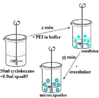
Figure 2-7 shows the schematic illustration of the emulsion method. First, 50 mL of cyclohexane as organic phase containing 0.9 ml sorbitan trioleate (Span 85) was stirred at a certain rotation speed for 2-3 min, and then 10.5 ml of aqueous phase containing pH 8.0 McIlvaine buffer (0.2 M Na2HPO4 and 0.1 M citric acid),
enzyme (from 1 mg to 50 mg) and 1 ml of poly(ethyleneimine) with a molecular weight of 1,300 Da was added to form water-in-oil microdroplets. After 15 min of agitation, the emulsion was stabilized, droplets remained a constant size after this period. Next 0.5 ml sebacoyl chloride (C10H16Cl2O2) as a crosslinker in 10 ml
cyclohexane was added to react with poly(ethyleneimine) at the interface of all droplets simultaneously and the mixture was agitated for 5 min to mature the membranes. The reaction was stopped by diluting with 50 ml cyclohexane. Supernatant was discarded. Microcapsules were rinsed with 50 ml of cyclohexane to remove any remaining organic residues like hydrolyzed sebacoyl chloride. The microcapsules were then transferred to a Buchner filter funnel and rinsed with Milli-Q water to remove water soluble residues. Small molecules such as hydrolyzed sebacoyl chloride, hydrochloric acid and unreacted poly(ethyleneimine) on the surfaces and inside the microcapsule cores were rinsed off.
Figure 2- Schematic illustration of the emulsion method.
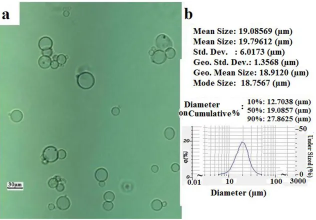
This method can prepare small microcapsules ranging from a few microns to 50 µm. The properties of produced microcapsules are discussed in Chapter 4. As agitation influences mass transfer in a reactor, this mass transfer can be considered as a crucial parameter for determining the diameter of microcapsules. In general, the diameter of produced microcapsules decreased with increased agitation speed. Microcapsules prepared by the emulsion method have a relatively small size yet a larger size distribution than those prepared by the vibration nozzle method (encapsulator). The size and size distribution of microcapsules prepared by the emulsion method is shown in Figure 2-8.
Figure 2- Optical micrograph of microcapsules prepared by the emulsion method with pitched blade turbine (a); size distribution of microcapsules obtained from particle size analyzer (b).
2.5 Encapsulator microencapsulation method
The vibration nozzle (or encapsulator) method of microencapsulation is based on the laminar jet break-up technique. The Inotech encapsulator has been used for cell immobilization with different types of alginates. We reported for the first time that PEI microcapsules could likewise be prepared by this encapsulator.
Kondo’s [19] US Patent 2 379 817 was issued in 1945 as the initial nozzle-based microencapsulation technology. This program has led to the development of a two-fluid gravity-flow apparatus producing relatively large capsules. Immobilization of living cells or biocatalysts in hydrogels is a well-established technique in a broad, increasing range of different applications. Sodium alginate is the most used polysaccharide as a polymer matrix due to its good biocompatibility and its easy gelling reaction [20-23]. Our work is the first to demonstrate the ability to permanently encapsulate enzymes in PEI microcapsules with an encapsulator. No microencapsulation technique hitherto had permitted the simultaneous production of beads with a narrow size distribution, a high production rate, a satisfactory level of material utilization under mild and nontoxic conditions, under completely sterile conditions and with the ability to scale up [21].
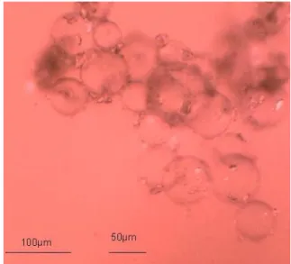
The vibration nozzle technique can be used to produce monodisperse beads of >200 µm in diameter, depending on how the application is scaled up. The break-up of laminar jets is well investigated, but most of the analysis end at the point where the diameter of the neck decreases under a certain value, since mean diameter of microcapsules is about double of nozzle diameter. We tried to break the neck and replaced it with a homemade nozzle with a diameter of 10 µm to produce smaller microcapsules. However, the produced microcapsules were not as small as expected and were lightly aggregated.
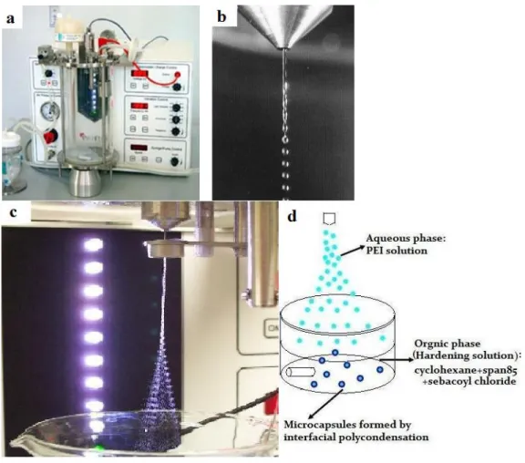
This method was performed with a commercial encapsulation device (Inotech IE-50R encapsulator, Figure 2-9 a). The encapsulator was equipped with a 100 µm
nozzle (Figure 2-9 c). Figure 2-9 b shows beads generated from a 500 µm nozzle. Microdroplets were produced by pumping a 10 mL solution containing pH 8.0 McIlvaine buffer and 5% (v/v) PEI with MW of 750 kDa (PEI 750) through the nozzle while applying vibrations at a given frequency. Formed microdroplets fell into a hardening solution containing 50 mL of cyclohexane, 0.7 mL of span 85 and 0.4 mL of sebacoyl chloride, which was agitated by a magnetic stir bar. The scheme of the vibration nozzle method is shown in Figure 2-9 d.
The homemade nozzle is a glass needle with a nozzle diameter of 10 µm. In general, the bead diameter of a Newtonian liquid is about twice the diameter of the nozzle. However capsules we obtained from the nozzle of 10 µm were much bigger than 20 µm and large microcapsules were attached to several small microcapsules (Figure 2-10). As shown in Figure 2-10, microcapsules aggregated and had a large size distribution.
2.6 Scheme of enzyme microcapsules
The fabrication process for enzyme microcapsules is similar to that of empty microcapsules. The only difference is the enzyme-containing aqueous phase with PEI. Given that enzyme molecules contain primary amine groups that can react with the crosslinker, enzymes after the microencapsulation process can be found embedded both in the membranes and within the core (Figure 2-11). A low molecular weight of PEI (1.3 kDa) was used for the emulsion method, yet only a high molecular weight of PEI (750 kDa) can produce strong, intact and spherical
microcapsules using the vibration nozzle method. According to the pore size measurement described in Chapter 4, the membranes of microcapsules were semi-permeable and allowed diffusion of small molecules such as PEI with a molecular weight of 1.3 kDa (PEI1.3), but not large molecules such as laccase (67 kDa) and PEI with a molecular weight of 750 kDa (PEI 750). Microcapsules prepared by the emulsion method only contained enzymes and water molecules after rinsing with Milli-Q water since PEI 1.3 molecules were washed off (Figure 2-11 Emulsion method). However, large microcapsules prepared with high molecular weight PEI by the vibration nozzle method contained PEI 750 as well as enzyme and water molecules; only the very small molecules such as hydrochloric acid and hydrolyzed sebacoyl chloride could pass through and be rinsed away (Figure 2-11 Vibration nozzle method).
PEI interacts with laccase, and its presence in the core phase decreased the enzymatic activity of laccase, which is discussed in Chapter 4. The toxicity of PEI increased with increased molecular weight [24, 25], suggesting that PEI 750 is more toxic than PEI 1.3. In addition, the yield of the emulsion method is about twice that of the vibration nozzle method. In comparison, the emulsion was deemed more suitable as an enzyme encapsulation method for our biosensor platform. Given that the size of microcapsules influences the immobilization quality, microcapsules prepared by the vibration nozzle method were too big to be immobilized into paper pulp.
Figure 2- Scheme of the encapsulator and vibration nozzle method. Inotech IE-50R encapsulator (a); Beads generated from 500 µm nozzle (b); Microdroplets generated from 100 µm nozzle at optimal condition (c); Scheme of PEI microcapsules formation by vibration nozzle method (d).
Figure 2- Optical micrograph of microcapsules prepared by an encapsulator equipped with a homemade nozzle with a diameter of 10 µm.
2.7 Preparation of paper strips
In the universal paper-making method, a dilute suspension of wood pulp in water is drained through a screen and the interwoven pulp is laid down on the screen. Suspended water is removed from the sheet by pressing and drying in order to make paper. The paper handsheets were prepared in our lab using simple equipments. Hard wood paper pulp from Paprican was used as substrate for the preparation of handsheets, including empty paper (without microcapsules, as negative control), enzyme paper (containing enzyme microcapsules), BV paper (used in BV test, containing microcapsules soaked in BCIN and NBT solution) and microcapsule paper (containing empty microcapsules).
Paper pulp was suspended in water or buffer depending on the experiment, and then agitated to form a homogeneous suspension before transferring it to a Buchner filtration funnel for quick drainage. Similarly, empty microcapsules or enzyme microcapsules suspension in water or buffer were mixed with a homogeneous pulp suspension, gently agitated, and then transferred to a Buchner filtration funnel for drainage. Wet paper was dried by purging with N2. Dried paper handsheets were cut
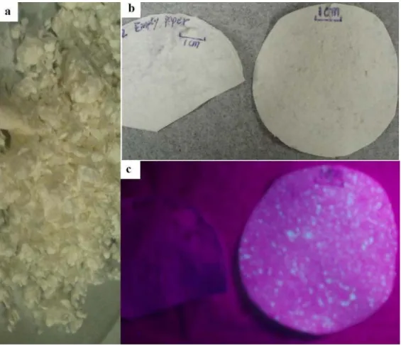
and kept in separated tubes, which were later used for the colorimetric test and storage stability test. These paper strips are only for single use and can facilitate detection purposes by signaling a color change. Figure 2-12 shows hard wood paper pulp before paper-making (a) and papers with or without microcapsules labeled by fluorescamine (b, c), in which microcapsules were labeled by fluorescamine;
emitting green color under ultraviolet excitation (Figure 2-12 c right hand paper). This paper contained microcapsule aggregates, suggesting that capsules were not uniformly dispersed in paper pulp before filtration. Generally speaking, with this technique it is possible to achieve a better dispersion yet not to obtain monodispersed microcapsules. Thus this kind of colorimetric paper strip generates uneven intensity, which in practice improves visual ability.
Figure 2- Dry bleached hard wood pulp from Paprican (a) and papers with microcapsules labeled by fluorescamine (right side in b and c) and without microcapsules (left in b and c).
2.8 References
1. M.L. Hitchman, Measurement of Dissolved Oxygen1978, London: Wiley-interscience.
2. T.W. Rejdaa, J.D.N. Bunschoten and G.A.J. Kalis, Dissolved oxygen measurement with an improved amperometric cell Analytica Chimica Acta, 1986. 190: p. 275-279.
3. Y.H. Lee and G.T. Tsao, Advances in Biochemical Engineering. Dissolved oxygen electrodes. Vol. 13. 1979, New York: Springer-Verlag.
4. Retrived from http://www.rankbrothers.co.uk/prod1.htm on Febrary 8th 2012.
5. J. Pawley, Handbook of Biological Confocal Microscopy. 3rd ed2006, New York: Plenum Press.
6. M. Müller, Introduction to confocal fluorescence microscopy. 2nd ed2005, Bellingham, Washington: SPIE press. 115.
7. N.S. Claxton, T.J. Fellers and M.W. Davidson, Microscopy, Confocal, in Encyclopedia of Medical Devices and Instrumentation2006, John Wiley & Sons, Inc.
8. M. Minsky, Microscopy apparatus 1961: US patent 3013467 (A).
9. W.B. Amos and J.G. White, How the Confocal Laser Scanning Microscope entered Biological Research. Biology of the Cell, 2003. 95(6): p. 335-342.
10. G.J. Brakenhoff, P. Blom and P. Barends, Confocal Scanning Light Microscopy with High Aperture Immersion Lenses. Journal of microscopy, 1979. 117: p. 219-232.
11. G.J. Brakenhoff, H.T.M. van der Voort, E.A. van Spronsen, W.A.M. Linnemans and N. Nanninga, Three-dimensional chromatin distribution in neuroblastoma nuclei shown by confocal scanning laser microscopy. Nature, 1985. 317(6039): p. 748-749.
12. C.J.R. Sheppard and T. Wilson, Effect of spherical aberration on the imaging properties of scanning optical microscopes. Applied Optics, 1979. 18(7): p. 1058-1063.
13. D.K. Hamilton and T. Wilson, Scanning optical microscopy by objective lens scanning Journal of Physics E: Scientific Instruments, 1986. 19(1): p. 52-54.
14. R. Bhavane, Nanoparticle Agglomerates for Pulmonary Drug Delivery, in Health science center at Houston2006, University of Texas. p. 36.
15. U. Ziegler, A.G. Bittermann and M. Hoechli. Introduction to Confocal Laser
Scanning Microscopy (LEICA). Retrived from
http://www.zmb.uzh.ch/resources/download/CLSM.pdf, on Febrary 8th 2012.
16. I. Treviranus. Laser diffraction theory. User training course for LA-950 Horiba 2010; Available from: www.horiba.com/us/particle
17. D. Poncelet, T. Alexakis, d.S.B. Poncelet and R. Neufeld, Microencapsulation within crosslinked polyethyleneimine membranes. Journal of Microencapsulation, 1994. 11(1): p. 31-40.
18. S. Benita, ed. Microencapsulation. Methods and Industrial Applications. Drugs and the Pharmaceutical Sciences, ed. J. Swarbrick. Vol. 73. 1996, Marcel Dekker Inc.: New Tork. 640.
19. A. Kondo, History and classification of microencapsulation, in Microcapsules processing and technology, J.W.V. Valkenburg, Editor 1979, Marcel Dekker: New York.
20. H. Brandenberger, D. Nussli, V. Piech and F. Widmer, Monodisperse particle production: A method to prevent drop coalescence using electrostatic forces. Journal of Electrostatics, 1999. 45(3): p. 227-238.
21. D. Serp, E. Cantana, C. Heinzen, U.V. Stockar and I.W. Marison, Characterization of an encapsulation device for the production of monodisperse alginate beads for cell immobilization. Biotechnology and Bioengineering, 2000. 70(1): p. 41-53. 22. M. Homar, Scaron, D. uligoj, scaron, Ga, scaron and M. perlin, Preparation of
microcapsules with self-microemulsifying core by a vibrating nozzle method. Journal of Microencapsulation, 2007. 24(1): p. 72 - 81.
23. A.W. Martinez, S.T. Phillips, E. Carrilho, S.W. Thomas, H. Sindi and G.M. Whitesides, Simple telemedicine for developing regions: Camera phones and paper-based microfluidic devices for real-time, off-site diagnosis. Analytical Chemistry, 2008. 80(10): p. 3699-3707.
24. D. Fischer, T. Bieber, Y. Li, H.-P. Elsässer and T. Kissel, A Novel Non-Viral Vector for DNA Delivery Based on Low Molecular Weight, Branched Polyethylenimine:
Effect of Molecular Weight on Transfection Efficiency and Cytotoxicity. Pharmaceutical Research, 1999. 16(8): p. 1273-1279.
25. D. Fischer, Y. Li, B. Ahlemeyer, J. Krieglstein and T. Kissel, In vitro cytotoxicity testing of polycations: influence of polymer structure on cell viability and hemolysis. Biomaterials, 2003. 24(7): p. 1121-1131.
![Figure 2- Idealized laser diffraction setup (angle of scatter VS. particle size) [16]](https://thumb-eu.123doks.com/thumbv2/123doknet/7658125.238240/47.918.251.777.145.342/figure-idealized-laser-diffraction-setup-angle-scatter-particle.webp)