SYLVAIN CHAUVETTE
SLOW-WAVE SLEEP: GENERATION AND
PROPAGATION OF SLOW WAVES, ROLE IN
LONG-TERM PLASTICITY AND GATING
Thèse présentée
à la Faculté des études supérieures et postdoctorales de l’Université Laval dans le cadre du programme de doctorat en Neurobiologie
pour l’obtention du grade de Philosophiae Doctor (Ph.D.)
DÉPARTEMENT DE PSYCHIATRIE ET DE NEUROSCIENCES FACULTÉ DE MÉDECINE
UNIVERSITÉ LAVAL QUÉBEC
2013
Résumé
Le sommeil est connu pour réguler plusieurs fonctions importantes pour le cerveau et parmi celles-ci, il y a le blocage de l’information sensorielle par le thalamus et l’amélioration de la consolidation de la mémoire. Le sommeil à ondes lentes, en particulier, est considéré être critique pour ces deux processus. Cependant, leurs mécanismes physiologiques sont inconnus. Aussi, la marque électrophysiologique distinctive du sommeil à ondes lentes est la présence d’ondes lentes de grande amplitude dans le potentiel de champ cortical et l’alternance entre des périodes d’activités synaptiques intenses pendant lesquelles les neurones corticaux sont dépolarisés et déchargent plusieurs potentiels d’action et des périodes silencieuses pendant lesquelles aucune décharge ne survient, les neurones corticaux sont hyperpolarisés et très peu d’activités synaptiques sont observées. Tout d'abord, afin de mieux comprendre les études présentées dans ce manuscrit, une introduction générale couvrant l'architecture du système thalamocortical et ses fonctions est présentée. Celle-ci comprend une description des états de vigilance, suivie d'une description des rythmes présents dans le système thalamocortical au cours du sommeil à ondes lentes, puis par une description des différents mécanismes de plasticité synaptique, et enfin, deux hypothèses sur la façon dont le sommeil peut affecter la consolidation de la mémoire sont présentées.
Puis, trois études sont présentées et ont été conçues pour caractériser les propriétés de l'oscillation lente du sommeil à ondes lentes. Dans la première étude (chapitre II), nous avons montré que les périodes d'activité (et de silence) se produisent de façon presque synchrone dans des neurones qui ont jusqu'à 12 mm de distance. Nous avons montré que l'activité était initiée en un point focal et se propageait rapidement à des sites corticaux voisins. Étonnamment, le déclenchement des états silencieux était encore plus synchronisé que le déclenchement des états actifs.
L'hypothèse de travail pour la deuxième étude (chapitre III) était que les états actifs sont générés par une sommation de relâches spontanées de médiateurs. Utilisant différents enregistrements à la fois chez des animaux anesthésiés et chez d’autres non-anesthésiés, nous avons montré qu’aucune décharge neuronale ne se produit dans le néocortex pendant les états silencieux du sommeil à ondes lentes, mais certaines activités synaptiques peuvent
être observées avant le début des états actifs, ce qui était en accord avec notre hypothèse. Nous avons également montré que les neurones de la couche V étaient les premiers à entrer dans l’état actif pour la majorité des cycles, mais ce serait ainsi uniquement pour des raisons probabilistes; ces cellules étant équipées du plus grand nombre de contacts synaptiques parmi les neurones corticaux. Nous avons également montré que le sommeil à ondes lentes et l’anesthésie à la kétamine-xylazine présentent de nombreuses similitudes.
Ayant utilisé une combinaison d'enregistrements chez des animaux anesthésiés à la kétamine-xylazine et chez des animaux non-anesthésiés, et parce que l'anesthésie à la kétamine-xylazine est largement utilisée comme un modèle de sommeil à ondes lentes, nous avons effectué des mesures quantitatives des différences entre les deux groupes d'enregistrements (chapitre IV). Nous avons trouvé que l'oscillation lente était beaucoup plus rythmique sous anesthésie et elle était aussi plus cohérente entre des sites d’enregistrements distants en comparaison aux enregistrements de sommeil naturel. Sous anesthésie, les ondes lentes avaient également une amplitude plus grande et une durée plus longue par rapport au sommeil à ondes lentes. Toutefois, les ondes fuseaux (spindles) et gamma étaient également affectées par l'anesthésie.
Dans l'étude suivante (Chapitre V), nous avons investigué le rôle du sommeil à ondes lentes dans la formation de la plasticité à long terme dans le système thalamocortical. À l’aide de stimulations pré-thalamiques de la voie somatosensorielle ascendante (fibres du lemnisque médial) chez des animaux non-anesthésiés, nous avons montré que le potentiel évoqué enregistré dans le cortex somatosensoriel était augmenté dans une période d’éveil suivant un épisode de sommeil à ondes lentes par rapport à l’épisode d’éveil précédent et cette augmentation était de longue durée. Nous avons également montré que le sommeil paradoxal ne jouait pas un rôle important dans cette augmentation d'amplitude des réponses évoquées. À l’aide d'enregistrements in vitro en mode cellule-entière, nous avons caractérisé le mécanisme derrière cette augmentation et ce mécanisme est compatible avec la forme classique de potentiation à long terme, car il nécessitait une activation à la fois les récepteurs NMDA et des récepteurs AMPA, ainsi que la présence de calcium dans le neurone post-synaptique.
La dernière étude incluse dans cette thèse (chapitre VI) a été conçue pour caractériser un possible mécanisme physiologique de blocage sensoriel thalamique survenant pendant le sommeil. Les ondes fuseaux sont caractérisées par la présence de potentiels d’action calcique à seuil bas et le calcium joue un rôle essentiel dans la transmission synaptique. En utilisant plusieurs techniques expérimentales, nous avons vérifié l'hypothèse que ces potentiels d’action calciques pourraient causer un appauvrissement local de calcium dans l'espace extracellulaire ce qui affecterait la transmission synaptique. Nous avons montré que les canaux calciques responsables des potentiels d’action calciques étaient localisés aux synapses et que, de fait, une diminution locale de la concentration extracellulaire de calcium se produit au cours d’un potentiel d’action calcique à seuil bas spontané ou provoqué, ce qui était suffisant pour nuire à la transmission synaptique.
Nous concluons que l'oscillation lente est initiée en un point focal et se propage ensuite aux aires corticales voisines de façon presque synchrone, même pour des cellules séparées par jusqu'à 12 mm de distance. Les états actifs de cette oscillation proviennent d’une sommation de relâches spontanées de neuromédiateurs (indépendantes des potentiels d’action) et cette sommation peut survenir dans tous neurones corticaux. Cependant, l’état actif est généré plus souvent dans les neurones pyramidaux de couche V simplement pour des raisons probabilistes. Les deux types d’expériences (kétamine-xylazine et sommeil à ondes lentes) ont montré plusieurs propriétés similaires, mais aussi quelques différences quantitatives. Nous concluons également que l'oscillation lente joue un rôle essentiel dans l'induction de plasticité à long terme qui contribue très probablement à la consolidation de la mémoire. Les ondes fuseaux, un autre type d’ondes présentes pendant le sommeil à ondes lentes, contribuent au blocage thalamique de l'information sensorielle.
Abstract
Sleep is known to mediate several major functions in the brain and among them are the gating of sensory information during sleep and the sleep-related improvement in memory consolidation. Slow-wave sleep in particular is thought to be critical for both of these processes. However, their physiological mechanisms are unknown. Also, the electrophysiological hallmark of slow-wave sleep is the presence of large amplitude slow waves in the cortical local field potential and the alternation of periods of intense synaptic activity in which cortical neurons are depolarized and fire action potentials and periods of silence in which no firing occurs, cortical neurons are hyperpolarized, and very little synaptic activities are observed. First, in order to better understand the studies presented in this manuscript, a general introduction covering the thalamocortical system architecture and function is presented, which includes a description of the states of vigilance, followed by a description of the rhythms present in the thalamocortical system during slow-wave sleep, then by a description of the mechanisms of synaptic plasticity, and finally two hypotheses about how sleep might affect the consolidation of memory are presented.
Then, three studies are presented and were designed to characterize the properties of the sleep slow oscillation. In the first study (Chapter II), we showed that periods of activity (and silence) occur almost synchronously in neurons that are separated by up to 12 mm. The activity was initiated in a focal point and rapidly propagated to neighboring sites. Surprisingly, the onsets of silent states were even more synchronous than onsets of active states.
The working hypothesis for the second study (Chapter III) was that active states are generated by a summation of spontaneous mediator releases. Using different recordings in both anesthetized and non-anesthetized animals, we showed that no neuronal firing occurs in the neocortex during silent states of slow-wave sleep but some synaptic activities might be observed prior to the onset of active states, which was in agreement with our hypothesis. We also showed that layer V neurons were leading the onset of active states in most of the cycles but this would be due to probabilistic reasons; these cells being equipped with the most numerous synaptic contacts among cortical neurons. We also showed that slow-wave sleep and ketamine-xylazine shares many similarities.
Having used a combination of recordings in ketamine-xylazine anesthetized and non-anesthetized animals, and because ketamine-xylazine anesthesia is extensively used as a model of slow-wave sleep, we made quantitative measurements of the differences between the two groups of recordings (Chapter IV). We found that the slow oscillation was much more rhythmic under anesthesia and it was also more coherent between distant sites as compared to recordings during slow-wave sleep. Under anesthesia, slow waves were also of larger amplitude and had a longer duration as compared to slow-wave sleep. However, spindles and gamma were also affected by the anesthesia.
In the following study (Chapter V), we investigated the role of slow-wave sleep in the formation of long-term plasticity in the thalamocortical system. Using pre-thalamic stimulations of the ascending somatosensory pathway (medial lemniscus fibers) in non-anesthetized animals, we showed that evoked potential recorded in the somatosensory cortex were enhanced in a wake period following a slow-wave sleep episode as compared to the previous wake episode and this enhancement was long-lasting. We also showed that rapid eye movement sleep did not play a significant role in this enhancement of response amplitude. Using whole-cell recordings in vitro, we characterized the mechanism behind this enhancement and it was compatible with the classical form of long-term potentiation, because it required an activation of both NMDA and AMPA receptors as well as the presence of calcium in the postsynaptic neuron.
The last study included in this thesis (Chapter VI) was designed to characterise a possible physiological mechanism of thalamic sensory gating occurring during sleep. Spindles are characterized by the presence of low-threshold calcium spikes and calcium plays a critical role in the synaptic transmission. Using several experimental techniques, we verified the hypothesis that these calcium spikes would cause a local depletion of calcium in the extracellular space which would impair synaptic transmission. We showed that calcium channels responsible for calcium spikes were co-localized with synapses and that indeed, local extracellular calcium depletion occurred during spontaneous or induced low-threshold calcium spike, which was sufficient to impair synaptic transmission.
We conclude that slow oscillation originate at a focal point and then propagate to neighboring cortical areas being almost synchronous even in cells located up to 12 mm
apart. Active states of this oscillation originate from a summation of spike-independent mediator releases that might occur in any cortical neurons, but happens more often in layer V pyramidal neurons simply due to probabilistic reasons. Both experiments in ketamine-xylazine anesthesia and non-anesthetized animals showed several similar properties, but also some quantitative differences. We also conclude that slow oscillation plays a critical role in the induction of long-term plasticity, which very likely contributes to memory consolidation. Spindles, another oscillation present in slow-wave sleep, contribute to the thalamic gating of information.
Foreword
The following thesis is presented as a collection of scientific articles published, submitted for publication, or in final steps prior to submission. The first chapter is composed of a general introduction describing the theoretical concepts and experimental strategies that were used in the presented studies. Then the core of this thesis is represented by five scientific articles and the first three articles focus on the slow oscillation found either during natural slow-wave sleep or under ketamine-xylazine anesthesia. We describe the mechanisms of generation and propagation of this type of oscillation. Out of the two other studies composing the core of this thesis, one is about the synaptic plasticity induced by the slow oscillation of slow-wave sleep, and the last article presents a mechanism of thalamic gating during another type of sleep oscillation, namely the spindle, which is also present during slow-wave sleep. A brief review of the main results and a general discussion finalizes this thesis. The bibliography used for both the general introduction and the general discussion is presented at the very end of the thesis, while the bibliography for each articles (chapter II-VI) is presented at the end of the text of the corresponding paper, with a formatting of the reference list in accordance to the rules of the journal where the paper was published or submitted.
I would like to take the opportunity to express my gratitude to my thesis supervisor, Prof. Igor Timofeev, who, about ten years ago, trusted me enough to give me the opportunity to work in his lab as an undergraduate student although my marks were rather low at that time. Since then, a fruitful collaboration developed and our productivity gave me the opportunity to get fellowships from different organisms during both my master [Centre de Recherche en Neuroscience (CRN)] and my PhD [Savoy Foundation (refused) and Canadian Institutes of Health Research (CIHR)], and to publish several studies. It also gave me the opportunity to receive four excellence prices for different poster presentations (2003, 2004, 2007, and 2012) and a travel award for the Federation of European Neuroscience Societies (FENS) forum held in Amsterdam (2010). Without his critics, discussions, and support, these studies would not have been possible.
I would also like to thank Prof. Maxim Volgushev, Prof. Maxim Bazhenov, Josée Seigneur, Sofiane Boucetta, Sylvain Crochet, Mikhail Mukovski, Francis Lajeunesse, Alex
Ferescko, Kriszta Kovacs, Laszlo Grand, Courtney Pinard, and Maxime Lemieux for their great collaboration that resulted in several completed studies that are already published or will be soon submitted for publication. I would like to thank Pierre Giguère (now retired) and Sergiu Ftomov for their excellent technical support. I would also like to thank all my colleagues during these almost ten years in this lab with whom, even if we did not have official scientific collaboration per se, we had several fruitful and nice discussions that really help in improving my studies.
Following, is the chronological list of scientific publications (published or submitted) in which I have contributed during the progress of my postgraduate studies:
1. Crochet S, Chauvette S, Boucetta S, Timofeev I (2005) Modulation of synaptic transmission in neocortex by network activities. The European journal of neuroscience 21:1030-1044.
2. Volgushev M, Chauvette S, Mukovski M, Timofeev I (2006) Precise long-range synchronization of activity and silence in neocortical neurons during slow-wave oscillations [corrected]. J Neurosci 26:5665-5672.
3. Mukovski M, Chauvette S, Timofeev I, Volgushev M (2007) Detection of active and silent states in neocortical neurons from the field potential signal during slow-wave sleep. Cereb Cortex 17:400-414.
4. Chauvette S, Volgushev M, Mukovski M, Timofeev I (2007) Local origin and long-range synchrony of active state in neocortex during slow oscillation. In: Mechanisms of spontaneous active states in the neocortex (Timofeev I, ed), pp 73-92. Kerala, India: Research Signpost.
5. Volgushev M, Mukovski M, Chauvette S, Timofeev I (2007) Detection of active and silent states in neocortical networks. In: Mechanisms of spontaneous active states in the neocortex (Timofeev I, ed), pp 93-122. Kerala, India: Research Signpost.
6. Boucetta S, Chauvette S, Bazhenov M, Timofeev I (2008) Focal generation of paroxysmal fast runs during electrographic seizures. Epilepsia 49:1925-1940.
7. Chauvette S, Volgushev M, Timofeev I (2010) Origin of Active States in Local Neocortical Networks during Slow Sleep Oscillation. Cerebral Cortex 20:2660-2674.
8. Chauvette S, Crochet S, Volgushev M, Timofeev I (2011) Properties of Slow Oscillation during Slow-Wave Sleep and Anesthesia in Cats. The Journal of Neuroscience 31:14998-15008.
9. Timofeev I, Chauvette S (2011) Thalamocortical Oscillations: Local Control of EEG Slow Waves. Current Topics in Medicinal Chemistry 11:2457-2471.
10. Volgushev M, Chauvette S, Timofeev I (2011) Long-range correlation of the membrane potential in neocortical neurons during slow oscillation. In: Progress in Brain Research (Van Someren EJW, Van Der Werf YD, Roelfsema PR, Mansvelder HD, Lopes Da Silva FH, eds), pp 181-199: Elsevier.
11. Chen J-Y, Chauvette S, Skorheim S, Timofeev I, Bazhenov M (2012) Interneuron-mediated inhibition synchronizes neuronal activity during slow oscillation. The Journal of physiology 590:3987-4010.
12. Chauvette, S, Seigneur J, Timofeev I (2012) Sleep Oscillations in the Thalamocortical System Induce Long-Term Neuronal Plasticity. Neuron 75(6): 1105-1113.
13. Boucetta S, Crochet S, Chauvette S, Seigneur J, Timofeev I (in press) Extracellular Ca2+ fluctuations in vivo affect afterhyperpolarization potential and modify firing patterns of neocortical neurons. Experimental Neurology
14. Ferecsko AS, Seigneur J, Chauvette S, Kovacs K, Lajeunesse F, Sik A, Timofeev I (in preparation for submission) Low-threshold calcium spike dependent gating in thalamus.
Cette thèse est dédiée à mon père, Marcel Chauvette, qui a toujours cru en moi et m’a toujours soutenu dans les moments plus difficiles et à ma sœur, Chantal Chauvette, malheureusement décédée beaucoup trop jeune, mais qui malgré tout, demeure encore aujourd’hui une source quotidienne de courage et d’inspiration.
Table of Contents
Résumé ... i Abstract ... iv Foreword ... vii List of figures ... 14 List of abbreviations... 17 Chapter I ... 18 1.0 General introduction ... 191.1 Architecture of thalamocortical system ... 19
1.1.1 Thalamus ... 19
1.1.2 Thalamic reticular nucleus ... 21
1.1.3 Neocortex ... 22
1.1.3.1 Pyramidal neurons ... 22
1.1.3.2 Non-pyramidal neurons, interneurons ... 24
1.1.3.3 Electrophysiological types of cortical neurons ... 24
1.1.3.4 Correlation between electrophysiology and morphology ... 25
1.1.3.5 Laminar and columnar organisation of cortex... 29
1.1.3.6 Synaptic connections in neocortex ... 30
1.2 States of vigilances ... 32
1.2.1 Wake... 32
1.2.2 Rapid eye movement sleep ... 33
1.2.3 Slow-wave sleep ... 34
1.2.4 Thalamocortical oscillations during slow-wave sleep ... 35
1.2.4.1 Infra-slow oscillation ... 35
1.2.4.2 Slow oscillation ... 35
1.2.4.3 Delta oscillation ... 38
1.2.4.4 Spindle oscillation ... 39
1.2.4.5 Beta-Gamma activities ... 41
1.2.4.6 Ripples (very fast oscillations, >100 Hz) ... 43
1.3 Synaptic plasticity ... 44
1.3.1 Short-term synaptic plasticity ... 44
1.3.1.1 Short-term depression ... 45
1.3.1.2 Short-term facilitation ... 46
1.3.1.2 Differences between in vivo and in vitro studies ... 47
1.3.2 Mid-term plasticity ... 47
1.3.3 Long-term synaptic plasticity ... 48
1.3.3.1 Long-term depression ... 48
1.3.3.2 Long-term potentiation ... 49
1.3.4 Augmenting responses, a form of plasticity ... 49
1.4 Plasticity in different states of vigilance, learning, and memory ... 50
1.4.1 Sleep and synaptic homeostasis hypothesis ... 51
1.4.2 Memory consolidation during slow-wave sleep hypothesis ... 54
1.5 General objectives of the thesis and experimental approaches ... 58
2.0 Precise long-range synchronization of activity and silence in neocortical neurons during
slow-wave oscillation... 60
2.1 Résumé en français ... 61
2.2 Abstract... 62
2.3 Introduction ... 63
2.4 Materials and Methods ... 65
2.5 Results ... 68 2.6 Discussion ... 74 2.7 Acknowledgements... 77 2.8 References ... 78 2.9 Figures ... 82 Chapter III ... 89
3.0 Origin of active states in local neocortical networks during slow sleep oscillation. ... 90
3.1 Résumé en français ... 91
3.2 Abstract... 92
3.3 Introduction ... 93
3.4 Material and methods ... 95
3.5 Results ... 98
3.5.1 Depth profile of field potentials, current sinks and sources during natural slow-wave sleep... 98
3.5.2 Depth distribution of firing at the onset of active state in simultaneously recorded multiunit activity during slow-wave sleep ... 100
3.5.3 Activity onset in simultaneously recorded nearby neurons during slow oscillation and slow-wave sleep ... 101
3.5.4 Synaptic buildup at the onset of active states ... 104
3.6 Discussion ... 106
3.6.1 Cortical origin of active states during slow sleep oscillation. ... 107
3.6.2 Origin of activity: What drives the first neuron to generate the first spike? ... 107
3.6.3 Origin of current sinks and sources during slow oscillation. ... 110
3.6.4 Conclusion: a scenario for active state onset. ... 111
3.7 Funding and acknowledgments... 112
3.8 References ... 113
3.9 Figures ... 119
3.10 Supplemental information ... 137
Chapter IV ... 142
4.0 Properties of slow oscillation during slow-wave sleep and anesthesia in cats. ... 143
4.1 Résumé en français: ... 144
4.2 Abstract... 145
4.3 Introduction ... 146
4.4 Material and methods ... 148
4.5 Results ... 152
4.5.1 Less power in the slow and spindle range, but more in the high frequency range during ketamine-xylazine anesthesia than in natural sleep. ... 153
4.5.2 Stronger rhythmicity of neuronal activity during anesthesia than in natural sleep. ... 154
4.5.3 Higher coherence of slow oscillation between different regions during ketamine-xylazine anesthesia than during SWS. ... 154
4.5.4 Silent states are more prominent during anesthesia than in SWS. ... 155
4.6 Discussion ... 157
4.6.1 Effects of anesthesia on cortical neurons ... 157
4.6.2 Anesthesia, sleep, consciousness ... 159
4.7 Acknowledgment ... 161
4.8 References ... 162
4.9 Figures ... 168
Chapter V ... 176
5.0 Sleep oscillations in the thalamocortical system induce long-term neuronal plasticity177 5.1 Résumé en Français ... 178
5.2 Abstract... 179
5.3 Introduction ... 180
5.4 Results ... 182
5.4.1 Evoked responses are potentiated after slow-wave sleep ... 182
5.4.2 REM sleep does not play a significant role in the enhancement of response .... 182
5.4.3 Intracellular responses during wake – slow-wave sleep – wake transitions ... 183
5.4.4 In vitro, only the full sleep-like pattern of stimulation replicates the in vivo results ... 184
5.4.5 Mechanisms of the enhancement of responses during the full sleep-like stimulation ... 185 5.5 Discussion ... 186 5.6 Experimental Procedures ... 189 5.7 References ... 194 5.8 Acknowledgements... 198 5.9 Figures ... 199 5.10 Supplemental Information ... 209 5.10.1 Supplemental figures ... 209 Chapter VI ... 212
6.0 Low-threshold calcium spike dependent gating in thalamus. ... 213
6.1 Résumé en français ... 214 6.2 Abstract... 215 6.3 Introduction ... 216 6.4 Results ... 217 6.5 Discussion ... 222 6.6 Acknowledgments ... 224 6.7 References ... 225 6.8 Figures ... 230
6.9 Online Methods and Supplementary Materials: ... 239
6.9.1 Material and methods: ... 239
6.9.2 Supplemental Figure ... 246
Chapter VII ... 247
7.0 General conclusion... 248
7.1 Summary of the results ... 248
7.2 Technical considerations ... 252
7.3 Final Remarks ... 252
List of figures
Chapter I
Figure I- 1 The thalamocortical system ... 23 Figure I- 2 Neuronal firing pattern could be modulated by the network activity. ... 27 Figure I- 3 Modulation of extracellular calcium concentration affects intrinsic
excitability and firing patterns. ... 28 Figure I- 4 Oscillations in thalamocortical system... 34
Chapter II
Figure II- 1 Active and silent states in 4 simultaneously recorded neurons and in the EEG. ... 82 Figure II- 2 Two methods of state detection. ... 84 Figure II- 3 Clusters of active and silent states in simultaneously recorded cells. ... 86 Figure II- 4 Population analysis of the onsets of active and silent states in
simultaneously recorded neurons. ... 88
Chapter III
Figure III- 1 Depth profile of the LFP during natural slow-wave sleep... 119 Figure III- 2 Alternating pattern of current sinks and sources during natural
slow-wave sleep revealed with current-source density analysis. ... 120 Figure III- 3 Depth distribution of neuronal firing during slow-wave sleep. ... 121 Figure III- 4 Cells from deep layers fire earlier than other cells at the onset of active
state. ... 123 Figure III- 5 Onsets of active states in local neuronal constellations. ... 125 Figure III- 6 Depth profile of activity onset in simultaneously recorded neurons:
Population analysis. ... 127 Figure III- 7 The high variability of active state onsets in cells during natural sleep.
... 129
Figure III- 8 Progressive build up vs. sharp transitions from silent to active states. . 130 Figure III- 9 Membrane potential fluctuations increase just before the onset of active
state. ... 131 Figure III- 10 Earlier involvement in activity is associated with slower transitions to
active states. ... 132 Figure III- 11 Intrinsically-bursting cells are leading the onset of active states. ... 134 Figure III- 12 Comparison of the depth profiles of field potential and intracellular
events during slow oscillation. ... 136 Figure III- S1 Sigmoid-fitting method for active state onset is robust and reliable. .. 137 Figure III- S2 Diverse sequential order of involvement of neighboring cells in active
states. ... 139 Figure III- S3 Difference in delays of active state onset and developed active states. 141
Chapter IV
Figure IV-1 Calculation of the amplitude of slow oscillation in the membrane
potential during transitions from silent to active states. ... 168 Figure IV-2 Fragments of continuous electrographic recordings during waking,
slow-wave sleep and ketamine-xylazine anesthesia. ... 169 Figure IV-3 Typical field potential and intracellular recordings from different cortical areas during natural slow-wave sleep and ketamine-xylazine anesthesia. ... 170 Figure IV-4 Spectral composition of local field potentials is different during slow-wave
sleep and anesthesia. ... 171 Figure IV-5 Rhythmicity of slow waves in local field potential and membrane
potential is higher during ketamine-xylazine anesthesia than in SWS. ... 172 Figure IV-6 Higher coherence of slow oscillation during ketamine-xylazine anesthesia than in slow-wave sleep. ... 173 Figure IV-7 Silent states are more prominent during anesthesia than in slow-wave
sleep (SWS). ... 174
Chapter V
Figure V- 1 Amplitude of evoked potential responses (N1) to medial lemniscus stimuli throughout sleep-wake periods. ... 199 Figure V- 2 Late REM sleep does not potentiate somatosensory evoked potential in a
following wake episode. ... 201 Figure V- 3 Intracellularly recorded evoked responses are enhanced after a period of
slow-wave sleep... 202 Figure V-4: Slow-wave sleep pattern of synaptic stimulation combined to intracellular hyperpolarization pulses induces long-term potentiation in vitro. ... 203 Figure V-5: Absence of long-term potentiation after wake pattern of stimulation in
vitro. ... 205 Figure V- 6 Properties of long-term plasticity induced by sleep pattern of stimulation.
... 207
Figure V-S1: Field potential evoked responses are enhanced after a period of slow-wave sleep. ... 209 Figure V-S2: Stimulating protocol to investigate effects of steady-state synaptic
Chapter VI
Figure VI-1 Strong reduction in cortical response amplitude to pre-thalamic stimuli during spindles. ... 230 Figure VI-2 Responses and failure rates in the VPL thalamic nucleus to electrical
stimulation of the medial lemniscus during spindles and interspindle lulls. ... 232 Figure VI-3 Increased failure rate following an LTS in thalamocortical (TC) neurons
of the VPL nucleus in vivo. ... 233 Figure VI-4 Evoked LTS induces an increase of synaptic failures from lemniscal
stimulations and Ca2+ depletion in the vicinity of neuron. ... 234 Figure VI-5 Specificity of LTS dependent increase in synaptic failure rates. ... 235 Figure VI-6 [Ca2+]o dynamics probed in a FEM model. ... 236
Figure VI-7 Electron microphotographs showing co-localization of Cav3.1 subunit in
dendrites of TC neurons and Cav3.3 subunit in dendrites of a reticular (RE)
neuron with synapses. ... 238 Figure VI-S1 Responses and failures of a reticular neuron to lemniscal stimuli in vivo.
... 246
Chapter VII
List of abbreviations
ACSF Artificial cerebro-spinal fluid
AMPA alpha-amino-3-hydroxy-5-methyl-4-isoxazolepropionic acid EEG Electroencephalogram
EMG Electromyogram EOG Electro-oculogram
EPSP Excitatory postsynaptic potential FRB Fast-rhythmic-bursting
FS Fast-Spiking
GABA Gamma-amino butyric acid IB Intrinsically-bursting
Ih Hyperpolarization-activated cation current
Ileak Potassium leak current
IPSP Inhibitory postsynaptic potential IT Low-threshold calcium current
LDT Laterodorsal tegmental nucleus LFP Local field potential
LTD Long-term depression LTP Long-term potentiation LTS Low-threshold calcium spike NMDA N-Methyl-D-aspartic acid
PPT pedunculopontine tegmental nucleus RE Thalamic reticular nucleus
REM Rapid eye movement
RS Regular-spiking
sAHP Short afterhyperpolarization SWS Slow-wave sleep
1.0 General introduction
This first chapter is a general introduction to the concepts and findings that will be described in the following chapters. First a description of the architecture of the thalamocortical system, which is composed of the thalamus, the thalamic reticular nucleus, and the neocortex, will be provided. Then the electrographic activities during the three states of vigilance (wake, rapid-eye-movement sleep, and slow-wave sleep) will be briefly described with a focus in describing the thalamocortical oscillations occurring during slow-wave sleep. Subsequently, a section will be dedicated at describing the different forms of synaptic plasticity, and finally, the last section will be presenting two hypotheses about the mechanisms of plasticity related to states of vigilances, their possible role in learning and memory.
1.1 Architecture of thalamocortical system
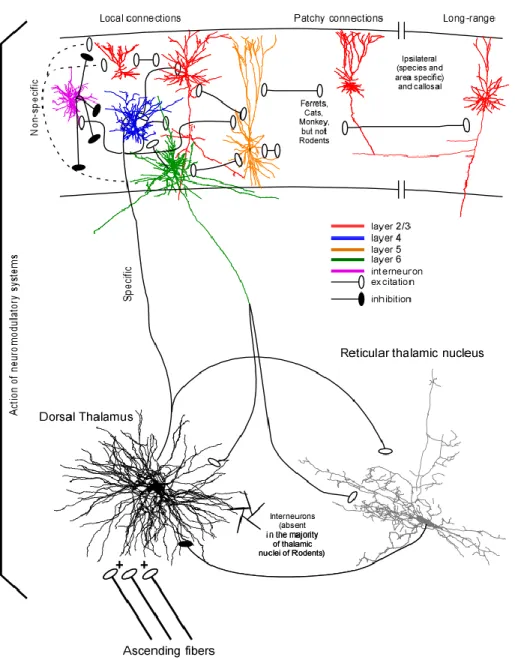
The thalamocortical (TC) system is the site of generation of many different types of oscillatory rhythms with distinct mechanisms. The TC network is organised in a loop (Fig. I.1) where three structures are implicated: the dorsal thalamus, the thalamic reticular nucleus (RE), and the neocortex. The complete organisation will be discussed in details in the following sections, but briefly, the thalamus receives information from ascending pathway and gives glutamatergic projections to both the neocortex and the thalamic reticular nucleus. The neocortex processes the information received and send glutamatergic axons to both TC and RE neurons, but with a greater impact on RE neurons. Then RE neurons send GABAergic projections to TC cells.
1.1.1 Thalamus
The thalamus of different mammals is composed of TC relay neurons, some (20-30 %) local inhibitory interneurons, and glial cells, however for rodents, interneurons would be present only in the lateral geniculate nucleus (visual thalamus). It is composed of specific and non-specific nuclei. The specific nuclei relay information from periphery and
project mainly to a given cortical region, while non-specific nuclei have widespread cortical projections.
The dorsal thalamus is composed of multiple specific nuclei and it receives information from ascending sensory pathway such as medial lemniscus for somatosensory information, optic tract for visual information, inferior colliculus for auditory information, brachium conjunctivum for motor information from cerebellum. The dorsal thalamus also receives information from the brainstem modulatory systems (cholinergic, norepinephrinergic, serotoninergic, etc.) (reviewed in (Steriade et al., 1997)). Typically, for thalamic relay neurons, ascending pathways (driving inputs) form large synapses with multiple release sites on proximal dendrites, while cortico-thalamic projections (modulator inputs) preferentially arrive to distal dendrites (Liu et al., 1995a; Sherman, 2005). The same logic applies for higher order thalamic nuclei, however with an inverse role where corticothalamic inputs from cortical layer V become the driving inputs that project on proximal dendrites and other inputs arriving mainly on distal dendrites (Sherman, 2005). An example of that is seen for layer Vb neurons of somatosensory (barrel) cortex that form giant synapses on proximal dendrites of thalamic neurons of the posteromedial nucleus (POm) (Groh et al., 2008). Two different chemically defined types of TC neurons have been described, which are the calbindin and parvalbumin containing TC neurons (reviewed in (Jones, 2001)). Parvalbumin-containing TC neurons project in a topographically ordered fashion to layer IV of the cortex and form the thalamic core projections, while calbindin-containing TC neurons have widespread projections to superficial cortical layers (mainly layer I) which are not limited by cortical areas boundaries; these projections represent the thalamic matrix projections (reviewed in (Jones, 2001)). While calbindin-positive TC neurons are found in all thalamic nuclei, parvalbumin-positive are found in much higher concentration in specific TC nuclei (Jones, 2001).
Thalamocortical neurons are not interconnected by chemical synapses, but one study suggests the presence of gap junctions (electrical synapses) between TC neurons in cats (Hughes et al., 2002). Electrophysiologically, TC neurons possess some characteristic features. They are equipped with low-threshold calcium current (IT) which needs
hyperpolarisation for a certain amount of time (80-150 ms) to deinactivate (Llinas and Jahnsen, 1982; Jahnsen and Llinas, 1984). TC neurons also possess a hyperpolarisation-activated depolarizing current (Ih) (McCormick and Pape, 1990). Those currents are
important for the generation of intrinsic delta rhythm (described in a following section). They are also important to characterise the two firing mode of TC neurons, which are tonic firing and bursting. When TC neurons are depolarised either by a current injection or by the presence of some neuromodulators (acetylcholine, noradrenaline, serotonin), they start to fire action potential, and the more these neurons are depolarised, the more they fire; this is the tonic mode (McCormick, 1992). However, when TC neurons are hyperpolarised sufficiently, either via intracellular current injection or by active inhibition, Ih is activated
and the neuron starts to depolarise to a point where IT is also activated. The activation of
this current leads to low-threshold calcium spike (LTS) and on top of this calcium spike, usually many sodium action potentials (up to 7) take place at high frequencies (up to 250 Hz). Thus TC neurons can transmit information following either excitation, where they fire in tonic mode or at the end of inhibition where they respond with a LTS in the bursting mode.
1.1.2 Thalamic reticular nucleus
The thalamic reticular nucleus is composed exclusively of GABAergic neurons (Houser et al., 1980; Oertel et al., 1983), all of them projecting to the correspondent thalamic nucleus after giving one or two collaterals (Scheibel and Scheibel, 1966; Yen et al., 1985; Liu et al., 1995b). These neurons are also equipped with IT and Ih, however Ih is
much weaker in RE neurons than in TC cells (Blethyn et al., 2006). As for TC cells, RE neurons fire in a tonic mode when depolarised and in a bursting mode when hyperpolarised. However they can generate more action potential during a LTS (up to 40) and with much higher frequency (up to 500 Hz) than TC neurons can do. RE neurons are interconnected with gap junction (Landisman et al., 2002; Fuentealba et al., 2004; Blethyn et al., 2008). The sources of afferents to the RE thalamic nucleus are the collaterals of TC and corticothalamic fibers (Fig. I.1), all of which pass through the RE nucleus (Jones, 1985). Both of these projections are glutamatergic and thus excitatory. The vast majority of corticothalamic fibers originate from layer VI small pyramidal neurons and project exclusively to relay (specific) nuclei and to the RE nucleus (Fig. I.1); other corticothalamic
fibers originate from layer V pyramidal neurons and these projections target both specific and non specific thalamic nuclei, but not RE neurons (reviewed in (Jones, 2007)). Layer VI terminals form 60% and TC terminals form 30% of synapses on RE thalamic neurons. However, EPSCs originating from TC neurons are faster rising and larger in amplitude as compared to those originating from corticothalamic fibers (Liu and Jones, 1999). Minimal stimulations of corticothalamic fibers evoked EPSCs that are 2.4 times greater in RE than in relay neurons; and the quantal size of EPSCs is also 2.6 times greater in RE neurons than in TC neurons. Also, GluR4 subunits labeled at corticothalamic synapses on RE neurons outnumbered those on relay cells by 3.7 times (Golshani et al., 2001). Thus, the excitatory influence of corticothalamic fibers on RE neurons is much larger than their influence on TC neurons. Therefore, both TC and corticothalamic axons produce efficient activation of RE neurons.
1.1.3 Neocortex
The neocortical tissue is composed of neuronal and glial cells commonly called neurons and glia or neuroglia. Neocortical neurons are the principal elements of neocortex. They receive information from periphery, integrate the received signals, and send the received information to executive structures. Two major groups of neurons compose neocortex. These are pyramidal cells and interneurons.
1.1.3.1 Pyramidal neurons
In fixed tissue, pyramidal neurons have a pyramidal shape; in the unfixed tissue they usually look ovoid. Pyramidal neurons constitute about 80 % of all cortical neurons (DeFelipe and Farinas, 1992). All pyramidal neurons are projecting neurons with long axons. Their distinct morphological feature is the presence of a long apical dendrite that arise from the upper pole of the neuronal body toward cortical surface giving rise to several oblique branches, which in turn give second, third, (etc.) order of branches. In most of pyramidal neurons, their apical dendrite reaches cortical layer I. The base of the neuronal body gives rise to several basal dendrites oriented either horizontally or downward. Basal dendrites produce also branches. In large pyramidal cells, the diameter of the basal dendritic field covers up to 500 µm wide. Usually the peri-somatic dendrites of pyramidal neuron is not covered with spines, but tens of microns away from cellular body the density
of spines dramatically increases, and it fades away toward the distal portions of the dendritic arbor. The axon of pyramidal cells is oriented downward; it originates from the base of neuronal body or from the very proximal part of basal dendrites. Giving off several short-range local branches within the surrounding tissue, the axon leaves neocortex to innervate other cortical and subcortical structures (DeFelipe and Farinas, 1992).
Figure I- 1 The thalamocortical system
1.1.3.2 Non-pyramidal neurons, interneurons
All non-pyramidal neurons are also called interneurons and most of them exert an inhibitory action on their target. Interneurons are a very heterogeneous population of neocortical cells with diverse morphological, physiological, and molecular features. Detailed description of interneurons morphological and physiological types could be found in recent reviews (Somogyi et al., 1998; Markram et al., 2004). All known neocortical interneurons are local-circuit cells with axon arborizing within neocortex either in vertical direction forming a neocortical column or horizontally. Different types of interneurons form synapses at different locations on their target: proximal dendrites, distal dendrites, soma, axon initial segment, etc. Out of all known cortical interneuronal types, only the spiny stellate cells, which have a star-shaped dendritic arbor and are located in layer IV, possess spines and exert a glutamatergic excitatory action. The major inputs on these spiny stellate neurons are from axons of specific thalamic nuclei. Spiny stellate neurones project preferentially to cortical layers II-III (Fig. I-1).
1.1.3.3 Electrophysiological types of cortical neurons
There are billions of neurons composing the neocortex, and they can be classified in at least four electrophysiological groups: Regular-Spiking (RS), Intrinsically-Bursting (IB), Fast-Spiking (FS), and Fast-Rhythmic-Bursting (FRB, also called chattering cells) neurons (Connors and Gutnick, 1990; Gray and McCormick, 1996; Steriade et al., 1998c; Steriade et al., 2001). All these electrophysiological types of neurons can be observed in vitro (FRB can be seen only with artificial cerebrospinal fluid that contains physiological levels of extracellular calcium (Brumberg et al., 2000)) as well as in vivo under anesthesia, or in vivo without anesthesia in any state of vigilance (Steriade et al., 2001). However, their electrophysiological type is dynamic as it might change according to the extracellular milieu or states of vigilance (Steriade, 2001b; Steriade et al., 2001; Boucetta et al., (in press)). In addition, they can all be observed in all cortical layers (Steriade et al., 1998c; Timofeev et al., 2000a; Cardin et al., 2005; Chauvette et al., 2010). RS cells are the most common type of cells recorded in neocortex (Connors and Gutnick, 1990). These cells are characterized by spike frequency adaptation in response to depolarizing current pulses and
by the absence of burst response (McCormick et al., 1985). IB cells are characterized by the presence of an initial burst that can be followed either by other bursts or by single spikes (McCormick et al., 1985; Connors and Gutnick, 1990; Nunez et al., 1993). Within a burst, spikes tend to decrease in amplitude and bursts occur at 5-15 Hz (Connors and Gutnick, 1990). The intraburst frequency could reach up to about 200 Hz (Nunez et al., 1993). The intraburst frequency of FRB cells is between 300 and 600 Hz, and the interburst frequency ranges from 20 to 50 Hz, but is mainly between 30 and 40 Hz (Gray and McCormick, 1996; Steriade et al., 1998c; Steriade et al., 2001; Cardin et al., 2005). The FRB cells differ from IB cells by their regular interspike intervals within a burst while the IB cells generally display a longer first interspike interval. In addition, the spikes of FRB cells show a much larger short afterhyperpolarization (sAHP), while spikes of IB and RS show only a slight sAHP (Steriade et al., 1998c). The spikes of FS and FRB cells are thin, while spikes of RS and IB are wider (Steriade et al., 2001). The FS cells are characterized by a linear current-frequency response to depolarizing current pulses with tonic firing reaching up to 800 Hz, and they don’t show spike frequency adaptation (Connors and Gutnick, 1990; Gray and McCormick, 1996).
1.1.3.4 Correlation between electrophysiology and morphology
Many authors have tried to correlate electrophysiology with neuronal morphology (McCormick et al., 1985; Chagnac-Amitai et al., 1990; Connors and Gutnick, 1990; Gray and McCormick, 1996; Timofeev et al., 2000a; Steriade, 2004). RS cells were thought to be either pyramidal cells or spiny stellate cells while FS were thought to be aspiny non-pyramidal neurons, thus interneuron (Connors and Gutnick, 1990; Gray and McCormick, 1996). There was also a study that showed that in layer V, IB cells are large pyramidal cells (Chagnac-Amitai et al., 1990). FRB, also called chattering cells, were initially thought to be layer II/III pyramidal cells (Gray and McCormick, 1996), but later they were identified as pyramidal cell of any layer or basket cell (Steriade et al., 1998c; Steriade, 2004). Further studies showed that electrophysiological properties of neurons can be modulated by a change in the network activity (Boucetta et al., (in press)) (Fig. I-2) or by changing the extracellular calcium concentration (Boucetta et al., (in press)) (Fig. I-3). The proportion of
IB cells observed in cortical slab preparation was increased with 39% of cells recorded being IB, while 15 to 20% are usually observed in intact cortex of anesthetized animals (Timofeev et al., 2000b). The presence of norepinephrine and acetylcholine also transforms the burst firing mode of a cell into a tonic firing mode (Wang and McCormick, 1993). Thus cells are able to change their firing pattern. In addition, by injecting slight sustained depolarization in the cells, some neurons converted their burst firing into a RS pattern. It is also important to note that these depolarisations can occur naturally with transition from slow-wave sleep to wakefulness or to REM sleep, and thus, the change in state of vigilance can change the firing pattern of neurons (Steriade, 2001a, b, 2004). This could be an explanation for the increased proportion of IB cells in cortical slabs (Timofeev et al., 2000b) or in slices (40-60%) (Nishikawa and MacIver, 2000) in which cells are more hyperpolarized, and it could also explain the lower percentage (4 %) of IB cells during wakefulness, a state in which the neurons are more depolarized (Steriade et al., 2001). The change in extracellular calcium concentration to a physiological range (1.0 to 1.2 mM) in artificial cerebrospinal fluid (ACSF) allowed to record cells with FRB pattern (Brumberg et al., 2000) while with traditional ACSF, no FRB cells were reported in vitro. Also, in vivo, with microdialysis technique, a diminution in the extracellular calcium concentration to approximately 0.8 mM transformed RS cells into IB cells (Fig. I-3). It is worth to note that extracellular calcium concentration naturally varies in this range during slow oscillation (Heinemann et al., 1977; Massimini and Amzica, 2001; Crochet et al., 2005). Transitions from RS to FRB to FS were also described (Steriade et al., 1998c) and these transitions were ascribed to the persistent sodium current (INa(p)) and to a reduction in fast voltage- and
calcium-dependant potassium conductances (Traub et al., 2003). Up to now, all FS cells recorded and stained were found to be interneurons, but for the three other types, no association between morphology and electrophysiology can be made since the firing pattern is modulated by different factors including the extracellular potassium concentration, the membrane potential, the network activity/state, the presence of neuromodulators, and the extracellular calcium concentration.
Figure I- 2 Neuronal firing pattern could be modulated by the network activity.
Local field potential and intracellular recording performed in cortical area 5 of a cat anesthetized with ketamine-xylazine. Depolarizing current pulses of 1.0 nA were applied to the fast-rhythmic-bursting neuron during active and silent network states. Note that FRB pattern of firing was seen only during active network states. (Figure from (Boucetta et al., (in press)))
Figure I- 3 Modulation of extracellular calcium concentration affects intrinsic excitability and firing patterns.
A, neuronal excitability was tested by injection of depolarizing current pulses of variable intensity. Examples of responses shown for three different neurons during high, control, and low conditions of [Ca2+]o . B, plots showing the number of action potentials elicited by
intracellularly applied current pulses of different intensity. Note a decrease in the number of action potentials as [Ca2+]o increases and the bursting response during low [Ca2+]o
conditions for the second (middle column) and third neuron (right column). (Figure from (Boucetta et al., (in press)))
1.1.3.5 Laminar and columnar organisation of cortex
Despite a large complexity, neocortex has a stereotyped organization. The normal cortex has a specific cytoarchitecture, being horizontally organized into six laminae (Baillarger, 1840) and vertically into groups of synaptically linked cells, called neocortical minicolumns, that represent the basic processing units of the mature neocortex, and which are further grouped together by short-range horizontal connections into cortical columns (Mountcastle, 1957, 1997; Buxhoeveden and Casanova, 2002a, b; Kaas, 2012).
Similar laminar cortical organization is found across multiple species. In cats the total thickness of neocortex in somatosensory and parietal areas in fixed and dry sections was estimated to be around 1.7-1.8 mm (Hassler and Muhs-Clement, 1964). However, neuronal recordings in vivo demonstrated the presence of neuronal activities until the depth of 2.3 mm (Mountcastle, 1957) suggesting that unfixed normal cortical tissue in cats could have a thickness of about 2.3 mm. Multiple original studies and reviews provided a description of cortical layers. We provide here only a brief version of such description done by (Creutzfeldt, 1995).
The molecular layer I contains almost exclusively GABAergic (mainly Cajal-Retzius)
neurons (more than 90%) (Winer and Larue, 1989) and consists mainly of extensions of apical dendrites from pyramidal neurons and horizontally oriented axons.
The external granular layer II contains small pyramidal neurons and numerous
interneurons.
The external pyramidal layer III contains predominantly small and medium sized
pyramidal neurons, as well as non-pyramidal neurons with vertically oriented intracortical axons. Layers I through III are the main target of interhemispheric corticocortical afferents, and layer III is the principal source of corticocortical afferents including callosal projections. Layers II and III are not easily distinguishable even from dried and fixed sections and many authors refer to layer II/III without any distinction between the two.
The internal granular layer IV contains different types of stellate and pyramidal neurons,
and is the main target of thalamocortical afferents as well as intra-hemispheric corticocortical afferents.
The internal pyramidal layer V contains large pyramidal neurons (as the Betz cells in the
primary motor cortex), as well as interneurons, and it is the principal source of efferent to tectum, brainstem and spinal cord. A subgroup of layer V pyramidal neurons is callosaly projecting, another group of neurons forms large and efficient synapses on thalamocortical neurons from higher order thalamic nuclei.
The multiform layer VI contains few large pyramidal neurons and many small spindle-like
pyramidal and multiform neurons. The layer VI sends efferent fibers to the thalamus establishing a very precise reciprocal interconnection between the cortex and the thalamus and primarily modulating the activity of thalamocortical neurons (see thalamocortical oscillations).
The cortical firing is generally sparser in supragranular layers as compared to infragranular ones (see figure III-3, also reviewed in (Barth and Poulet, 2012)).
1.1.3.6 Synaptic connections in neocortex
The synaptic connectivity in the neocortex is very dense. Each pyramidal cell receives 5000 to 60000 synapses (Cragg, 1967; DeFelipe and Farinas, 1992; Mountcastle, 1998; Somogyi et al., 1998). Local-circuit synapses have been estimated to account for as many as 70 % of the synapses present in some areas of the cortex (Szentagothai, 1965; Gruner et al., 1974; Douglas and Martin, 2007) and pyramidal cells constitute about 80 % of the total number of neocortical neurons (DeFelipe and Farinas, 1992). Most of inhibitory synapses are located in the perisomatic region and most of excitatory synapses are located on dendrites and dendrites spines (DeFelipe and Farinas, 1992). According to the cable theory of neuron (Rall, 1977), synapses that are located closer to the place of generation of action potential (axon hillock in most of the cases, but in some occasions in dendritic triggering zones) have a stronger influence on action potential generation than synapses located remotely. However, the influence of remotely connected synapses on the generation of action potentials might be significantly facilitated by a variety of dendritic intrinsic currents (Spencer and Kandel, 1961; Wong et al., 1979; Benardo et al., 1982; Llinas, 1988; Turner et al., 1991; Amitai et al., 1993; Magee and Johnston, 1995; Schwindt and Crill, 1995; Crill, 1996; Huguenard, 1996; Pape, 1996; Larkum and Zhu, 2002) and simultaneous or close time-related activation of several synapses (Markram et al., 1997a; Azouz and
Gray, 2000; Palva et al., 2000; Wang et al., 2000; Stuart and Hausser, 2001). Shunting effects of network activities on cortical neurons (Borg-Graham et al., 1998; Hirsch et al., 1998) and in particular on their dendrites might significantly influence the expression of the abovementioned phenomena. In addition to thalamic inputs (see above), corpus callosum neurons, connecting the two hemispheres of the cerebrum, provide inputs to neocortical areas. These neurons are located mainly in cortical layers II/III but also in infragranular layers, among them layer V, in different neocortical areas (Porter and White, 1986; Barbaresi et al., 1989; Barbaresi et al., 1994; Cisse et al., 2003). The other inputs to a given cortical area come from multiple ipsilateral cortical fields. For example associative cortical area 5 receives ipsilateral cortical inputs from anterior parietal cortex, motor cortex, somatosensory cortex, visual cortex, posterior to area 5 fields of suprasylvian gyrus, and to a lesser extent projections from dorsolateral prefrontal cortex, cingular, retrosplenial, insular cortices (Avendano et al., 1988). A given intracortical excitatory presynaptic axon forms from one to eight synaptic contacts with postsynaptic neurons (Markram et al., 1997b; Krimer and Goldman-Rakic, 2001) that elicit excitatory postsynaptic potentials from 0.1 to 10 mV, with a total mean of about 1 mV (Thomson et al., 1995; Buhl et al., 1997; Markram et al., 1997b; Feldmeyer et al., 1999; Krimer and Goldman-Rakic, 2001; Crochet et al., 2005). Similarly to RE neurons, a network of inhibitory interneurons in the neocortex is coupled via electrotonic synapses (Galarreta and Hestrin, 1999; Gibson et al., 1999).
The vertical cortical organization is complex and basically each layer is connected to each layer. However, some connections are more numerous and more powerful than others (reviewed in (Thomson and Lamy, 2007)). Briefly, when sensory inputs from thalamus arrive to layer IV, layer IV neurons excite layer III, which in turn excite layer III, layer IV and layer V neurons. Layer V neurons excite other layer V neurons, layer III neurons, and layer VI neurons. Layer VI neurons excite layer IV neurons (Thomson and Bannister, 2003; Douglas and Martin, 2004; Wester and Contreras, 2012) (Fig. I-1). This morphological background is reflected in consecutive pattern of activation of cortical tissue (Contreras and Llinas, 2001).
There are important species related differences in intracortical organization among species of mammals. These differences include basic features that are determinant of the emergent activity such as the density of neurons, patterns of connectivity, density of synaptic connections, cell types, proportion of inhibitory / excitatory cells, etc (Thomson et al., 2002). There are also differences in horizontal connectivity. Intracortical network forms horizontal patchy connections in cats, ferrets and primates, but not in rodents (Fig. I-1, Reviewed in (Sanchez-Vives et al., 2007).
1.2 States of vigilances
There are three states of vigilance, which are wake, rapid eye movements (REM, also called paradoxical) sleep, and slow-wave sleep (SWS, also called non-REM). The earliest electrophysiological study of brain activities demonstrated that the waking state is characterized by low amplitude fast waves, sleep is dominated by large amplitude slow waves, but a part of sleep (called fair sleep in that study) was characterized by low amplitude fast waves (Blake and Gerard, 1937). Modern formal electrophysiological description of states of vigilance requires the evaluation of three criteria: electroencephalogram (EEG) or local field potential (LFP), electromyogram (EMG), and electro-oculogram (EOG). These states are also characterized by the presence or the absence of different neuromodulatory systems in different states of vigilance. At the neuronal level, the membrane potential of cortical neurons during waking state and REM sleep is relatively depolarized (around -62 mV); during SWS cortical neurons oscillate between depolarizing (mean around -62 mV) and hyperpolarizing (mean around -70 mV) states (Steriade et al., 2001; Timofeev et al., 2001a).
1.2.1 Wake
From an electrophysiological point of view, the waking state is characterized by activated cortical LFP and/or EEG recordings (low amplitude fast waves), by the presence of a muscle tone (usually irregular), and might show eye movements. Physiologically, wake is characterized by the activation of brain ascending activating systems which is composed of several neuromodulators. Among major contributors are cholinergic, serotoninergic, and
norepinephrinergic systems, but, dopamine, glutamate, histamine, and orexin are also involved in promoting wakefulness (Wright et al., 2012). Cholinergic neurons located in the pedunculopontine (PPT) and laterodorsal tegmental nuclei (LDT) project to the thalamus and excite thalamocortical cells facilitating arousal and information transfer to the cerebral cortex (McCormick and Bal, 1997). Cholinergic neurons located in the BF, project to the cortex to promote behavioral and cortical arousal (Metherate et al., 1992; Steriade, 1992; Alam et al., 1999; Strecker et al., 2000). Activation of dopaminergic neurons located primarily in the substantia nigra and in the ventral tegmental areas which project to the striatum and frontal cortex, are important for behavioral arousal, reward seeking, and movement (reviewed in (Wright et al., 2012)). The locus coeruleus is composed of norepinephrine (noradrenergic) neurons that project to the forebrain and the cerebral cortex and are involved in attention, enhancing cortical activation, and behavioral and emotional arousal (Greene et al., 2009; Tully and Bolshakov, 2010). During slow-wave sleep, locus coeruleus neurons were recently shown to fire preferentially during the frontal cortex transition from silent to active states (Eschenko et al., 2012). Norepinephrine neurons also project to motor neurons providing excitatory drive to the motor neuron pool and therefore enhanced neuromuscular activity. Serotonin neurons, localized in the brain stem raphe nuclei project to the cortex and spinal cord to promote brain, emotional, and neuromuscular arousal (Hale and Lowry, 2011).
1.2.2 Rapid eye movement sleep
Rapid eye movement sleep, also referred as paradoxical sleep, is also characterized by low amplitude fast waves in cortical LFP and/or EEG recordings (similar to wake). However, a key electrophysiological feature is the absence of muscle tone in EMG recordings (except muscles of respiratory system and occasional twitches) and the presences of rapid ocular saccades in EOG recordings. Serotoninergic and norepinephrinegic systems are not activated during REM sleep, but cholinergic system is active (reviewed in (Steriade and McCarley, 2005)).
1.2.3 Slow-wave sleep
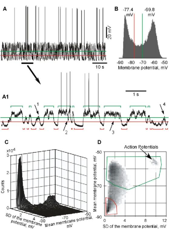
Slow-wave sleep (SWS) is characterized by the presence of large amplitude slow waves in cortical LFP and/or EEG recordings, a steady muscle tone, and the absence of eye movements. Intracellular recordings of neocortical neurons show a bimodal distribution of the membrane potential due to the presence of active (depolarized, Up) and silent (hyperpolarized, Down) states, which are present only during SWS and under certain types of anaesthesia. SWS properties will be largely discussed throughout this thesis.
Figure I- 4 Oscillations in thalamocortical system.
a. Frequency band of oscillations recorded in the thalamocortical system. b. Segment of intracellular recording (black) recorded during slow-wave sleep in cat’s associative cortex. The signal was then filtered for slow oscillation (0.1-1 Hz, red trace). c. Segment of intracellular recording (black) recorded during slow-wave sleep in cat’s somatosensory cortex. A segment filtered for spindle (7-15 Hz, green). From (Timofeev and Chauvette, 2011)
1.2.4 Thalamocortical oscillations during slow-wave sleep
The frequencies of thalamocortical oscillations range from as slow as 0.02 Hz to as fast as 500 Hz (Fig. I-4). These different rhythms and their functionality were largely reviewed previously (Timofeev and Bazhenov, 2005; Bazhenov and Timofeev, 2006; Timofeev and Chauvette, 2011). Therefore, thalamocortical oscillations are grouped in several frequency bands, for some of which the mechanisms of generation are known.
1.2.4.1 Infra-slow oscillation
This type of oscillatory activity has a period within the range of tens of seconds to a minute (Aladjalova, 1957). Very little is known about the underlying mechanisms of these oscillations but at least some of the factors responsible for their generation could depend on non-neuronal dynamics, such as changes in CO2 concentration (Nita et al., 2004).
Infra-slow activities likely have a cortical origin given that they can be recorded from neocortical slabs (Aladjalova, 1962). Infra-slow oscillation is correlated with the magnitude of faster EEG oscillations, and infra-slow oscillation might be implied in controlling the gross cortical excitability (Vanhatalo et al., 2004).
1.2.4.2 Slow oscillation
During slow-wave sleep (SWS) and some types of anesthesia the dominant activity is generated with a frequency (0.3 - 1 Hz), termed slow oscillation (Steriade et al., 1993b; Steriade et al., 1993a; Steriade et al., 2001; Timofeev et al., 2001a). In both, anesthetized (Metherate and Ashe, 1993; Contreras and Steriade, 1995) and naturally sleeping animals (Steriade et al., 2001; Timofeev et al., 2001a; Okun et al., 2010), during slow oscillation the entire cortical network (both excitatory and inhibitory neurons) alternates between silent (Hyperpolarizing, or Down) and active (Depolarizing, or Up) states that correspond to depth-positive and depth-negative waves of local field potentials, respectively (Fig. I-4 b). Active states are characterized by cellular firing and faster frequencies (Mukovski et al., 2007; Volgushev et al., 2007). Silent states are periods of disfacilitation and are mediated mainly by a potassium current, likely the leak current (Timofeev et al., 2001a). During
silent states, both RE and TC neurons are hyperpolarized like cortical neurons; during active states RE neurons are depolarized and either fire in tonic mode or reveal spindle-like high frequency spike-bursts (Contreras and Steriade, 1995; Contreras et al., 1996a; Fuentealba et al., 2005). By contrast, TC neurons display rhythmic IPSPs with spindle frequency and occasional rebound spike-bursts (Contreras and Steriade, 1995; Timofeev and Steriade, 1996; Timofeev et al., 2001b).
Most studies point to a cortical origin of slow oscillation because slow oscillation is present in cortical slab in vivo (Timofeev et al., 2000b), in cortical brain slice with modified ACSF (Sanchez-Vives and McCormick, 2000; Sanchez-Vives et al., 2007), in animals with kainic acid thalamic lesion (two days after the lesion) (Steriade et al., 1993a), and absent in decorticated cats (Timofeev and Steriade, 1996). Inactivating a small cortical region disrupt the synchrony between sites on each side of the inactivation site (Amzica and Steriade, 1995), and layer V was shown to be critical for the lateral propagation of slow oscillation in thalamocortical slice preparations (Wester and Contreras, 2012).
Intracellular recordings from cortical neurons during states of vigilance demonstrated that an alternation of active and silent states occurs only during SWS and not in any other state of vigilance (Steriade et al., 2001; Timofeev et al., 2001a; Mukovski et al., 2007; Rudolph et al., 2007). Similar alternations of active and silent states in neocortical neurons were recorded in associative, motor, somatosensory, and visual cortices (see chapter IV; (Chauvette et al., 2011)). Waking state and REM sleep show a single mode distribution of membrane potential and silent states were absent during these states of vigilance (Matsumura, 1979; Steriade et al., 2001). In contrast to these observations, large amplitude fluctuations of membrane potential (unlikely states) were found during quiet wakefulness in mice barrel cortex (Crochet and Petersen, 2006; Poulet and Petersen, 2008; Gentet et al., 2010). In these studies, states of vigilance were not identified formally. However, in the available example [(Poulet and Petersen, 2008), see their Fig. 2B], the large amplitude membrane potential fluctuations occur only when slow waves are present in local field potential recordings, suggesting that at this time the mice was either in the