Case Report
Contribution of the Clinical and Histopathological
Features in the Positive Diagnosis of the
Juvenile Polyposis Syndrome -
Poaty H
1,2*, Batamba Bouya L
1, Gassaye D
1,3, Mavoungou Biatsi K
4,
Lumaka Zola A
5and Peko JF
1,41Faculty of Health Sciences, Marien Ngouabi University, Brazzaville, Congo 2National Research Institute on Health Sciences, Brazzaville, Congo
3Gastroenterology Service, CHU de Brazzaville, Congo 4Morbid Anatomy Service, CHU de Brazzaville, Congo
5National Biomedical Research Institute (INRB), Kinshasa, DR Congo
*Address for Correspondence: Henriette Poaty, Faculty of Health Sciences, Marien Ngouabi University, BP 2672, Brazzaville, Congo, Tel: +002-420-6 68-657-61; ORCID : orcid.org/0000-0003-2114-6415 ;
E-mail:
Submitted: 08 January 2018; Approved: 12 February 2018; Published: 14 February 2018
Cite this article: Poaty H, Batamba Bouya L, Gassaye D, Mavoungou Biatsi K, Lumaka Zola A, et al.
Contribution of the Clinical and Histopathological Features in the Positive Diagnosis of the Juvenile Polyposis Syndrome. American J Genet Genom. 2018;1(1): 001-004.
Copyright: © 2018 Poaty H, et al. This is an open access article distributed under the Creative
Commons Attribution License, which permits unrestricted use, distribution, and reproduction in any medium, provided the original work is properly cited.
American Journal of
Genetics & Genomics
SCIRES Literature - Volume 1 Issue 1 - www.scireslit.com Page - 002
American Journal of Genetics & Genomics
INTRODUCTION
Juvenile Polyposis Syndrome (JPS) is a rare condition characterized by the presence of the juvenile hamartomatous polyps mostly located in the gastrointestinal tract [1]. It predisposes aff ected individuals to an increased risk of Colorectal Cancer (CRC) and less frequently to extra gastrointestinal malignancies, especially in adulthood [1-4]. Th e clinical manifestation of the disease appears aft er birth, especially in young subjects. Th e germline mutation occurs in BMPR1A (Bone Morphogenetic Protein Receptor Type-1A) gene mapped on chromosome band 10q23.2 or in SMAD4 (Mothers against decapentaplegic homolog 4) located on chromosome band 18q21.2 [2,5,6]. Both genes are tumor suppressors and are involved in the Transforming Growth Factor-Beta (TGF-β) signaling pathways [4,6]. Th e challenge in this condition is the rapid recognition of the disease and the detection of the mutation in the index patient and the family members in order to perform radiologic, endoscopic examinations and rapid polypectomy and to prevent partially cancer [6]. But the search of mutation is initially guided by elements in favor of the hereditary character of the aff ection such as in lynch syndrome (other predisposed CRC aff ection) [7]. Th e interrogatory in search of familial history of polyps or cancers, the pedigree, the endoscopic examination and the clinical criteria, are the fi rst guiding elements for JPS positive diagnosis [4]. Th e histological analysis in research of typical features of JPS also helps the clinicians to recognize the condition [8,9]. We report here a clinical observation whose suggestive endoscopic and clinic histological phenotype in favor of JPS have motivated the research of the mutations in BMPR1A and SMAD4 genes.
PATIENT PRESENTATION
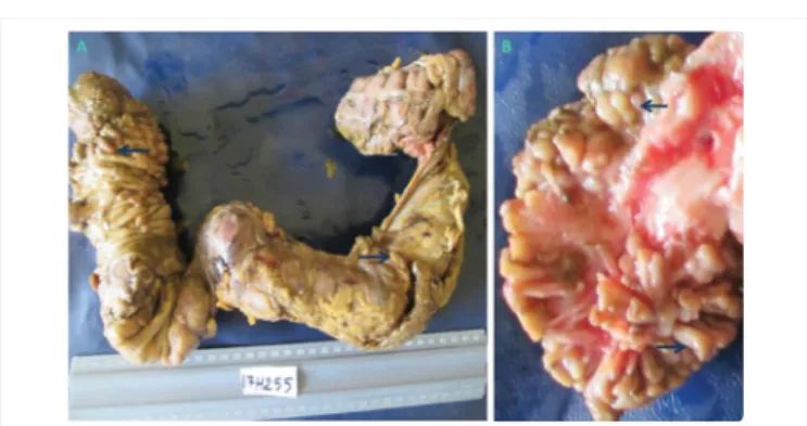
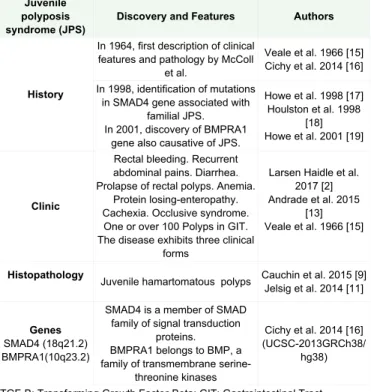
He is a 25-year-old congolese man, single, with a father who died from a CRC (Figure 1). Its disease begins at the age of 15 years-old by recurrent diarrhea, rectal bleeding, persistent abdominal pains, abdominal bloating followed by cachexia and occlusive syndrome. Th e barium enema and the colonoscopy showed numerous polyps along the colon. Th e tumor macroscopic examination found approximately more than 50 sessile and pedunculated polyps of diff erent sizes (Figure 2). Histological analysis (17H255A2) exhibited evocative lesions of JPS and a slight dysplasia (Figure 3). Direct sequencing (performed in Human Genetic Center of University hospital of Leuven, Belgium) identifi ed a heterozygous mutation in SMAD4 defi ned as « c.1229_1230delAG », exon 10 (ENSE00003694477) with the pathogenic variant « p.Ser411Leufs*17 » (of the abnormal predicted protein). We concluded to a juvenile polyposis coli form with a slight dysplasia in young man carrying SMAD4 germline mutation. Th e patient underwent a surgical cure (partial colectomy). He has been doing well for two years and is followed by a multidisciplinary team.
DISCUSSION
Juvenile polyposis syndrome appears in three clinical forms: juvenile infantile polyposis, generalized juvenile Polyposis and juvenile colorectal polyposis [5]. Th e approximate annual incidence is 1:16,000 to 1:100,000 in the population [2,5,10,11]. Family history is found in 20 to 50% of the aff ected patients [1,4,5,9]. Clinical criteria defi ned by Jass et al. in 1998 and revised in 2000 by World Health Organization (WHO) are used to facilitate the recognition of JPS (Table 1) [3,5,6,11,12]. Diagnosis may be suspected in presence of one criterion. Our patient met the three clinical criteria guidelines (Table 1), and presented the juvenile colonic polyposis form. According to publications data, JPS is manifested by constant presence of juvenile hamartomatous polyps [5,9]. Th e number of polyps varies from 1 to over 100, and they are most frequently located in the distal colon (such as in our patient) and the rectum [2,4,11]. Others gastrointestinal locations are stomach, duodenum, small intestine [3,4,13]. Polyps can appear at any age (from childhood through adulthood), most oft en during the adolescence (around 12-14 years-old) [11,13,14]. JPS
ABSTRACT
Juvenile Polyposis Syndrome (JPS) is a rare genetic disease characterized by the presence of the juvenile hamartomatous polyps. The condition is caused by germline mutation in the BMPR1A or the SMAD4 genes and it is inherited in an autosomal dominant manner. It predisposes affected persons to a high risk of malignant tumors, mainly colorectal and stomach cancers. The confi rmation of the diagnosis is based on genetic analysis. But at fi rst, family history, pedigree, clinical criteria and histopathological analysis guide to an inherited disease. We present here a Congolese patient with suggestive clinical and histopathological features which lead to the JPS.
Keywords: Juvenile polyposis syndrome; Clinic; Histopathology; Genetic Disease; Hereditary Cancer
Figure 1: Family pedigree of patient. Proband (arrow) with JPS having a father died for colorectal cancer.
Figure 2: Macroscopic aspect of resected colon tumor Presence of multiples pedunculated and sessile polyps (> 50) of different sizes in the colon.
SCIRES Literature - Volume 1 Issue 1 - www.scireslit.com Page - 003
American Journal of Genetics & Genomics
is the most common hereditary form of polyposis in the childhood, the protein losing-enteropathy and the prolapse of rectal polyps are frequently observed in children (Genetic home reference 2013) [4,6,10,13]. JPS is also accompanied by other nonspecifi c symptoms among which the rectal bleeding and the recurrent abdominal pains are the most common revealing signs [6,13]. We have in (Table 2) reported the main clinical manifestations [2,14,15]. Th e histological analysis which is practiced without diffi culties in African hospitals is essential in the approach of positive diagnosis. Concerning our patient, the exhibited tumor histological profi le (Figure 3) was consistent with literature data [1,8,15]. Typically, the microscopic aspects show the juvenile polyps (Figure 3A) with a characteristic architecture: Glands are more oft en spherical, separated, dilated, cystic and rich in mucus (Figure 3B, C) [3,8,11]. Th ey are associated with lamina propia edema, massive infi ltrating infl ammatory cells (Figure 3B, C) [2,4,11]. Th e stroma is dense with dystrophic blood vessels [1,3,6]. Juvenile hamartomatous polyps are identifi ed by histological examination in 88-100% of cases [13]. Genetically, JPS is a Mendelian pathology [3,6,16-19]. Th e condition is inherited from one parent (as in our Report) in an autosomal dominant manner (in 75% of patients), except in cases of de novo mutation reported in 25% of JPS (Genetic home reference 2013) [9,14]. BMPR1A and SMAD4 mutations are detected approximately in 40 to 60% of patients [14]. Both genes are TGF-β signaling proteins which initiate cell cycle arrest and growth inhibition [14,16]. Polyps in JPS can degenerate and the approximate risk of developing CRC varies between 38 to 50%, In case of multiple polyps, especially in adults [3,5,11,14]. Hamartomatous polyposis including JPS is responsible approximately for 1% of all CRC [5]. Th e occurrence cancer risk in the JPS (when the mutation is known in the family) requires the early screening of asymptomatic family members and the polypectomy is highly recommended. Regular endoscopy
monitoring (every 2 to 3 years) of persons carrying a BMPR1A or a SMAD4 germline mutation is recommended starting at 10 to 15 years-old [3,5,9,16].
CONCLUSION
Th e combination of endoscopy, pedigree, clinical criteria and histological analyzes is at fi rst a good approach to spot JPS. It allows to guide towards a pathology inherited and especially to signify the presence of polyps (and cancer) for a rapid surgical management.
ACKNOWLEDGEMENT
Th e authors are grateful to Th eophile chomienne for the English proofreading
AUTHORS’ CONTRIBUTIONS
GD: Endoscopic examination; GD, BBL, JFP, MK, LZA: Review and data analysis; HP: Data analysis and manuscript preparation.
REFERENCES
1. Ahmed A, Alsaleem B. Non familial Juvenile polyposis syndrome with exon 5 novel mutation in SMAD 4 gene. Case Rep Pediatr. 2017; 2017: 5321860. https://goo.gl/ZteC1D
2. Larsen H J, Howe JR. Juvenile polyposis syndrome. Editor gene reviews. Seattle (WA): University of Washington, Seattle.1993-2018. https://goo.gl/8pwj8e
3. B. Buecher. Les formes hereditaires des cancers colorectaux, hors syndrome de lynch et polyposes adenomateuses. La lettre de lHepato-gastroenterologue. 2009; 6: 222-227. https://goo.gl/Kebxvp
4. Bronner MP. Gastrointestinal inherited polyposis syndromes. Mod Pathol. 2003; 16: 359-365. https://goo.gl/CVLURx
5. Alimi A, Weeth-Feinstein LA, Stettner A, Caldera F, Weiss JM. Overlap of Juvenile polyposis syndrome and cowden syndrome due to de novo chromosome 10 deletion involving BMPR1A and PTEN: implications for treatment and surveillance. Am J Med Genet A. 2015; 167: 1305-1308. https://goo.gl/mjeUMh
Figure 3: Image of histological section of juvenile polyps (Hematoxylin and Eosin-stained)
A) Juvenile polyps (examination No 17H255A2) showing increased of cellular elements, lamina propia and proliferative glands
B) and C) Excess of small, cystical and spherical proliferative glands with abundant mucus. Lamina propria with peri-glandular edema, infl ammatory cells and dystrophic blood vessels.
Table 1: Clinical criteria of juvenile polyposis syndrome [2,3,6,11,12]
Number Clinical criteria
1 Multiple juvenile polyps in the colorectum, more than fi ve.
2 Patient with a family history of juvenile polyposis syndrome,
regardless the number of juvenile polyps and their site.
3 Multiple juvenile polyps throughout the intestinal tract with exclusion
of the other causes of digestive polyposis.
Table 2: Natural history and phenotype features in juvenile polyposis syndrome. Juvenile
polyposis syndrome (JPS)
Discovery and Features Authors
History
In 1964, fi rst description of clinical features and pathology by McColl
et al.
Veale et al. 1966 [15] Cichy et al. 2014 [16] In 1998, identifi cation of mutations
in SMAD4 gene associated with familial JPS. In 2001, discovery of BMPRA1
gene also causative of JPS.
Howe et al. 1998 [17] Houlston et al. 1998
[18] Howe et al. 2001 [19]
Clinic
Rectal bleeding. Recurrent abdominal pains. Diarrhea. Prolapse of rectal polyps. Anemia.
Protein losing-enteropathy. Cachexia. Occlusive syndrome.
One or over 100 Polyps in GIT. The disease exhibits three clinical
forms
Larsen Haidle et al. 2017 [2] Andrade et al. 2015
[13] Veale et al. 1966 [15]
Histopathology
Juvenile hamartomatous polyps Cauchin et al. 2015 [9]Jelsig et al. 2014 [11]
Genes
SMAD4 (18q21.2) BMPRA1(10q23.2)
SMAD4 is a member of SMAD family of signal transduction
proteins. BMPRA1 belongs to BMP, a family of transmembrane
serine-threonine kinases
Cichy et al. 2014 [16] (UCSC-2013GRCh38/
hg38)
SCIRES Literature - Volume 1 Issue 1 - www.scireslit.com Page - 004
American Journal of Genetics & Genomics
6. Huber AR, Findeis-Hosey JJ, Whitney-Miller CL. Hereditary gastrointestinal polyposis syndromes: a review including newly identifi ed syndromes. J Gastroint Dig Syst. 2013; 3: 155. https://goo.gl/NTAZEz
7. Poaty H, Aba Gandzion C, Soubeyran I, Gassaye D, Peko JF, Nkoua Bon JB, et al. The identifi cation of lynch syndrome in congolese colorectal cancer patients. Bull Cancer. 2017; 104: 831-839. https://goo.gl/zs1N3t
8. Van hattem WA, Langeveld D, De Leng WW, Morsink FH, van Diest PJ, Iacobuzio-Donahue CA, et al. Histologic variations in juvenile polyp phenotype correlate with genetic defect underlying juvenile polyposis. Am J Surg Pathol. 2011; 35: 530-536. https://goo.gl/CWTcr8
9. Cauchin E, Touchefeu Y, Matysiak-Budnik T. Hamartomatous tumor in the gastrointestinal tract. Gastrointest Tumors. 2015; 2: 65-74. https://goo.gl/qAHUvU
10. Hansraj N, Safta A, Alaish SM. Altered mental status as a presentation of Juvenile polyposis syndrome. J Ped Surg Case Report. 2015; 3: 566-569. https://goo.gl/XeRo3X
11. Jelsig AM, Qvist N, Brusgaard K, Nielsen CB, Hansen TP, Ousager LB. Hamartomatous polyposis syndromes: a review. Orphanet J Rare Dis. 2014; 9: 101. https://goo.gl/1UdeXM
12. Jass JR, Williams CB, Bussey HJ, Morson BC. Juvenile polyposis-a precancerous condition. Histopathology. 1988; 13: 619-630. https://goo.gl/WwxoJh
13. Andrade DO, Ferreira AR, Bittencourt PF, Ribeiro DF, Silva RG, Alberti LR. Clinical, epidemiologic, and endosopic profi les in children and adolescents with colonic polyps in two referencs centers. Arq Gastroenterol. 2015; 52: 303-310. https://goo.gl/uNPgtK
14. Durno CA. Colonic polyps in children and adolescents. Can J Gastroenterol. 2007; 21: 233-239. https://goo.gl/Z7eK85
15. Veale AM, McColl I, Bussey HJ, Morson BC. Juvenile polyposis coli. J Med Genet. 1966; 3: 5-16. https://goo.gl/yU2Bxi
16. Cichy W, Klincewicz B, Plawski A. Juvenile polyposis syndrome. Arch Med Sci. 2014; 10: 570-577. https://goo.gl/H88UPA
17. Howe JR, Roth S, Ringold JC, Summers RW, Jarvinen HJ, Sistonen P, et al. Mutations in the SMAD4/DPC4 gene in juvenile polyposis. Science. 1998; 280: 1086-1088. https://goo.gl/8KDv9k
18. Houlston R, Bevan S, Williams A, Young J, Dunlop M, Rozen P, et al. Mutations in DPC4 (SMAD4) cause juvenile polyposis syndrome, but only account for a minority of cases. Hum Mol Genet. 1998; 7: 1907-1912. https://goo.gl/pThRRy
19. Howe JR, Bair JL, Sayed MG, Anderson ME, Mitros FA, Petersen GM, et al. Germline mutations of the gene encoding bone morphogenetic protein receptor 1A in juvenile polyposis. Nat Genet. 2001; 28: 184-187. https://goo.gl/xVhWdB