RhoA Inhibition Is a Key Step in Pituicyte Stellation Induced by A
1-Type
Adenosine Receptor Activation
Lia Rosso,1 Brigitta Peteri-Brunbäck,1 Valérie Vouret-Craviari,2 Christophe Deroanne,2 Jean-Denis Troadec,1
Sylvie Thirion,1 Ellen Van Obberghen-Schilling,2 and Jean-Marc Mienville1
1Laboratoire de Physiologie Cellulaire et Moléculaire, Université de Nice-Sophia Antipolis, Nice, France 2Centre Antoine Lacassagne, Université de Nice-Sophia Antipolis, Nice, France
ABSTRACT
Pituicyte stellation in vitro represents a useful model with which to study morphological changes that occur in vivo in these cells during times of high neurohypophysial hormone output. This model has helped us establish the hypothesis of a purinergic regulation of pituicyte morphological plasticity. We first show that ATP induces stellation in 37% of pituicytes, an effect that is secondary to the metabolism of ATP to adenosine. Adenosine-induced stellation of pituicytes appears to be mediated by A1-type receptors. The effect is independent of
intracellular calcium and does not involve the mitogen-activated protein kinase pathway. The basal (nonstellate) state of pituicytes depends on tonic activation of a Rho GTPase because both C3 transferase (a Rho inhibitor) and Y-27632 (an inhibitor of p160Rho kinase) can induce stellation. Lysophosphatidic acid, a Rho activator, blocks the morphogenic effect of adenosine dose-dependently. Using a specific RhoA pull-down assay, we also show that downregulation of activated RhoA is the key event coupling A1 receptor activation to pituicyte
stellation, via F-actin depolymerization and microtubule reorganization. Finally, both vasopressin and oxytocin can prevent or reverse adenosine-induced stellation. The effects of vasopressin, and those of high concentrations of oxytocin, are mediated through Vla receptors. Placed within the context of the relevant literature, our data
suggest the possibility of a purinergic regulation of pituicyte morphological plasticity and subsequent modulation of hormone release, with these hormones providing a negative feedback mechanism.
KEYWORDS : neurohypophysis ; purines ; vasopressin ; oxytocin ; cytoskeleton
INTRODUCTION
The morphological plasticity of glial cells in the hypothalamus-neurohypophysis (HNH) axis may represent an important regulatory system of neurohypophysial hormone release. During physiological conditions such as lactation or dehydration, i.e., conditions that require a high output of oxytocin (OXT) or vasopressin (AVP), morphological changes are observed at the level of both hypothalamic astrocytes and neurohypophysial
pituicytes. For instance, astrocytes withdraw from between oxytocinergic neurons throughout the hypothalamus, which, through an increase in the number of contacts between neuronal membranes, may promote excitability and optimize hormone release (reviewed in Theodosis and Poulain, 1987, 1993). In the neurohypophysis, pituicytes retract from perivascular spaces, facilitating access of the secreting terminals to the basal lamina and release of their hormonal contents into the blood (reviewed in Theodosis and Macvicar, 1996; Hatton, 1999). Explant cultures represent a convenient means of studying the mechanisms of morphological plasticity in pituicytes. With appropriate stimulation, these cells can switch from a flat, processless shape to a rounded, stellate morphology in a manner reminiscent of that occurring in vivo upon stimulation of the HNH axis (reviewed by Hatton, 1988). One such stimulation that is now well established concerns activation of β-adrenergic receptors, which has been shown to induce stellation in pituicyte explants (Bicknell et al., 1989), as well as corresponding morphological changes in a whole neural lobe preparation (Luckman and Bicknell, 1990). Several lines of evidence also suggest the possibility of a purinergic regulation of pituicyte plasticity, including the presence on these cells of purinergic receptors whose function is unresolved (Loesch et al., 1999; Troadec et al., 1999), the observation that ATP can be released from neurohypophysial terminals (Gratzl et al., 1980; Zimmermann, 1994; Sperlágh et al., 1999), and previous data suggesting that adenosine can induce stellation of rat pituicytes (Miyata et al., 1999) and astrocytes (Abe and Saito, 1998). We therefore studied the effects of endogenous and pharmacological ligands of purinergic receptors on pituicyte stellation in vitro and sought to
gain insight into the receptor subtypes and transduction mechanisms involved herein. To extend the
physiological relevance of our findings, we also studied the action of AVP and OXT on stellation, suggesting a model for the concerted regulation of morphological plasticity of pituicytes in vivo. Most of these results have been reported at two recent meetings (Rosso et al., 2001a,b).
MATERIALS AND METHODS Pituicyte Explant Cultures
Adult Wistar rats (150-200 g) were anesthetized with CO2 and decapitated in accordance with French/ European
ethical guidelines. For each culture, 4-6 hypophyses were placed in Hank's balanced salt solution (HBSS) (Gibco #14025) supplemented with 10 mM HEPES, 0.5 mg ml-1 bovine serum albumin (BSA), 100 U ml-1 penicillin,
and 100 µg ml-1 streptomycin. The posterior lobe was separated from the anterior and intermediate lobes under a
dissecting microscope and was divided into ~20 pieces. For morphological studies, each piece of tissue was placed in a 35-mm plastic dish coated with 0.05 mg ml-1 collagen and containing DMEM medium (Gibco
#52100) supplemented with 1.2 gl-1 NaHCO
3 and 10% fetal calf serum (FCS). For Rho assays, five tissue pieces
were plated in 60-mm dishes. Cultures were maintained at 37°C in an incubator supplied with a [5% CO2/95%
air] humidified atmosphere. The medium was replaced every 2-3 days. All experiments were performed on cell cultures after 8-12 days in vitro, at which time a monolayer of pituicytes had spread 5-10 mm from the explant. Control experiments (n = 10) performed as previously published by this laboratory (Troadec et al., 1999) showed that >95% of the cells were GFAP-positive.
Morphological Analysis
Because of the inhibitory effect of serum on stellation, cells were switched to a serum-free medium 1 h before experiments, a standard procedure followed by most investigators (e.g., Ramsell and Cobbett, 1997; Abe and Saito, 1998). Unless otherwise stated, all drugs or their vehicle as control were added 1 h (37°C) before image acquisition. The latter was performed with either a CCD camera (Dage MTI) and Axon Imaging Workbench 2.2 software for kinetic studies, or a digital still camera and Adobe Photoshop software for cell counting. Digitized images were coded with respect to treatment in order to perform blind counting. Cells were considered stellate when they displayed ≥2 processes (Ramsell and Cobbett, 1997). For each culture dish, the proportion of stellate cells was assessed by counting 100-200 cells at 10× magnification (Fig. 1A) over five arbitrarily chosen areas 0.9 × 0.7 mm wide, and taking the resulting average. Differences between treatment groups were evaluated with analysis of variance followed by a Bonferroni post hoc test, with significance set at P < 0.05.
Calcium Imaging Intracellular Ca2+ ([Ca2+]
i) was measured with the ratiometric, membrane-permeable, fluorescent probe Fura-2
AM. Briefly, pituicyte cultures were incubated 45 min at 37°C in the presence of 5 µM Fura-2 AM + 0.01% pluronic acid. During experiments, a culture dish was continuously superfused with Ringer's solution (3 ml/min). These experiments were performed on the stage of an inverted microscope (Zeiss ICM 405) equipped with a Xe lamp and a rotating filter set allowing 350/380-nm excitation. Axon Imaging Workbench 2.2 software was used to drive the filter wheel, acquire fluorescence images and process data. For any given experiment, fluorescence signals were averaged from 10-15 cells defined as "regions of interest". Free [Ca2+]
i was estimated from a
calibration procedure using a "zero-Ca" solution (3 mM EGTA + 2 µM ionomycin) and a Ca-saturated solution (3 mM CaCl2 + 2 µM ionomycin). F350/380 ratios were converted to free [Ca2+]i using the Grynkiewicz
equation (see Hatton et al., 1992). Drugs were applied locally via a miniperfusion system. ERK and p38 Phosphorylation Assays (Western Blot Analysis)
Serum-free cultures were stimulated with ATP or adenosine (or vehicle) for 10 min, washed in phosphate-buffered saline (PBS) at 4°C, then exposed to a lysis buffer (20 mM Tris, 137 mM NaCl, 2 mM EDTA, 25 mM β-glycerophosphate, 1 mM Na-orthovanadate, 2 mM NaPPi, 1 mM PMSF, 10% glycerol, 2% Triton X-100; pH 7.5) supplemented with the protease inhibitors aprotinin (2 µg/ml), leupeptin (10 µM), and AEBSF (1 mM). Cell lysates were scraped off culture dishes and centrifuged (14,000g; 10 min at 4°C) to recover cytosolic proteins from the supernatant. Proteins were then quantified based on Bradford reaction (Bio-Rad kit). Because of the low yield (~0.4 µg/ml protein) of our samples, proteins were precipitated with 5 vol/vol acetone. The resulting samples were stored 30 min at -80°C, subsequently centrifuged (8,500g; 3 min at 4°C), and pellets were resuspended in adequate volumes of standard denaturing buffer. Proteins (25 µg/lane) were separated by sodium dodecyl sulfate-polyacrylamide gel electrophoresis (SDS-PAGE) electrophoresis at 100 V for 1 h on 10%
acryl-amide gels, transferred to nitrocellulose membranes (Amersham's Hybond-C), and stained with Ponceau red to verify even transfer. The membranes were then saturated with 5% skim milk in PBS containing 0.1% Tween 20 for 1 h at RT, and incubated with primary antibodies diluted 1:2,000 for anti-phospho p38 (M8177 from Sigma) and 1/10,000 for anti-phospho ERK (M8159 from Sigma) in PBS containing 1% skim milk and 0.1% Tween 20. After 3 rinses in PBS/Tween 20, membranes were incubated for 1 h at RT with secondary antibody (goat anti-mouse coupled to horseradish peroxidase [HPO]) diluted 1:5,000 in the same PBS-skimmed milk solution. After secondary antibody removal, blots were developed using the enhanced chemi-luminescence (ECL) detection system (Amersham).
Rho GTPase Activity Assay
The RhoA assay was performed on pituicytes under the following conditions, using 5 60-mm culture dishes per condition: (1) control, i.e., 70 min without serum; (2) 60 min without serum followed by 10-min exposure to 10 µM adenosine; and (3) positive control, i.e., 60 min without serum followed by 10 min in the presence of 10% serum. The cells were lysed in buffer A (25 mM HEPES [pH 7.3], 150 mM NaCl, 5 mM MgCl2, 0.5 mM EGTA,
0.5% Triton X-100, 4% glycerol, 20 mM β-glycerophosphate, 10 mM NaF, 2 mM Na-orthovanadate, 5 mM dithiothreitol, and protease inhibitors) for 10 min at 4°C; the Triton X-100 insoluble material was removed by centrifugation (10 min; 10,000 rpm), and the lysates were incubated for 40 min at 4°C with 20 µg of bacterially produced GST-RBD (glutathione-S-transferase-Rho binding domain), which was bound to glutathione-coupled Sepharose beads (Ren et al., 1999). Beads were washed 4 times in buffer A, resuspended in Laemmli buffer, and proteins were separated by SDS-PAGE on 12% acrylamide gels. RhoA was detected by Western blotting, using a specific antibody (Rho A (26C4): sc-418, Santa Cruz Biotechnology). Before incubation with the beads, 50-µl aliquots were removed from each sample for determination of total RhoA.
Cytoskeleton Labeling
Actin filaments were labeled with FITC-coupled phalloidin (100 ng/ml), and microtubules were labeled with an anti-β-tubulin monoclonal antibody (Sigma) diluted 1:200. After exposure to specific control and test conditions as indicated, the cells were fixed with 3% paraformaldehyde + 2% sucrose at 37°C, washed 3 times in PBS, permeabilized for 4 min in PBS + 0.2% Triton, and again rinsed three times in PBS and saturated 15 min in PBS + 5% serum. Primary antitubulin antibody was applied for 2 h in a wet chamber at room temperature. After four rinses and a new saturation with PBS + 5% serum for 15 min, the cells were incubated with anti-mouse IgG secondary antibody (Alexa 594, Molecular Probes, Eugene, OR) with or without FITC-coupled phalloidin for 1 h at RT. After four rinses, the cells were mounted on slides in the presence of Citifluor for observation on an epifluorescence microscope.
Drugs
All drugs that were used are listed in Table 1. We carefully checked that neither the solvents nor the receptor antagonists or enzyme inhibitors that we tested had any significant effect of their own on pituicytes (≤5% stellation). All drugs were from Sigma except bestatin (Boehringer-Roche), PP2 (Calbiochem), and the following compounds, which were given to us by the indicated persons: C3 toxin, P. Boquet (INSERM U-452, Nice, France); Y-27632, A. Yoshimura (Yoshitomi Pharmaceutical Industries, Osaka, Japan); SR 49059 and SR 121463, C. Serradeil-Le Gal (Sanofi-Synthélabo, Toulouse, France); CL 12-27, M. Manning (Medical College of Ohio, Toledo, OH).
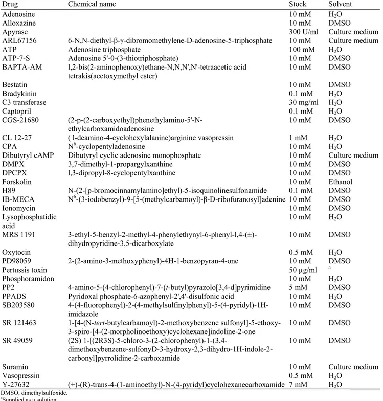
TABLE 1. List of drugs used
Drug Chemical name Stock Solvent
Adenosine 10 mM H2O
Alloxazine 10 mM DMSO
Apyrase 300 U/ml Culture medium
ARL67156 6-N,N-diethyl-β-γ-dibromomethylene-D-adenosine-5-triphosphate 10 mM Culture medium
ATP Adenosine triphosphate 100 mM H2O
ATP-7-S Adenosine 5'-0-(3-thiotriphosphate) 10 mM DMSO
BAPTA-AM l,2-bis(2-aminophenoxy)ethane-N,N,N',N'-tetraacetic acid
tetrakis(acetoxymethyl ester) 10 mM DMSO
Bestatin 10 mM DMSO Bradykinin 0.1 mM H2O C3 transferase 30 mg/ml H2O Captopril 0.1 mM H2O CGS-21680 (2-p-(2-carboxyethyl)phenethylamino-5'-N-ethylcarboxamidoadenosine 10 mM DMSO CL 12-27 ( l-deamino-4-cyclohexylalanine)arginine vasopressin 1 mM H2O CPA N6-cyclopentyladenosine 10 mM H 2O
Dibutyryl cAMP Dibutyryl cyclic adenosine monophosphate 10 mM Culture medium
DMPX 3,7-dimethyl-1-propargylxanthine 10 mM DMSO
DPCPX l,3-dipropyl-8-cyclopentylxanthine 10 mM DMSO
Forskolin 10 mM Ethanol
H89 N-(2-[p-bromocinnamylamino]ethyl)-5-isoquinolinesulfonamide 0.1 mM DMSO
IB-MECA N6-(3-iodobenzyl)-9-[5-(methylcarbamoyl)-β-D-ribofuranosyl]adenine 10 mM DMSO
Ionomycin 10 mM DMSO Lysophosphatidic acid 10 mM H2O MRS 1191 3-ethyl-5-benzyl-2-methyl-4-phenylethynyl-6-phenyl-l,4-(±)-dihydropyridine-3,5-dicarboxylate 10 mM DMSO Oxytocin 0.5 mM H2O PD98059 2-(2-amino-3-methoxyphenyl)-4H-1-benzopyran-4-one 10 mM DMSO Pertussis toxin 50 µg/ml a Phosphoramidon 10 mM H2O PP2 4-amino-5-(4-chlorophenyl)-7-(t-butyl)pyrazolo[3,4-d]pyrimidine 5 mM DMSO
PPADS Pyridoxal phosphate-6-azophenyl-2',4'-disulfonic acid 10 mM H2O
SB203580 4-(4-fluorophenyl)-2-(4-methylsulfinylphenyl)-5-(4-pyridyl)-1H-imidazole 10 mM DMSO SR 121463 1-[4-(N-tert-butylcarbamoyl)-2-methoxybenzene sulfonyl]-5-ethoxy-3-spiro-[4-(2-morpholinoethoxy)cyclohexane]indoline-2-one 10 mM DMSO SR 49059 (2S) 1-[(2R3S)-5-chloro-3-(2-chlorophenyl)-1-(3,4- dimethoxybenzene-sulfonyD-3-hydroxy-2,3-dihydro-1H-indole-2-carbonyl]pyrrolidine-2-carboxamide 10 mM DMSO
Suramin 10 mM Culture medium
Vasopressin 0.5 mM H2O
Y-27632 (+)-(R)-trans-4-(1-aminoethyl)-N-(4-pyridyl)cyclohexanecarboxamide 7 mM H2O
DMSO, dimethylsulfoxide.
aSupplied as a solution.
RESULTS
ATP and Adenosine Effects on Pituicyte Stellation
Our initial experiments indicated that ATP was able to induce pituicyte stellation (Fig. 1B,D). However, this effect was not reproduced by the nonhydrolyzable analogue ATP-γ-S (Fig. 1D). Given the presence of ecto-ATPases on the outer surface of the pituicyte plasma membrane (Thirion et al., 1996), we surmised that the effects of ATP might be due to its hydrolytic products. Indeed, adenosine (as well as ADP and AMP; not shown) was found to induce a more extensive stellation of pituicytes and at a lower concentration than ATP (Fig. 1A,C,D). This difference might reflect partial hydrolysis of ATP, as the effects of the latter were substantially slower (~60 min) than those of adenosine (~30 min; Fig. 1B,C). To test our hypothesis, we performed two experiments. First, we coapplied ATP with apyrase to accelerate the rate of ATP hydrolysis artificially; under
these conditions, the effects observed with ATP were significantly stronger than when it was applied alone (Fig. 1D). Second, we coapplied ATP with ARL67156, an ecto-ATPase inhibitor; in these conditions conversely, the effect of ATP was completely blocked (Fig. 1D), clearly demonstrating that hydrolysis was a necessary step for ATP to induce stellation. In fact, the nonselective P2 receptor (i.e., ATP receptor) antagonists PPADS and suramin failed to block the effect of ATP (Fig. 1D), whereas an A1 (i.e., adenosine receptor) antagonist was
effective (see below).
Adenosine Receptor Subtype Specificity
We then carried out a pharmacological characterization to identify the adenosine receptor subtypes involved in the stellation process. The results are summarized in Figure 2A. CPA, an A1 agonist, and IB-MECA, an A3
agonist, induced stellation to an extent comparable to that obtained with adenosine, while CGS-21680, an A2A
agonist, was without effect. Confirming this latter result, DMPX, an A2A antagonist, did not prevent
adenosine-induced stellation. As there are no A2B-selective agonists (Ralevic and Burnstock, 1998), we tested a relatively
specific A2B antagonist, alloxazine, which failed to block adenosine-induced stellation. Surprisingly, DPCPX, an
A1 antagonist, completely blocked the effects of both adenosine and IB-MECA (Fig. 2A), suggesting that either
IB-MECA or DPCPX exert nonspecific effects on A1 or A3 receptors, respectively. However, the fact that MRS
1191, an A3 antagonist, failed to block the effects of either adenosine or IB-MECA (Fig. 2A) strongly suggests
that IB-MECA has a nonspecific action at A1 receptors. In an attempt to optimize the selectivity of DPCPX by
reducing its concentration, we tested 100 nM DPCPX against 10 µM adenosine (n = 8), CPA (n = 3) or IB-MECA (n = 3). Unfortunately, at this dose the antagonist failed to block either agonist (≥75% stellation), probably because its Ki is too close to the KD of the agonists. DPCPX (10 µM) also completely blocked the
effects of ATP (Fig. 2A), ADP and AMP (not shown), a further argument in favor of an indirect, adenosine-mediated effect of ATP.
Transduction Mechanisms Involved in Adenosine Receptor Activation
Having identified the receptor subtype involved, we aimed to investigate the transduction mechanism
responsible for the morphogenic effects of purinergic agonists. We have previously reported that adenosine can raise ([Ca2+]
i) in pituicytes (Peteri-Brunbäck et al., 2001). Using the consistency of bradykinin responses as a
standard, we found that adenosine induces either a strong [Ca2+]
i response, a weak response or no response at all,
with each of these occurrences observed in roughly equal proportions (~1/3 cells; Fig. 3A,B,C). We thus tested whether a Ca2+ signal could mediate the induction of stellation. Contradicting this hypothesis, we found that the
Ca2+ ionophore ionomycin was without effect (Fig. 2B). Furthermore, preincubation of the cells with the
membrane-permeable Ca2+ chelator BAPTA-AM for 50 min failed to prevent the morphogenic effect of
adenosine (Fig. 2B).
We have also reported that in pituicytes adenosine is able to stimulate mitogen-activated protein kinase (MAPK) pathways (Peteri-Brunbäck et al., 2001), which are known to be involved in cell differentiation. Figure 3D shows results obtained with both adenosine and ATP, which has previously been shown to stimulate MAP kinases in astrocytes via P2Y receptors (Neary et al., 1996). Nevertheless, we ruled out involvement of these pathways in
pituicyte stellation, since neither PD98059, an inhibitor of MEK (MAP/ ERK [extracellular signal-regulated] kinase), nor SB203580, an inhibitor of p38 kinase, antagonized the morphogenic effect of adenosine (Fig. 2B), CPA, or IB-MECA (not shown).
It has previously been suggested that a raise in cytoplasmic cAMP is an important intermediary step in pituicyte stellation (Ramsell and Cobbett, 1997). We have confirmed that both forskolin (10 µM), an adenyl-cyclase activator, and dibutyryl cAMP (1 mM), a cell-permeant analogue of cAMP, can induce stellation (68 ± 4% and 57 ± 6%, respectively; n = 8 each; P < 0.05 compared with control). These results may seem at odds with the effects of A1 receptor stimulation, which have been described as transducing through Gi protein activation,
inhibition of adenylcyclase and cAMP decrease (Ralevic and Burnstock, 1998). This, together with the fact that neither pertussis toxin, a Gi/o protein inhibitor, nor H89, a protein kinase A (PKA) inhibitor, blocked
Fig. 1. Effects of ATP and adenosine on rat pituicyte stellation. A: Cultured pituicytes in control conditions (serum-free medium; left) and 1 h after treatment with 10 µM adenosine (Ado; right). B,C: Photographs of cultured pituicytes (fixed field) taken after exposure to ATP (B) or adenosine (C) for the time indicated in upper right corner (in min). D: Summary of results (mean ± SE) obtained in the various conditions indicated. Apyrase, ARL67156, PPADS, and suramin were applied 10 min before ATP. Concentrations: ATP, ATP-γ-S, and PPADATP-γ-S, 100 µM; adenosine, 10 µM; apyrase, 30 U ml-1; ARL67156 and suramin, 300 µM. Apyrase alone did not induce any
significant stellation. In this and subsequent graphs, the number on top of each bar represents the number of culture dishes counted. a, significantly different from control; b, significantly different from ATP. A, 500 × 500 µm (×10 lens), B, C, 200 × 200 µm (×32 lens).
Fig. 2. Pharmacology of purinergic induction of rat pituicyte stellation. A: Pharmacology of adenosine receptor subtypes involved in stellation. Antagonists (DPCPX, DMPX, alloxazine, and MRS-1191) were applied 10 min before agonists. a, significantly different from CGS-21680; b, significantly different from CPA. B: Pharmacology of transduction pathways involved in adenosine receptor activation. BAPTA-AM was applied 50 min, PD98059 and SB203580 30 min, pertussis toxin (PTX) 12 h, and H89 15 min before adenosine application. All drugs were applied at 10 µM except ATP, 100 µM; ionomycin, 1 µM; PTX, 0.1 µg ml-1 and H89, 0.3 µM. PD98059 and
SB203580 were also tested at 30 µM and yielded similar results. BAPTA-AM alone did not induce any significant stellation. *Significantly different from BAPTA-AM.
Involvement of RhoA Inactivation in Adenosine-induced Pituicyte Stellation
As it has previously been shown that astrocyte stellation is controlled by a member of the Rho family of proteins (Ramakers and Moolenaar, 1998), which are small GTPases involved in cytoskeleton rearrangement (Machesky and Hall, 1996), we tested whether this mechanism could transduce the morphogenic effects of adenosine in pituicytes. It should be noted that the "default," i.e., the basal state of cultured glia corresponds to the
maintenance of actin stress fibers under the tonic activation of Rho (Ramakers and Moolenaar, 1998), implying that stellation should be the result of Rho inactivation. We found that Rho indeed was active in pituicytes because stellation could be induced with C3 transferase (Fig. 4), a Clostridium botulinum toxin that specifically inactivates Rho (Chardin et al., 1989; Machesky and Hall, 1996; Ramakers and Moolenaar, 1998). Further, similar results were obtained with Y-27632 (Fig. 4), a specific inhibitor of a Rho-associated protein kinase named p160ROCK and known to be the main downstream mediator of cytoskeletal changes (Uehata et al., 1997). Conversely, when we treated pituicytes for 10 min with lysophosphatidic acid (LPA), a powerful Rho activator (Ramakers and Moolenaar, 1998), the drug dose-dependently prevented adenosine-mediated stellation (Fig. 4). By suggesting competition for a common signaling pathway, this result provides preliminary evidence in favor of our initial hypothesis that adenosine induces stellation via the Rho pathway. In contrast to LPA, the Src family kinase inhibitor PP2 failed to block adenosine-induced stellation (Fig. 4; see Discussion).
To confirm Rho involvement, we performed pulldown assays to monitor the activity of RhoA and thereby determine whether it was a target for the inhibitory action of adenosine in pituicytes. Figure 5 shows that the activated form of RhoA is greatly decreased after 10 min in the presence of adenosine. As expected, in control conditions, i.e., in serum-free medium, a basal activity is detected, which apparently suffices to maintain the fusiform shape of pituicytes. Conversely, addition of serum, which contains high amounts of LPA, enhances RhoA activity.
Cytoskeletal Rearrangements During Adenosine-induced Pituicyte Stellation
Next, we investigated the cytoskeletal changes that occur in pituicytes during adenosine-induced stellation. These changes consisted basically of actin rearrangement, with disappearance of stress fibers, and specific reassembly of microtubules within newly formed processes (Fig. 6), similar to results obtained by Miyata et al. (1999) upon stimulation with dibutyryl cAMP. We also compared the kinetics of the morphogenic effects elicited by adenosine and Y-27632. In both cases, significant alterations of actin fibers could be seen after 20 min of treatment, while the whole process appeared to be completed within 30 min (Fig. 7). These results suggest that the limiting step in the time course of the stellation process lies between Rho inhibition and depolymerization of actin fibers. Thus, whatever the nature of the coupling mechanism between A1 receptor
activation and RhoA inactivation, this initial step appears to be relatively fast, as also indicated by the shorter response time observed for adenosine-induced repression of activated RhoA (Fig. 5).
Fig. 3. Effects of adenosine on pituicyte [Ca2+]i mobilization (A-C) and on MAP kinase pathways (D). Application of adenosine to pituicytes induced either a strong [Ca2+]
i response comparable to that of bradykinin (A), a relatively weak response (B), or no
response at all (C). Graphs show average [Ca2+]
i levels from 9 (A), 11 (B), and 10 (C) experiments (note that each experiment itself is the
average of 10-15 cells; see Methods). D: Both ATP and adenosine increase the activated (phosphorylated) form of p38 and ERK proteins. Similar results were obtained in a duplicate experiment for p38-P, and in two more experiments for ERK-P.
Fig. 4. Involvement of Rho GTPase in rat pituicyte stellation. Lysophosphatidic acid (LPA) and PP2 were applied 10 min before adenosine. C3 toxin was applied for 24 h (Ramakers and Moolenaar, 1998); therefore, we also included a control count at this time point. Concentrations: Adenosine, 10 µM; LPA, as indicated (in µM); C3, 20 µg ml-1; Y-27632, 7 µM; PP2, 5 µM.
*Significantly different from adenosine (0.1 µM LPA is also significantly different from 1 µM LPA).
Fig. 5. Western blot showing adenosine-induced inhibition of RhoA activity in pituicytes. RhoA activity was detected with a pull-down assay; total RhoA is shown for reference (see Materials and Methods). Adenosine (10 µM) or serum were applied for 10 min before cell lysis. Similar results were obtained in a duplicate experiment.
Effects of AVP and OXT
We then tested whether AVP and OXT might provide a negative feedback for the action of adenosine, and discovered that both hormones were capable of preventing, and more interestingly reversing, the effects of adenosine (Fig. 8). It should be mentioned that Espelt et al. (1998) previously found, using a different culture technique, that long-term treatment with AVP or OXT results in pituicytes devoid of processes. Comparing hormone potency, we noted that 10 nM OXT reversed stellation in only 17% cells, compared with 86% with 1 nM AVP (n = 4; not shown). In order to elucidate the receptor subtype involved in these hormone effects, we tested SR 49059, a specific V1a antagonist, and SR 121463, a specific V2 antagonist (Serradeil-Le Gal, 1998).
Whereas SR 121463 had no effect, SR 49059 antagonized the effects of both AVP and OXT (Fig. 8).
Confirming involvement of V1a receptors, complete inhibition of stellation reversal was obtained with 100 nM
SR 49059 against 10 nM AVP (72 ± 6% stellation; n = 4). In view of the unavailability of specific V1b
antagonists, we tested whether CL 12-27, a relatively specific V1b peptidic agonist (Derick et al., 2001), would
mimic the effects of AVP. In concentrations up to 25 nM, CL 12-27 did not reverse adenosine-induced stellation, which was obtained only at 1 µM, a concentration well beyond the threshold of selectivity of CL 12-27 for V1b
versus other receptor subtypes (Derick et al., 2001). Finally, in view of the potential breakdown of AVP to potent metabolites (reviewed by Barberis and Tribollet, 1996), we checked for the effects of AVP in the presence of a cocktail of peptidase inhibitors (10-5 M bestatin + 10-6 M captopril + 10-5 M phosphoram-idon). No
difference was seen in the effects of AVP alone or in the presence of peptidase inhibitors (Fig. 8), suggesting that AVP per se reverses adenosine-induced stellation.
Fig. 6. Adenosine-induced reorganization of pituicyte cytoskeleton. Microphotographs show fluorescent labeling of F-actin (A,B) and tubulin (C,D) in control (A,C,E) and after exposure to 10 µM adenosine for 30 min (B,D,F). E,F: Actin/tubulin double-labeling overlays. Scale bar = 40 µm.
Fig. 7. Kinetics of pituicyte F-actin copolymerization in the presence of 10 µM adenosine (top) or 7 µM of the p160ROCK inhibitor Y-27632 (bottom) at the times indicated (min). Scale bar = 50 µm. [Color figure can be viewed in the online issue, which is available at www.interscience.wiley.com]
Fig. 8. Blockade of adenosine-induced pituicyte stellation by vasopressin (AVP) and oxytocin (OXT). Cells were either pretreated with 500 nM hormone for 10 min (second and third bars) or exposed to 100 nM hormone for 20 min after stellation was induced with 10 µM adenosine (Ado) for 40 min (all remaining bars). No significant difference in stellation was found between 40 and 60 min adenosine (P = 0.17; see also Fig. 1C). SR antagonists (both at 100 nM) and peptidase inhibitors were applied 5 min before hormones. CL 12-27 was applied at 25 nM following the same protocol as for AVP and OXT. a, significantly different from adenosine; b, significantly different from AVP 100; c, significantly different from OXT 100.
DISCUSSION
Our results suggest the existence of a purinergic regulation of morphological plasticity in pituicytes. A likely scenario would involve release of ATP from neurosecretory vesicles and nerve swellings of the HNH axis (Gratzl et al., 1980; Zimmermann, 1994; Sperlágh et al., 1999), hydrolysis of the nucleotide to adenosine by local ecto-nucleotidases (Thirion et al., 1996), and stimulation of pituicyte A1 receptors followed by Rho
inactivation and depolymerization of actin stress fibers. The fact that the effects we observed with ATP are secondary to adenosine formation is demonstrated by several arguments: (1) the longer delay observed for these effects is more likely to involve time-dependent hydrolysis than different receptor kinetics; (2) these effects were insensitive to P2 receptor antagonists and completely blocked by DPCPX, an A1 receptor antagonist; (3)
ATP-γ-S may be due to a direct effect on A1 receptors (Ralevic and Burnstock, 1998); and (4) enhancing ATP
hydrolysis with apyrase increased the effects of the nucleotide, while preventing endogenous hydrolysis with an ecto-ATPase inhibitor blocked them.
All adenosine receptors are coupled to heterotrimeric G proteins, A1 receptor activation being known to
transduce either via the Gi pathway, leading to adenyl-cyclase inhibition, or via the Gq pathway, leading to IP3
production and Cai2+ mobilization (Ralevic and Burnstock, 1998). In our case, involvement of Cai2+ was clearly
ruled out and cAMP stimulation rather than inhibition induced stellation; moreover, pertussis toxin, a Gi/o protein
inhibitor, failed to block adenosine's effects. Similarly, involvement of a Gs protein is unlikely, since PKA
inhibition did not prevent adenosine-induced stellation, and Abe and Saito (1998) demonstrated the absence of any change in cAMP concentration during adenosine-induced stellation of cortical astrocytes. Thus far, we then can postulate two coexisting pathways capable of inducing stellation, one used by adenosine, which is cAMP independent, and one used, e.g., by catecholamines (see below), which is cAMP dependent.
We then attempted to correlate these effects with our recent finding that adenosine activates MAPK pathways in pituicytes (Peteri-Brunbäck et al., 2001). Indeed, Neary et al. (1996) showed that ATP, via P2 receptors, can both stimulate MAP kinases and induce stellation in astrocytes, suggesting a possible link between these events. In contrast, Abe and Saito (1998) demonstrated that in cortical astrocytes adenosine-induced stellation was not coupled to MAPK pathways. The latter results, therefore, are entirely consistent with our own, given the failure of MAPK specific inhibitors to prevent adenosine-induced stellation in pituicytes.
Finally, we have gathered conclusive evidence that pituicyte morphological plasticity is under the control of RhoA GTPase, and that adenosine most likely induces stellation by inhibiting this protein. It is intriguing that the opposite seems to occur in neurons, in which A1 receptor activation leads to stress fiber formation via the Rho
kinase pathway (Thevananther et al., 2001). Thus, A1/Rho coupling may constitute an important component of
differentiation of the neuronal versus glial lineage. On the other hand, Abbracchio et al. (2001) have shown that in astrocytoma cells selective activation of A3-type adenosine receptors by a chloro derivative of IB-MECA
induces the formation of actin stress fibers via Rho stimulation. Although it may not be relevant to compare transformed astrocytes with normal pituicytes, the potential discrepancy that adenosine would elicit conflicting effects if both A1 and A3 receptors were coexpressed in a given cell type might be resolved based on different
affinity constants for adenosine, which might allow selective activation according to specific physiological demands (Ralevic and Burnstock, 1998). From these considerations, it is clear that the next important step will be to determine which adenosine receptor subtypes are expressed in various cell lineages (e.g., glial vs neuronal) or sublineages (e.g., astrocytes vs pituicytes).
The other important issue concerns the mode of coupling of these receptors to the Rho pathway. Our results are novel with respect to the fact that A1 receptor-mediated stellation is effected via Rho inhibition. Recent reports
(Arthur et al., 2000) indicate that RhoA activity is suppressed after integrin engagement by a Src-dependent mechanism involving tyrosine phosphorylation and stimulation of p190RhoGAP (a GTPase-activating protein that inactivates Rho). We do not believe that this is the case in our system since the Src family kinase inhibitor PP2 had no effect on adenosine-induced stellation. This conclusion is further supported by our finding that the response to adenosine could be overcome by treatment with the Rho activator LPA. This result would not be expected if activation of a RhoGAP were responsible for Rho inhibition by adenosine.
The postulate that all adenosine receptors are coupled to heterotrimeric G proteins is consistent with the idea that Rho activity may be modulated by extracellular signals through heterotrimeric G proteins (Seasholtz et al., 1999). We already excluded coupling with a Gi/o or Gs protein for A1 receptor-mediated stellation, even though it
has been shown that PKA can inhibit Rho in lymphocytes (Lang et al., 1996). We also noted earlier that A1
receptors may be coupled to Gq proteins. Interestingly, Safavi-Abbasi et al. (2001) hypothesized that activation
of the Gq/protein kinase C (PKC) pathway may lead to RhoA inhibition in astrocytes. Thus, we plan to test
whether this is the mechanism by which adenosine acting at A1 receptors induces pituicyte stellation, in which
case we predict that a calcium-independent type of PKC would be involved.
It should be noted that adenylcyclase-coupled catecholamine stimulation of β-adrenergic receptors (and subsequent cytosolic cAMP increase) also constitutes a route for pituicyte stellation (Hatton, 1999). This is particularly relevant because neurohypophysial hormones are in high demand under stress conditions. One can only surmise that at least two distinct pathways may operate in parallel. Given our present knowledge, it is possible to propose a concerted model accounting for the relationships between changes in pituicyte morphology and neurohypophysial hormone release. Thus, an initial stimulus (e.g., hypovolemia, hypertonicity) will trigger a primary response consisting of hypothalamic neuron firing and primary hormonal release from the
neurohypophysis; this might be aided secondarily by pituicyte morphological remodeling under the action of circulating catecholamines. Simultaneously, the primary release of secretory vesicles should also result in a high enough (tens of micromolar) concentration of extracellular ATP (Troadec et al., 1998) available to
ecto-nucleotidases for adenosine synthesis and further enhancement of the glial morphogenic process. In addition, it has been shown that ATP directly acting on P2X2 receptors of neurohypophysial terminals can enhance AVP
release (Troadec et al., 1998).
Thus, it appears that these events may represent a powerful amplification system tailored to the requirements of specific physiological situations. To abide by homeostatic principles, however, any such system must be endowed with a stop signal. In this regard, we have shown that AVP and OXT can reverse pituicyte stellation induced by adenosine, thereby potentially providing a negative feedback signal. These effects apparently are mediated through V1a receptors, which is consistent with the presence of functional V1 receptors on pituicytes,
and with the fact that OXT might weakly activate these receptors (Hatton et al., 1992). In the hypothalamus, astrocytes change shape specifically in response to OXT (Theodosis et al., 1986). However, it is uncertain whether these changes, observed at the electron microscopic level, correspond to stellation or, on the contrary, elongation. Although the second case would be consistent with our results, we cannot exclude that astrocytes and pituicytes might behave differently. It remains to be determined whether reversal of stellation by AVP and OXT takes place via the same signal transduction pathways as induction of stellation by adenosine.
ACKNOWLEDGMENTS
The authors are grateful to Drs. Patrice Boquet, Akiko Yoshimura, Claudine Serradeil-Le Gal and Maurice Manning for the gift of their compounds. We also acknowledge Drs. Laurent Counillon, Benoît Dérijard, Michel Bidet, and Philippe Poujeol for their help and insightful discussions.
REFERENCES
Abbracchio MP, Camurri A, Ceruti S, Cattabeni F, Falzano L, Giam-marioli AM, Jacobson KA, Trincavelli L, Martini C, Malorni W, Fiorentini C. 2001. The A3 adenosine receptor induces cytoskeleton rearrangement in human astrocytoma cells via a specific action on Rho proteins. Ann N Y Acad Sci 939:63-73.
Abe K, Saito H. 1998. Adenosine stimulates stellation of cultured rat cortical astrocytes. Brain Res 804:63-71.
Arthur WT, Petch LA, Burridge K. 2000. Integrin engagement suppresses RhoA activity via a c-Src-dependent mechanism. Curr Biol 10:719-722.
Barberis C, Tribollet E. 1996. Vasopressin and oxytocin receptors in the central nervous system. Crit Rev Neurobiol 10:119-154.
Bicknell RJ, Luckman SM, Inenaga K, Mason WT, Hatton GI. 1989. Beta-adrenergic and opioid receptors on pituicytes cultured from adult rat neurohypophysis: regulation of cell morphology. Brain Res Bull 22:379-388.
Chardin P, Boquet P, Madaule P, Popoff MR, Rubin EJ, Gill DM. 1989. The mammalian G protein rhoC is ADP-ribosylated by Clostridium
botulinum exoenzyme C3 and affects actin microfilaments in Vero cells. EMBO J 8:1087-1092.
Derick S, Cheng LL, Stoev S, Guillon G, Manning M. 2001. Functional and pharmacological characterization of a specific vasopressin Vlb agonist. Presented at the World congress on neurohypophysial hormones, September 8-12, 2001, Bordeaux, France, p 1-15.
Espelt MV, Bilinski M, Tramezzani JH. 1998. Hormone uptake and morphological changes in cultured pituicytes exposed to oxytocin and vasopressin. Biocell 22:103-108.
Gratzl M, Torp-Pedersen C, Daertt D, Treiman M, Thorn NA 1980. Isolation and characterization of secretory vesicles from bovine neurohypophyses. Hoppe-Seylers Z Physiol Chem 361:1615-1628.
Hatton GI. 1988. Pituicytes, glia and control of terminal secretion. J Exp Biol 139:67-79.
Hatton GI. 1999. Astroglial modulation of neurotransmitter/peptide release from the neurohypophysis: present status. J Chem Neuroanat 16:203-222.
Hatton GI, Bicknell RJ, Hoyland J, Bunting R, Mason WT. 1992. Arginine vasopressin mobilises intracellular calcium via V1-receptor
activation in astrocytes (pituicytes) cultured from adult rat neural lobes. Brain Res 588:75-83.
Lang P, Gesbert F, Delespine-Carmagnat M, Stancou R, Pouchelet M, Bertoglio J. 1996. Protein kinase A phosphorylation of RhoA mediates the morphological and functional effects of cyclic AMP in cytotoxic lymphocytes. EMBO J 15:510-519.
Loesch A, Miah S, Burnstock G 1999. Ultrastructural localisation of ATP-gated P2X2 receptor immunoreactivity in the rat
hypothalamo-neurohypophysial system. J Neurocytol 28:495-504.
Luckman SM, Bicknell RJ. 1990. Morphological plasticity that occurs in the neurohypophysis following activation of the magnocellular neurosecretory system can be mimicked in vitro by beta-adrenergic stimulation. Neuroscience 39:701-709.
Machesky LM, Hall A. 1996. Rho: a connection between membrane receptor signalling and the cytoskeleton. Trends Cell Biol 6:304-310.
Miyata S, Furuya K, Nakai S, Bun H, Kiyohara T. 1999. Morphological plasticity and rearrangement of cytoskeletons in pituicytes cultured from adult rat neurohypophysis. Neurosci Res 33:299-306.
Neary JT, Rathbone MP, Cattabeni F, Abbracchio MP, Burnstock G. 1996. Trophic actions of extracellular nucleotides and nucleosides on glial and neuronal cells. Trends Neurosci 19:13-18.
Peteri-Brunbäck B, Rosso L, Munzenhuter C, Troadec J-D, Thirion S, Mienville J-M, Poujeol P, Bidet M. 2001. Voies de signalisation impliquées dans l'activation des récepteurs purinergiques des astrocytes neurohypophysaires (pituicytes) en culture primaire. Presented at the 5ème Colloque de la société des neurosciences, May 28-31, 2001, Toulouse, France, p C-34.
Ralevic V, Burnstock G 1998. Receptors for purines and pyrimidines. Pharmacol Rev 50:413-492.
Ramakers GJA, Moolenaar WH. 1998. Regulation of astrocyte morphology by RhoA and lysophosphatidic acid. Exp Cell Res 245:252-262.
Ramsell KD, Cobbett P. 1997. Serum uncouples elevation of cyclic adenosine monophosphate concentration from cyclic adenosine monophosphate dependent morphological changes exhibited by cultured pituicytes. Neurosci Lett 226:41-44.
Ren XD, Kiosses WB, Schwartz MA. 1999. Regulation of the small GTP-binding protein Rho by cell adhesion and the cytoskeleton. EMBO J 18:578-585.
Rosso L, Peteri-Brunbäck B, Munzenhuter C, Thirion S, Troadec J-D, Bidet M, Poujeol P, Mienville J-M. 2001a. Rôle des récepteurs purinergiques dans la stellation des astrocytes neurohypophysaires (pituicytes) en culture primaire. Presented at the 5ème colloque de la société des neurosciences, May 28-31, 2001, Toulouse, France, p C-39.
Rosso L, Peteri-Brunbäck B, Vouret-Craviari V, Deroanne C, Bidet M, Poujeol P, Van Obberghen-Schilling E, Mienville J-M. 2001b. Reciprocal regulation of neurohypophysial astrocyte (pituicyte) stellation by purinergic agents and neurohypophysial hormones: role of the small G-protein RhoA. Presented at the World congress on neurohypophysial hormones, September 8-12, 2001, Bordeaux, France, p 2-40.
Safavi-Abbasi S, Wolff JR, Missler M. 2001. Rapid morphological changes in astrocytes are accompanied by redistribution but not by quantitative changes of cytoskeletal proteins. Glia 36:102-115.
Seasholtz TM, Majumdar M, Brown JH. 1999. Rho as mediator of G protein-coupled receptor signaling. Mol Pharmacol 55:949-956.
Serradeil-Le Gal C. 1998. Nonpeptide antagonists for vasopressin receptors. Pharmacology of SR 121463A, a new potent and highly selective V2 receptor antagonist. Adv Exp Med Biol 449:427-438.
Sperlágh B, Mergl Z, Juranyi Z, Vizi ES, Makara GB. 1999. Local regulation of vasopressin and oxytocin secretion by extracellular ATP in the isolated posterior lobe of the rat hypophysis. J Endocrinol 160:343-350.
Theodosis DT, Macvicar B. 1996. Neurone-glia interactions in the hypothalamus and pituitary. Trends Neurosci 19:363-367.
Theodosis DT, Poulain DA. 1987. Oxytocin-secreting neurones: a physiological model for structural plasticity in the adult mammalian brain. Trends Neurosci 10:426-430.
Theodosis DT, Poulain DA. 1993. Activity-dependent neuronal-glial and synaptic plasticity in the adult mammalian hypothalamus. Neuroscience 57:501-535.
Theodosis DT, Montagnese C, Rodriguez F, Vincent JD, Poulain DA. 1986. Oxytocin induces morphological plasticity in the adult hypothalamo-neurohypophysial system. Nature 322:738-740.
Thevananther S, Rivera A, Rivkees SA. 2001. A1 adenosine receptor activation inhibits neurite process formation by rho kinase-mediated
pathways. NeuroReport 12:3057-3063.
Thirion S, Troadec J-D, Nicaise G 1996. Cytochemical localization of ecto-ATPases in rat neurohypophysis. J Histochem Cytochem 44: 103-111.
Troadec J-D, Thirion S, Nicaise G, Lemos JR, Dayanithi G. 1998. ATP-evoked increases in [Ca2+]
i and peptide release from rat isolated
neurohypophysial terminals via a P2X2 purinoceptor. J Physiol (Lond) 511:89-103.
Troadec J-D, Thirion S, Petturiti D, Bohn MT, Poujeol P. 1999. ATP acting on P2Y receptors triggers calcium mobilization in primary
Uehata M, Ishizaki T, Satoh H, Ono T, Kawahara T, Morishita T, Tamakawa H, Yamagami K, Inui J, Maekawa M, Narumiya S. 1997. Calcium sensitization of smooth muscle mediated by a Rho-associated protein kinase in hypertension. Nature 389:990-994.


![Fig. 3. Effects of adenosine on pituicyte [Ca 2+ ] i mobilization (A-C) and on MAP kinase pathways (D)](https://thumb-eu.123doks.com/thumbv2/123doknet/6338058.166960/8.892.110.513.576.950/fig-effects-adenosine-pituicyte-mobilization-map-kinase-pathways.webp)


