HAL Id: dumas-01103404
https://dumas.ccsd.cnrs.fr/dumas-01103404
Submitted on 14 Jan 2015HAL is a multi-disciplinary open access archive for the deposit and dissemination of sci-entific research documents, whether they are pub-lished or not. The documents may come from teaching and research institutions in France or abroad, or from public or private research centers.
L’archive ouverte pluridisciplinaire HAL, est destinée au dépôt et à la diffusion de documents scientifiques de niveau recherche, publiés ou non, émanant des établissements d’enseignement et de recherche français ou étrangers, des laboratoires publics ou privés.
Fractures du rachis thoraco-lombaires traitées
orthopédiquement chez le sujet de moins de 18 ans :
évolution dans le plan coronal et sagittal : à propos de
48 cas
Audrey Angelliaume
To cite this version:
Audrey Angelliaume. Fractures du rachis thoraco-lombaires traitées orthopédiquement chez le sujet de moins de 18 ans : évolution dans le plan coronal et sagittal : à propos de 48 cas. Médecine humaine et pathologie. 2014. �dumas-01103404�
1 Université Bordeaux 2 – Victor SEGALEN
U.F.R des Sciences Médicales
Année 2014 Thèse n°3058 THESE
Pour le
DIPLÔME D’ETAT DE DOCTEUR EN MEDECINE Présentée et soutenue publiquement
Le 26 septembre 2014 Par Audrey ANGELLIAUME Née le 21 août 1986 à Bergerac
FRACTURES DU RACHIS THORACO-LOMBAIRE TRAITEES ORTHOPEDIQUEMENT CHEZ LE SUJET DE MOINS DE 18 ANS.
EVOLUTION DANS LES PLANS CORONAL ET SAGITTAL. A PROPOS DE 48 CAS.
Directeur de thèse :
Monsieur le Docteur LEFEVRE Yan
Membres du jury :
Monsieur le Professeur VITAL Jean-Marc………Président Monsieur le Professeur GILLE Olivier……….Juge Monsieur le Professeur SALES-DE-GAUZY Jérôme………Juge Monsieur le Docteur AUNOBLE Stéphane………..Juge Monsieur le Docteur TOURNIER Clément………Juge Monsieur le Docteur PONTAILLER Jean-Roger……….Juge
Rapporteur de thèse :
2 Table des matières
Introduction ... 10
References ... 12
Part I: Post-trauma coronal spinal deformity after conservative treatment of thoracolumbar spinal fracture in children and adolescents. ... 13
Introduction ... 13
Materials and Methods ... 13
Figure 1: Measurements of local and regional Cobb angle. ...14
Results ... 14
Figure 3: Example of right thoracic scoliosis 4 years after T5 fracture. ...16
Analyses between subgroups (Table 3) ...17
Discussion ... 19
Conclusion ... 21
References ... 22
Part II: Thoracolumbar spinal fracture treated conservatively in children and adolescent, outcomes in sagittal plane ... 24
Introduction ... 24
Materials and Methods ... 24
Figure 4: Measurements of local kyphosis (LK), T4-T12 kyphosis and L1-L5 lordosis and C7 plumbline. Figure 5; Measurements of L3-L5 lordosis and all pelvic parameters (PI, PT and SS). ...25
Results ... 26
Table 3: Evolution of sagittal parameters between time at accident and last follow up, thoracic fracture. ...26
Table 4: Evolution of sagittal parameters between time at accident and last follow up, lumbar fracture. ...27
Table 5: Subgroups Risser grade 0, 1 and 2, thoracic fracture results. ...27
Table 6: Subgroups Risser grade 3, 4 and 5, thoracic fracture results ...28
Table 7: Subgroups Risser grade 0, 1 and 2, lumbar fracture results. ...28
Table 8: Subgroups Risser grade 3, 4 and 5, lumbar fracture results. ...28
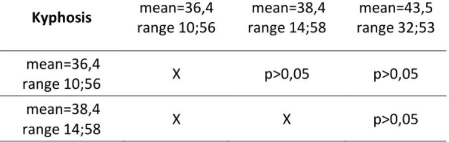
Tables 9: Comparison according to initial LK, thoracic fracture results ...29
Tables 10: Comparison according to initial LK, lumbar fracture results. ...29
Discussion ... 30
3
References ... 33
Part III: Is vertebral post-fracture local deformity a predictive factor of final spinal regional deformity? ... 35
Introduction ... 35
Materials and Methods ... 35
Figure 6: measurements on AP radiograph: Cobb angle and the two lateral height of fractured VB. ...36
Figure 7: measurements on lateral view: lordosis and anterior and posterior height of fractured VB. ...37
Table 11: Results for thoracic and lumbar fractures in coronal plane. ...38
Table 13: Results for thoracic fractures’ subgroup in coronal plane. ...39
Table 14: Results for lumbar fractures’ subgroup in coronal plane. ...39
Table 15: Results for lumbar fractures’ subgroup in sagittal plane. ...39
Table 16: Results for thoracic fractures’ subgroup in sagittal plane. ...39
Table 17: Analyses between RD according to VI values, thoracic fracture in coronal plane. ...40
Table 18: Analyses between RD according to VI values, lumbar fracture in coronal plane. ...40
Table 19: Analyses between RD according to VI values, thoracic fracture in sagittal plane. ...40
Table 20: Analyses between RD according to VI values, lumbar fracture in sagittal plane. ...41
Discussion ... 41 Conclusion ... 42 References ... 43 Conclusion ... 44 Références ... 46 Annexes communes: ... 46
Table 1: Demographic data. ... 46
Table 2 : Level injury ... 49
4 Remerciements
A mon directeur de thèse,
Monsieur le Docteur Yan Lefèvre Praticien Hospitalo-Universitaire
Service de chirurgie infantile, CHU Pellegrin
Je vous remercie très sincèrement de tout ce que vous avez fait et continuez de faire pour moi. Travailler à vos côtés est un plaisir, merci de toujours vous soucier de moi, de me soutenir et de m’apprendre votre métier avec autant de patience et de passion. J’espère vous rendre fier. Soyez assuré de mon respect et de ma gratitude.
A mon président de thèse,
Monsieur le Professeur Jean-Marc Vital
Professeur des Universités-Praticien Hospitalier Chef du pôle de chirurgie, CHU Pellegrin
Chef de service de l’unité rachis 1, CHU Pellegrin Membre de l’académie Nationale de Médecine
Merci de me faire l’honneur de présider ce travail. Les mois que j’ai passés dans votre service ont été riches de nombreux enseignements et resteront dans ma mémoire. Veuillez trouver dans ce travail le témoignage de mon profond respect et de ma gratitude.
A mon rapporteur de thèse,
Monsieur le Professeur Olivier Gille
Professeur des Universités-Praticien Hospitalier Unité rachis 1, CHU Pellegrin
Merci d’avoir pris du temps pour travailler avec moi, pour m’apprendre votre spécialité avec la rigueur et l’efficacité qui vous caractérisent. Soyez assuré de mon profond respect.
5
A mon jury de thèse,
Monsieur le Docteur Jean-Roger Pontailler Praticien Hospitalier
Service de chirurgie infantile, CHU Pellegrin
Merci pour la gentillesse et la bienveillance avec laquelle vous partagez votre savoir. J’ai hâte de poursuivre l’aventure à vos côtés pour profiter de votre expérience et de vos conseils avisés.
Vivement le 1er janvier 2019…
Monsieur le Docteur Clément Tournier Praticien Hospitalier
Service de chirurgie orthopédique et traumatologie, CHU Pellegrin
Travailler à tes côtés a été autant un plaisir qu’un honneur. Tu es pour moi un modèle à bien des égards, j’espère un jour être à la hauteur de ton enseignement. Merci pour tout.
Monsieur le Professeur Jérôme Sales-De-Gauzy Professeur des Universités-Praticien Hospitalier Chef de service de chirurgie infantile, CHU Purpan
Merci d’avoir accepté de juger ce travail. J’espère avoir le plaisir et l’honneur de travailler à vos côtés. Veuillez trouver dans ce travail le témoignage de mon profond respect.
Monsieur le Docteur Stéphane Aunoble
Maître de conférences des Universités et Praticien Hospitalier Unité d’Orthopédie et de Traumatologie, Rachis 2, CHU Pellegrin
Merci d’avoir accepté de juger ce travail. Soyez assuré de mon respect et de ma gratitude.
6
A ma sœur et mon frère,
Pour m’inspirer à chaque moment de ma vie. Merci de me faire rire, de m’apporter tant de bonheur et de fierté d’être votre sœur, grandir à vos côtés est un bonheur de tous les instants. Je vous aime de tout mon cœur.
A mes parents,
Pour être là depuis le début, pour m’avoir soutenue, encouragée, supportée…Merci de m’avoir toujours laissée être libre. Je vous aime.
A mes grands-pères que j’aurais voulu mieux connaitre.
A mes grands-mères pour être des modèles de travail et de vie. Je vous dédie ce
travail.
A tous les chirurgiens de l’hôpital pédiatrique,
Maya Loot, un soutien précieux, une amie bienveillante, merci de me faire profiter de tes expériences.
Aurore Bouty, pour m’avoir supportée, accompagnée et pour me rappeler tous les jours combien la rigueur est importante dans notre métier.
Monsieur le Professeur Eric Dobremez, Monsieur le Professeur Pierre Vergnes, Monsieur le Docteur Ramirez-Del-Villar, Monsieur le Docteur Frédéric Lavrand.
A monsieur le Docteur Bruno Zipoli,
Merci d’avoir cru en moi dès le début. Vous m’avez prise sous votre aile et depuis je ne l’ai pas quittée. Merci de toujours être là pour m’écouter et pour me dispenser des conseils avisés. J’espère un jour être digne de tout ce que vous avez fait pour moi. Je vous souhaite tout le bonheur que vous méritez.
A monsieur le Professeur Vincent Pointillart,
7
A mes maîtres d’internat,
Qui m’ont inspirée et qui m’ont fait partager l’amour du métier. Madame le Docteur Cernier,
Monsieur le Docteur Ribeyre, Monsieur le Professeur Fabre, Monsieur le Docteur Ibrahim Obeid.
A mes chefs de clinique,
Bertie, Julios Pallaros et sa palette murale, Fred, Motion, Louis, Rémi, le BDD, Julien. Vous n’avez pas été nombreux mais tellement exceptionnels. Mille mercis à vous de m’avoir appris mon métier avec autant de passion, de talent et toujours dans la bonne humeur. J’espère un jour être digne de votre enseignement et aussi m’occuper de vos enfants…
A tous mes amis,
Ceux de la première heure :
Mister Luke, témoin de ma thèse. Tu es l’ami dont tout le monde rêve. T’avoir à mes côtés est un plaisir, un réconfort et une grande fierté. Je te souhaite le bonheur et la réussite que tu mérites.
Emilie, ton courage a toujours été un exemple pour moi. Merci de m’être restée fidèle toutes ces années, tu es un repère dans ma vie. Que ton bonheur dure toujours.
Baptiste T., tu es un modèle, un grand frère bienveillant. Je te remercie de m’avoir obligée à me dépasser, de m’avoir montré qu’à cœur vaillant rien d’impossible. Obrigada por tudo amigo.
A vous trois, pour toutes ces années de bonheur partagé, merci d’avoir toujours été là pour moi, merci de m’avoir rappelé l’importance du dépassement de soi. Je suis tellement fière et heureuse de vous avoir auprès de moi. Je vous souhaite à tous les trois tout le bonheur que vous méritez.
A ma famille bordelaise :
Delphine, toi qui m’a si souvent remise en selle (dans tous les sens du terme !!)…Merci pour les soirées, les poneys, les verres vidés et tous ces bons moments qui n’ont pas de prix. Mille mercis de rendre ma vie géniale.
8 Léa, ma colocataire préférée. Merci d’être une amie fidèle, une confidente de choix. Merci pour les tartes au citron, les bouteilles de vin, les kiris…Je te souhaite beaucoup de bonheur et plein de p’tits chats.
DDC, ma copine, ne change rien. Merci de me faire rire, de me supporter, de me laisser être comme je suis et de partager mes écarts à la normale.
JMP, il n’y a pas de mot pour raconter ce qu’on a partagé, et c’est peut être mieux ainsi. Ce qui est sûr c’est que sans toi ça n’aurait pas été aussi facile. J’espère un jour avoir (l’immense) plaisir de retravailler avec toi. Ne change rien t’es au top. BMP.
A tous les autres :
Jean-Ma, Christelle, Zahia et leur passion pour la faune marine, Wendy, Alhem et sa capacité à monopoliser les micros, Cheval Fougueux, La Jousse, Maxime, et tous les autres.
Merci pour tous les bons moments.
A Bibine,
Merci d’être une amie fidèle, une personne sur qui je peux compter. Merci de m’avoir ouvert l’esprit et fait partager un peu de ta folie. Je te souhaite tout le bonheur que tu mérites.
A Amaury, Mimi, Chacha,
Merci de toujours être là bien que les équidés, eux, ne le soient plus.
A tous ceux qui ont partagé un semestre avec moi :
CDI, Sexy Deby, Swagman, Chouchou, Rouf, Knaki, Wrobi, Cogniet et minie-Cogniet, Vanessa, Vargasm, Paul Precaire, Aquel, Mi-homo, Nodimar, Anais, Romain, Pierre, Khalil.
9
A Patou,
Merci d’être toujours là, de toujours trouver les mots. Je ne te remercierai jamais assez pour tout. Je te dédie tout mon bonheur et te souhaite le meilleur.
A toutes les secrétaires des services d’orthopédie du 6ème
, 7ème et 8ème étage qui m’ont tant aidée.
A toutes les équipes paramédicales que j’ai pu croiser au cours de mon internat,
merci de m’avoir aidée, parfois encouragée et souvent supportée.
A Carlos et Prisca, vous qui m’avez montré que rien n’est possible sans le travail et
la persévérance, je vous dédie ce travail.
A Caillotte, Herko, Fifty, Perfect et Very Much merci pour toutes les joies et
l’affection que vous me donnez.
10
Introduction
Les fractures du rachis thoraco-lombaire sont rares en pédiatrie, moins de 4% des fractures de l’enfant (1-4) ; elles peuvent survenir de la naissance jusqu’à la fin de la croissance. Elles intéressent un squelette en croissance comprenant des zones cartilagineuses vertébrales non ossifiées, qui sont des zones de fragilité chez l’enfant. La part variable des différents éléments constituant la vertèbre (cartilage, os, moelle hématopoïétique) se modifie et les modes de faillite mécanique sont spécifiques à chaque âge pour un traumatisme équivalent. Les zones de vulnérabilité sont les cartilages neuro-centraux chez le tout petit, les cartilages de croissance des plateaux vertébraux chez l’enfant d’âge moyen et la jonction listel marginal-corps vertébral chez l’adolescent.
La croissance vertébrale est bien connue. Chaque corps vertébral compte deux zones de croissance : les physes des plateaux vertébraux qui permettent la croissance en hauteur du corps vertébral et les cartilages neuro-centraux qui unissent le corps vertébral aux pédicules et déterminent la taille du canal rachidien. La poursuite de la croissance du corps vertébral longitudinalement et dans le sens antéropostérieur dépend de points d’ossification secondaires qui apparaissent après la naissance. Cette croissance osseuse est responsable du phénomène de remodelage, spécifique à l’enfant, qui permet la « correction » de la déformation fracturaire après un traumatisme de la colonne vertébrale. La croissance rachidienne se prolonge durant la deuxième décennie de vie, avec un pic majeur au moment de la puberté, c’est pourquoi le traitement orthopédique des fractures du rachis thoraco-lombaire peut être proposé jusqu’à la fin de la croissance. Ce remodelage osseux peut être spectaculaire, en particulier dans le plan sagittal, cependant il demeure très difficile à anticiper. Plus encore, la déformation dans le plan coronal et son évolution, soumise aux mêmes capacités de remodelage, sont peu étudiées, et laissent une zone d’ombre importante sur les fractures tassements vertébrale générant une déformation dans le plan coronal.
A cela s’ajoute le risque de lésion des zones de croissance osseuses et le phénomène d’épiphysiodèse post-traumatique qui pourrait également intervenir dans l’évolution des courbures post-traumatiques. L’épiphysiodèse asymétrique dans le plan coronal, ou antérieure dans le plan sagittal, rendrait impossible le remodelage osseux dû à la croissance et, entraînerait des troubles de la statique rachidienne à type de scoliose dans le plan coronal et d’hypercyphose dans le plan sagittal. Ainsi, il est admis qu’il existe un risque de cyphose et/ou de scoliose après une fracture du rachis thoraco-lombaire chez l’enfant et l’adolescent (1, 2, 5, 6).
Bien que la littérature se soit intéressée aux fractures du rachis thoraco-lombaire chez l’enfant, il y a peu d’articles qui rapportent un suivi à long terme dans le plan coronal et dans le plan sagittal de ces fractures traitées orthopédiquement en période de croissance.
11 Ce travail avait pour objectif initial l’étude des déformations post-traumatiques dans le plan coronal et leurs évolutions. Le risque « scoliogène » des fractures tassements traitées orthopédiquement devait être caractérisé avec, si possible, la mise en évidence de facteurs de risque en rapport (âge, maturité osseuse, niveau lésionnel, nombre de fractures…).
L’ensemble des données recueillies a permis l’analyse dans le plan coronal et dans le plan sagittal des déformations post-traumatiques et de leurs évolutions respectives. Si l’évolution dans le plan frontal et dans le plan sagittal semblent a priori liée, les auteurs ont choisi, dans un souci de clarté, de diviser l’analyse en trois parties, sous forme de trois articles scientifiques qui ont pour objectif de répondre aux questions suivantes :
- Quelle est l’évolution des déformations vertébrales post-traumatiques en période de croissance dans le plan frontal lors d’un traitement orthopédique ?
- Quel est l’impact des déformations vertébrales post-traumatiques en période de croissance sur l’équilibre sagittal du rachis lors d’un traitement orthopédique ? - Quel est le lien entre la déformation initiale de la ou les vertèbre(s) fracturée(s) et
la déformation finale de la colonne vertébrale ?
Les trois articles sont rapportés ad integrum, comme soumis aux éditeurs, au prix de certaines redondances, obligatoires dans la mesures où il s’agit de la même série de patients. Ils ont été
12
References
1. Lascombes P. Fractures du rachis thoraco-lombaire. In: médical S, editor. Fractures de l'enfant. 2002. p. 301-12.
2. Parisini P, Di Silvestre M, Greggi T. Treatment of spinal fractures in children and adolescents: long-term results in 44 patients. Spine. 2002;27(18):1989-94.
3. Karlsson MK, Moller A, Hasserius R, Besjakov J, Karlsson C, Ohlin A. A modeling capacity of vertebral fractures exists during growth: an up-to-47-year follow-up. Spine. 2003;28(18):2087-92.
4. Kraus R, Stahl JP, Heiss C, Horas U, Dongowski N, Schnettler R. Fractures of the thoracic and lumbar spine in children and adolescents. Der Unfallchirurg. 2013;116(5):435-41.
5. Denis F. Thoracolumbar Spine Trauma. Moe's Textbook of Scoliosis and Other Spinal Deformities. 3rd ed1995. p. 430-50.
6. Vaccaro AR, Silber JS. Post-traumatic spinal deformity. Spine. 2001;26(24 Suppl):S111-8.
13 Part I: Post-trauma coronal spinal deformity after conservative treatment of
thoracolumbar spinal fracture in children and adolescents. Long-term results in 48 patients.
Introduction
Spine injuries in children are relatively rare and represent a small percentage of overall injuries to children (range 0.3-4%)(1-6), they are less common than in adults because of the greater mobility and elasticity of the pediatric spine and the smaller mass of the child’s body. The incidence of pediatric spine injuries has been reported to be 2-5% of all spine injuries (1) and occur in the thoracic or lumbar spine in 20-60% of injuries(4). Conservative treatment remains the most common treatment because children and adolescents have strong bones with excellent healing potential and usually reconstitute loss of vertebral height. Child spinal injuries are determined by the presence of growth plates. There are three groups of cartilage involved in vertebral growth, which allow the three dimensional growth of the vertebrae: the cartilage of the vertebral endplate, the neurocentral cartilage, and the ring apophysis. The cartilage of the vertebral endplate is responsible for the growth in height, like the growth plate of a long bone. In this way a traumatic separation of the vertebral endplate can account for abnormal growth because of premature epiphyseal fusion, in the manner described by Salter and Harris in long bones. This is one hypothesis to explain coronal spinal deformity after vertebral fracture. Post-trauma scoliosis are well known (1, 2, 7, 8) but, to our knowledge, there is no report which focuses on the coronal plane analysis after vertebral fracture. The aim of the present study was to evaluate radiological and clinical findings of coronal spinal balance, after conservative treatment of spinal fracture.
Materiel and Methods
A tricentric (2 hospitals, 3 departments of surgery) retrospective study was performed between 1996 and 2014. A computer search was initiated to find all patients under 18 years old who were hospitalized because of lumbar and/or thoracic vertebra fracture. Patients who underwent surgery during the first year after the accident, patients with neurological deficiency and patients with pre-existing scoliosis or spinal malformation were excluded. Fractures were classified according to AO spine fracture classification. All patients’ demographic data are summarized in table 1 (p.46).
Radiological measurements. All angular measurements were calculated by the method of
Cobb (9) with Surgimap software and, were done by a single person to decrease measurement bias. On anteroposterior (AP) radiographs, local and regional Cobb angles (figure 1) were measured to quantify the deformity of each fractured vertebra and the deformity between vertebrae at the upper and lower limits of the curve respectively. The difference between final and initial local Cobb angles was called local deformity (LD); and the same difference
14 regarding regional Cobb angles was called regional deformity (RD). All measurements were done at the moment of the accident, authors using full spine radiograph with brace or cast and at last follow up with another full spine radiograph without contention; all radiograph were done in standing position. Scoliosis was defined according to the Scoliosis Research Society (SRS) by a Cobb angle above 10° and, vertebra rotation was asserted by Nash and Moe method.
Analysis. Several analyses were done according to the initial Risser grade, the number of
vertebrae fractured (single fracture versus several stepped fractures) and the level of injury (thoracic versus lumbar). The first analysis was made in each group to characterize LD and RD. The second analysis was made to compare subgroups between them. The third analysis was to search for a correlation between sagittal and coronal deformity.
Telephone interview. The authors attempted to contact each patient included in the study.
Patients were asked about their pain, their professional and physical activities, their consumption of painkillers, and the consequences of back pain in their daily activities.
Statistical analysis. Data were presented as mean and range. A Student t-test was performed
to compare series. A difference of P<0.05 was regarded as a statistical significant difference. A Pearson test was used to look for a correlation between two series.
Figure 1: Measurements of local and regional Cobb angle.
Results
The computer search found 320 patients in center 1 (2 departments of surgery) and 41 in center 2. After application of exclusion criteria and verification of patients’ files, 48 patients were included, 23 males and 25 females (figure 2, p.50), with a total of 84 vertebrae. There were 43 thoracic fractures (T4-T12) and 41 lumbar fractures (L1-L5) (Table 2, p.49); 89.6% are A1 fracture according to Magerl classification. All fractures were treated conservatively,
15 85.4% had a brace and 14,6% underwent reduction of the fracture and a cast. The mean age at the inclusion was 12.3 years old (range 2.6-17.1). Mean follow-up was 49 months (range 12-210) and 62% of cases had a Risser grade 4 or 5 at the end of the follow-up.
Coronal deformity
Evolution of local deformity
Mean initial local Cobb angle (ILCA) was 3.05° (range 0;19) and mean final local Cobb angle (FLCA) was 2.6° (range 0;10). LD decreased by -0.3° but without any statistical difference between FLCA and ILCA.
Evolution of regional deformity
Mean initial regional Cobb angle (IRCA) was 5.9° (range 1;14) and mean regional final Cobb angle (FRCA) was 8.4° (range 1;34). RD increased by +2.5° with a statistical (p=0.01) difference between FRCA and IRCA.
Final scoliosis
At the end of the follow-up, 11 patients (23%) had a scoliosis with a Cobb angle above 10° and associated rotation (figure 3); according to Nash and Moe method, rotation was characterized as + in 5 patients and as ++ in 6. Ten individuals had a Cobb angle at 20° or less and one had a Cobb angle of 20°. None of these 11 patients presented an IRCA above 10° on the initial AP radiograph.
16
Figure 3: Example of left lumbar scoliosis 4 years after L2 fracture.
Predictive factors of coronal deformity
The following statistical analyses were performed making subgroups to try to find predictive factors of coronal deformity.
Subgroup results: skeletal maturity, initial Risser grade
Patients were divided into two groups, group 1 with patients who had a Risser grade of 0, 1 or 2 and group 2 who had a Risser grade of 3, 4 or 5. Initially, both groups were comparable regarding the following data: age, gender, mechanism of injury, follow-up. In group 2, FRCA (mean=9.9°, range 1;19) was statistically higher that the IRCA (mean=5.8°, range 1;13) (p=0.03). On the contrary, there was no statistical difference between IRCA and FRCA in group 1. There was also no statistical difference between ILCA and FLCA in each group.
17
Subgroup results: single fracture versus several staged fractures
Two groups were made regarding the number of fractures, group U, with a single vertebra fracture and group S, with several staged fractures. Initially, both groups were comparable regarding the following data: age, gender, initial Risser grade, mechanism of injury and follow-up. In group U, FRCA (mean = 9.0°, range 1;34) was statistically higher than IRCA (mean=5.4°, range 1;14) (p=0.02). There was no statistical difference regarding IRCA and FRCA in group S. There was also no statistical difference between ILCA and FLCA in each group.
Subgroup results: level of injury, thoracic versus lumbar
Patients were divided into two groups regarding the level of injury, group T, thoracic vertebra fracture and group L, lumbar vertebra fracture. Initially, both groups were comparable regarding the following data: age, gender, mechanism of injury and follow up. But, there was a statistical difference between the two groups regarding the initial Risser grade (p=0.003); in group T 83.3% were Risser 0, 1 or 2 whereas in group L there were only 50% of Risser 0, 1 or 2. In group L, FRCA (mean=10.6°, range 1;34) was statistically higher than IRCA (mean=5.9°, range 1;13) (p=0.007). The same analysis in group T did not find any statistical difference. There was also no statistical difference between ILCA and FLCA in each group.
18
ILCA p FLCA p IRCA p FRCA p LD p RD p
Group 1 : Risser 0, 1, 2 n=51 mean=3,2 range 0;19 p>0,05 mean=2,2 range 0;10 p>0,05 mean=6,7 range 1;14 p>0,05 mean=7,9 range 0;34 p>0,05 mean=-1,0 range-18;6 2>1 p=0,006 mean=1,9 range -12;28 p>0,05 Group 2 : Risser 3, 4, 5 n=33 mean=2,9 range 0;10 mean=3,2 range 0;8 mean=5.8 range 1;13 mean=9.9 range 1;19 mean=0,9 range -4;5 mean=3,8 range -4;12 Thoracic (T) n=43 mean=3,4 range 0;19 p>0,05 mean=2,2 range 0;8 p>0,05 mean=5,6 range 1;14 p>0,05 mean = 6,2 range : 1;19 L>T p=0,02 mean=-1,3 range -18;5 L>T p=0,003 mean=0,5 range -12;9 L>T p=0,02 Lumbar (L) n=41 mean=2,6 range 0;8 mean=3,1 range 0;10 mean=6 range 1;13 mean = 10,6 range : 1;34 mean=0,9 range -4;6 mean=4,6 range -4;28 Single fracture (U) n=32 mean=3,5 range 1;19 p>0,05 mean=3,1 range 0;10 p>0,05 mean=5,3 range 1;14 p>0,05 mean = 9.0 range 1;34 p>0,05 mean=0,7 range -5;6 U>S p=0,03 mean=3,6 range -3;28 p>0,05 Several fractures (S) n=52 mean=2,4 range 0;8 mean=1,9 range 0;8 mean=6,5 range 1;13 mean=7,4 range 0;19 mean=-1,1 range -18;5 mean=0,9 range -12;12
19
Correlation with sagittal deformity
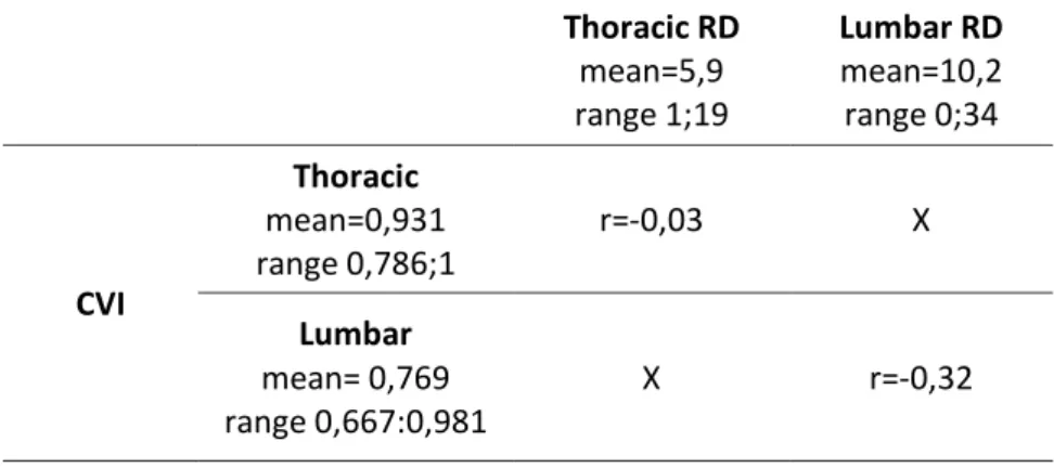
Our analyses reported no statistical correlation between coronal and sagittal deformity at the last follow-up, nor in thoracic (r=-0.2) or lumbar area (r=0.05).
Others analyses
Clinical findings
Thirty-two patients were contacted by telephone, 81% reported back pain; but 87.5% revealed that back pain did not disturb their daily life and did not require any medication. Only one patient had stopped his professional activity, 31 of the 32 patients (97%) continued their professional or student life as before.
Mechanism of injury
The mechanism of injury was sport accidents in 50%, motor vehicle accidents (MVA) in 26% and falls (accidents or suicide attempts) in the last 23%.
Associated lesions
There were 42% of associated injuries in our series. Associated skull and long bone fractures were present in 33% of all cases, abdominal and/or thoracic injuries in 18.75%.
Discussion
The present series of 84 vertebrae confirms that conservative treatment of wedge compression fractures without neurological lesion ensures stabilization with moderate residual deformity. It confirms that patients with the most skeletal immaturity have greater bone remodeling power. But, in 11 cases restoration of height is inconclusive. Prevalence of scoliosis in our series is 23% whereas prevalence of idiopathic scoliosis (IS) is 0.47-15.3% (10-13), decreasing to 0.2-3% for a Cobb angle superior to 30° (11, 14). This result suggests that a fracture of the thoracolumbar spine is a risk factor of scoliosis. Other publications find scoliosis after conservative treatment of spinal fractures. Parisini et al. (1), in a series of 44 patients report four (9%) burst fractures (one T5, two L1 and one L3) treated conservatively with a mean final Cobb angle of 20°. Karlsson et al. (5) have two individuals (8.3%), in a series of 24 patients, who developed a scoliosis with 11° and 13° Cobb angles. Gnanenthiran
et al. (15) find only one (2%) scoliosis, in a series with 50 patients, with a Cobb angle of 14°.
The authors note that, in our series and in literature, post-traumatic scoliosis has small Cobb angles. The hypothesis to explain the incapacity to reconstitute loss of vertebral height in immature patients is the post-traumatic epiphysiodesis, as it may be encountered in long bones (16).
An interesting result in the analysis of the whole series is the improvement of local deformity whereas regional deformity worsens. The phenomenon of epiphysiodesis does not explain this result while the Hueter and Volkmann principle could. They describe how increased pressure on the end plate of bone retards growth (Hueter) and conversely, reduced pressure accelerates growth (Volkmann). Indeed, we can imagine that initial LD takes time to
20 heal and during this time an asymmetric growth occurs worsening the RD. Once the deformity reaches certain proportions, the effect of gravity on lever arm created continues to be a major deforming force. Using this principle, scoliotic curves have been reproduced on animal studies. Braun et al.(17) create an idiopathic type of deformity in goats by applying forces across the spine and find wedging of the vertebrae similar to that seen in scoliosis in humans. Similarly, Mente (18, 19) and Stokes (20, 21) in separate studies on rat tail models, not only create scoliosis, but also succeed in correcting it reversing the forces. Another explanation is the involvement of the intervertebral disc in the curve increase. Schlösser et al. (22) study the contribution of VB and intervertebral discs to three-dimensional (3-D) spinal deformity in adolescent idiopathic scoliosis (AIS); they conclude that the discs contribute more to 3-D deformity than the bony structure, because of the decreased stiffness of intervertebral fibrocartilage as compared to bony vertebrae. Modi et al.(23) in a series of 150 adolescent AIS conclude that wedging in disc and body increase with progression of the scoliosis and disc wedging is more profound in the lumbar area. This could explain why, in our series, lumbar fractures have a higher deformity rate than thoracic fractures. But, all of these studies are done on patients with AIS, not with post-trauma spinal deformity. Moreover, literature has varying results regarding disc lesion after VB fracture. Karlsson et al. (5) report no decreased in disc height at the fracture level but just a modification of nucleus pulposus intensity on the T2-weighted MRI. Whereas Kertulla et al. (24) find 57% of disc degeneration with a strong association between endplate damage and adjacent intervertebral disc degeneration. The hypothesis of intervertebral disc involvement in post-trauma spinal deformity has to be studied further to know its role in final spinal deformity.
The analysis regarding the level of injury shows that lumbar fractures are more responsible for coronal deformity than thoracic fractures; this could be explained by the fact that lumbar vertebrae are more mobile than thoracic vertebrae which are attached to the rib, making a rigid assembly less sensitive to deformity. Literature seems to report the same tendency as our series (1).
Our analysis according to initial Risser grades underscores that the group with a Risser grade 3 or above has FRCA higher than IRCA whereas there is no difference in the group with a Risser grade of 2 or below. This confirms that healing potential allows the restoration of the loss of vertebral height in immature patients. There are other publications which find that the cut-off point of a Risser grade 2 allows good restoration of vertebral wedging (2, 25)
The difference in group with a single fracture where FRCA is higher than IRCA is not found in the group with several fractures. Local deformity is higher in group with a single fracture than in group with several fractures. Authors make the hypothesis that when there is just one fracture, the deformity is more important whereas when several vertebrae are involved the energy of the trauma is absorbed in a greater area, making the deformity is smaller. Indeed, even if there is no statistical difference regarding ILCA between the two groups, ILCA is higher in group with unique fracture than in group with several fractures.
In our study, there is no correlation between coronal and sagittal deformity at the last follow-up; these results confirm that coronal analysis must be taken into account when the
21 treatment of vertebral fracture is decided. Indeed, to be concerned only with sagittal plane does not seem sufficient and satisfactory.
The weakness in our study is the retrospective design responsible for a heavy loss of information and patients. The strength of this study is the follow-up and the radiographic evaluation by the same individual at both baseline and at follow-up.
Our series finds a high rate of back pain, but a functional satisfying result, because patients do not feel limited in their daily life and need no pain-relief medication. We find varying values in literature; 12% to 57% of back pain after conservative treatment (1, 5, 24, 26). The rate of back pain in adolescents without back injury is high too: 7% to 70% (27-29) depending on the definition of pain and study design; this makes the interpretation of our rate difficult because back pain is multifactorial and it is difficult to identify the precise origin. Initially,authors wanted to search a correlation between coronal deformity and back pain but, because of the high rate found, it was not possible to conclude.
Our series finds the same common mechanisms of injury as in literature (2, 6, 30-32): sports accidents, MVA, pedestrian-vehicle accidents, and falls with varying proportions according to publications.
Values concerning associated lesions are similar with those of literature: 42% to 65%
of patients have associated injuries with 5% to 50% of skull or long bone fractures and 20% to 37% of visceral (thoracic and/or abdominal) injuries (3, 7, 32).
Conclusion
There is a higher prevalence of scoliosis in the population which sustains thoracolumbar spinal fractures. Risser grade 3 or above, lumbar fractures and single fractures seem to be responsible for more severe coronal deformation.
22
References
1. Parisini P, Di Silvestre M, Greggi T. Treatment of spinal fractures in children and adolescents: long-term results in 44 patients. Spine. 2002;27(18):1989-94.
2. Lascombes P. Fractures du rachis thoraco-lombaire. In: médical S, editor. Fractures de l'enfant2002. p. 301-.
3. Rush JK, Kelly DM, Astur N, Creek A, Dawkins R, Younas S, et al. Associated injuries in children and adolescents with spinal trauma. J pediatr orthop. 2013;33(4):393-7. 4. Sayama C, Chen T, Trost G, Jea A. A review of pediatric lumbar spine trauma.
Neurosurg focus. 2014;37(1):E6.
5. Karlsson MK, Moller A, Hasserius R, Besjakov J, Karlsson C, Ohlin A. A modeling capacity of vertebral fractures exists during growth: an up-to-47-year follow-up. Spine. 2003;28(18):2087-92.
6. Kraus R, Stahl JP, Heiss C, Horas U, Dongowski N, Schnettler R. Fractures of the thoracic and lumbar spine in children and adolescents. Der Unfallchirurg. 2013;116(5):435-41.
7. Denis F. Thoracolumbar Spine Trauma. Moe's Textbook of Scoliosis and Other Spinal Deformities. 3rd ed1995. p. 430-50.
8. Vaccaro AR, Silber JS. Post-traumatic spinal deformity. Spine. 2001;26(24 Sl):S111-8.
9. Cobb JR. The problem of the primary curve. J Bone Joint Surg Am. volume. 1960;42-A:1413-25.
10. H. Bracq MC. Dépistage, examen clinique et radiographie conventionnelle de la scoliose idiopathique. In: Médical S, editor. Scoliose Idiopathique1997. p. 71-2.
11. Lonstein JE. Idiopathic Scoliosis. Moe's Textbook of Scoliosis and Other Spinal Deformities1995. p. 219-21.
12. Konieczny MR, Senyurt H, Krauspe R. Epidemiology of adolescent idiopathic scoliosis. J Child Orthop. 2013;7(1):3-9.
13. De Souza FI, Di Ferreira RB, Labres D, Elias R, De Sousa AP, Pereira RE.
Epidemiology of adolescent idiopathic scoliosis in students of the public schools in Goiania-GO. Acta Ortop Bras. 2013;21(4):223-5.
14. Kane WJ, Moe JH. A scoliosis-prevalence survey in Minnesota. Clin Orthop Rel Res. 1970;69:216-8.
15. Gnanenthiran SR, Adie S, Harris IA. Nonoperative versus operative treatment for thoracolumbar burst fractures without neurologic deficit: a meta-analysis. Clin Orthop Rel
Res. 2012;470(2):567-77.
16. Zhang H, Sucato DJ. Unilateral pedicle screw epiphysiodesis of the neurocentral synchondrosis. Production of idiopathic-like scoliosis in an immature animal model. J Bone
23 17. Braun JT, Ogilvie JW, Akyuz E, Brodke DS, Bachus KN, Stefko RM. Experimental scoliosis in an immature goat model: a method that creates idiopathic-type deformity with minimal violation of the spinal elements along the curve. Spine. 2003;28(19):2198-203. 18. Mente PL, Aronsson DD, Stokes IA, Iatridis JC. Mechanical modulation of growth for the correction of vertebral wedge deformities. J Orthop Res: official publication of the
Orthopaedic Research Society. 1999;17(4):518-24.
19. Mente PL, Stokes IA, Spence H, Aronsson DD. Progression of vertebral wedging in an asymmetrically loaded rat tail model. Spine. 1997;22(12):1292-6.
20. Stokes IA. Analysis of symmetry of vertebral body loading consequent to lateral spinal curvature. Spine. 1997;22(21):2495-503.
21. Stokes IA, Spence H, Aronsson DD, Kilmer N. Mechanical modulation of vertebral body growth. Implications for scoliosis progression. Spine. 1996;21(10):1162-7.
22. Schlosser TP, van Stralen M, Brink RC, Chu WC, Lam TP, Vincken KL, et al. Three-Dimensional Characterization of Torsion and Asymmetry of the Intervertebral Discs versus Vertebral Bodies in Adolescent Idiopathic Scoliosis. Spine. 2014.
23. Modi HN, Suh SW, Song HR, Yang JH, Kim HJ, Modi CH. Differential wedging of vertebral body and intervertebral disc in thoracic and lumbar spine in adolescent idiopathic scoliosis - A cross sectional study in 150 patients. Scoliosis. 2008;3:11.
24. Kerttula LI, Serlo WS, Tervonen OA, Paakko EL, Vanharanta HV. Post-traumatic findings of the spine after earlier vertebral fracture in young patients: clinical and MRI study.
Spine. 2000;25(9):1104-8.
25. Pouliquen JC, Kassis B, Glorion C, Langlais J. Vertebral growth after thoracic or lumbar fracture of the spine in children. J pediatr orthop.1997;17(1):115-20.
26. Moller A, Hasserius R, Redlund-Johnell I, Ohlin A, Karlsson MK. Nonoperatively treated burst fractures of the thoracic and lumbar spine in adults: a 23- to 41-year follow-up.
Spine J : official journal of the North American Spine Society. 2007;7(6):701-7.
27. Bjorck-van Dijken C, Fjellman-Wiklund A, Hildingsson C. Low back pain, lifestyle factors and physical activity: a population based-study. J Rehabil Med. 2008;40(10):864-9. 28. Grauers A, Topalis C, Moller H, Normelli H, Karlsson M, Danielsson A, et al. Prevalence of Back Problems in 1069 Adults With Idiopathic Scoliosis and 158 Adults Without Scoliosis. Spine. 2014.
29. Fontecha CG, Balague F, Pellise F, Rajmil L, Aguirre M, Pasarin M, et al. Low back pain in adolescents: is quality of life poorer in those seeking medical attention? Spine. 2011;36(17):E1154-61.
30. B. Yaszay BAA. Pediatric spinal trauma. Spine Secrets Plus. 2nd ed2012. p. 304-10. 31. Erfani MA, Pourabbas B, Nouraie H, Vadiee I, Vosoughi AR. Results of fusion and instrumentation of thoracic and lumbar vertebral fractures in children: a prospective ten-year study. Musculoskelet Surg. 2014.
32. Mahan ST, Mooney DP, Karlin LI, Hresko MT. Multiple level injuries in pediatric spinal trauma. J Trauma. 2009;67(3):537-42.
24 Part II: Thoracolumbar spinal fracture treated conservatively in children and
adolescents, outcomes in sagittal plane Long term follow up in 48 patients.
Introduction
Spine injuries in children are relatively rare and represent a small percentage of overall injuries in children (range 0.3-4%)(1-6), there are less common than in adults because of the greater mobility and elasticity of the pediatric spine and the smaller mass of the child’s body. The incidence of the pediatric spine injuries has been reported to be 2-5% of all spine injuries (1) and occur in the thoracic or lumbar spine from 20% to 60%(4). Conservative treatment remains the most common because children and adolescents have strong bones with excellent healing potential and usually reconstitute loss of vertebral height. Although spine injuries in children have received an increasing coverage in the literature, there are, to our knowledge, few reports which focusing on the post-trauma sagittal balance whereas it is associated with pain, neurologic symptoms and poor quality of life. Adjacent anatomical regions of the spine and sacro-pelvis are interdependent (7) , and their relationship results in a stable and balanced posture (8, 9). Therefore, it would be logical that post-trauma local kyphosis (LK) bring hyperlordosis and maybe modify pelvic parameters as adaptive behaviors, as shown in aging spine. The aim of the present study is to confirm that vertebral fractures are responsible for LK and to investigate the sagittal balance looking at compensation mechanisms as increased lumbar lordosis and modification of pelvic parameters.
Materials and Methods
A tricentric (2 hospitals, 3 departments of surgery) retrospective study was performed between 1996 and 2014. An informatics search was initiated to find all patients under 18 years old who were hospitalized because of lumbar and/or thoracic vertebra fracture. Patients who underwent surgery during the first year after the accident, patients with neurologic deficiency and patient with pre-existing scoliosis or spinal malformation were excluded. Fractures were classified according to Magerl classification. All patients’ demographic data are summarized in table 1 (p.46).
Radiological measurements (figure 1 and 2). All angular measurements were calculated by a
modification of the Cobb technique (10) with Surgimap software, there were done by a single person to decrease measurements bias. On lateral plain radiographs, LK was measured to quantify the deformity of each fractured vertebra; thoracic kyphosis (T4-T12) and L1-L5 lumbar lordosis were measured to quantify the regional deformity (RD) after thoracic and lumbar fracture respectively. The L3-L5 lumbar lordosis and pelvic parameters (pelvic tilt (PT), sacral slope (SS), pelvic incidence (PI)) were measured to quantify adaptive spinal and pelvic behaviors. At last follow up, the C7 plumbline was measured to characterize the global
25 sagittal balance. The measurement was assumed positive if directed forwards and negative if directed backwards. All measurements were done at the moment of the accident, authors using full spine radiograph with brace or cast and at last follow up with another full spine radiograph without contention; all radiograph were done in standing position.
Analysis. First analyses were done to search an evolution between time at fracture and last
follow up looking at all the measurements. Second analyses were done according to the initial Risser grade to know if evolution was different according to skeletal maturity. Last analyses were done according to the initial LK to search if higher initial LK was responsible for higher RD and modification of pelvic parameters and C7 plumbline.
Telephone interview. Authors attempted to contact each patient included in the study. Patients
were asked about their pain, their professional and sport activities, and their painkillers’ consumption.
Statistical analysis. Data are presented as mean and range. A Student t test was performed to
compare series. A difference of P<0.05 was regarded as a statistical significant difference.
Figure 4: Measurements of local kyphosis (LK), T4-T12 kyphosis and
L1-L5 lordosis and C7 plumbline.
Figure 5; Measurements of L3-L5 lordosis and all pelvic parameters (PI, PT and SS).
26
Results
The informatics search found 320 patients in center 1 (2 departments of surgery) and 41 in center 2. After application of exclusion criteria and verification of patients’ files, 48 patients were included (figure 2, p.50), 23 males and 25 females. There were a total of 84 vertebrae, 43 thoracic fractures (T4-T12) and 41 lumbar fractures (L1-L5) (Table 2, p49); 89.6% are A1 fracture according to Magerl classification. All fractures were treated conservatively, 85.4% had a brace and 14,6% underwent reduction of the fracture and a cast. Mean age at the inclusion was 12.3 years old (range 2.6-17.1). Mean follow up was 49 months (range 12-210) and, 62% had a Risser grade 4 or 5 at the end of the follow up.
Sagittal deformity, global analysis: thoracic fracture (T1-T12) (Table 3), lumbar fracture (L1-L5) (Table 4).
Initially, the two groups were comparable regarding the following data: age, gender, initial Risser grade, mechanism of injury and follow up. These analyses confirmed the remodeling power of young person and reported a statistically increased lordosis (L1-L5 and L3-L5) after lumbar fracture.
Table 3: Evolution of sagittal parameters between time at accident and last follow up, thoracic fracture.
Kyphosis Local Kyphosis Thoracic Lordosis L1-L5 Pelvic Tilt Sacral Slope Incidence Pelvic C7 Plumb line (mm)
Initial (I) 11,6 (1;22) 35,8 (14;65) 34,9 (21;56) 10,1 (1;22) 37 (18;65) 47,9 (32;88) -2,5 (-38,6;38,1) Final (F) 8,2 (0;18) 39,6 (14;66) 34,2 (20;41) 9(1;22) 36,1 (15;57) 46,3 (31;69) -3,6 (-36,1;49) p I>F p=0,0007 p>0,05 p>0,05 p>0,05 p>0,05 p>0,05 p>0,05
27
Table 4: Evolution of sagittal parameters between time at accident and last follow up, lumbar fracture. Local Kyphosis L1-L5 Lordosis L3-L5
Lordosis Pelvic Tilt
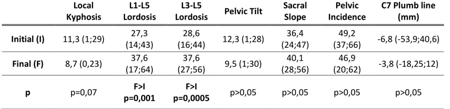
Sacral Slope Pelvic Incidence C7 Plumb line (mm) Initial (I) 11,3 (1;29) 27,3 (14;43) 28,6 (16;44) 12,3 (1;28) 36,4 (24;47) 49,2 (37;66) -6,8 (-53,9;40,6) Final (F) 8,7 (0,23) 37,6 (17;64) 37,6 (27;56) 9,5 (1;30) 40,1 (28;56) 46,9 (20;62) -3,8 (-18,25;12) p p=0,07 F>I p=0,001 F>I p=0,0005 p>0,05 p>0,05 p>0,05 p>0,05
Sagittal deformity: Analyses according to skeletal maturity, initial Risser grade (Tables 5, 6, 7 and 8). Initially, both groups were comparable regarding the following data: age,
gender, mechanism of injury and follow-up. Results are the same than in the analysis of the whole series: it confirmed the remodeling power of young person and found an increased lordosis after lumbar fracture.
All analyses report no modification of C7 plumbline and pelvic parameters at follow up except the increased SS after lumbar fracture in group with Risser grade at 2 or less.
Table 5: Results of subgroup Risser grade 0, 1 and 2, thoracic fractures.
Local
Kyphosis Kyphosis
L1-L5
Lordosis Pelvic Tilt Sacral Slope
Pelvic Incidence C7 Plumb line (mm) Initial (I) 12,2 (1;22) 34,5(14;60) 35,9 (25;56) 10,3 (1;28) 38,1 (31;65) 47,1 (34;88) -1,2 (-38,6;38,1) Final (F) 7,9 (1;18) 38,3(14;66) 34,6 (26;41) 6,8 (1;16) 36,6 (17;47) 45,8 (31;62) -3,4 (-45,4;49) p I>F p=0,0004 p>0,05 p>0,05 p>0,05 p>0,05 p>0,05 p>0,05
28
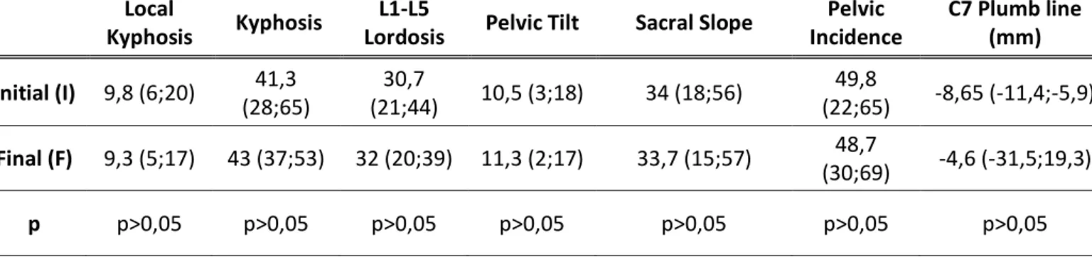
Table 6: Results of subgroup Risser grade 3, 4 and 5, thoracic fractures.
Local
Kyphosis Kyphosis
L1-L5
Lordosis Pelvic Tilt Sacral Slope
Pelvic Incidence C7 Plumb line (mm) Initial (I) 9,8 (6;20) 41,3 (28;65) 30,7 (21;44) 10,5 (3;18) 34 (18;56) 49,8 (22;65) -8,65 (-11,4;-5,9) Final (F) 9,3 (5;17) 43 (37;53) 32 (20;39) 11,3 (2;17) 33,7 (15;57) 48,7 (30;69) -4,6 (-31,5;19,3) p p>0,05 p>0,05 p>0,05 p>0,05 p>0,05 p>0,05 p>0,05
Table 7: Results of subgroup Risser grade 0, 1 and 2, lumbar fractures.
Local
Kyphosis
L1-L5 Lordosis
L3-L5
Lordosis Pelvic Tilt Sacral Slope
Pelvic Incidence C7 Plumb line (mm) Initial (I) 13,5 (4;28) 26,8 (14;43) 26,6 (16;39) 10,3 (1;28) 34,9 (24;42) 46,2 (36;66) -7,8 (-18,2;-2,1) Final (F) 10,2 (1;23) 35,1 (17;52) 38,9 (29;56) 6,8 (1;16) 42,3 (28;56) 47,8 (20;62) -0,1 (-5:5,3) p p>0,05 F>I p=0,02 F>I p=0,002 p>0,05 F>I p=0,02 p>0,05 p>0,05
Table 8: Results of subgroup Risser grade 3, 4 and 5, lumbar fractures.
Local
Kyphosis
L1-L5 Lordosis
L3-L5
Lordosis Pelvic Tilt Sacral Slope
Pelvic Incidence C7 Plumb line (mm) Initial (I) 8,6 (1;29) 28,4 (25;31) 28,7 (22;44) 16,4 (3;24) 39 (28;47) 55,2 (39;65) -5,7 (-53,9;40,6) Final (F) 6,9 (1;17) 42,3 (26;64) 34,8 (27;46) 13,7 (6;30) 36,5 (30;41) 46,7 (35;61) -7,5 (-18,2;12) p p>0,05 F>I p=0,05 p>0,05 p>0,05 p>0,05 p>0,05 p>0,05
Comparison according to initial LK (Tables 9 and 10). Initial LK was statistically different
29
Tables 9: Comparison according to initial LK, thoracic fracture.
LK FK Final PI Final PT Final SS Final C7
plumbline (mm) group 1 mean=7 range 1;10 mean=41 range 26;53 mean=46,6 range 31;68 mean=10 range 1;17 mean=36,6 range 17;57 mean=-3,2 range -45,4;29 group 2 mean=15,7 range 12;22 mean=40,4 range 24;65 mean=46 range 30;61 mean=8,8 range 1;22 mean=37,8 range 29;47 mean=-4,2 range -36,1;49 p p<0,05 p>0,05 p>0,05 p>0,05 p>0,05 p>0,05
Tables 10: Comparison according to initial LK, lumbar fracture.
LK FL Final PI Final PT Final SS Final C7
plumbline (mm) group 1 mean=5,5 range 1;9 mean=37,3 range 27;50 mean=51,3 range 35;62 mean=10 range 1;22 mean=39,7 range 30;3 mean=-5,4 range -15;5,3 group 2 mean=16,6 range 10;29 mean=37,8 range 29;56 mean=43,1 range 20;56 mean=9,1 range 2;30 mean=39,8 range 28;56 mean=-1,9 range -18,3;12 p p<0,05 p>0,05 p>0,05 p>0,05 p>0,05 p>0,05 Other analyses Clinical findings
Thirty-two patients were contacted by telephone in which 81% reported back pain; but 87.5% revealed that back pain did not disturb their daily life and did not require any medication. Only one patient had stopped his professional activity, 31 of the 32 patients (97%) continued their professional or student life as before.
Mechanism of injury
The mechanism of injury was 50% sport accidents, 26% motor vehicle accidents (MVA) and 23% falls (accidents or suicide attempts).
Associated lesions
There were 42% associated injuries in our series. Associated skull and long bone fractures were present in 33% of all cases, abdominal and/or thoracic injuries in 18.75%.
30
Discussion
All analyses report no sagittal imbalance at follow up. Measurements of both C7 plumbline and pelvic parameters show not difference between the moment of the fracture and the last follow up. Our results confirm that young patients have a great growth potential to correct post trauma LK and therefore, that conservative treatment is a treatment of choice of wedge compression thoracolumbar fractures.
The weakness in our study is the retrospective design responsible for heavy loss of information and patients. Measurement of kyphosis suffers from quality of radiograph because of pulmonary interpositions; to decrease the bias authors choose to measure T4-T12 kyphosis. The strength of this study is the follow-up, the radiographic evaluation by the same individual at both baseline and follow-up. Authors do not use absolute values because there are various values of kyphosis and lordosis in the general population (11); but these values are stable during childhood (9, 12, 13) allowing comparison between initial and final values. Moreover, thoracic kyphosis is similar for males and females until 40 years old (14) and this is why our analyses are not done according to gender. Authors choose to use L3-L5 lumbar lordosis to search hyperlordosis as a compensation mechanism of post-fracture LK. Indeed, it was not possible to use the L1-L5 lordosis because, in our series, there are too many L1 and L2 fractures with a loss of anterior vertebral height which underestimates lordosis. Moreover, 70% of L1-L5 lumbar lordosis is below L3 (15).
Thoracic fractures are responsible for eight degrees of LK at the end without modification of regional kyphosis, lordosis, pelvic parameters and C7 plumbline. This means that a LK at 8° or less in thoracic spine has no consequence on sagittal spinopelvic balance in young person.
Results regarding lumbar fractures reveal that LK decreases but nine degrees remain. Moreover, the difference between initial and final LK is statistically significant in the thoracic area whereas it is not in the lumbar one. Authors make the hypothesis that healing potential is higher in thoracic spine because compressive forces are smaller than in lumbar spine. Indeed, lumbar vertebrae support the entire weight of the body; Roaf (16) has proposed a law to explain that. Studying Scheuermann’s disease he describes a vicious cycle regarding the progression of kyphosis. According to it, a minimal wedging of the vertebrae would produce abnormal compressive force in the vertebral end plate, which would increase the wedging, as per the Hueter and Volkmann’s law, and thus produce further abnormal forces.
The nine degrees of LK which remain in lumbar area are not responsible for loss of lordosis because L1-L5 lordosis is equal at L1-L3 lordosis. Measurement of LK reflects the wedging of bony structure whereas L1-L5 and L3-L5 lordosis include both bony and soft structures. This result suggests that other structures than bone are involved in spinal adaptive behavior. Intervertebral discs have probably a role in the evolution of spinal curve, like in scoliosis and adult spine, but it is not described in pediatric population. Modi et al. (17) in a series of 150 adolescent idiopathic scoliosis (AIS) conclude that disc wedging and body are increasing with progression on scoliosis and, disc wedging is more profound in lumbar area.
31 This result could explain why in our series lumbar fractures have a significant increase in lordosis whereas thoracic curvatures are stable. Schlösser et al. (18) study the contribution of VB and intervertebral discs to the three-dimensional (3-D) spinal deformity in AIS; they conclude that discs contribute more to 3-D deformity than the bony structure, because of the decreased stiffness of intervertebral fibrocartilage as compared to bony vertebrae. Analyses according to the values of initial LK confirm that other structures than bone are involved in the final RD because no differences are found regarding final RD, pelvic parameters and C7 plumbline between the two groups whereas initially the two groups have a statistical difference concerning the initial bony LD.
It remains less than ten degrees in both thoracic and lumbar area but regional curvature is modified only in lumbar one. To explain this, authors suggest that lumbar spine is more mobile than thoracic because of the ribcage. The greater mobility of lumbar vertebrae makes them probably more sensitive to deformity.
Our analyses find no modification in pelvic parameters at follow up except the SS in lumbar fracture when children have a Risser grade 2 or less. The absence of pelvic parameters’ modification, particularly the PT, means that adaptive mechanisms after vertebral fracture in children only concern vertebral column and not pelvis. The increased SS in lumbar fracture is explained by the increased lumbar lordosis. Surprisingly this result is only found in patients with Risser grade 2 or less. Our results regarding sagittal balance (PP, PI, SS, Thoracic kyphosis, Lumbar lordosis) are similar to those in the literature in general population (7, 19).
Our series find a hight rate of back pain, but a functional satisfying result, because patients do not feel limited in their daily life and need no pain medication. There are varying values in literature, 12% to 57% of back pain after conservative treatment (1, 5, 20, 21). The rate of back pain in adolescents without back injury is hight too : 7% to 70% (21-23) depends on the definition of pain and study design; that’s makes difficult the interpretation of our rate because back pain is multifactorial and it’s difficult to identify the precise origin. Initially, authors wanted to search a correlation between sagittal deformity and back pain but, because of the high rate found, it was not possible to conclude.
Our series find the same common mechanism of injury as in the literature (2, 6, 25-27): sport accident, MVA, pedestrian-vehicle accidents, and falls with varying proportions according to publications.
Values concerning associated lesion are similar with those of the literature: 42% to 65% of patients have associated injuries with 5% to 50% of skull or long bones fracture and 20% to 37% of visceral (thoracic and/or abdominal) injuries (3, 26, 28)
32
Conclusion
Our study confirms that young patients have a great growth potential to correct post trauma LK and report no sagittal imbalance at follow up. Lumbar fractures seem to be responsible for more adaptive responses than thoracic ones. Our results suggest that other structures than bone are involved in spinal adaptive behavior; intervertebral discs have probably a role in the evolution of sagittal balance.
33
References
1. Parisini P, Di Silvestre M, Greggi T. Treatment of spinal fractures in children and adolescents: long-term results in 44 patients. Spine. 2002;27(18):1989-94.
2. Lascombes P. Fractures du rachis thoraco-lombaire. In: médical S, editor. Fractures de l'enfant2002. p. 301-.
3. Rush JK, Kelly DM, Astur N, Creek A, Dawkins R, Younas S, et al. Associated injuries in children and adolescents with spinal trauma. J Pediatr Orthop. 2013;33(4):393-7. 4. Sayama C, Chen T, Trost G, Jea A. A review of pediatric lumbar spine trauma.
Neurosurg focus. 2014;37(1):E6.
5. Karlsson MK, Moller A, Hasserius R, Besjakov J, Karlsson C, Ohlin A. A modeling capacity of vertebral fractures exists during growth: an up-to-47-year follow-up. Spine. 2003;28(18):2087-92.
6. Kraus R, Stahl JP, Heiss C, Horas U, Dongowski N, Schnettler R. [Fractures of the thoracic and lumbar spine in children and adolescents]. Der Unfallchirurg. 2013;116(5):435-41.
7. Mac-Thiong JM, Labelle H, Berthonnaud E, Betz RR, Roussouly P. Sagittal spinopelvic balance in normal children and adolescents. Eur Spine Journal. 2007;16(2):227-34.
8. Berthonnaud E, Dimnet J, Roussouly P, Labelle H. Analysis of the sagittal balance of the spine and pelvis using shape and orientation parameters. J Spinal Disord Tech. 2005;18(1):40-7.
9. Bernhardt M, Bridwell KH. Segmental analysis of the sagittal plane alignment of the normal thoracic and lumbar spines and thoracolumbar junction. Spine. 1989;14(7):717-21. 10. Cobb JR. The problem of the primary curve. J Bone Joint Surg Am volume. 1960;42-A:1413-25.
11. Stagnara P, De Mauroy JC, Dran G, Gonon GP, Costanzo G, Dimnet J, et al. Reciprocal angulation of vertebral bodies in a sagittal plane: approach to references for the evaluation of kyphosis and lordosis. Spine. 1982;7(4):335-42.
12. Boseker EH, Moe JH, Winter RB, Koop SE. Determination of "normal" thoracic kyphosis: a roentgenographic study of 121 "normal" children. J Pediatr Orthop. 2000;20(6):796-8.
13. Cil A, Yazici M, Uzumcugil A, Kandemir U, Alanay A, Alanay Y, et al. The evolution of sagittal segmental alignment of the spine during childhood. Spine. 2005;30(1):93-100.
14. Fon GT, Pitt MJ, Thies AC, Jr. Thoracic kyphosis: range in normal subjects. Am J
Roentgenol. 1980;134(5):979-83.
15. Kobayashi T, Atsuta Y, Matsuno T, Takeda N. A longitudinal study of congruent sagittal spinal alignment in an adult cohort. Spine. 2004;29(6):671-6.
16. Roaf R. Vertebral growth and its mechanical control. J Bone Joint Surg Br volume. 1960;42-B:40-59.
34 17. Modi HN, Suh SW, Song HR, Yang JH, Kim HJ, Modi CH. Differential wedging of vertebral body and intervertebral disc in thoracic and lumbar spine in adolescent idiopathic scoliosis - A cross sectional study in 150 patients. Scoliosis. 2008;3:11.
18. Schlosser TP, van Stralen M, Brink RC, Chu WC, Lam TP, Vincken KL, et al. Three-Dimensional Characterization of Torsion and Asymmetry of the Intervertebral Discs versus Vertebral Bodies in Adolescent Idiopathic Scoliosis. Spine. 2014.
19. Mac-Thiong JM, Labelle H, Roussouly P. Pediatric sagittal alignment. Eur Spine J. 2011;20 (Suppl 5):S586–S590
20. Moller A, Hasserius R, Redlund-Johnell I, Ohlin A, Karlsson MK. Nonoperatively treated burst fractures of the thoracic and lumbar spine in adults: a 23- to 41-year follow-up. Spine J: official journal of the North American Spine Society. 2007;7(6):701-7.
21. Kerttula LI, Serlo WS, Tervonen OA, Paakko EL, Vanharanta HV. Post-traumatic findings of the spine after earlier vertebral fracture in young patients: clinical and MRI study.
Spine. 2000;25(9):1104-8.
22. Bjorck-van Dijken C, Fjellman-Wiklund A, Hildingsson C. Low back pain, lifestyle factors and physical activity: a population based-study. J Rehabilit Med. 2008;40(10):864-9. 23. Grauers A, Topalis C, Moller H, Normelli H, Karlsson M, Danielsson A, et al. Prevalence of Back Problems in 1069 Adults With Idiopathic Scoliosis and 158 Adults Without Scoliosis. Spine. 2014.
24. Fontecha CG, Balague F, Pellise F, Rajmil L, Aguirre M, Pasarin M, et al. Low back pain in adolescents: is quality of life poorer in those seeking medical attention? Spine. 2011;36(17):E1154-61.
25. B. Yaszay BAA. Pediatric spinal trauma. Spine Secrets Plus. 2nd ed2012. p. 304-10. 26. Erfani MA, Pourabbas B, Nouraie H, Vadiee I, Vosoughi AR. Results of fusion and instrumentation of thoracic and lumbar vertebral fractures in children: a prospective ten-year study. Musculoskelet Surg. 2014.
27. Mahan ST, Mooney DP, Karlin LI, Hresko MT. Multiple level injuries in pediatric spinal trauma. J Trauma. 2009;67(3):537-42.
28. Denis F. Thoracolumbar Spine Trauma. Moe's Textbook of Scoliosis and Other Spinal Deformities. 3rd ed1995. p. 430-50.