ii DÉVELOPPEMENT D’OUTILS DE DÉTECTION DE CAMPYLOBACTER SPP.
par
Melissa Arango Giraldo
Mémoire présenté au Département de biologie en vue de l’obtention du grade de maître en sciences (M.sc.)
FACULTÉ DES SCIENCES UNIVERSITÉ DE SHERBROOKE
iii Le 26 octobre 2017
le jury a accepté le mémoire de Madame Melissa Arango Giraldo dans sa version finale.
Membres du jury Professeur François Malouin
Directeur de recherche Département de biologie
Melissa Buzinhani Évaluatrice interne Laboratoires Foodchek Inc.
Professeur Ryszard Brzezinski Évaluateur interne Département de biologie
Professeur Sébastien Rodrigue Président-rapporteur Département de biologie
iv
SOMMAIRE
Campylobacter est la principale cause de diarrhée alimentaire chez les humains dans les pays industrialisés et la consommation de viande de volaille contaminée est la principale source d'infection. L'incidence élevée de la diarrhée causée par Campylobacter, leur durée et leurs séquelles potentielles leur confèrent une grande importance socioéconomique. Le dépistage de la bactérie dans les aliments est essentiel pour assurer un meilleur contrôle de la sécurité alimentaire et réduire le risque de campylobactériose. Les méthodes de culture traditionnelles pour la détection de Campylobacter spp. sont lentes et fastidieuses. Il existe un besoin de méthodes rapides, spécifiques et sensibles, tout en restant très faciles à utiliser.
Un anticorps polyclonal qui reconnaît les trois espèces de Campylobacter les plus couramment impliquées dans les maladies humaines (C. jejuni, C. coli, C. lari) a été développé et évalué pour être utilisé dans les tests de détection. La spécificité de l'anticorps polyclonal produit a été évaluée contre différentes souches de Campylobacter spp. et des espèces non cibles. Les résultats ont montré que le sérum polyclonal reconnaissait spécifiquement toutes les souches de Campylobacter testées et aucune réaction croisée avec des espèces non ciblées a été détectée. Ces résultats indiquent que ce sérum polyclonal serait utile comme test de dépistage pour la détection de Campylobacter spp.
v
REMERCIEMENTS
J'adresse mes remerciements aux personnes qui m'ont aidé dans la réalisation de ce mémoire. En premier lieu, je remercie mon directeur de recherche François Malouin ainsi que ma codirectrice Melissa Buzinhani pour le temps qu’ils m’ont consacré, ils m'ont guidé dans mon travail et m'ont aidé à trouver des solutions pour avancer. Je remercie François pour sa patience et sa bonne humeur constante ; Melissa pour sa douceur et gentillesse.
Merci à mes conseillers Ryszard Brzezinski et Sébastien Rodrigue, pour leurs sages conseils. À tous les membres du laboratoire Malouin, spécialement Céline Ster, Eric Brouillette et Julie Côté-Gravel, pour toute leur aide et soutien.
Je remercie également tous les membres des Laboratoires Foodchek Inc. pour tous les beaux moments passés ensemble.
Je remercie le Fonds de recherche du Québec – Nature et technologies (FRQNT) pour le soutien financier.
Finalement, je tiens à remercier mes chers parents, mon frère et mon conjoint pour leur support moral, encouragements et amour inconditionnel.
vi
TABLE DES MATIÈRES
SOMMAIRE ... iv
REMERCIEMENTS ... v
TABLE DES MATIÈRES ... vi
LISTE DES TABLEAUX ... viii
LISTE DES FIGURES ... ix
CHAPITRE 1 ... 1 INTRODUCTION ... 1 1.1 Le genre Campylobacter ... 1 1.1.1 Historique ... 1 1.1.2 Caractéristiques générales ... 2 1.2 Caractéristiques métaboliques ... 2
1.2.1 Acquisition et métabolisme du carbone et de l’azote ... 2
1.2.2 Acides aminés ... 3
1.2.3 Intermédiaires du cycle de l’acide citrique ... 4
1.2.4 Acides gras à chaîne courte ... 4
1.3 Campylobacteriose ... 5
1.4 Réservoirs de Campylobacter ... 5
1.5 Campylobacter chez le poulet ... 6
1.6 Pathogenèse ... 6
1.6.1 Facteurs de virulence ... 7
1.6.2 Flagelle ... 7
1.6.3 Adhésion ... 8
1.6.4 Cytolethal Distending Toxin (CDT) ... 8
vii
1.8 Prévention ... 9
1.9 Détection, isolement et confirmation de Campylobacter ... 10
1.10 Normes ... 11
1.11 Technologie immuno-chromatographie magnétique ... 11
1.12 Projet de maîtrise ... 13 CHAPITRE 2 ... 15 ARTICLE 1 ... 15 2.1. Avant-propos ... 15 2.2. Contributions ... 15 2.3. Résumé de l’article ... 16 2.4 Insertion de l’article ... 16 ABSTRACT ... 18 IMPORTANCE ... 18 INTRODUCTION ... 18
MATERIALS AND METHODS ... 20
RESULTS ... 29 DISCUSSION ... 37 ACKNOWLEDGEMENTS ... 40 REFERENCES ... 40 CHAPITRE 3 ... 44 DISCUSSION ... 44 CONCLUSION GÉNÉRALE ... 48 BIBLIOGRAPHIE ... 49
viii
LISTE DES TABLEAUX
Chapitre 2 – Article 1
Table 1 List of bacterial strains…………... 22 Table 2 Homology percentages of potential target proteins compared to
Campylobacter jejuni………. 30
Table 3 Antigenic B-cell epitopes of C. jejuni CstA identified using
ix
LISTE DES FIGURES
Liste des figures : Chapitre 1
Figure 1 Technologie immuno-chromatographique magnétique..…….. 13
Liste des figures : Chapitre 2
Figure 1 A) Relative expression of cfrA, cstA and flgC in C. jejuni, C. coli and C. lari. B) Relative expression of cfrA in C. jejuni, C. coli and C. lari in cultures supplemented with 6 mM of
2,2′-bipyridyl (+) or not(-)... 31
Figure 2 PCR detection of the gene coding for CstA in Campylobacter spp. ... 32
Figure 3 Transmembrane topology of C. jejuni CstA ……….…………. 34
Figure 4 Caracterization of the polyclonal serum by ELISA…….………. 35
Figure 5 Western blot analysis of C. jejuni, C. coli, C. lari and E. coli
1 1 CHAPITRE 1 INTRODUCTION 1.1 Le genre Campylobacter 1.1.1 Historique
Il semblerait que le premier constat concernant Campylobacter a été fait par Theodore Escherich en 1886 qui a observé et décrit des bactéries spiralées non cultivables (King and Adams, 2008). Ensuite, Campylobacter a été identifié pour la première fois en 1906, lorsque deux vétérinaires Britanniques ont rapporté la présence d’un « grand nombre d'organismes particuliers » dans le mucus utérin d'une brebis enceinte (Skirrow, 2006). En 1913, McFadyean et Stockman ont isolé ces microorganismes à partir des fœtus bovins avortés. Plus tard, en 1927, Smith et Orcutt ont isolé un groupe de bactéries à partir des fèces de bétail avec diarrhée et l’ont nommé Vibrio jejuni (Vandamme, 2000). Dû à la faible composition en guanine et cytosine de leur ADN, leur métabolisme non fermentaire et leur nature micro-aérophile, le genre Campylobacter a été proposé pour la première fois en 1963 par Sebal et Véron, les distinguant ainsi des Vibrio spp. (On, 2001).
2 1.1.2 Caractéristiques générales
Les bactéries appartenant au genre Campylobacter sont des bacilles mobiles, à Gram négatif et non sporulées. Ces bactéries sont dotées d’un flagelle polaire situé à l’une des extrémités ou aux deux extrémités, lui conférant un mouvement en vrille et une forme effilée en « S » qui est caractéristique du genre (Penner, 1988). Dans les vieilles cultures et celles qui ont été exposées à l’air pendant une période prolongée, la bactérie peut apparaître sous forme sphérique ou coccoïde qui correspond à un état dormant viable mais non cultivable (Portner et al, 2007). Quelques espèces de Campylobacter (C. coli, C. jejuni, C. lari et C. upsaliensis) sont thermophiles et croissent de façon optimale à 42 °C (Allos, 2001). Le pH optimal pour la croissance de Campylobacter spp. est de 6.5-7.5. Elles ne survivent pas en dessous d'un pH de 4,9 et au-dessus d’un pH de 9,0. Ces bacilles sont essentiellement micro-aérophiles et se développent mieux dans une atmosphère à basse tension d'oxygène (5% d'O2, 10% de CO2 et 85% de N2) (Garénaux et al., 2008).
1.2 Caractéristiques métaboliques
1.2.1 Acquisition et métabolisme du carbone et de l’azote
Campylobacter possède des propriétés métaboliques qui la distinguent clairement des autres bactéries entéropathogéniques. La plus frappante c’est son catabolisme des carbohydrates restreint. Contrairement à la plupart des bactéries, Campylobacter n’a pas la capacité d’utiliser les carbohydrates en tant que source de carbone (Parkhill et al., 2000). Campylobacter est donc considérée
3 comme une bactérie non saccharolytique. En plus de ne pas posséder les transporteurs nécessaires à l’assimilation des sucres comme le glucose et le galactose, il lui manque également plusieurs enzymes clés impliquées dans la voie glycolytique. Par exemple, elle ne possède aucune forme de glucokinase pour phosphoryler le glucose extracellulaire. Elle est aussi privée de 6-phosphofructokinase qui catalyse la phosphorylation du fructose-6-phosphate en fructose-1,6-diphosphate pendant la glycolyse (Velayudhan and Kelly, 2002).
Il semble que Campylobacter serait capable de cataboliser quelques molécules comme le glycérol-3-phosphate, ce qui requière la présence des portions finales de la voie glycolytique (Hofreuter et al., 2006). D’un autre côté, cette bactérie possède les enzymes de la partie non oxydative du cycle des pentoses phosphate. Par contre, il lui manque tout la partie oxydative du cycle. Ce qui exclut la possibilité qu'elle soit en mesure de métaboliser les pentoses par cette voie (Line et al., 2010).
1.2.2 Acides aminés
Étant donné la nature assacharolytique de Campylobacter, l’utilisation d’acides aminés joue un rôle essentiel dans l’alimentation du métabolisme central de cette dernière. Toutefois, seulement quelques acides aminés glucogéniques sont dégradés par ce pathogène et supportent sa prolifération. Les acides aminés sont utilisés dans un ordre séquentiel : serine, aspartate, asparagine et glutamate. La proline peut également être métabolisée lorsque les autres nutriments sont épuisés (Wright et al., 2009).
4 1.2.3 Intermédiaires du cycle de l’acide citrique
Toutes les voies mentionnées plus tôt mènent à la production de pyruvate, fumarate, oxaloacetate ou 2-oxoglutarate. Ces derniers alimentent directement le cycle de l’acide citrique. Cela montre que Campylobacter dépend fortement du cycle de l’acide citrique pour ses besoins en énergie. De plus, cette bactérie est capable de transporter différents intermédiaires du cycle de l’acide citrique et de les utiliser directement comme source de nutriments. Par exemple, le succinate, le fumarate et le malate peuvent être transportés par DcuA et DcuB, deux transporteurs C4-dicarboxylate (Guccione et al., 2008). Campylobacter est connue pour être capable d’utiliser le pyruvate comme source principale de carbone. Par contre, aucun transporteur de pyruvate n’a pas encore été identifié. Le mécanisme par lequel le pyruvate est transporté dans la cellule est donc présentement inconnu (Stahl and Butcher, 2012).
1.2.4 Acides gras à chaîne courte
Des grandes quantités d’acides gras à chaîne courte (AGCC) sont retrouvées dans l’intestin. Ils sont les principaux produits de la fermentation bactérienne colique des glucides et des acides aminés (Duncan et al., 2004). Les AGCC sont rapidement absorbés par la muqueuse colique ou métabolisés par les bactéries présentes dans l’intestin. L’AGCC le plus retrouvé dans l’intestin est l’acétate, avec des petites quantités de lactate, propionate et butyrate également présentes (Belenguer et al., 2011). Des études suggèrent que Campylobacter est seulement capable de transporter et métaboliser l’acétate et le lactate (Thomas et al., 2011).
5 1.3 Campylobacteriose
Campylobacter est la cause majeure de gastroentérite bactérienne au monde (EFSA, 2008). La maladie infectieuse causée par les membres de ce genre bactérien est appelée campylobactériose. Après l'ingestion de la bactérie, Campylobacter adhère et envahit les cellules épithéliales qui recouvrent le tractus gastro-intestinal, induisant une réponse inflammatoire puissante (Backert et al., 2013). Il en résulte une diarrhée modérée à sévère qui peut être accompagnée de sang dans les selles, de crampes abdominales et de fièvre. La maladie peut durer jusqu'à dix jours, et la plupart des malades se rétablissent sans traitement. Alors que la campylobactériose est généralement caractérisée par une gastro-entérite, elle peut également conduire à la septicémie, à l'arthrite post-infectieuse, au syndrome de Guillain-Barré ou au syndrome de Miller Fisher (Goldstein et al., 2016). De plus, Campylobacter a récemment été associée à des maladies intestinales inflammatoires telles que la maladie de Crohn et la colite ulcéreuse (Kaakoush et al., 2014). Présentement, les espèces le plus fréquemment signalées comme à l’origine de maladies humaines sont C. jejuni, C. coli ainsi que C. lari.
1.4 Réservoirs de Campylobacter
Campylobacter peut être retrouvé dans les animaux de production alimentaire, comme la volaille, le bœuf, le porc et le mouton. Des études ont indiqué que la prévalence de la colonisation de Campylobacter chez les bovins est de 0% à 80% et de 20% chez les moutons (Moore et al, 2005). Par contre, la volaille et les produits de volaille constituent la source majeure de Campylobacter. En effet, la volaille cause de 50% -70% des infections à Campylobacter. La volaille
6 comprend les poulets de chair, les poules pondeuses, les dindes, les canards et les autruches (Nachamkin et al, 2008). Alors que l'ingestion de volailles contaminées est le principal mode d'infection dans les pays industrialisés, l'ingestion d'eau contaminée est généralement responsable des infections à Campylobacter dans les pays en voie de développement (Kaakoush et al., 2015).
1.5 Campylobacter chez le poulet
Campylobacter colonise principalement la volaille et se retrouve de façon prédominante dans le cécum et le côlon, où la population de Campylobacter peut atteindre 106-108 UFC/g de cécum (Meade et al., 2009). La température corporelle naturelle des espèces aviaires (40-42 °C) constitue un environnement idéal pour la croissance de Campylobacter (Hamrita et Conway, 2017). Dans les troupeaux de volailles, la colonisation naturelle des poussins se produit dans les 2 à 3 semaines suivant l’éclosion par une contamination horizontale à partir de l'environnement et les oiseaux restent habituellement colonisés à vie (Sahin et al., 2003). Lors de l’éviscération des poulets, il peut avoir rupture de l’intestin et le microorganisme peut se propager par contamination croisée dans la viande. Des études ont estimé que jusqu'à 98% de la viande de poulet de détail aux États-Unis et 60%-80% en Europe est contaminée par C. jejuni. La peau et les abats de poulet, contiennent les concentrations de Campylobacter les plus élevées (Bull et al., 2006).
7 1.6.1 Facteurs de virulence
Les mécanismes de virulence spécifiques de Campylobacter spp. n’ont pas encore été élucidés, probablement en raison du manque de similitude de la pathogenèse de Campylobacter et d'autres agents pathogènes (Guerry, 2007). La mobilité médiée par le flagelle, l’adhésion à la muqueuse intestinale, la capacité d’invasion et la production de toxines ont été identifiées comme facteurs de virulence (Silva et al., 2011). Il est connu que le flagelle est requis pour la colonisation du petit intestin, la bactérie se déplace ensuite jusqu’à l’organe cible, le colon (Poly and Guerry, 2008). L’invasion, qui cause de l’inflammation cellulaire, est probablement due à la production de cytotoxines. De plus, l’habilité du pathogène à atteindre le tractus intestinal est en part due à la résistance aux acides gastriques et aux sels biliaires (Van Deun et al., 2007). Plusieurs études ont signalé une meilleure capacité de colonisation de l’humain après un passage à travers la volaille (Cawthraw et al., 1996).
1.6.2 Flagelle
La motilité est essentielle à la survie dans les différentes conditions chimiotactiques rencontrées dans le tractus gastro-intestinal et pour la colonisation de l'intestin grêle (Guerry, 2007). Campylobacter montre une motilité inhabituelle, en particulier dans les substances visqueuses. Cela a été attribué à la présence d'un ou deux flagelles polaires et à la forme hélicoïdale des cellules. Le flagelle de Campylobacter est composé de deux flagellines hautement homologues, FlaA et FlaB. Le gène flaA semble être primordial pour l’invasion des cellules épithéliales. Il a été observé qu’une mutation dans ce gène mène à une forte réduction de la motilité (Guerry, 2007). Tandis qu’une mutation dans
8 flaB ne semble avoir aucun impact. Le gène flaA est responsable de l’expression de l’adhérence, la colonisation du tractus gastro-intestinal et l’invasion des cellules hôtes (Jain et al., 2008). Les mécanismes d’invasion de Campylobacter dans le poulet et dans les lignées cellulaires humaines sont similaires, mais pas identiques. Par exemple, C. jejuni survit intracellulairement dans les cellules épithéliales humaines T84 mais ne survit pas dans les entérocytes primaires du poulet (Van Deun et al., 2007).
1.6.3 Adhésion
L’adhérence de Campylobacter aux cellules épithéliales gastro-intestinales de l’hôte est primordiale pour la colonisation. Cette adhérence est médiée par plusieurs adhésines présentes sur la surface de la bactérie (Jin et al., 2001). L’adhésion de Campylobacter à la fibronectine est médiée par CadF, une protéine de liaison à la fibronectine (Konkel et al., 1997). La liaison à la fibronectine déclenche un processus de signalisation qui mène à l’activation des GTPases Rac1 et Cdc42 qui induisent l’internalisation de Campylobacter (Monteville et al. 2003).
1.6.4 Cytolethal Distending Toxin (CDT)
Campylobacter produit différentes cytotoxines (Schulze et al., 1998). Par contre, seulement la CDT a été étudiée en détail. L'holotoxine CDT fonctionne comme une toxine de type "AB2". CdtB est l'unité toxique active "A", tandis que CdtA et CdtC composent les unités "B2". Les unités B2 sont requises pour la liaison de la toxine aux cellules cibles et pouvoir ainsi livrer CdtB à l'intérieur de la cellule. Une
9 fois la toxine transportée dans le noyau de la cellule, la sous-unité CdtB, qui est une DNase, provoque des ruptures double brin de l’ADN ce qui entraîne un arrêt du cycle cellulaire au stade G2 / M et l'apoptose (Méndez-Olvera et al., 2016).
1.7 Prévalence et coûts
Autant dans les pays industrialisés que dans les pays en voie de développement, Campylobacter provoque plus de cas de diarrhée que les bactéries du genre Salmonella et Shigella. Annuellement, elle affecte environ 1% de la population en Europe (Denny et al., 2007). Au Canada et aux États-Unis, elle affecte 26,7 et 13 habitants sur 100 000, respectivement (Ailes et al., 2008). En 2012, il y a eu 14% d’augmentation dans l’incidence des maladies d’origine alimentaire causées par Campylobacter aux États-Unis (CDC, 2013). Le coût de la campylobactériose pour les systèmes de santé publique et la perte de productivité en Europe est estimé par l’autorité Européenne de sécurité des aliments (EFSA) à environ 2,4 milliards d’euros par année (EFSA, 2012). Aux États-Unis, cette perte est estimée à 1.3 - 6.8 milliards de dollars annuellement (Scharff et al., 2012). Dans les pays développés, plus de 90% des cas de campylobacteriose se produisent pendant l’été en raison des viandes insuffisamment cuites dans le barbecue. Les gens de toutes les tranches d’âge peuvent être affectées, mais plus particulièrement les enfants en bas de quatre ans et les jeunes adultes de 15 à 44 ans (Heredia et al., 2009).
1.8 Prévention
Comme les poulets représentent la source majeure d'infections humaines dans le monde développé, il a été proposé qu’afin de diminuer l'incidence de la
10 campylobactériose, la colonisation aviaire doit être combattue (Meunier et al., 2016). Il a été prédit qu’une diminution de la colonisation par Campylobacter de la volaille de 2-log10 réduirait de 30 fois les infections humaines. Beaucoup de recherches se sont concentrées sur la compréhension de la colonisation de la volaille par Campylobacter, car même une petite réduction pourrait avoir un impact extrêmement positif sur la santé humaine (Rosenquist et al., 2003).
1.9 Détection, isolement et confirmation de Campylobacter
La sensibilité de Campylobacter à l’oxygène et aux radicaux libres a mené au développement de plusieurs milieux sélectifs contenant un ou plusieurs désoxygénants, comme le sang, le fer ferreux et le pyruvate. L’efficacité de plusieurs milieux sélectifs, comme le Bolton Broth (BB), le Campylobacter enrichment broth (CEB) et le Preston Broth (PB) a été comparée (Baylis et al., 2000). L’incorporation de l’enzyme oxyrase dans ces milieux sélectifs, est particulièrement efficace pour réduire les niveaux d’oxygène et améliorer l’isolement de Campylobacter dans des échantillons naturellement contaminés (Abeyta et al.,1997).
La méthode standard la plus récente (ISO 10272, 2006) pour la détection et l’isolement de Campylobacter, utilise le mCCDA (Bolton et al., 1984) comme agar sélectif. Le bouillon Bolton (Bolton and Robertson, 1982) est utilisé pour l'étape d'enrichissement. La suspension est initialement incubée à 37 ° C dans une atmosphère micro aérophile pendant 4-6h, et par la suite à 41,5 ° C pendant 40-48h. Ensuite, un ensemencent sur mCCDA est effectué. Cependant, cette méthode n’est pas couramment utilisée dans les laboratoires dû à la difficulté à cultiver Campylobacter et à conserver les cultures de référence.
11 Plusieurs méthodes alternatives plus rapides ont été développées pour détecter et confirmer la présence de Campylobacter. Certaines utilisent l’hybridation in situ en fluorescence (FISH) (Lehtola et al., 2006), des tests d’agglutination au latex (Wilma et al.,1992) ou des méthodes d’enrichissement physique (filtration) qui permettent la séparation de Campylobacter des autres organismes présents dans les matrices alimentaires (Baggerman and Koster, 1992). Il existe également des méthodes qui utilisent la réaction en chaîne par polymérase (PCR) (Debretsion et al.,2007).
1.10 Normes
Malgré la grande incidence des diarrhées causées par Campylobacter, ce n’est qu’en 2011 que l’U.S. Department of Agricultucure (USDA) et le Food Safety and Inspection Service (FSIS) ont annoncé la mise en place de normes microbiologiques appliquables aux carcasses de poulets et dindes, pour ce qui concerne la contamination par Campylobacter. La norme a été établie à huit échantillons positifs acceptables par ensemble de 51 échantillons pour les jeunes poulets et trois échantillons positifs acceptables par ensemble de 56 échantillons pour les dindes (FSIS Notice Number 54-12, 2011).
1.11 Technologie immuno-chromatographie magnétique
Les laboratoires Foodchek Inc. (St-Hyacinthe, QC, Canada) se spécialisent dans le développement et la commercialisation de tests de détection rapide des
12 bactéries pathogènes dans les aliments. La détection de Campylobacter dans les aliments est difficile en raison de l’état sublétal et le faible nombre dans lequel se retrouve la bactérie ainsi que par la haute concentration de la flore indigène. L’utilisation de tests rapides de détection nécessite l’enrichissement sélectif de Campylobacter afin qu’il puisse être détecté. L’enrichissement se fait dans le bouillon Bolton. Une fois l’échantillon enrichi, il est déposé dans un test (cassette). Le principe de la méthode repose sur le système MICT (Magnetic Immuno-Chromatographic Technology). La cassette est constituée de particules super paramagnétiques conjuguées à un anticorps spécifique qui lie un antigène cible du pathogène. L’échantillon se déplace par capillarité dans la cassette et les bactéries cibles se lient aux anticorps conjugués aux particules super paramagnétiques. Cela forme un complexe immun antigène-anticorps, ce complexe immun est attrapé par les anticorps de la zone de capture qui reconnaissent également l’antigène cible. Il en résulte une accumulation de particules magnétiques dans cette zone. Les nanoparticules sont ensuite excitées par un champ magnétique créé par un électro-aimant. Le signal magnétique émis par les nanoparticules est détecté par l’appareil de lecture MICT. En aval, il y a une "ligne de contrôle" qui contient des anticorps qui reconnaissent la partie Fc des anticorps et agit de façon à vérifier que le test a été exécuté correctement (voir Figure 1).
13 Figure 1 – Technologie immuno-chromatographique magnétique
1.12 Projet de maîtrise
Mon projet de recherche en partenariat avec l’Université de Sherbrooke et les Laboratoires Foodchek Inc. visait à donner de meilleurs outils à l’industrie et aux agences gouvernementales pour prévenir la campylobactériose. L’objectif général était de développer un test de dépistage de Campylobacter simple, fiable et économique, qui ne nécessiterait pas d’investissement important en équipement ou de longue formation pour le mettre en place et l’utiliser dans l’industrie.
14 Mon hypothèse de travail était de développer un anticorps capable de reconnaître efficacement les 3 espèces de Campylobacter le plus souvent impliquées dans des maladies humaines, soit C. jejuni, C. coli et C. lari.
Pour vérifier cette hypothèse, nous avons élaboré des objectifs spécifiques. Le premier objectif spécifique consistait à trouver une protéine conservée et hautement homologue chez les trois espèces cible de Campylobacter. Le deuxième objectif était de trouver une région immunogène dans la protéine choisie afin de produire un sérum polyclonal. Finalement, le dernier objectif était de caractériser le sérum polyclonal obtenu et d’évaluer sa pertinence comme outil de détection de Campylobacter spp.
15
CHAPITRE 2
ARTICLE 1
2.1. Avant-propos
Dans la présente étude, nous voulions développer un anticorps capable de reconnaître efficacement les 3 espèces de Campylobacter le plus souvent impliquées dans des maladies humaines, soit C. jejuni, C. coli et C. lari afin qu’il soit utilisé dans des tests de détection des contaminations et la prévention des campylobactérioses. Les résultats de nos travaux seront soumis pour publication au journal « Applied and Environmental Microbiology » (AEM).
2.2. Contributions
Melissa Arango Giraldo (1ère auteure) : Élaboration des protocoles. Expérimentatrice principale qui a généré et analysé les résultats. Rédaction de la première version de l’article.
Céline Ster (2ème auteure) : Élaboration des protocoles, suivi et analyse des résultats.
Eric Brouillette (3ème auteur) : Responsable de l’immunisation des souris.
Melissa Buzinhani (4ème auteure) : Chercheuse co-responsable du projet, analyse des résultats.
16 résultats, supervision dans la rédaction de l’article.
2.3. Résumé de l’article
Un anticorps polyclonal qui reconnaît les trois espèces de Campylobacter les plus couramment impliquées dans les maladies humaines (C. jejuni, C. coli, C. lari) a été développé et évalué pour être utilisé dans des tests de détection. Une analyse bio-informatique a permis de trouver trois protéines présentes dans ces trois espèces qui montraient une grande homologie des acides aminés entre les espèces cibles de Campylobacter et une faible homologie à d'autres espèces non cible : Ferric Enterobactin Uptake Receptor (CfrA), Carbon Starvation Protein (CstA) et Flagellar Hook-Associated Protein (FlgC). Ces protéines ont ensuite été analysées à l’aide de critères établis tels la présence de régions immunogènes et leur expression relative. Cette analyse a permis de choisir la CstA comme cible pour la production d'anticorps polyclonaux. La présence et l'expression de cette protéine ont été vérifiées dans plusieurs souches de Campylobacter. La spécificité de l'anticorps polyclonal produit a été évaluée contre 30 Campylobacter spp. et 22 espèces non cibles. L'ELISA a montré que le sérum immun polyclonal reconnaissait toutes les souches de Campylobacter testées et qu'il n'y avait aucune réaction croisée avec des espèces non cibles. Les résultats indiquent que cet anticorps polyclonal est utile comme outil de dépistage pour la détection de Campylobacter spp.
17
Development and characterization of polyclonal antibodies for specific detection of Campylobacter spp
Melissa Arango Giraldo1,2, Céline Ster1, Eric Brouillette1, Melissa Buzinhani2, François Malouin1
1 Département de biologie, Faculté de sciences, Université de Sherbrooke, Sherbrooke, QC, Canada
2 Laboratoires Foodchek Inc., St-Hyacinthe, QC, Canada
Running title: Polyclonal antibody for detection of Campylobacter spp
ABSTRACT
Campylobacter is the leading cause of food-borne diarrhea in humans in the developed world and consumption of contaminated poultry meat is the main source of infection. A polyclonal antibody that recognizes the three most commonly reported species of Campylobacter involved in this disease (C. jejuni, C. coli, C. lari) was developed and evaluated for use in detection tests. Bioinformatic analysis allowed the identification of three proteins based on their high amino acid homology in the three target species but a weak homology for non-target species. The Ferric Enterobactin Uptake Receptor (CfrA), the Carbon Starvation Protein (CstA), and the Flagellar Hook-Associated Protein (FlgC) were further analyzed using established criteria such as the presence of immunogenic regions and their relative expression. This analysis led to the selection of the carbon starvation protein (CstA) as the most promising target protein for polyclonal antibody production. The presence and expression of CstA were verified in several strains of Campylobacter. The specificity of the resulting
anti-18 CstA polyclonal antibody was also evaluated against 30 Campylobacter spp., including poultry isolates, as well as 22 non-Campylobacter spp. ELISA showed that the polyclonal immune serum recognized all Campylobacter strains tested and revealed no cross-reaction with non-target species. These results suggest that this anti-CstA polyclonal antibody could be useful for the detection of Campylobacter spp in food detection assays.
IMPORTANCE
The high incidence of Campylobacter diarrhea, their duration and potential sequelae confer them a major socio-economic importance. Screening for this bacterium in food is essential to ensure better control of food safety and thus reduce the risk of campylobacteriosis. Traditional cultivation methods for the detection of Campylobacter spp. are slow and tedious. There is a need for rapid and easy to use methods for the specific and sensitive detection of Campylobacter spp. We report the production of a polyclonal antibody that shows a strong potential for the detection of Campylobacter spp. in food detection tests.
INTRODUCTION
Bacteria belonging to Campylobacter genus are mobile, Gram negative and non-sporulated bacilli. These bacteria are endowed with a polar flagellum located at one end or at both ends, giving them a spin-like movement and a "S" shape that is characteristic of the genus (Penner, 1988). In old cultures and those that have been exposed to air for a prolonged period, the bacterium may appear in
19 spherical or coccoid form that corresponds to a viable but non-culturable (VBNC) dormant state. This microorganism is fastidious because of its relatively slow growth (generation time of 1 hour under optimum conditions) and the need for rigorous culture conditions. Indeed, Campylobacter is a micro aerophilic bacterium, it needs an oxygen concentration of 5% and carbon dioxide of 3-10% (Bhunia, 2008). Some Campylobacter species (C. coli, C. jejuni, C. lari and C. upsaliensis) are thermophilic and grow optimally at 42 ° C (Allos, 2001). This bacterium uses amino acids rather than carbohydrates as energy source (Vandamme et al, 2010). Animals are the main reservoir of Campylobacter, it can be found in chickens, rabbits, birds, cows, pigs and even domesticated animals.
Campylobacter is the major cause of bacterial gastroenteritis in the world (EFSA, 2008). The infectious disease caused by members of this bacterial genus is called campylobacteriosis (Altekruse et al, 1999). Characteristics symptoms of infection include watery diarrhea, inflammatory enterocolitis, abdominal pain, fever, nausea and vomiting (Allos, 2001). Currently, the species most frequently reported as causing human diseases are C. jejuni, C. coli and C. lari. Most outbreaks are foodborne and poultry products are a major source of Campylobacter (Bhunia, 2008). In developed and developing countries, it causes more cases of diarrhea than bacteria of the genus Salmonella and Shigella. Annually, it affects about 1% of the population in Europe (Denny et al, 2007). In Canada and the United States, it affects 13 and 26.7 of every 100,000 habitants, respectively (Ailes et al, 2008). In 2012, there was a 14% increase in the incidence of foodborne illness caused by Campylobacter in the United States (CDC, 2013).
The high incidence of Campylobacter diarrhea, their duration and potential sequelae make them of great socioeconomic importance. The cost of campylobacteriosis to public health systems and loss of productivity in Europe is
20 estimated by the European Food Safety Authority (EFSA) at around € 2.4 billion per year (EFSA, 2012). Detection of this bacteria in food becomes essential to ensure better control of food safety and thus reduce the risk of campylobacteriosis. In this study, a polyclonal antibody that specifically recognizes the three most commonly reported Campylobacter species causing human disease (C. jejuni, C. coli, C. lari) was developed for use in detection tests.
MATERIAL AND METHODS
Selection of target proteins for antibody production
Identification of potential target genes. The genomes of Campylobacter jejuni
(ACN78426.1), Campylobacter coli (AAC45421.1) and Campylobacter lari (WP_012661200.1) were compared using the SEED bioinformatics tool (http://pubseed.theseed.org/). This software allows comparative analysis and annotation of genomes. SEED also allows to divide the genes into functional subsystems (Overbeek et al., 2014). Using SEED subsystems, genes present in the target species and involved in metabolic functions that are likely to be expressed in most conditions were selected for detailed investigations.
Verification of amino acid identity. Amino acid identity on the complete
sequence of common proteins was verified by BLASTp (https://blast.ncbi.nlm.nih.gov/Blast.cgi). BLAST finds regions of similarity between biological sequences. The program compares nucleotide or protein sequences to sequence databases and calculates the statistical significance. (Ye J. et al, 2006)
21
Evaluation of the relative expression level of target genes by qPCR
RNA extractions of C. jejuni ATCC 33292, C. coli ATCC 49941 and C. lari ATCC BAA-1060 were performed with RNeasy kit (QIAGEN). Total RNA was converted to cDNA using random hexamers primers and Superscript II reverse transcriptase (Invitrogen). 1 µg of RNA, 0.5 mM of dNTPs and 50 ng of random hexamers were heated at 65°C for 5 min. After cooling to room temperature, RT 5X buffer and dithiothreitol (DTT) were added. The reaction was heated for 2 min at 42°C. Then, 200 U of Superscript II were added and the reaction proceeded for 10 min at 25°C, followed by 50 min at 42°C. Finally, the reverse transcriptase was inactivated at 70°C for 15 min. cDNA was purified by using QIAquick PCR purification kit (QIAGEN). Specific primers for real-time PCR were designed using PrimerQuest tool (Integrated DNA Technologies). Real-time PCR was performed using a Stratagene Mx3000P (Stratagene, La Jolla, CA) as follows: the reaction mixture consisted of 10 μl of real-time PCR Master Mix,100 nM of primers, 1 uL cDNA 125 uM dNTPs and 0,5 U of JumpStart Taq DNA Polymerase (Sigma). The final volume of the mixture was adjusted to 20 μl with the addition of DNase- and RNase-free H2O. Amplification was started at 95°C for 30 s as the first step, followed by 35 cycles of PCRs: at 95°C for 15 s, at 55°C for 30 s, and at 72°C for 1 min, successively. Gene expression levels were determined using the 16S rRNA as internal control gene. Delta Ct was calculated and results are expressed as 1/2- ΔCt. qPCR was also done with cultures treated for 4 h with 6mM of 2,2′-bipyridyl (Sigma-Aldrich, Oakville, ON) to create an iron-deficient medium.
Ubiquity evaluation of candidate protein by PCR. Whole genomic sequences
of bacterial strains (Table 1) were extracted with the GenElute Bacterial Genomic DNA Kit (Sigma-Aldrich, Oakville, ON), according to the recommendations of the manufacturer. Although virtual PCR were performed to demonstrate the ubiquity of the target proteins in all Campylobacter spp. sequenced genomes,
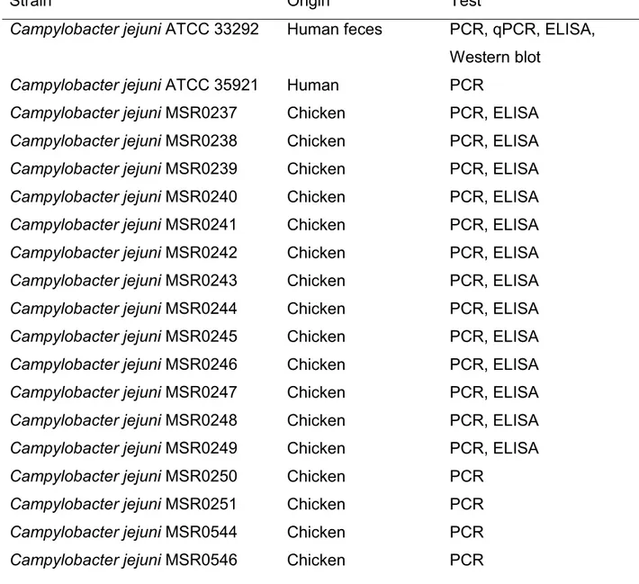
22 experimental PCRs were performed to verify the presence of selected gene in 44 different strains of Campylobacter (Table 1). Each 25 μl reaction mixture consisted of 10 ng of genomic DNA, 0.5 μM primers and 0.005 U Q5 High-Fidelity DNA Polymerase. PCRs were carried out using 95°C for 30 s, followed by 35 cycles of 10 s at 98°C, 20 s at 65°C, and 40 s at 72°C, and finished with a 2 min incubation at 72°C. Products were subsequently resolved on a 1% agarose gel.
TABLE 1. List of bacterial strains
Straina Origin Test
Campylobacter jejuni ATCC 33292 Human feces PCR, qPCR, ELISA, Western blot
Campylobacter jejuni ATCC 35921 Human PCR
Campylobacter jejuni MSR0237 Chicken PCR, ELISA Campylobacter jejuni MSR0238 Chicken PCR, ELISA Campylobacter jejuni MSR0239 Chicken PCR, ELISA Campylobacter jejuni MSR0240 Chicken PCR, ELISA Campylobacter jejuni MSR0241 Chicken PCR, ELISA Campylobacter jejuni MSR0242 Chicken PCR, ELISA Campylobacter jejuni MSR0243 Chicken PCR, ELISA Campylobacter jejuni MSR0244 Chicken PCR, ELISA Campylobacter jejuni MSR0245 Chicken PCR, ELISA Campylobacter jejuni MSR0246 Chicken PCR, ELISA Campylobacter jejuni MSR0247 Chicken PCR, ELISA Campylobacter jejuni MSR0248 Chicken PCR, ELISA Campylobacter jejuni MSR0249 Chicken PCR, ELISA Campylobacter jejuni MSR0250 Chicken PCR
Campylobacter jejuni MSR0251 Chicken PCR Campylobacter jejuni MSR0544 Chicken PCR Campylobacter jejuni MSR0546 Chicken PCR
23 Campylobacter jejuni MSR0545 Ovine PCR
Campylobacter coli ATCC 33559 Pig feces PCR Campylobacter coli ATCC 49941 API System
bioMerieux
PCR, qPCR, ELISA, Western blot
Campylobacter coli MSR0197 Turkey PCR, ELISA Campylobacter coli MSR0198 Turkey PCR, ELISA Campylobacter coli MSR0264 Chicken PCR
Campylobacter coli MSR0265 Turkey PCR
Campylobacter coli MSR0266 Chicken PCR, ELISA Campylobacter coli MSR0267 Chicken PCR, ELISA Campylobacter coli MSR0268 Chicken PCR, ELISA Campylobacter coli MSR0270 Chicken PCR, ELISA Campylobacter coli MSR0271 Chicken PCR, ELISA Campylobacter coli MSR0272 Chicken PCR
Campylobacter coli MSR0273 Swine PCR, ELISA Campylobacter coli MSR0274 Chicken PCR, ELISA Campylobacter coli MSR0275 Chicken PCR
Campylobacter coli MSR0276 Swine PCR, ELISA Campylobacter coli MSR0278 Chicken PCR
Campylobacter coli MSR0279 Turkey PCR
Campylobacter lari ATCC 35221 Herring gull cloacal swab
PCR, ELISA Campylobacter lari ATCC 35222 Dog feces PCR, ELISA
Campylobacter lari ATCC 35223 Child feces PCR, qPCR, ELISA, Western blot
Campylobacter lari ATCC 43675 Human feces PCR, ELISA Campylobacter lari ATCC BAA-1060 Human feces PCR, ELISA Campylobacter lari MSR0554 Swine PCR, ELISA
Acinetobacter baumannii UdeS ELISA
24
Alcaligenes faecalis UdeS ELISA
Bacillus subtulis ATCC 6051 N/A ELISA
Bacillus cereus ATCC 11778 Derived from Bacillus cereus ATCC 9634
ELISA
Bacillus cereus UdeS ELISA
Enterobacter aerogenes ATCC 35029
BBL Microbiology Systems
ELISA Enterobacter cloacae ATCC 23355 Stanford University ELISA
Enterococcus gallinarium UdeS ELISA
Escherichia coli ATCC 25922 Clinical isolate ELISA
Escherichia coli K12 UdeS ELISA
Escherichia coli K12 AB 2847 N/A ELISA, Western blot Klebsiella oxytoca ATCC 43165 Clininal isolate ELISA
Klebsiella pneumoniae ATCC 13883 NCTC ELISA
Klebsiella pneumoniae UdeS ELISA
Proteus mirabilis ATCC 25933 Human vagina ELISA
Proteus vulgaris ATCC 13315 NCTC ELISA
Pseudomonas aeruginosa ATCC 27853
Blood culture ELISA
Pseudomonas aeruginosa UdeS ELISA
Salmonella typhimurium ATCC 14028
Heart and liver chicken tissue
ELISA Serratia marcescens ATCC 8100 NR Smith ELISA a
The ATCC strains were obtained from the American Type Culture Collection (Manassas, VA, USA), the MSR strains from the Foodchek Inc. culture collection (St-Hyacinthe, QC, Canada), and the UdeS strains from F. Malouin (Université de Sherbrooke, Sherbrooke, QC, Canada).
25
Peptide selection for antibody production
Subcellular localization prediction. The subcellular localization of proteins was
predicted using CELLO (http://cello.life.nctu.edu.tw), an approach based on multi-class SVM multi-classification system. CELLO uses four types of sequence coding schemes: the amino acid composition, the dipeptide composition, the partitioned amino acid composition and the sequence composition based on the physico-chemical properties of amino acids. (Yu CS et al, 2006)
Epitope prediction of protein antigens. Potentially immunogenic regions were
predicted by using the BCPreds server 1.0 (http://ailab.ist.psu.edu/bcpred/). This server uses an antigenic peer scale of amino acids. This scale assigns a value to each dipeptide. The peaks of amino acids are generated by decomposing the peptides. The database consists of 872 positive epitopes and 872 negative epitopes (Chen J et al., 2007).
Transmembrane topology of the target protein. Transmembrane topology of
target protein was checked using TMHMM online tool (http://www.cbs.dtu.dk/services/TMHMM/). TMHMM is a tool for predicting the topology of membrane proteins using a hidden Markov model. It predicts transmembrane helices and discriminates the soluble proteins of membrane proteins with a high degree of precision (Moller S et al., 2001).
Immunization of mice. The 40 amino acids peptide
GIQKIMPYEEGNKVANAVSHVAAVNIQSQKIKDLEFKLNN-NH2 was synthesized by Biomatik Inc. (Cambridge, ON, Canada) with a purity of 96.25% and was used as antigen for immunization. Upon receipt, the peptide was suspended in sterile
26 water at a concentration of 5 mg/mL and stored at -80°C until day of use. For the preparation of the immunization dose, the peptide was mixed and suspended in PBS containing 20% of the EMULSIGEN®-D oil-in-water emulsion adjuvant to obtain a final dose of 20 µg of polypeptide per injection in 100 µl. Four female CD-1 mice were injected 3 times in the back of the neck at week 0, 3 and 5, and blood samples taken before the first immunization (preimmune sera) and 10 days after the last injection (immune sera). The blood aliquots were allowed to clot at room temperature for an hour, and were then centrifuged at 10,000 g for 10 min at 4°C. The sera were harvested and kept at -20°C until subsequent analysis.
Serum characterization
Preparation of bacterial fractions. Bacterial lysates were prepared by
suspending ~107 cells of 30 different C. jejuni, C. coli and C. lari strains and 22 non-Campylobacter species (strains that were used in ELISA are identified in TABLE 1) in 1 mL of carbonate-bicarbonate buffer and boiled for ten minutes. Bacterial membranes were prepared by suspending cells from a 50 mL culture of C. jejuni, C. coli, C. lari or E. coli in 7 mL of 10 mM HEPES buffer, pH 7.4, and lysed by French pressure cell disruption. The lysed cell preparation was centrifuged at 10,000 g for 10 min at 4°C to remove cell debris and intact cells. The membranes were collected by ultracentrifugation at 100,000 g for 1 h at 4°C (Beckman, Ti70.1 rotor). The pellet was suspended in 500 L 10 mM HEPES, pH 7.4. Membrane protein concentrations were determined by using micro BCA Protein Assay kit (Thermo Fisher Scientific, Rockford, IL, USA).
ELISA. Reactivity between the serum and the peptide used for immunization as
27 determined by ELISA. Peptide and membranes were diluted at 10 mg/mL in carbonate-bicarbonate buffer for use in ELISA plates (100 μl/well). The plates were incubated overnight at 4°C followed by incubation for 1 h at 37°C with 5% milk. Then, incubation with primary antibody dilution 1:15,000 – 1:405,000 for the peptide and 1:500 for bacterial membranes. The preimmune serum diluted at 1:1,500 was used as negative control. Then, incubation with a secondary antibody solution (HRP-conjugated anti-mouse immunoglobulins; dilution 1:10,000) at 37°C for 1 h; all antibodies were diluted with PBST containing 1% milk and 0.05% Tween 20. After color development and cessation of the reaction, the optical density (OD) value of each well was measured using microplate reader at a wavelength of 450 nm. For bacterial lysates, the same procedure was used except that primary antibody was diluted 1:1,500.
PAGE and electroblotting. A total amount of 20 μg of bacterial membranes was
added to a mixture containing 6Χ Laemmli loading buffer (4% sodium dodecyl sulfate, 10% 2-mercaptoethanol, 20% glycerol, 0.004% bromophenol blue and 0.125 M Tris HCl; pH 6.8). The samples were heated at 95°C for 10 min. Samples were run on 4–15% gel at 110 V for 1 h 45 min using Towbin buffer with SDS (25 mM Tris Base, 192 mM glycine, 0.1% SDS; pH 8.3). Following completion of the PAGE run, samples were transferred to a PVDF membrane (Millipore; Darmstadt, Germany) using Towbin buffer. Membranes were transferred at 100 V for 1 h.
Western blot. PVDF membranes were blocked for 1 h in 5 % milk/TBST,
incubated in primary antibody (polyclonal serum 1:750) overnight, washed with TBST, incubated in secondary antibody (HRP-conjugated anti-mouse immunoglobulins; dilution 1: 10,000) for 1 h, washed with TBST, and revealed with Pierce DAB substrate kit (ThermoFisher Scientific).
28
Protein digestions and mass spectrometry. Protein digestion and mass
spectrometry analyses were performed by the Proteomics Platform of the Université de Laval Research Center (Quebec, Qc, Canada). Bands of interest were extracted from gels after PAGE as described above and placed in 96-well plates and then washed with water. Tryptic digestion was performed on a liquid handling robot (MultiProbe, Perkin Elmer) according to the manufacturer’s specifications and to the protocol of Shevchenko et al (1996) with the modifications suggested by Havlis et al (2003 ). Briefly, proteins were reduced with 10mM DTT and alkylated with 55mM iodoacetamide. Trypsin digestion was performed using 126nM of modified porcine trypsin (Sequencing grade, Promega, Madison, WI) at 37°C for 18h. Digestion products were extracted using 1% formic acid, 2% acetonitrile followed by 1% formic acid, 50% acetonitrile. The recovered extracts were pooled, vacuum centrifuge dried and then resuspended into 12 µl of 0.1% formic acid and 5 µl were analyzed by mass spectrometry.
Peptide samples were separated by online reversed-phase (RP) nanoscale capillary liquid chromatography (nanoLC) and analyzed by electrospray mass spectrometry (ES MS/MS). The experiments were performed with a Ekspert NanoLC425 (Eksigent) coupled to a 5600+ mass spectrometer (Sciex, Framingham, MA, USA) equipped with a nanoelectrospray ion source. Peptide separation took place on a picofrit column (Reprosil 3u, 120A C18, 15 cm x 0.075 mm internal diameter, New Objective, Woburn, Ma). Peptides were eluted with a linear gradient from 5-35% solvent B (acetonitrile, 0.1% formic acid) in 35 minutes, at 300 nL/min. Mass spectra were acquired using a data dependent acquisition mode using Analyst software version 1.7. Each full scan mass spectrum (400 to 1250 m/z) was followed by collision-induced dissociation of the twenty most intense ions. Dynamic exclusion was set for a period of 12 sec and a tolerance of 100 ppm.
29
Data base searching. All MS/MS samples were analyzed using Mascot (Matrix
Science, London, UK; version 2.5.1). Mascot was set up to search the TAX_Campylobacter_jejuni_197_201603018 database (unknown version, 91006 entries), CP_CampyColi_ci_195_20161031 database (unknown version, 3501 entries) and CP_CampyLari_ci_201_20161031 database (unknown version, 1711 entries) indicating that the peptides have been generated by digestion with trypsin. Mascot was searched with a fragment ion mass tolerance of 0,100 Da and a parent ion tolerance of 0,100 Da. Carbamidomethyl of cysteine was specified in Mascot as a fixed modification. Deamidation of asparagine and glutamine and oxidation of methionine were specified in Mascot as variable modifications.
Criteria for protein identification. Scaffold (version Scaffold_4.5.1, Proteome
Software Inc., Portland, OR) was used to validate MS/MS based peptide and protein identifications. Peptide identifications were accepted if they could be established at greater than 95,0 % probability by the Scaffold Local FDR algorithm. Protein identifications were accepted if they could be established at greater than 95,0 % probability and contained at least 1 identified peptide. Protein probabilities were assigned by the Protein Prophet algorithm (Nesvizhskii, Al et al Anal. Chem. 2003;75(17):4646-58). Proteins that contained similar peptides and could not be differentiated based on MS/MS analysis alone were grouped to satisfy the principles of parsimony.
RESULTS
30 Bioinformatic analyses using SEED allowed the identification of proteins commonly found in three Campylobacter species of interest. These proteins were analyzed by Blast using established criteria such as the presence of regions with high homology between the three target Campylobacter species and low homology to non-target species. This analysis led to the selection of three proteins specific to the 3 Campylobacter species: the carbon starvation protein (CstA), the Ferric enterobactin uptake receptor (CfrA), and the Flagellar hook-associated protein (FlgC) (Table 2).
TABLE 2. Similarity percentages of potential target proteins compared to Campylobacter jejuni
a Percentages are relative to C. jejuni
Relative expression of the selected genes was determined by qPCR. Results showed that the gene coding for CfrA, an iron-related protein (Miller et al, 2009), was weakly expressed and only in the presence of 2,2′-bipyridyl, an iron chelator used to create an iron-deficient environment (Figure 1B). As for the cstA and flgC genes, both were well expressed in the Campylobacter target species (Figure 1A).
Protein C. coli C. lari
Carbon starvation protein A (CstA) 94%a 83% Ferric enterobactin uptake receptor (CfrA) 92% 83% Flagellar hook-associated protein (FlgC) 98% 90%
31
Figure 1. A) Relative expression of cfrA, cstA and flgC in C. jejuni ATCC 33292,
C. coli ATCC 49941 and C. lari ATCC 35223. B) Relative expression of cfrA in C. jejuni ATCC 33292, C. coli ATCC 49941 and C. lari ATCC 35223 in cultures supplemented with 6 mM of 2,2′-bipyridyl (+) or not (-). Significant differences in comparison to the untreated control are shown by asterisks. Statistical analysis was performed using non-parametric one-way ANOVA: ns, no-significant; *, p<0.05; **, p<0.01; ***, p<0.001.
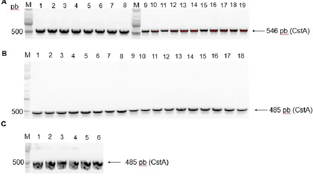
The ubiquity of cstA was demonstrated by experimental PCR. All the 44 Campylobacter species and strains tested were positive for the presence of the cstA gene (Figure 2).
32
Figure 2. PCR detection of the gene coding for CstA in Campylobacter species.
A) Gene cstA amplification in C. jejuni strains; Lane M: 1kb plus ladder; Line 1: C. jejuni ATCC 33292; Line 2: C. jejuni ATCC 35921; Lines 3-16: C. jejuni MSR0237-MSR0251; Lines 17-19: C. jejuni MSR0544-MSR0546 B) in C. coli strains; Line 1: C. coli ATCC 33559; Line 2: C. coli ATCC 49941; Lines 3-4: C. coli MSR0197-MSR0198; Lines 5-9: C. coli MSR0264-MSR0268; Lines 10-16: C. coli MSR0270-MSR0276; Lines 17-18: C. coli MSR0278-MSR0279 and C) in C. lari strains; Line 1: C. lari ATCC 35221; Line 2: C. lari ATCC 35222; Line 3: C. lari ATCC 35223; Line 4: C. lari ATCC 43675; Line 5: C. lari ATCC BAA-1060; Line 6: C. lari MSR0554.
Peptide selection for antibody production
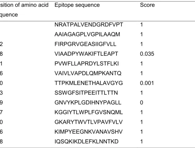
The subcellular localization of CstA was predicted using CELLO. The CELLO output gave significant reliability for inner membrane (4.677). Then, potentially immunogenic regions of CstA were predicted by using the BCPreds server 1.0. A
33 total of 13 epitopes were predicted from 703 amino acids. The predicted epitopes are shown in Table 3.
TABLE 3. Antigenic B-cell epitopes of C. jejuni CstA identified using the BCPreds server
Transmembrane topology of the CstA protein was checked using TMHMM online tool. The TMHMM server showed that residues 27-30, 110-118, 182-190, 241-254, 307-320, 387-464, 531-539 and 595-660 presented outside region, residues 7-26, 31-53, 87-109, 119-138, 159-181, 191-213, 218-240, 255-277, 284-306, 321-343, 364-386, 465-487, 508-530, 540-563, 575-594 and 661-683 were within the transmembrane and residues 1-6, 54-86, 139-158, 214-217, 278-283, 344-363, 488-507, 563-574 and 684-703 were inside the region of the protein. Position of amino acid
sequence
Epitope sequence Score
66 NRATPALVENDGRDFVPT 1 94 AAIAGAGPLVGPILAAQM 1 212 FIRPGRVGEASIIGFVLL 1 238 VIAADPYWAKIFTLEAPT 0.035 271 PVWFLLAPRDYLSTFLKI 1 296 VAIVLVAPDLQMPKANTQ 1 350 TTPKMLENETHALAVGYG 0.001 413 SSWGFSITPEEITTLTTN 1 499 GNVYKPLGDIHNYPAGLL 0 537 KGGIYTLWPLFGVSNQML 1 570 GKARYTWVTLVPAVFVLV 1 596 KIMPYEEGNKVANAVSHV 1 618 IQSQKIKDLEFKLNNTKD 1
34 TMHMM suggested the presence of 16 TM helix. A consensus predicted topology is presented in Figure 3.
Figure 3. Transmembrane topology of C. jejuni CstA. Probability for
transmembrane helix, inside or outside. * Target peptide position.
Epitopes were blasted to find a sequence specific for Campylobacter. The best result was obtained with this peptide sequence: 593GIQKIMPYEEGNKVANAVSHVAAVNIQSQKIKDLEFKLNN633 (region showed by an asterisk on Figure 3).
This peptide contains two epitopes listed in position 596 and 618 of TABLE 3. The 194 firsts BLAST results of this sequence represented the target species of Campylobacter with similarity percentages between 100%-70%, followed by Arcobacter skirrowii with 68%, Arcobacter lanthieri with 66% and Arcobacter butzleru with 62%.
35
Serum characterization
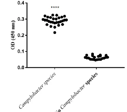
ELISA. ELISA against the peptide used for immunization showed a strong signal
resulting in an antibody titer of>1:405,000 (Figure 4A). ELISA against bacterial membranes showed a signal with Campylobacter species and no signal with E. coli (Figure 4B). ELISA with bacterial lysates showed a signal for 30 members of genus Campylobacter species and no signal for 22 non-Campylobacter strains (Figure 4C).
36
Figure 4. Caracterization of the polyclonal serum by ELISA. A) Reactivity of
peptide in ELISA with preimmune (PI) and immun sera. B) Reactivity of bacterial membranes in ELISA with premmune (PI) and immun sera diluted 1:500. C) Reactivity of bacterial lysates in ELISA with the immun serum diluted 1:1,500. Bacterial strains used are listed in Table 1. Statistical analysis was performed using non-parametric one-way ANOVA: ns, no-significant; *** p ≤ 0.001, **** p ≤ 0.0001.
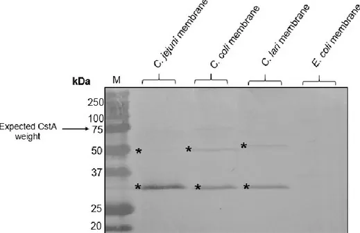
Western blot and mass spectrometry. All Campylobacter bacterial membranes
gave a signal on a Western blot. There was no signal for bacterial membranes prepared from E. coli (Figure 5). The expected band should correspond to a molecular weight of 75 kDa. The bands revealed were not at the expected molecular weight. Bands were sent for analysis by mass spectrometry. Considering a minimum of 1 peptide with a threshold of 95%, 5 and 6 exclusive unique peptides for CstA were found in the C. jejuni and C. coli 50 kDa bands,
37 respectively. The result of analysis shows that the target protein is present in the 50 kDa band.
Figure 5. Western blot analysis of reaction of C. jejuni ATCC 33292, C. coli
ATCC 49941, C. lari ATCC 35223 and E. coli K12 AB2847 membranes with polyclonal serum diluted 1:750. M Precision Plus ProteinTM, * Bands analyzed by mass spectrometry.
DISCUSSION
The methods typically employed to evaluate the presence of Campylobacter in food samples are based on the Horizontal Method for Detection and Enumeration of Campylobacter spp. (ISO 10272, 2006). These cultivation procedures are lengthy and tedious. Furthermore, due to the overgrowth of competitive bacteria on agar plates, sometimes it is difficult to identify Campylobacter colonies. Developing a polyclonal antibody gives an interesting tool that can be used in immunological tests that would allow a much faster and specific detection than microbiological methods for Campylobacter spp.
38 In this study three Campylobacter proteins were chosen as potential target for antibody production: the ferric enterobactin uptake receptor (CfrA), the carbon starvation protein (CstA), and the flagellar hook-associated protein (FlgC). These proteins showed a high amino acid homology between target Campylobacter species (sTable 2) and low homologies to other species.
The ferric enterobactin receptor CfrA is an outer membrane receptor responsible for high-affinity iron acquisition in C. jejuni by assimilating ferric enterobactin (Palyada et al., 2004). CfrA could plays several other functions during the intestinal colonization by Campylobacter. For example, CfrA is involved in the reactivity of C. jejuni to the catecholamine hormone (Ximin et al., 2009). C. jejuni carbon starvation protein A (CstA) is also involved in peptide utilization, motility agglutination, and It plays a role in stimulating murine dendritic cells (Rasmussen et al., 2013). Finally, FlgC is a structural component of the flagellar basal body (Konkel et al., 2004). It has been shown that an orthologous FlgC of Salmonella enterica plays a significant role in the binding ability to the intestinal mucosa of poultry (Shippy DC et al., 2014).
The qPCR analyses allowed to discard CfrA from the potential targets because of its apparently low expression. To show that CfrA was indeed weakly expressed and that this was not an artefact, qPCRs with cultures treated with 2,2′-bipyridyl, an iron chelator, were made to create an iron-deprived environment to induce expression of iron-regulated proteins. Treatment with 2,2′-bipyridyl effectively increased the abundance of CfrA’s cDNA.
39 Our experiments suggest that genes coding for CstA and FlgC were expressed at similar levels in C. jejuni and C. coli. In C. lari, cstA seems to be relatively more expressed than flgC. Studies have shown that CstA plays a role in the stimulation of murine dendritic cells (Rasmussen et al, 2013). For this characteristic, and for its good expression in C. lari, this protein was chosen as the target for antibody production. Furthermore, the ubiquity of the cstA gene among Campylobacter strains was demonstrated par PCR (Figure 2).
The polyclonal serum produced showed a great reactivity against the peptide. Furthermore, ELISA with bacterial membranes and lysates showed that the polyclonal immune serum recognized 30 Campylobacter strains and there was no cross-reaction with 22 non-target species.
Finally, the Western blot with polyclonal serum against membranes of C. jejuni, C. coli and C. lari revealed a large band of about 30 kDa and a weaker band of 50 kDa. The expected band size for CstA was 75 kDa. Mass spectrometry confirmed the presence of CstA in the 50 kDa band of C. jejuni and C. coli. The western blot with E. coli membranes did not show a signal, thus confirming the specificity of the serum. These results indicate that this polyclonal serum is useful as a screening test for the detection of Campylobacter spp.
However, the use of polyclonal antibodies is prone to batch-to-batch variability. Now that we identified a well-known target, a monoclonal antibody can be developed. In this way, all batches will be identical, increasing consistency and standardization of experimental procedures and results. Moreover, hybridomas are a constant and renewable source once created.
40 Other than the production of a polyclonal serum, this study allowed the identification of a specific, conserved and immunogenic peptide to Campylobacter spp: 593GIQKIMPYEEGNKVANAVSHVAAVNIQSQKIKDLEFKLNN633. Linear epitopes are an important tool for clinical applications and they can be used in a wide range of tests because they are quickly synthesized. In addition, production costs using chemical synthesis are lower and easier than recombinant expression of complete proteins. Finally, specific peptides of pathogenic bacteria reduce risks associated with the use of the entire pathogen.
As Campylobacter continues to be the leading cause of food-borne diarrhea in humans in the developed world. Efforts to reduce or eliminate Campylobacter contamination should become a priority. The results of this study contribute to progress in this direction and are a step towards the development of methods for detecting this pathogen.
ACKNOWLEDGEMENTS
This work was partly funded through the ENGAGE program supported by the Natural Sciences and Engineering Research Council (NSERC) of Canada.
REFERENCES
Ailes, E.; Demma, L.; Hurd, S.; Hatch, J.; Jones, T.F.; Vugia, D.; Cronquist, A.; Tobin-D’Angelo, M.; Larson, K.; Laine, E.; Edge, K.; Zansky, S.; Sallan, E (2008). Continued decline in the incidence of Campylobacter infections Ailes. FoodNet 1996–2006. Foodborne Pathog. Dis.5, 329–337
41 Allos, B. M. (2001). Campylobacter jejuni infections: Update on emerging issues and trends. Clinical Infectious Diseases, 32(8), 1201-1206.
Altekruse, S.F.; Stern, N.J.; Fields, P.I.; Swerdlow, D.L. (1999) Campylobacter jejuni and emerging foodborne pathogen. Emerg. Infect. Dis. 5, 28–35.
Bhunia A. K. (2008). “Campylobacter and arcobacter,” in Foodborne Microbial Pathogens: Mechanisms and Pathogenesis, ed. Bhunia A. K., editor. (New York, NY: Springer), 217–226
CDC (2013). Incidence and trends of infection with pathogens transmitted commonly through food-foodborne diseases active surveillance network, 10 United States Sites, 1996–2012; U.S. Department of Health and Human Services Centers for Disease Control and Prevention: Atlanta, GA, USA; pp. 283–287. Chen J, Liu H, Yang J, Chou K (2007) Prediction of linear B-cell epitopes using amino acid pair antigenicity scale. Amino Acids 33: 423-428.
Denny, J.; Boelaert, F.; Borck, B.; Heur, O.E.; Ammon, A.; Makela, P. (2007). Zoonotic infection in Europe: Trends and figures—A summary of the EFSA-ECDC annual report. Eur. Surviell. 51.
EFSA (2012). EFSA explains zoonotic diseases: Campylobacter
EFSA: The community summary report on trends and sources of zoonoses, zoonotic agents and food-borne outbreaks in the European Union in 2008. EFSA J 2010, 1496.
Havlis J, Thomas H, Sebela M, Shevchenko A. (2003). Fast-Response Proteomics by Accelerated In-Gel Digestion of Proteins. Anal. Chem. 75(6);1300-1306.
ISO - International Organization for Standardization. ISO 10272 microbiology of food and animal feeding stuff - horizontal method for detection and enumeration of Campylobacter spp. - Part 1: enrichment method; part 2: enumeration method.