Year 2019
Thesis N° 199
Neuropsychiatric manifestations
in idiopathic Parkinson’s disease
THESIS
PRESENTED AND DEFENDED PUBLICLY ON 11/07/2019
BY
Mr
. Raymond
KLEVOR
Born on 07 January 1991 in Ghana
TO OBTAIN A MEDICAL DOCTORATE
KEYWORDS
Parkinson’s Disease – Neuropsychiatric Manifestations – Fluctuations
L-Dopa - Quality of Life
JURY
M
PmeP.
Mr.
M
PmeP.
Mr.
F. ASRI
Professor of Psychiatry
M. CHRAA
Associate Professor of Neurophysiology
F. MANOUDI
Professor of Psychiatry
H. GHANNANE
Professor of Neurosurgery
CHAIRPERSON
SUPERVISOR
JUDGES
HIPPOCRATIC OATH
At the time of being admitted as a member of the medical profession:
i solemnly pledge to dedicate my life to the service of humanity;
the health and well-being of my patient will be my first consideration;
i will respect the autonomy and dignity of my patient;
i will maintain the utmost respect for human life;
i will not permit considerations of age, disease or disability, creed, ethnic
origin, gender, nationality, political affiliation, race, sexual orientation, social
standing or any other factor to intervene between my duty and my patient;
i will respect the secrets that are confided in me, even after the patient has
died;
i will practice my profession with conscience and dignity and in accordance
with good medical practice;
i will foster the honour and noble traditions of the medical profession;
i will give to my teachers, colleagues, and students the respect and gratitude
that is their due;
i will share my medical knowledge for the benefit of the patient and the
advancement of healthcare;
i will attend to my own health, well-being, and abilities in order to provide
care of the highest standard;
i will not use my medical knowledge to violate human rights and civil
liberties, even under threat;
i make these promises solemnly, freely and upon my honour.
UNIVERSITE CADI AYYAD
FACULTE DE MEDECINE ET DE PHARMACIE MARRAKECH
Doyens Honoraires : Pr. Badie Azzaman MEHADJI
: Pr. Abdelhaq ALAOUI YAZIDI ADMINISTRATION
Doyen : Pr. Mohammed BOUSKRAOUI
Vice doyen à la Recherche et la Coopération : Pr. Mohamed AMINE
Vice doyen aux Affaires Pédagogiques : Pr. Redouane EL FEZZAZI
Secrétaire Générale : Mr. Azzeddine EL HOUDAIGUI
Professeurs de l’enseignement supérieur
Nom et Prénom Spécialité Nom et Prénom Spécialité
ABKARI Imad Traumato- orthopédie FINECH Benasser Chirurgie – générale ABOU EL HASSAN
Taoufik
Anésthésie- réanimation
FOURAIJI Karima Chirurgie pédiatrique ABOUCHADI Abdeljalil Stomatologie et chir
maxillo faciale GHANNANE Houssine Neurochirurgie ABOULFALAH Abderrahim Gynécologie- obstétrique
GHOUNDALE Omar Urologie
ABOUSSAIR Nisrine Génétique HAJJI Ibtissam Ophtalmologie
ADERDOUR Lahcen Oto- rhino-
laryngologie
HOCAR Ouafa Dermatologie
ADMOU Brahim Immunologie JALAL Hicham Radiologie
AGHOUTANE El Mouhtadi Chirurgie pédiatrique KAMILI El Ouafi El Aouni
Chirurgie pédiatrique
AIT AMEUR Mustapha Hématologie
Biologique
KHALLOUKI Mohammed
Anesthésie- réanimation
AIT BENALI Said Neurochirurgie KHATOURI Ali Cardiologie
AIT BENKADDOUR Yassir Gynécologie- obstétrique
KHOUCHANI Mouna
Radiothérapie
AIT-SAB Imane Pédiatrie KISSANI Najib Neurologie
AKHDARI Nadia Dermatologie KOULALI IDRISSI
Khalid
Traumato- orthopédie ALAOUI Mustapha Chirurgie- vasculaire
péripherique
KRATI Khadija Gastro- entérologie
AMINE Mohamed Epidémiologie- clinique
LAGHMARI Mehdi Neurochirurgie
AMMAR Haddou Oto-rhino-laryngologie LAKMICHI
Mohamed Amine
Urologie
AMRO Lamyae Pneumo- phtisiologie LAOUAD Inass Néphrologie
ARSALANE Lamiae Microbiologie
-Virologie
LOUZI Abdelouahed
Chirurgie – générale
ASMOUKI Hamid Gynécologie-
obstétrique
MADHAR Si Mohamed
Traumato- orthopédie
ASRI Fatima Psychiatrie MANOUDI Fatiha Psychiatrie
BEN DRISS Laila Cardiologie MANSOURI Nadia Stomatologie et chiru
maxillo faciale BENCHAMKHA Yassine Chirurgie réparatrice et
plastique MOUDOUNI Said Mohammed Urologie BENELKHAIAT BENOMAR Ridouan
Chirurgie - générale MOUFID Kamal Urologie
BENJILALI Laila Médecine interne MOUTAJ Redouane Parasitologie
BOUAITY Brahim Oto-rhino-
laryngologie
MOUTAOUAKIL Abdeljalil
Ophtalmologie
BOUCHENTOUF Rachid Pneumo- phtisiologie NAJEB Youssef Traumato- orthopédie
BOUGHALEM Mohamed Anesthésie -
réanimation
NARJISS Youssef Chirurgie générale BOUKHIRA Abderrahman Biochimie - chimie NEJMI Hicham Anesthésie-
réanimation
BOUMZEBRA Drissi Chirurgie
Cardio-Vasculaire
NIAMANE Radouane
Rhumatologie
BOURROUS Monir Pédiatrie NOURI Hassan Oto rhino laryngologie
BOUSKRAOUI Mohammed
Pédiatrie OUALI IDRISSI
Mariem
Radiologie
CHAFIK Rachid Traumato- orthopédie OULAD SAIAD
Mohamed
Chirurgie pédiatrique
CHAKOUR Mohamed Hématologie
Biologique
QACIF Hassan Médecine interne
CHELLAK Saliha Biochimie- chimie QAMOUSS
Youssef
Anésthésie- réanimation CHERIF IDRISSI EL
GANOUNI Najat
Radiologie RABBANI Khalid Chirurgie générale
CHOULLI Mohamed Khaled
Neuro pharmacologie RAFIK Redda Neurologie
EL ADIB Ahmed Rhassane Anesthésie- réanimation
SAIDI Halim Traumato- orthopédie
EL ANSARI Nawal Endocrinologie et
maladies métaboliques SAMKAOUI Mohamed Abdenasser Anesthésie- réanimation
EL BARNI Rachid Chirurgie- générale SAMLANI
Zouhour
Gastro- entérologie
EL BOUCHTI Imane Rhumatologie SARF Ismail Urologie
EL BOUIHI Mohamed Stomatologie et chir maxillo faciale
SORAA Nabila Microbiologie - Virologie EL FEZZAZI Redouane Chirurgie pédiatrique SOUMMANI
Abderraouf
Gynécologie- obstétrique
EL HAOURY Hanane Traumato- orthopédie TASSI Noura Maladies infectieuses
EL HATTAOUI Mustapha Cardiologie YOUNOUS Said Anesthésie-
réanimation
EL HOUDZI Jamila Pédiatrie ZAHLANE Mouna Médecine interne
EL KARIMI Saloua Cardiologie ZOUHAIR Said Microbiologie
ELFIKRI Abdelghani Radiologie ZYANI Mohammed Médecine interne
ESSAADOUNI Lamiaa Médecine interne
Professeurs Agrégés
Nom et Prénom Spécialité Nom et Prénom Spécialité
ABIR Badreddine Stomatologie et
Chirurgie maxillo faciale
GHAZI Mirieme Rhumatologie
ADALI Imane Psychiatrie HACHIMI
Abdelhamid
Réanimation médicale
ADARMOUCH Latifa Médecine
Communautaire (médecine préventive, santé publique et hygiène)
HAROU Karam Gynécologie-
obstétrique
AISSAOUI Younes Anesthésie -
réanimation HAZMIRI Fatima Ezzahra Histologie – Embryologie - Cytogénéque
AIT BATAHAR Salma Pneumo-
phtisiologie
IHBIBANE fatima Maladies Infectieuses
ALJ Soumaya Radiologie KADDOURI Said Médecine interne
ATMANE El Mehdi Radiologie LAKOUICHMI Mohammed
Stomatologie et Chirurgie maxillo faciale
BAIZRI Hicham Endocrinologie et
maladies métaboliques
LOUHAB Nisrine Neurologie
BASRAOUI Dounia Radiologie MAOULAININE Fadl
mrabih rabou
Pédiatrie (Neonatologie)
BASSIR Ahlam Gynécologie-
obstétrique
MARGAD Omar Traumatologie
-orthopédie
BELBACHIR Anass Anatomie-
pathologique
MATRANE Aboubakr Médecine nucléaire
BELBARAKA Rhizlane Oncologie
médicale
MEJDANE Abdelhadi Chirurgie Générale
BELKHOU Ahlam Rhumatologie MLIHA TOUATI
Mohammed
Oto-Rhino - Laryngologie BENHIMA Mohamed Amine Traumatologie -
orthopédie
MOUAFFAK Youssef Anesthésie - réanimation BENJELLOUN HARZIMI Amine Pneumo-
phtisiologie
MOUHSINE Abdelilah Radiologie
BENLAI Abdeslam Psychiatrie MSOUGGAR
Yassine
Chirurgie thoracique
BENZAROUEL Dounia Cardiologie NADER Youssef Traumatologie -
orthopédie
BOUKHANNI Lahcen Gynécologie-
obstétrique
OUBAHA Sofia Physiologie
BOURRAHOUAT Aicha Pédiatrie RADA Noureddine Pédiatrie
BSISS Mohamed Aziz Biophysique RAIS Hanane Anatomie
pathologique
CHRAA Mohamed Physiologie RBAIBI Aziz Cardiologie
DAROUASSI Youssef Oto-Rhino -
Laryngologie
ROCHDI Youssef Oto-rhino- laryngologie
DRAISS Ghizlane Pédiatrie SAJIAI Hafsa Pneumo- phtisiologie
EL AMRANI Moulay Driss Anatomie SALAMA Tarik Chirurgie pédiatrique
EL HAOUATI Rachid Chirurgie Cardio- vasculaire
SEDDIKI Rachid Anesthésie -
Réanimation
EL IDRISSI SLITINE Nadia Pédiatrie SERGHINI Issam Anesthésie -
Réanimation
EL KHADER Ahmed Chirurgie
générale
TAZI Mohamed Illias
Hématologie- clinique
EL MEZOUARI El Moustafa Parasitologie Mycologie
ZAHLANE Kawtar Microbiologie - virologie EL MGHARI TABIB Ghizlane Endocrinologie et maladies métaboliques
ZAOUI Sanaa Pharmacologie
EL OMRANI Abdelhamid Radiothérapie ZARROUKI Youssef Anesthésie - Réanimation
FADILI Wafaa Néphrologie ZEMRAOUI Nadir Néphrologie
FAKHIR Bouchra Gynécologie-
obstétrique
ZIADI Amra Anesthésie -
réanimation
FAKHRI Anass Histologie-
embyologie cytogénétique ZIDANE Moulay Abdelfettah Chirurgie Thoracique Professeurs Assistants
Nom et Prénom Spécialité Nom et Prénom Spécialité
ABDELFETTAH Youness Rééducation et Réhabilitation Fonctionnelle
ELOUARDI Youssef Anesthésie réanimation
ABDOU Abdessamad Chiru Cardio
vasculaire
ELQATNI Mohamed Médecine interne
AIT ERRAMI Adil Gastro-entérologie ESSADI Ismail Oncologie Médicale
AKKA Rachid Gastro -
entérologie
FDIL Naima Chimie de
Coordination Bio-organique
ALAOUI Hassan Anesthésie -
Réanimation
FENNANE Hicham Chirurgie Thoracique
AMINE Abdellah Cardiologie GHOZLANI Imad Rhumatologie
ARABI Hafid Médecine physique
et réadaptation fonctionnelle
HAJJI Fouad Urologie
ARSALANE Adil Chirurgie
Thoracique
HAMMI Salah Eddine Médecine interne
ASSERRAJI Mohammed Néphrologie Hammoune Nabil Radiologie
AZIZ Zakaria Stomatologie et chirurgie maxillo faciale
JALLAL Hamid Cardiologie
BAALLAL Hassan Neurochirurgie JANAH Hicham Pneumo- phtisiologie
BABA Hicham Chirurgie
générale
LAFFINTI Mahmoud Amine
BELARBI Marouane Néphrologie LAHLIMI Fatima Ezzahra
Hématologie clinique
BELFQUIH Hatim Neurochirurgie LALYA Issam Radiothérapie
BELGHMAIDI Sarah OPhtalmologie LOQMAN Souad Microbiologie et
toxicologie environnementale
BELHADJ Ayoub Anesthésie
-Réanimation
MAHFOUD Tarik Oncologie médicale
BELLASRI Salah Radiologie MILOUDI Mohcine Microbiologie -
Virologie
BENANTAR Lamia Neurochirurgie MOUNACH Aziza Rhumatologie
BENNAOUI Fatiha Pédiatrie NAOUI Hafida Parasitologie
Mycologie BOUCHENTOUF Sidi
Mohammed
Chirurgie générale
NASSIH Houda Pédiatrie
BOUKHRIS Jalal Traumatologie -
orthopédie
NASSIM SABAH Taoufik Chirurgie Réparatrice et Plastique
BOUTAKIOUTE Badr Radiologie NYA Fouad Chirurgie Cardio -
Vasculaire
BOUZERDA Abdelmajid Cardiologie OUERIAGLI NABIH
Fadoua
Psychiatrie
CHETOUI Abdelkhalek Cardiologie OUMERZOUK Jawad Neurologie
CHETTATI Mariam Néphrologie RAISSI Abderrahim Hématologie clinique
DAMI Abdallah Médecine Légale REBAHI Houssam Anesthésie -
Réanimation
DOUIREK Fouzia Anesthésie-
réanimation
RHARRASSI Isam Anatomie-patologique
EL- AKHIRI Mohammed Oto- rhino- laryngologie
SAOUAB Rachida Radiologie
EL AMIRI My Ahmed Chimie de Coordination bio-organnique
SAYAGH Sanae Hématologie
EL FAKIRI Karima Pédiatrie SEBBANI Majda Médecine
Communautaire (médecine préventive, santé publique et hygiène)
EL HAKKOUNI Awatif Parasitologie mycologie
TAMZAOURTE Mouna Gastro - entérologie EL HAMZAOUI Hamza Anesthésie
réanimation
EL KAMOUNI Youssef Microbiologie Virologie
ZBITOU Mohamed Anas
Cardiologie
ELBAZ Meriem Pédiatrie ELOUARDI Youssef Anesthésie
réanimation LISTE ARRÉTÉÉ LE 22/04/2019
To my parents who believe in me
To an older brother who found me
To a younger sister who longs to see me
And, to the many few whom stance and circumstance
Have brought my way.
Our paths have crossed per fate or per chance;
It’s the same anyway
Because my life without you – I can’t imagine!
So, thank you for the eyes that have gazed
And, not only gazed,
For we have shed many a tear together
And, we have shared our hearts together.
Still, I can’t imagine.
So, thank you for the ears that listened
To the heart broken, and the soul weakened.
Thank you for the hands that were strong
And the words that became a song
From the burning flames of your hearts.
Pr. Fatima ASRI
The glow of your person – that is what touched our hearts as your students.
It was a beautiful experience to have had the opportunity to interne in
your department. A short experience with lasting memories. I am very
grateful that you agreed to preside over the jury. I feel lucky. It is an
honor, professor.
Thank you.
“Here comes the sun.” From Abbey Road by George Harisson
Pr. Mohamed CHRAA
This here can’t be enough. Maybe words indeed aren’t enough. Because
from the moment I approached you for my thesis you have been more than
I could ever imagine. You are exceptional. But again, words aren’t enough.
You are the person we want to be but might never come close to being
because if we get to be half the person you are, it would be one great
achievement of a lifetime. But alas, words! If only I had enough of them!
Thank you, sir.
"But truly, if I were not Alexander, I wish I were Diogenes." Alexander by
Plutarch
Pr. Fatiha MANOUDI
I can remember a role-playing session we had with you where we had to
bring to life a case of a young man in the throes of addiction. It is this vivid
picture that comes to mind each time we hear your name. It is that
liveliness that you infuse into us, that theatrical passion – the art,
performance that has left us with one certainty: we will make better
doctors because of you. It is an honor, professor.
Thank you.
“O body swayed to music, O brightening glance/ How can we know the
dancer from the dance?” Among School Children by W. B. Yeates
Pr. Houssine El GHANNANE
Thank you sir! We will always remember you fondly because of the way
you treat us – calmly, understandingly with that paternal touch. Yes, the
paternal touch. I remember when I came to petition your acceptance to be
on the panel – I really can’t forget this – you kept saying yes, yes,
yes…before I even landed. You knew what I was after before I made it
known, and you accepted before I even asked. We are lucky to have you as
our teacher.
Thank you.
“A learner with the simplest, a teacher of the thoughtfullest…”Song of
Myself by Whitman
Pr. Nisrine LOUHAB
How could I forget you, professor? I can still remember in my neurology
rotation, fourth year, I think, having to give a presentation on the
Cerebellar syndrome. And, from that day, I understood that simple things
make the difference because you were simply ready to give your best to us.
You have touched many lives than you might know, and indeed, professor,
I am one better because of you.
Thank you.
“One by one they came/ As far as the eye could see/ Each one somehow
touched By your generosity.” Thank You by Ray Boltz
“Someone will remember us, I say, even in another time.” A translated line
from Sappho’s Fragments.
Pr. Najib KISSANI
You yearn for knowledge. It is that verve with which you seek to know that
also pushes you to demand the best of us. You want us to be the best of
ourselves. You want us to challenge ourselves. You want us to go far in our
lives.
Thank you, sir.
“There's a divinity that shapes our ends/ Rough-hew them how we will.”
The Neurology and Neurosurgery teams
You might not know it but you are remembered with admiration. You are
an inspiration. You guys break your backs to mend others. May your
training be filled with joy, and may you succeed in all your endeavors.
Drs. Traoré, Jalami, Asmae, Khadija… Drs. Arsène, Far, Zakaria,
Hamidou, Davis…
Ms. Imane. Ms. Soukayna
Ms. Imane, you helped me start the work; Ms. Soukayna, you helped me
finish it. Thank you both.
“So long as men can breathe or eyes can see/ So long lives this, and this
gives life to thee.” Sonnet 18 by Shakespeare
My Year Group
I have had the singular opportunity to study with wonderful people. It’s
been one beautiful phenomenon. I wish I could mention all your names.
Alas!
Ahmed, Hajar, Suléman, Mouad, Mansour, Khadija, Imane, Naima,
Nassima, Btissam…
Ayméric, Namar, Germaine, Rougui,
Luke, Elizabeth, Mariata, Mike, Taweh
Béatrice, Adson
My Compatriots
I thank you guys for your support. You guys are amazing.
Dr. Keita, Dr. Samuel, Raphael
Thank you
And now
If I have erred or hurt anyone in the course of our encounters, either by
commission or by omission, I, here, humbly plead for forgiveness for it was
List of tables
Table I : Summary of Patient and Caregiver Distress NPI Scores (N = 79) Table II : Comparison of sex ratios in our study with other series
Table III : Comparison of most frequent Co-morbidities in our study with other series Table IV : Comparison of Prevalence of Parkinson’s disease before
50 years in our study with other series
Table V : Comparison of Age and Duration in our Patients with other Studies Table VI : Prevalence of Neuropsychiatric Symptoms in our
Sample compared to other Series
Table VII : Comparison of PDQ-8 Scores in our Sample with other Series Table VIII : Prevalence of Depression in our Samplecompared with other Series
Table IX : Comparison of Associations Found with Depression in Our Study and Other Studies Table X : Prevalence of Anxiety in Parkinson's Disease as Compared with the General Population Table XI : Comparison of Associations with Anxiety in Our Study and Other Studies
Table XII : Comparison of Prevalence of Hallucinations in Our Patients with Other Studies Table XIII : Comparison of Age, Onset and Duration in Patients with Hallucination and
Patients without Hallucinations
Table XIV : Comparison of the Prevalence of Apathy with Other Studies
Table XV : Prevalence of Impulse Control Symptoms in our Sample compared with other studies Table XVI : Prevalence of Dopamine Dysregulation Syndrom in our Sample compared with other
List of figures
Figure 1 : Flow chart representing the recruitment process of our ascertainment cohort Figure 2 : Distribution of Patients based on Gender
Figure 3 : Distribution of patients based on residence
Figure 4 : Distribution of patients based on whether or not they have health insurance Figure 5 : Distribution of Findings on Patient History
Figure 6 : Distribution of Patients Based on Age Groups
Figure 7 : Distribution of Patients Based on Age of Onset Groups
FIGURE 8 : Distribution of Patients Based on Onset before or after 50 years Figure 9 : Distribution of Patients Based on Duration Groups
Figure 10 : Distribution of Symptoms in our Patients Figure 11 : Prevalence of Neuropsychiatric Symptoms Figure 12 : Distress Scores for NPI Items
Figure 13 : Distribution of Number of Symptoms Reported
Figure 14 : Comparison of Number of Symptoms, Total NPI Score, and PDQ-8 Score in Female and Male Patients
Figure 15 : Distribution of Depression in Our Patients Figure 16 : Distribution of Depression in Females Figure 17 : Distribution of Depression in Males
Figure 18 : Distribution of Distress Scores in Males and Females Figure 19 : Distribution of Depression in Duration Groups
Figure 20 : Distribution of Patients Based on Age of Onset Groups
Figure 21 : Comparison of Onset and Duration in Patients with and those without Depression Figure 22 : Distribution of Caregiver Distress based on Gender of Patients
Figure 23 : Distribution of Caregiver Distress Scores based on Gender Figure 24 : Distribution of Response to L-Dopa Treatment
Figure 27 : Distribution of Anxiety in Females Figure 28 : Distribution of Anxiety in Males
Figure 29 : Comparison of Distress Scores in Males and Females
Figure 30 : Comparison of Onset and Duration in Patients with and those without Anxiety Figure 31 : Distribution of Anxiety in the Age of Onset Groups
Figure 32 : Distribution of Distress Score in Age of Onset Groups Figure 33 : Distribution of Anxiety in the Duration Groups
Figure 34 : Distribution of Distress Scores in the Duration Groups Figure 35 : Distribution of Caregiver Distress based on Gender Figure 36 : Distribution of Distress Scores based on Patient Gender Figure 37 : Distribution of Response to L-Dopa Treatment
Figure 38 : Comparison of PDQ-8 Scores between Patients with and those without Anxiety Figure 39 : Distribution of Psychosis in the Sample
Figure 40 : Comparison of Distress Scores in Males and Females
Figure 41 : Comparison of Distress Scores for Hallucinations in Males and Females Figure 42 : Comparison of Onset and Duration in Psychotic and Non Psychotic Patients Figure 43 : Distribution of Distress Scores in Age Groups
Figure 44 : Distribution of Psychosis in Duration Groups Figure 45 : Distribution of Distress Scores in Duration Groups
Figure 46 : Distribution Caregiver Distress based on the Gender of Patient
Figure 47 : Distribution of Caregiver Distress Scores based on Gender of Patients Figure 48 : Distribution of Response to Treatment
Figure 49 : Comparison of L-Dopa Dose in Psychotic and Non Psychotic Patients
Figure 50 : Comparison of PDQ-8 Scores between Patients with and those without Psychosis Figure 51 : Distribution of Apathy in the Sample
Figure 52 : Comparison of Apathy Scores across Gender
Figure 53 : Comparison of Age, Onset and Duration in Patients with and without Apathy Figure 54 : Distribution of Apathy in the Duration Groups
Figure 56 : Distribution of Caregiver Distress based on Gender Figure 57 : Distribution of Response to Treatment
Figure 58 : Comparison of PDQ-8 Scores between Patients with and those without Apathy Figure 59 : Distribution of Impulse Control Issues in our Sample
Figure 60 : Distribution of Dopamine Dysregulation Syndrome in our Sample Figure 61 : Distribution of Impulse Control Issues across Duration Groups Figure 62 : Distribution of Response to Treatment
Figure 63 : Comparison of PDQ-8 Scores between Patients with and those without Impulse Control Issues
Figure 64 : A transverse section of the midbrain depicting depigmentation of substantia nigra Figure 65 : Haematoxylin and eosin staining of the ventrolateral region of the substantia nigra Figure 66 : Immunohistochemical staining of α-synuclein
Figure 67. : The theorized progression of α-synuclein aggregation in Parkinson’s Disease Figure 68. : Molecular mechanisms of neurodegeneration in Parkinson’s disease
Figure. 69 : Parkinsonism-related changes in overall activity (‘rate model’) in the basal ganglia-thalamocortical motor circuit
I vowed to avoid abbreviations in this work to the best of my abilities. I have done that. But if any such is found in this work, it must have been an oversight. For that I’m sincerely sorry.
OUTLINE
INTRODUCTION 1
PATIENTS AND METHODS 5
I. Methods 6 1. Study Design 6 2. Statistical Analysis 8 3. Ethical Considerations 10 RESULTS 11 I. General Findings 12 1. Gender 12 2. Place of residence 12 3. Healthcare insurance 13 4. Patient history 13
5. Age, Age of onset, Disease duration 14
6. Clinical symptoms 16
II. Specific Findings 21
1. Depression 21 2. Anxiety 29 3. Psychotic Symptoms 37 4. Apathy 47 5. Behavioral Symptoms 53 DISCUSSION 56 I. General Findings 57 1. Gender 57 2. Place of Residence 58 3. Health Insurrance 59 4. Patient History 59
5. Age, Onset, Duration 60
6. Clinical Symptoms 62
II. Specific Findings 65
1. Depression in our Patients 65
2. Anxiety in our Patients 66
3. Psychotic Symptoms in our Patients 68
4. Apathy in our Patients 69
5. Behavioral Symptoms in our Patients 70
III. Methodological Limitations in Our Study 72
RECOMMENDATIONS 73
ANNEX 78
ABSTRACTS 98
Since James Parkinson’s findings on what he called “the shaking palsy”, several advancements have been made. His findings on what we now know as Parkinson’s disease have mostly stood the test of time [1]. Parkinson’s disease, more than a movement disorder, involves several other axes of dysfunction including cognitive and autonomic dysfunction, as well as, sleep disorders, sensory impairment and neuropsychiatric manifestations [2–5]. The later is the focus of our research.
Parkinson’s disease is a neurodegenerative disorder characterized essentially by the loss of dopaminergic neurons of the pars compacta of the locus niger of the basal ganglia [6, 7]. It is the second most common neurodegenerative disease after Alzheimer’s disease [8]. It is characterized by hallmark motor symptoms such as bradykinesia, rest tremor and rigidity, which together constitute the extrapyramidal syndrome [9 –12].
The neuropsychiatric manifestations of Parkinson’s disease are not only quite frequent, they could also antedate motor symptoms by several years [13]. Before motor symptoms appear, that is during the prodromal stage of the disease, the constellation of non motor symptoms may be the cause of errant diagnoses and several fruitless referrals [14, 15]. These non motor symptoms, especially the neuropsychiatric symptoms, have considerable impact on motor symptoms and quality of life, and considerably contribute to caregiver burden [16 – 20]. These symptoms could also be associated with medication [21, 22].
The incidence estimates of Parkinson disease range from 5 to over 35 new cases per 100,000 individuals yearly on a global scale [23]. The variation probably reflects differences in the demographics of the populations studied or in study methods. Parkinson’s disease is rare before 50 years of age [23], but the incidence increases 5 to 10-fold from the 6th to the 9th decade of life [23 – 25]. The global prevalence, conservatively estimated at 0.3% overall, likewise increases sharply with age to over 3% in those over 80 years of age [26].
In Morocco, Parkinson’s disease is estimated to be affecting 30 000 to 50 000 people, with an incidence of 4 000 new cases per year [27].
The number of people with Parkinson disease is expected to double [7] due to longer survival of patients with improved healthcare [28]. This has rather ominous socio-economic implications [29 – 34]. Mortality, however, remains high in patients with Parkinson’s disease [35].
The disease is twice as common in men as in women in most populations [25, 36], with a few exceptions: one study in Japan found no difference or even a female predominance [37]. A protective effect of female sex hormones, a sex-associated genetic mechanism or sex-specific differences in exposure to environmental risk factors might explain this male predominance, although disparities in healthcare could also contribute. The incidence also seems to vary within subgroups defined by race, ethnicity, genotype or environment [38, 39]. Shared genetic factors, exposure to environmental pollutants, as well as lifestyle could explain the differences [40]. In fact, Parkinson’s disease is more frequent in people exposed to pesticides and traumatic brain injury, and lower in smokers and caffeine users [41].
Progress has been made in understanding the neuropathology of Parkinson disease and its progression throughout the nervous system, as well as the molecular and neurophysiological mechanisms and perturbations underlying the disease and its symptoms. This has made it possible to synthesize efficacious therapies targeting specific points in the disease process. Of especial mention is the dopamine replacement therapy which comes as the first line of treatment in Parkinson’s disease. Also, innovatively conceived is the biotechnological approach to treatment, deep brain stimulation, which extends the therapeutic arsenal to surgical dimensions. Parkinson’s disease, therefore, enjoys that singular place of being the first and still unparalleled example of a neurodegenerative disease that can be effectively managed, leading to sustained symptom control and quality of life up to decades after disease onset [42].
Despite these advancements in the treatment of Parkinson’s disease, most patients express some level of dissatisfaction at outcomes. In the particular case of our patients, there are several complaints of non motor symptoms despite an effective control of motor symptoms. This has resulted in frustration among patients and their caregivers, and influences attitudes toward the disease.
Our hypothesis is that non motor symptoms, especially neuropsychiatric manifestations, are quite frequent in our patients. These manifestations also have an adverse impact on the quality of life of our patients.
The primary aim of our study, therefore, is to
• determine the prevalence of neuropsychiatric manifestations in Parkinson’s disease • determine their impact on the quality of life of our patients
And, secondarily, to
• investigate the fluctuations of these manifestations
• evaluate the response to treatment of these manifestations
I. Methods
1. Study Design
1.1. Study type
• A prospective, single center, descriptive and analytical study
1.2. Study duration
• Conducted over a period of 1 year from March 2018 to March 2019
1.3. Setting
• Specialized Movement Disorder consultation, Department of Neurology, Arrazi Hospital, Mohammed VI University Teaching Medical Center
1.4. Patients
a. Criteria of inclusion
• All patients followed up in the Department of Neurology during the period of study diagnosed with idiopathic Parkinson’s disease by a board-certified Neurologist in the department
• The diagnosis of Parkinson’s disease was based on the UK Parkinson’s Disease Society Brain Bank Criteria[12] (Annex IV, 1)
• Diagnosis should have been made at least 6 months prior to the research
b. Criteria of exclusion
1.5. Tools
• An initial structured interview was conducted to gather information pertaining to patient history, age, age of onset, duration of disease, and treatment
• A set of two questionnaires was administered to the patients and their caregivers during their appointment with a neurologist in the department
1. A 10-item version of the Neuro-Psychiatric Inventry (NPI) administered to patients’ caregivers by a trained interviewer from the department (Annex IV, 2)
o Positive responses were probed with structured questions focusing on specific features of the neuropsychiatric symptom. The informant rated the frequency of each symptom on a scale from 1 to 4, and the severity of the symptom on a scale from 1 to 3. A composite score, defined as the product of frequency and severity, yielding a score ranging from 1 to 12 for each item, was used in the analysis. A caregiver distress item was also included, where the caregiver was requested to score the level of emotional distress they experienced due to each symptom (level of distress: not at all, 1; minimal, 2;moderate, 3; severe, 4; very severe or extreme distress, 5).
2. A self-report questionnaire administered directly to patients, and where necessary, the help of the caregiver was sought. This questionnaire evaluated the response to treatment of a set of neuropsychiatric symptoms (Annex IV, 3)
• A third interview was conducted either in person, or over the phone for those living far away who could not come in, and involved the administration of the Parkinson’s Disease Quality of Life measure 8-item version(PDQ-8) directly to patients, and where necessary, the help of the caregiver was sought (Annex IV, 4) (FIG 1)
• The PDQ-8 aggregate score was found by calculating the sum of the score from each of the eight domains; this sum was then expressed as a percentage of the total maximum score possible:
Score from each domain x 100 Maximum score from each domain x 8
• Higher scores imply worse quality of life (QoL) outcomes.
• A Cronbach α ≥ 0.7 was considered an acceptable measure of internal reliability.
Figure 1. Flow chart representing the recruitment process of our ascertainment cohort
2. Statistical Analysis
• Data were first reported on a questionnaire sheet and later transferred on the SPSS program
• Descriptive and multivariate analyses were conducted using the SPSS software version 21 for Windows (SPSS Inc., Chicago, III., USA)
• For descriptive analysis, sample size, rates, ranges, means and standard deviations (SD) were calculated for the various variablesfrom each interview,first for the general sample • Variables studied included 10 items on the NPI scale (Delusions, Hallucinations, Aggressivity,
Depression, Anxiety, Euphoria, Apathy, Disinhibition, Irritability and Sleep disorders), and 7
PDQ-8 administered
Structured Interview + NPI Questionnaire
administered
Data base patients
All patients in the Parkinson's disease
data base (147)
79 met the inclusion criteria and honored their appointments
43 responded to subsequent calls
36 did not respond to calls
The remaining 68 patients either did not respond or honor
items on the treatment response questionnaire (Depression, Anxiety, Panic attacks, Apathy, Hallucinations, Impulse Control Symptoms and Cognitive impairment)
• Next, for each variable, the group of patients presenting a symptom was examined based on gender, age groups, onset groups, and duration groups
o As to gender, the sample was dichotomized into female and male subgroups
o As to age groups and onset groups, an arbitrary grouping created the following class intervals: <40, 40 – 49, 50 – 59, 60 – 69, ≥70 year age groups
o As to duration of disease groups, an arbitrary grouping created the following class intervals: <5, 5 – 9, 10 – 14, ≥15 year duration groups
• Student’s t-test was used for comparisons of normally distributed continuous data and χ2 test forcategorical variables
• To test whether patients with different NPI scores differed on demographic and clinical variables, comparisons of continuous data were carried out using one-way analysis of variance. In case of rejection of the nul hypothesis, the post-hoc Scheffé multiple comparison procedure was used to determine which group means were different from each other
• Bivariate correlation was performed where appropriate. The Pearson r was determined • In order to identify predictors of worse outcomes on quality of life, a multivariate analysis
was performed using a stepwise logistic regression, submitting all covariates that showed statistical significance
• A p-value <0.05 was considered significant
• We indicate that in this study, the tools used only allow for identification of symptoms, and not for specific diagnoses. Hence, the use of terms such as ‘Depression’, ‘Anxiety’, etc, should be understood as ‘depression symptoms’ and ‘anxiety symptoms’ respectively, and not specific diagnoses of depression and anxiety.
3. Ethical Considerations
• The research proposal was studied and approved by the Faculty Thesis Committee • All patients and their caregivers involved in the research were informed once they met the
I. General Findings
1. Gender
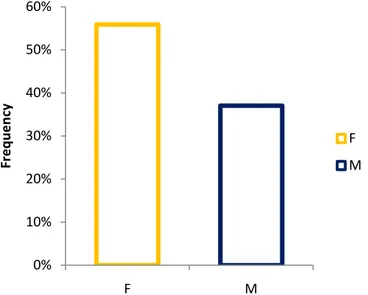
• The study comprised 79 patients in all, 36 of whom were females, representing 45.57% of the sample; and 43 were males, representing 54.43% of the sample. (FIG 2)
• The sex ratio was 1.19.
Figure 2: Distribution of Patients based on Gender
2. Place of residence
• Up to 64.56% of our patients lived in rural areas. (FIG 3)
Figure 3 : Distribution of patients based on residence
45,57% 54,43% F M 35,44% 64,56% Urban Rural
3. Healthcare insurance
• Only 15.19% of patients had healthcare insurance. (FIG 4)
Figure 4 : Distribution of patients based on whether or not they have health insurance
4. Patient history
• Up to 43.85% of our patients were followed up for some documentedmedical condition other than Parkinson’s disease at the time of diagnosis. (FIG 5)
Figure 5 : Distribution of Findings on Patient History
15,19% 84,81% Health Insurance No Health Insurance 0% 10% 20% 30% 40% 50% 60% Type 2
Diabetes High Blood Pressure ExposureToxic None Other
Type 2 Diabetes High Blood Pressure Toxic Exposure None
5. Age, Age of onset, Disease duration
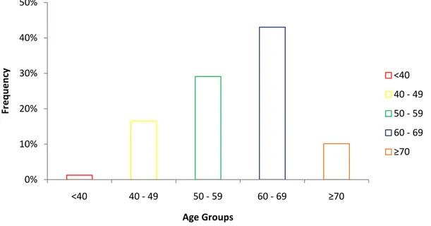
• The ages of the patients ranged from 28 to 78 years. A mean age of 59.04 years (SD = 9.43) was found in the sample.
• Patients aged 60 – 69 years were the most frequent with a relative frequency of 43%, whereas the least frequent age group was patients aged ≥ 70 years who had a frequency of 10.10%. (FIG 6)
Figure 6 : Distribution of Patients Based on Age Groups 0% 10% 20% 30% 40% 50% <40 40 - 49 50 - 59 60 - 69 ≥70 Fre qu en cy Age Groups <40 40 - 49 50 - 59 60 - 69 ≥70
• The age of onset ranged from 20 to 73 years. The mean age of onset was 52.57 years (SD = 9.91).
• Up to 43.00% of our sample had an age of onset within the 50 – 59 year range, while only 2.50% of patients had an age of onset of 70 years and above. (FIG 7)
Figure 7 : Distribution of Patients Based on Age of Onset Groups • Patients with age of onset < 50 years represented 35.44% of our sample. (FIG 8)
FIGURE 8 : Distribution of Patients Based on Onset before or after 50 years
• The duration of illness ranged from 6 months to 16 years. On average, the mean duration of illness was 6.52 years (SD = 3.81).
0% 10% 20% 30% 40% 50% <40 40 - 49 50 - 59 60 - 69 ≥70 <40 40 - 49 50 - 59 60 - 69 ≥70 35,44% 64,56% < 50 ≥50
• Patients with a disease duration of 5 – 9 years were the most frequent, with a frequency of 44.3%, whereas patient with a disease duration ≥ 15 years were the least frequent with a frequency of 6.3%. (FIG 9)
Figure 9 : Distribution of Patients Based on Duration Groups
• No difference was observed across genders in terms of age (p = 0.44), age of onset (p = 0.24), and duration (p = 0.30).
6. Clinical symptoms
• Out of the 79 patients evaluated with the NPI questionnaire, 94.94% presented at least one symptom. (FIG 10)
Figure 10 : Distribution of Symptoms in our Patients 0% 10% 20% 30% 40% 50% <5 5 - 9 10 - 14 ≥15 Fre qu en cy Duration Groups <5 5 - 9 10 - 14 ≥15 5,06% 94,94% No Symptom Symptoms
• The most frequent symptoms were depression (77%), irritability (67%), anxiety (57%) and sleep disorders (52%). The least frequent were apathy (16%), delusions (14%), and hallucinations (14%). (FIG 11)
Figure 11 : Prevalence of Neuropsychiatric Symptoms
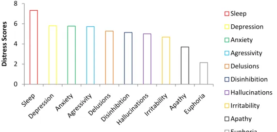
• Among patients reporting symptoms, the highest scores were recorded for sleep disorders depression, and anxiety. (FIG 12)
Figure 12 : Distress Scores for Neuropsychiatric Symptoms 0% 10% 20% 30% 40% 50% 60% 70% 80% 90% Fre qu en cy Neuropsychiatric Symptoms Depression Irritability Anxiety Sleep Disorders Agressivity Impulse Control Disinhibition Euphoria Dopamine Dysregulation Apathy Hallucination Delusion 0 2 4 6 8 Di st res s S co res Neuropsychiatric Symptoms Sleep Depression Anxiety Agressivity Delusions Disinhibition Hallucinations Irritability Apathy Euphoria
• Of the patients reporting symptoms, 90.58% of them presented more than one symptom. • The majority of patients reported 2 to 6 symptoms simultaneously. (FIG 13)
Figure 13 : Distribution of Number of Symptoms Reported in Patients
• The mean number of symptoms present was 3.67 (SD = 1.90); 4.36 (SD = 1.64) in the female group, and 3.09 (SD = 1.93) in the male group.
• A statistically significant difference was observed across genders with females reporting more symptoms than males (p = 0.02). (FIG 14)
• The mean total score was 22.57 (SD = 16.61) in the general sample, 26.69 (SD = 16.71) in the female group, and 19.12 (SD = 15.89) in the male group.
• A statistically significant difference was observed across genders with females reporting higher mean total scores than males (p = 0.04). (FIG 14)
• The mean PDQ-8 score was 43.11 (SD = 22.10), and ranged from 3.13 to 90.63.
• On the PDQ-8, female patients reported a statistically higher mean of 52.78 (SD = 19.99) than males 34.82 (SD = 20.78); p = 0.009. (FIG 14)
0% 5% 10% 15% 20% 25% 0 1 2 3 4 5 6 7 8 Fre qu en cy Number of Symptoms 0 1 2 3 4 5 6 7 8
Figure 14 : Comparison of Number of Symptoms, Total NPI Score, and PDQ-8 Score in Female and Male Patients
0 10 20 30 40 50 60 F M Number of Symptoms Total Score PDQ-39
p=0.02
p = 0.04
p =0.009
TABLE I : Summary of Patient and Caregiver Distress NPI Scores (N = 79)
All Patients Patients showing symptoms
Item score Care giver distress score Proportion with
non-zero score Item score Care giver distress score
NPI Item Mean SD Mean SD n(%) Mean SD Mean SD with symptom) n(% of patients
Delusions 0.73 2.27 0.33 1.23 11(13.92%) 5.27 3.74 4.33 1.63 6(54.55%) Hallucinations 0.7 1.9 0.30 1.14 11(13.92%) 5.00 2.15 4.00 1.55 6(54.55%) Aggressivity 1.81 3.23 0.57 1.60 25(31.65%) 5.72 3.26 5.00 0 9(36.00%) Depression 4.47 3.32 1.47 2.14 61(77.22%) 5.79 2.56 4.00 1.49 29(47.54%) Anxiety 3.28 3.91 1.19 2.00 45(56.96%) 5.76 3.54 4.09 1.35 23(51.11%) Euphoria 0.38 0.88 0.38 1.12 14(17.72%) 2.14 0.77 3.33 1.12 9(64.29%) Apathy 0.61 1.71 0.22 0.96 13(14.46%) 3.69 2.56 4.25 0.96 4(30.77%) Disinhibition 1.04 2.46 0.47 1.42 16(20.25%) 5.13 3.01 4.63 0.74 8(50.00%) Irritability 3.14 3.03 1.43 2.14 53(67.09%) 4.68 2.54 4.19 1.33 27(50.94%) Sleep 3.8 4.26 1.20 2.04 41(53.90%) 7.32 3.01 4.13 1.46 23(56.10%)
• A low correlation was observed between duration of disease and number of symptoms (r = 0.26, p = 0.02).
• No correlation was observed between total score and age (p = 0.59), and age of onset (p = 0.29), and duration (p = 0.15).
• No correlation was found between L-dopa dose and age (p = 0.67), or total score (p = 0.085). The correlation between dose and duration of disease approached significance (p = 0.06).
• No difference was found across genders with respect to response to treatment.
• A correlation was found between PDQ-8 scores and depression (p=0.046); the association with aggressivity approached significance (p=0.073).
II. Specific Findings
1.
Depression
1.1. Prevalence
• Depression was found in 61 patients, that is 77.22% of our sample. (FIG 15)
Figure 15 : Distribution of Depression in Our Patients
77,22% 22,78%
Depression No Depression
• With respect to gender, 34 patients were females, that is 94.44% of the female patients; and 27 were males, that is 62.79% of male patients. (FIG 16 & 17)
• There was a statistically significant difference in the distribution of depression in terms of gender (χ2= 11.15, df = 1, p = 0.01, ϕ = -0.376).
Figure 16 : Distribution of Depression in Females
Figure 17 : Distribution of Depression in Males
94,44% 5,56% Depression No Depression 62,79% 37,21% Depression No Depression
1.2. Score
• In the set of patients with depression, the average score was 5.79 (SD = 2.56).
• The difference in distress score across genders did not reach the level of statistical significance (p=0.567). (FIG 18)
Figure 18 : Distribution of Distress Scores in Males and Females
1.3. Age of Onset, Duration
• The mean age of onset of patients reporting depression was 51.18 years (SD = 9.40) whereas patients without depression had a mean age of onset of 57.28 years (SD = 10.40). A statistically significant difference was found between the two (p = 0.03).
• The mean duration of disease in patients reporting depression was 7.23 years (SD = 3.89). There was a statistically significant difference between patients reporting depression and those without depression in terms of duration, with patients reporting depression presenting a longer duration of disease (p < 0.0001).
5,4 5,5 5,6 5,7 5,8 5,9 6 6,1 F M Di st res s S co res F M
p=0.567
• Depression was most prevalent in patients with disease duration of 10 years and above (100%) and least prevalent in patients with disease duration less than 5 years (53.85%). (FIG 19)
Figure 19 : Distribution of Depression in Duration Groups
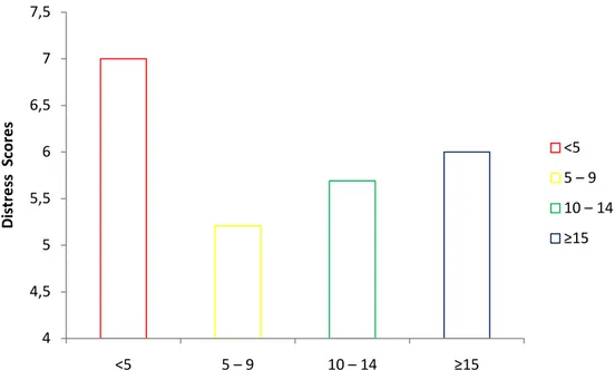
• The highest distress score was recorded in patients having a disease duration less than 5 years. (FIG 20)
Figure 20 : Distribution of Patients Based on Age of Onset Groups 50% 60% 70% 80% 90% 100% <5 5 – 9 10 – 14 ≥15 Fre qu en cy <5 5 – 9 10 – 14 ≥15 4 4,5 5 5,5 6 6,5 7 7,5 <5 5 – 9 10 – 14 ≥15 Di st res s S co res <5 5 – 9 10 – 14 ≥15
• There was no correlation between depression distress score and age (p=0.233).
• FIG 21 below shows that patients reporting depression have a younger age of onset and a longer duration of disease than patients without depression.
Figure 21: Comparison of Onset and Duration in Patients with and those without Depression
1.4. Other NPI items
• All patients experiencing delusions (100%) also reported depression.
• In terms of correlation, a moderate positive correlation was found between depression and anxiety(r= 0.57, p < 0.0001).
1.5. Caregiver Distress
• Up to 47.54% of cases of depression caused some level of distress for caregivers.
• The mean score was 4.00 (SD=1.49) and ranged from 1 to 5. The majority (17) of caregivers reported a maximum distress score of 5. That is 17 out of 29 caregivers, or 58.62% of those who reported some level of distress said it was extremely distressing.
• Breaking our findings down into gender groupings, we found that 19 of the 29 caregivers (65.51%) catered for female patients. That is 19 out of 34 females reporting depression, or 55.88%. The mean score was 4.11 (SD=1.37) with 11 out of the 19 (57.89%) reporting extreme distress. 0 10 20 30 40 50 60 70 Depression No Depression Ye ar s Onset Duration
p=0.03
p<0.0001
• The remaining 10 caregivers (34.48%) catered for male patients. That is 10 out of 27 males reporting depression, or 37.04%. The mean score was 3.80 (SD=1.75), with 6 out of the 10 (60.00%) reporting extreme distress. (FIG 22 & 23)
Figure 22 : Distribution of Caregiver Distress based on Gender of Patients
Figure 23 : Distribution of Caregiver Distress Scores based on Gender
• Caregiver distress score was better correlated withthe depression in male patients than that in female patients. (r= 0.54, p < 0.0001 vs.r= 0.38, p < 0.0001) 0% 10% 20% 30% 40% 50% 60% F M Fre qu en cy F M 3,6 3,8 4 4,2 F M Ca reg iv er D ist res s F M
p = 0.19
1.6. Response to Treatment
• Of patients reporting depression, 87.93% reported symptoms only during the off-state showing improvement during the on-state. The remaining 12.07% reported depression during the off and on states. (FIG 24)
• There was no statistically significant difference between the male and female groups with respect to response to dopamine therapy (χ2=0.36, df = 1, p = 0.55).
Figure 24 : Distribution of Response to L-Dopa Treatment
• Patients who responded to treatment did not differ from those without improvement of symptoms in age (p= 0.375), age of onset (p= 0.178), duration (p= 0.144), and L-dopa dose (p= 0.794). 87,93% 12,07%
Response to Treatment
Improvement No Improvement• Patients reporting depression received higher doses of L-dopa than those without depression (p = 0.011). (FIG 25)
Figure 25 : Comparison of L-Dopa Dose in Depressed and Non Depressed Patients
1.7. Impact on Quality of Life
• Depression distress scores were correlated to worse QoL outcomes (p=0.046). 0 200 400 600 800 Depression No Depression L-Do pa Do se s Depression No Depression
p = 0.011
2.
Anxiety
2.1. Prevalence
• Anxiety was reported in 56.96% of our sample. (FIG 26)
Figure 26 : Distribution of Anxiety in the Sample
• Anxiety was found in 66.67% of the female sample, and 48.83% of the male sample. Males and females did not differ significantly in the distribution of anxiety (χ2= 2.54, df=1, p >0.05). (FIG 27 & 28)
Figure 27 : Distribution of Anxiety in Females
56,96% 43,04% Anxiety No Anxiety 66,67% 33,33% Anxiety No Anxiety
Figure 28 : Distribution of Anxiety in Males
2.2. Scores
• In the set of patients reporting anxiety, the average score was 5.76 (SD = 3.54).
• There was no statistically significant difference between the mean distress score across genders (p=0.812). (FIG 29)
Figure 29 : Comparison of Distress Scores in Males and Females
48,83% 51,17% Anxiety No Anxiety 5,2 5,4 5,6 5,8 6 F M Di st res s S co res F M p=0.812
2.3. Age of Onset, Duration
• The mean age of onset of patients reporting anxiety was 52.40 years (SD= 10.15); and the mean duration of disease was 6.51 years (SD = 3.67).
• There was no statistical difference between patients reporting anxiety and those without anxiety in terms of age, age of onset, and duration. (FIG 30)
Figure 30 : Comparison of Onset and Duration in Patients with and those without Anxiety • Anxiety was most frequent in patients with age at onset of 70 years and more (87.5%), and
was least reported in patients in the 40 – 49 years group (46.15%). (FIG 31)
Figure 31 : Distribution of Anxiety in the Age of Onset Groups 0 10 20 30 40 50 60 Anxiety No Anxiety Ye ar s Age of Onset Duration
p=0.862
p=0.983
0% 20% 40% 60% 80% 100% 40 – 49 50 – 59 60 – 69 ≥70 Fre qu en cy 40 – 49 50 – 59 60 – 69 ≥70• The scores for anxiety was least (3.33, SD=2.58) in patients with age at onset of 40 – 49 years, and highest (6.13, SD=3.96) in patients in the 50 – 59 years group. (FIG 32)
Figure 32 : Distribution of Distress Score in Age of Onset Groups
• The only patient aged less than 40 years also reported anxiety with the maximum score of 12. • Our study found no correlation between anxiety distress scores and age of patients. • Patients with disease duration of at least 15 years had the least frequency of anxiety (40%),
while patients with disease duration from 10 to 14 years had the highest frequency (69.23%). (FIG 33)
Figure 33 : Distribution of Anxiety in the Duration Groups 0 1 2 3 4 5 6 7 40 – 49 50 – 59 60 – 69 ≥70 Di st re ss S co re 40 – 49 50 – 59 60 – 69 ≥70 0% 10% 20% 30% 40% 50% 60% 70% 80% <5 5 – 9 10 – 14 ≥15 Fre qu en cy <5 5 – 9 10 – 14 ≥15
• The highest mean distress score for anxiety of 7.46 (SD = 3.78) was recorded for patients with a disease duration of less than 5 years, whereas patients with a duration from 5 to 9 years had the least distress score of 4.71 (SD = 3.32). (FIG 34)
Figure 34 : Distribution of Distress Scores in the Duration Groups
2.4. Caregiver Distress
• Up to 51.11% of cases of anxiety caused some level of distress for caregivers.
• More caregivers catering for female patients (62.50%) reported distress as compared to caregivers of male patients (38.01%). (FIG 35)
Figure 35 : Distribution of Caregiver Distress based on Gender 0 1 2 3 4 5 6 7 8 <5 5 – 9 10 – 14 ≥15 Di st res s S co res <5 5 – 9 10 – 14 ≥15 0% 10% 20% 30% 40% 50% 60% 70% F M Fre qu en cy F M
• The mean score was 4.13 (SD=1.25) in the caregivers catering for female patients as compared to 4.00 (SD=1.60) in male patients. (FIG 36)
Figure 36 : Distribution of Distress Scores based on Patient Gender
• A moderate positive correlation was found between caregiver distress scores for anxiety and anxiety in patients (r= 0.58, p < 0.0001).
• The correlation between the caregiver distress scores for anxiety and anxiety in patients was stronger in male patients (r= 0.64, p < 0.0001) than in female patients (r= 0.50, p = 0.001).
3 3,5 4 4,5 F M Ca reg iv er D ist res s F M
p=0.827
2.5. Response to Treatment
• Of patients reporting anxiety, 83.64% experienced symptoms during the off state showing improvement of symptoms during the on state, while 16.36% reported symptoms during both on and off states. (FIG 37)
Figure 37 : Distribution of Response to L-Dopa Treatment
• There was no statistically significant difference between the male and female groups with respect to response to dopamine therapy (χ2= 0.30, df=1, p = 0.59).
• Patients who responded to treatment did not differ from those without improvement of symptoms in age (p= 0.986), age of onset (p= 0.868), duration (p= 0.2), and L-dopa dose (p= 0.706).
83,64% 16,36%
Improvement No Improvement
2.6. Impact on Quality of Life
• The difference between the PDQ-8 scores of patients with anxiety and those without did not reach statistical significance (p=0.395). (FIG 38)
Figure 38 : Comparison of PDQ-8 Scores between Patients with and those without Anxiety 0 10 20 30 40 50 Anxiety No Anxiety PDQ -8 Sc or e Anxiety No Anxiety
3.
Psychotic symptoms
3.1. Prevalence
• Psychotic symptoms were present in 22.78% of our patients.Only 5.04% of the sample reported both delusions and hallucinations. (FIG 39)
a. Delusions
• Delusions were present in 13.92% of the sample. In terms of gender, 16.67% of the female patients and 11.62% of the male patients reported delusions.
• There was no statistically significant difference in terms of gender (χ2= 0.42, p=0.519).
b. Hallucinations
• Hallucinations were present in 13.92% of the patient. In terms of gender, 19.44% of the female patients and 9.30% of the males patients reported hallucinations.
• There was no statistically significant difference in terms of gender (χ2= 1.68, p=0.195)
Figure 39 : Distribution of Psychosis in the Sample
22,78%
3.2. Scores a. Delusion
• In the set of patients presenting delusions, the average score was 5.27 (SD = 3.74).
• The difference in distress score across genders did not reach the level of statistical significance (p=0.807). (FIG 40)
Figure 40 : Comparison of Distress Scores in Males and Females 4,7 4,8 4,9 5 5,1 5,2 5,3 5,4 5,5 5,6 5,7 F M Di st res s S co res F M
p=0.807
b. Hallucination
• In the set of patients presenting hallucinations, the average score was 5.00 (SD = 2.14). • The difference in distress score across genders did not reach the level of statistical
significance (p=0.586). (FIG 41)
Figure 41 : Comparison of Distress Scores for Hallucinations in Males and Females 4,2 4,4 4,6 4,8 5 5,2 5,4 5,6 F M Di st res s S co res F M
p=0.586
3.3. Age of Onset, Duration
• The mean age of onset of patients reporting psychotic manifestations was 51.22 years (SD=12.64).
• The mean duration of disease was 8.06 years (SD=3.95).
• No statistically significant difference was found between the group of patients with psychotic manifestation and those without in terms of age (p=0.903) and age of onset (p=0.515). (FIG 42)
• Patients with psychotic manifestations had a relatively longer duration with a difference approaching significance (p=0.051). (FIG 42)
Figure 42 : Comparison of Onset and Duration in Psychotic and Non Psychotic Patients 0 10 20 30 40 50 60 Psychosis No Psychosis Ye ar s Onset Duration
p=0.515
p=0.051
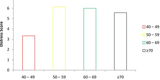
• In terms of scores, the composite score for psychotic manifestation was highest in the 60 – 69 year age groups, and least in patients aged over 70 years. (FIG 43)
Figure 43 : Distribution of Distress Scores in Age Groups
• The highest frequency of psychotic symptoms was recorded in patients with disease duration of at least 15 years (40%), and least in patients with duration less than 5 years (15.38%). (FIG 44)
Figure 44 : Distribution of Psychosis in Duration Groups 0 0,5 1 1,5 2 2,5 3 3,5 4 4,5 40 – 49 50 – 59 60 – 69 ≥70 Di st res s S co res 40 – 49 50 – 59 60 – 69 ≥70 0% 5% 10% 15% 20% 25% 30% 35% 40% 45% <5 5 – 9 10 – 14 ≥15 Fre qu en cy <5 5 – 9 10 – 14 ≥15
• Patients within the 10 – 14 year disease duration group recorded the highest mean distress score of 4.7 (SD=3.42), whereas patients within the 5 – 9 year disease duration group recorded the least mean distress score of 2.43 (SD=1.34). (FIG 45)
Figure 45 : Distribution of Distress Scores in Duration Groups 0 0,5 1 1,5 2 2,5 3 3,5 4 4,5 5 <5 5 – 9 10 – 14 ≥15 Di st re ss S co re <5 5 – 9 10 – 14 ≥15
3.4. Caregiver Distress
• Of all the patient reporting psychotic symptoms, 50% were distressful for caregivers. • In female patients reporting psychotic symptoms, 63.64% cause some level of distress to
caregivers as against 28.57% in male patients with psychotic symptoms. (FIG 48) • The mean caregiver distress score was 1.39 (SD=1.70).
• No statistically significant difference was observed across genders in terms of caregiver score (p=0.544). (FIG 46)
Figure 46 : Distribution Caregiver Distress based on the Gender of Patients 0% 10% 20% 30% 40% 50% 60% 70% F M Fre qu en cy F M
Figure 47 : Distribution of Caregiver Distress Scores based on Gender of Patients
3.5. Response to Treatment
• Of patients reporting psychotic manifestations 77.78% reported a stagnation of symptoms during the on state. The remaining 22.22% showed worsening of symptoms with L-Dopa treatment. (FIG 48)
Figure 48 : Distribution of Response to Treatment 0 0,2 0,4 0,6 0,8 1 1,2 1,4 1,6 1,8 F M Di st res s S co res F M 77,78% 22,22% Stagnation of Symptoms Worsening of Symptoms
p=0.544
• Patients whose symptoms did not get worse with L-Dopa had a latter onset of disease (p = 0.042).
• There was no statistically significant difference between patients who reported psychotic symptoms and those who did not in terms of L-Dopa dose (p=0.745). (FIG 49)
Figure 49 : Comparison of L-Dopa Dose in Psychotic and Non Psychotic Patients 0 200 400 600 Psychosis No Psychosis L-Do pa Do se s Psychosis No Psychosis
p=0.745
3.6. Impact on Quality of Life
• The difference between the PDQ-8 scores of patients with psychosis and those without did not reach statistical significance (p=0.128). (FIG 50)
Figure 50 : Comparison of PDQ-8 Scores between Patients with and those without Psychosis 0 10 20 30 40 50 Psychosis No Psychosis PDQ -8 Sc or e Psychosis No Psychosis
4.
Apathy
4.1. Prevalence
• Apathy was present in 16.46% of our sample. (FIG 51)
Figure 51 : Distribution of Apathy in the Sample
4.2. Score
• The average score was 3.69(SD =2.56); 3.83 (SD = 2.77) in the female group as against 3.57 (SD = 2.57) in the male group.
• The difference of distress score did not reach statistical significance across genders (p=0.863). (FIG 52)
Figure 52 : Comparison of Apathy Scores across Gender
16,46% 83,54% Apathy No Apathy 0 0,5 1 1,5 2 2,5 3 3,5 4 F M Di st res s S co res F M
p=0.863
4.3. Age of Onset, Duration
• There was no statistically significant difference between patients reporting apathy and those without apathy in terms of age of onset (p=0.116) and duration (p=0.107). (FIG 53)
Figure 53 : Comparison of Age, Onset and Duration in Patients with and without Apathy 0 10 20 30 40 50 60 Apathy No Apathy Ye ar s Onset Duration
p=0.116
p=0.107
• Apathy was most frequent in patients with disease duration of at least 15 years (40%), and least frequent in patients with disease duration less than 5 years (11.54%).(FIG 54)
• The highest mean distress score of 6.00 (SD=4.36)was recorded in patients within the 10 – 14 year duration group, whereas the lowest mean distress score of 2.33 (SD=1.53) was recorded in patients with duration less than 5 years.(FIG 55)
Figure 54 : Distribution of Apathy in the Duration Groups
Figure 55 : Distribution of Apathy Scores in Duration Groups 0% 5% 10% 15% 20% 25% 30% 35% 40% 45% <5 5 – 9 10 – 14 ≥15 Fre qu en cy <5 5 – 9 10 – 14 ≥15 0 1 2 3 4 5 6 7 <5 5 – 9 10 – 14 ≥15 Di st res s S co res <5 5 – 9 10 – 14 ≥15