Universite de Sherbrooke
Faculte de genie
Departement de genie chimique
DEVELOPMENT OF BIOACTIVE, POROUS SCAFFOLDS FOR THE VASCULARIZATION OF ENGINEERED TISSUES
DEVELOPPEMENT D'ECHAFAUDAGES POREUX, BIOACTIFS POUR LA VASCULARISATION DE TISSUS ARTIFICIELS
Thesis submitted for the degree of Doctor of Philosophy Chemical Engineering
Hanna BRAMFELDT
Sherbrooke, Quebec, CANADA February, 2008
1*1
Library and Archives Canada Published Heritage Branch 395 Wellington Street Ottawa ON K1A0N4 Canada Bibliotheque et Archives Canada Direction du Patrimoine de I'edition 395, rue Wellington Ottawa ON K1A0N4 CanadaYour file Votre reference ISBN: 978-0-494-37954-7 Our file Notre reference ISBN: 978-0-494-37954-7
NOTICE:
The author has granted a non-exclusive license allowing Library and Archives Canada to reproduce, publish, archive, preserve, conserve, communicate to the public by
telecommunication or on the Internet, loan, distribute and sell theses
worldwide, for commercial or non-commercial purposes, in microform, paper, electronic and/or any other formats.
AVIS:
L'auteur a accorde une licence non exclusive permettant a la Bibliotheque et Archives Canada de reproduire, publier, archiver,
sauvegarder, conserver, transmettre au public par telecommunication ou par Plntemet, prefer, distribuer et vendre des theses partout dans le monde, a des fins commerciales ou autres, sur support microforme, papier, electronique et/ou autres formats.
The author retains copyright ownership and moral rights in this thesis. Neither the thesis nor substantial extracts from it may be printed or otherwise reproduced without the author's permission.
L'auteur conserve la propriete du droit d'auteur et des droits moraux qui protege cette these. Ni la these ni des extraits substantiels de celle-ci ne doivent etre imprimes ou autrement reproduits sans son autorisation.
In compliance with the Canadian Privacy Act some supporting forms may have been removed from this thesis.
Conformement a la loi canadienne sur la protection de la vie privee, quelques formulaires secondaires ont ete enleves de cette these.
While these forms may be included in the document page count,
their removal does not represent any loss of content from the thesis.
Canada
Bien que ces formulaires
aient inclus dans la pagination, il n'y aura aucun contenu manquant.
ABSTRACT
The lack of vascular networks to support the growth, function, and survival of three-dimensional tissues represents one of the greatest challenges of tissue engineering today. This thesis project was designed to constitute the first part of a work whose final objective is the creation of functional micro-vessel networks within three-dimensional scaffold constructs for tissue engineering. Specifically, this part was aimed at creating functional scaffolds, that may be used in the continued studies towards achieving this long-term goal.
As a first step, three co-polymers on the form poly(s-caprolactone-co-D,L- lactic acid)-poly(ethylene glycol)-poly(E-caprolactone-co-D,L- lactic acid) were synthesized to provide mechanically stable biomaterials with controllable and tunable material properties. To achieve this, the monomer content in the side chains was varied and the resulting co-polymers were characterized using 'H-NMR, GPC, DSC, WAXS, and DMA1. Furthermore, the hydrolytic degradation profiles of samples
fabricated from melt-pressed films were studied over six months, as well as the influence of degradation on tensile strength. Based on obtained results and observations, two of the co-polymers were judged suitable, in terms of mechanical integrity and degradation profile, for further investigation.
To improve cell/polymer interactions, co-polymer films were modified by protein immobilization using imidoester chemistry. The effect of several protein preparations on smooth muscle cell (SMC) adhesion and proliferation was studied over six days in culture. Cells were counted at pre-set intervals, and further analyzed by scanning electron microscopy and fluorescent labelling of the differentiation marker a-actin. Immobilized proteins greatly enhanced adhering cell numbers, although for stimulation of SMC proliferation covalent immobilization was superior to physisorption.
1 'H-NMR: 'H-nuclear magnetic resonance; GPC: gel permeation chromatography; DSC: differential scanning calorimetry; WAXS: wide-angle x-ray spectrometry; and DMA: dynamic-mechanical analysis
RESUME
La construction d'un reseau vasculaire pouvant assurer la croissance, la fonction et la survie de tissus tridimensionnels represente actuellement l'un des defis les plus importants du genie tissulaire. Cette these fait partie d'un vaste projet ayant pour but de developper des m6thodes de culture de micro-vaisseaux sanguins au sein d'un echafaudage tridimensionnel synth6tique qui pourra servir en genie tissulaire en conditions de bioreacteur.
Trois copolymeres composes de poly(e-caprolactone-co-D,L-acide lactique)-PEG-poly(e-caprolactone-co-D,L-acide lactique) ont ete synthetises par la polymerisation par ouverture de cycle. La composition monomerique a ete variee et les polymeres obtenus ont ete caracterises par 'H-resonance magnetique nucleaire, chromatographie de permeation de gel, calorimetrie differentielle a balayage, diffraction des rayons X aux grands angles, et analyseur mecanique dynamique. De plus, la degradation hydrolytique des echantillons, fabriques par moulage a l'etat fondu, a ete etudiee, ainsi que l'impact d'une telle degradation sur l'endurance a la tension mecanique de ces materiaux. Considerant les resultats observes au niveau des proprietes mecaniques et leur evolution au cours des tests de degradation, deux des copolymeres ont ete selectionnes pour etre etudies davantage en lien avec leur possible utilisation en cultures cellulaire et tissulaire.
Dans l'objectif d'ameliorer les interactions cellules/materiaux, des films composes de ces deux copolymeres ont ete modifies en immobilisant des proteines a leur surface. L'effet de plusieurs proteines sur le comportement de cellules musculaires lisses {smooth muscle cells, SMC) a ete etudie au cours de cultures allant jusqu'a six jours. Les cellules ont ete comptees a des intervalles fixes, et caracterisees par la microscopie electronique a balayage ainsi que par le marquage fluorescent du marqueur de differentiation, oc-actine. La presence de proteines a ameliore de fa?on significative les interactions cellulaires, notamment au niveau de la proliferation qui etait davantage stimulee par l'utilisation de proteines immobilisees par liaison covalente.
Une technique basee sur des melanges polymeriques co-continus a ete appliquee a nos copolymeres. Les echafaudages fabriques par cette technique demontraient une porosite hautement interconnectee, dont les tallies de pores peuvent etre controlees par recuit. La permeabilite et la resistance a la compression des structures tridimensionnelles ont ete determinees. De plus, l'adhesion de SMC a des echafaudages modifies par proteines a indique une dependance des tailles de pores.
Les resultats de cette these demontrent que les echafaudages fabriques et modifies tel que specifie, pourraient trouver usage dans l'ingenierie de tissus a base de SMC. Ces cultures pourraient aussi servir a la vascularisation de tissus artificiels par voie de co-culture avec des cellules endotheliales.
GENERAL INTRODUCTION
The in vitro generation of human tissues for transplantation constitutes a long-standing dream in reparative and reconstructive medicine. Today, the regeneration of tissues such as cartilage[l], bone[2], and skin[3] is possible with available techniques. However, engineering (TE) of thick, metabolically demanding tissues is facing an important challenge, as the lack of functional vascular networks, able to support a growing cell mass, has proven to significantly limit tissue development^]. Such tissues, including the pancreas, liver, heart, and kidney, require intrinsic blood supply for long-term survival and development^]. To date, the focus of research has been placed mainly on promoting ingrowth from the pre-existing blood vessels at the implant site into the TE construct[6]. However, less attention has been given to the regeneration of viable tissue around pre-constructed vascular networks.
OBJECTIVES. This project was designed to constitute the first part of a work whose final objective will be the creation of functional vascular beds within three-dimensional scaffold constructs for tissue engineering. The main objective of the thesis was the development of three-dimensional, bioactive, porous scaffolds that may be used for advanced tissue culture in bioreactors. To this end, we wanted to:
1. Synthesize a group of biocompatible and modifiable co-polymers, with controllable degradation rates and mechanical properties. Characterize and evaluate these co-polymers for their applicability as scaffold materials.
2. Apply a biochemical surface-modification method pertaining to the optimization of interactions between the synthesized biomaterials and cells native to blood vessels, which may eventually serve to improve formation of mature vessel-like structures. Smooth muscle cells, an essential component of mature vasculature[7,8] was chosen for this purpose.
3. Fabricate bioactive, three-dimensional, porous scaffolds with highly interconnected porosity that may withstand long-term culture conditions in a perfusion bioreactor. To employ the surface modification technique developed to improve cell adhesion in a three-dimensional environment.
THESIS STRUCTURE. This thesis was written in a manner that substitutes the classical division into chapters by the scientific publications that are the results of this project. Some of these articles have been accepted for publication, whereas others are submitted for review. The details of the publications and their current status are available in the List of Publications. Below follows a brief description of their content.
Chapter 1, "Scaffold vascularization: a challenge for three-dimensional tissue
engineering", consists of a literature review of advancements in three-dimensional tissue
engineering with an emphasis on in vitro tissue culture, and the vascularization of porous scaffolds. Moreover, the effects of co-culture techniques as well as mechanical conditioning of tissue constructs within the confines of a bioreactor are discussed. Following this introductory chapter, the articles resulting from the work carried out within the framework of this project are presented in Chapters 2-5.
In Chapter 2, "Characterisation, degradation and mechanical strength of poly(D,L lactide-co-e-caprolactone)-poly(ethylene glycol)-poly(D,L-lactide-co-s-caprolactone)", the detailed characterization of three synthesized co-polymers with varying D,L-lactide and e-caprolactone content is described. These co-polymers were chosen due to their biocompatibility, and the ease with which their material properties can be altered to answer to the diverse set of demands placed on scaffold materials. The polymers were characterized using ^-nuclear magnetic resonance ('H-NMR), gel permeation chromatography (GPC), differential scanning calorimetry (DSC), wide-angle x-ray spectrometry (WAXS), and dynamic-mechanical analysis (DMA). The hydrolytic degradation of co-polymer films as well as the influence of degradation on the tensile strength of these co-polymers is reported. In addition, blends of these copolymers were prepared and the hydrolytic degradation profiles of films of different composition and thickness were compared. The results of this study can be seen in Chapter 3, "Blends as a strategy towards tailored hydrolytic degradation of
P(CL-co-D,L-LA)-PEG-P(CL-co-D,L-LA) copolymers ".
Chapter 4, "Enhanced smooth muscle cell adhesion and proliferation on
protein-modified polycaprolactone-based copolymers", describes a two-dimensional cell-culture
study carried out using films from two of the co-polymers that were judged most suitable for scaffold fabrication. In an effort to create bioactive materials, primary amine groups were introduced through aminolysis, followed by treatment with dimethyl pimelimidate. The thus
activated surfaces were subsequently reacted with several proteins (fibronectin, fibrinogen, and fibrin layers), which were covalently immobilized. Co-polymer films exhibiting physisorbed proteins were also included in the study. The effects of this surface modification on smooth muscle cell adhesion and proliferation were investigated.
In the last phase of this work, three-dimensional scaffolds were fabricated using a binary polymer blend technique, which resulted in highly interconnected pores and allowed close control of pore size through static annealing. With the aim of evaluating the applicability of such scaffolds in tissue engineering, the compressive strength and permeability of non-annealed and non-annealed structures were investigated. Moreover, scaffolds of two different pore sizes were fibronectin-modified using the method used in Chapter 4, and compared to evaluate cell adhesion and seeding efficiency of smooth muscle cells. The results are presented and discussed in Chapter 5, "Smooth muscle cell adhesion in surface-modified three-dimensional
co-polymer scaffolds prepared from co-continuous blends".
CONTRIBUTIONS. During the course of this thesis, I have had important help from several
people, to whom I am very grateful. The co-author of three of the publications making up this document, Dr Pierre Sarazin, has assisted in the execution and interpretation of results from DSC, WAXS, and DMA. The DMA analysis itself was carried out by Dr Patrice Cousin, Department of Civil Engineering, at the Universite de Sherbrooke. Pierre Sarazin has furthermore prepared all the binary polymer blends used for the fabrication of porous scaffolds, and taught me the techniques to carry out static annealing, and preparation of samples for SEM analysis. Finally, he contributed the idea to prepare co-polymer blend films to study the influence of such compositions and of film thickness on degradation.
The testing of compressive mechanical properties was carried out at Industrial Materials Institute in Boucherville, QC, where Dr Martin Bureau and Manon Plourde assisted in the use of the equipment. WAXS and 'H-NMR analysis were carried out by the designated
References
(1) Stock U, Vacanti JP. Tissue engineering: current state and prospects. Annu. Rev. Med. 2001;52:443-51.
(2) Middleton JC, Tipton AJ. Synthetic biodegradable polymers as orthopedic devices. Biomaterials 2000; 21:2335-2346.
(3) Balasubramani M, Kumar TR, Babu M. Skin substitutes: a review. Burns 2001; 27:534-544.
(4) Kannan RY, Salacinski HJ, Sales K, Butler P, Seifalian AM. The roles of tissue engineering and vascularisation in the development of micro-vascular networks: a review. Biomaterials 2005; 26:1857-1875.
(5) Borenstein JT, Terai H, King KR, Weinberg EJ, Kaazempur-Mofrad MR, Vacanti JP. Microfabrication technology for vascularized tissue engineering. Biomed. Microdevices 2002;4:167-175.
(6) Cassell OCS, Hofer S, Morrison WA, Knight KR. Vascularization of tissue-engineered grafts: the regulation of angiogenesis in reconstructive surgery and in disease states. Brit. J. Plastic Surg. 2002; 55:603-610.
(7) Drake CJ, Hungerford JE, Little CD. Morphogenesis of the first blood vessels. Morphogenesis: Cell. Interactions 1998; 857:155-179.
(8) Vailhe B, Vittet D, Feige JJ. In vitro models of vasculogenesis and angiogenesis. Lab. Invest. 2001;81:439-452.
LIST OF PUBLICATIONS
The thesis contains modified versions of the below publications.
• Bramfeldt, H., Vermette, P. Scaffold vascularization: A challenge for three-dimensional
tissue engineering. In preparation for Frontiers in Biomedicine.
• Bramfeldt, H., Sarazin, P., and Vermette, P. Characterisation, degradation and
mechanical strength ofpoly(D,L-lactide-co-e-caprolactone)-poly(ethylene glycol)-poly(D,L-lactide-co-s-caprolactone). Journal of Biomedical Materials, Part A, 2007;
83A : 503-511.
• Bramfeldt, H., Sarazin, P., and Vermette, P. Blends as a strategy towards tailored
hydrolytic degradation ofP(CL-co-D,L-LA)-PEG-P(CL-co-D,L-LA) copolymers.
Submitted to Polymer Stability and Degradation.
• Bramfeldt, H., Vermette, P. Enhanced smooth muscle cell adhesion and proliferation on
protein-modified polycaprolactone-based copolymers. Journal of Biomedical Materials,
Part A, accepted for publication. Reference number: JBMR-A-07-0467.R1
• Bramfeldt, H., Sarazin, P., Vermette, P. Smooth muscle cell adhesion in surface-modified
three-dimensional co-polymer scaffolds prepared from co-continuous blends. Submitted
ACKNOWLEDGEMENTS
First of all, I would like to extend a special thank you to my supervisor, Dr. Patrick Vermette. You have reinforced in me the values of independence and perseverance. You were never too busy to meet or to discuss. I truly feel I have become a better scientist under your supervision.
Dr. Pierre Sarazin, a post-doctoral fellow in our group (presently at Ecole Polytechnique in Montreal). I would like to thank you for everything you have helped me with: from the technical know-how and the pursuit of perfection to the constant asking of nagging questions and general merriment. Not only has your knowledge and clear-sightedness motivated me and helped me to restructure and refocus on many an occasion, but I find your philosophy on life, your curiosity, and your sense of humour very refreshing!
My very own Matej. What can I say? Without you, what would all this mean anyway? You have supported me and challenged me, and your good mood makes my good mood. I thank you also for all the weekends spent at home or outdoors, alone or in the company of good friends, exploring the most remote corners of Quebec and elsewhere. It has been a constant reminder of how much more there is to life than lab bench tops and evenings spent in the less stimulating company of primary cell cultures.
To my family: I have always been able to count on you. Thanks for cheering me on, for pretending to listen when I talk about my work, for the cross-words and the Christmas decorations, and for never telling me it was a bad idea to come here. Here's to many more years of me boring you with fascinating scientific details!
Much appreciation is extended to my friends and colleagues, the patient and fantastic people who have endured me on a close-to-daily basis for these four years. Without you I would have given up a long time ago. Good luck to you all in the future, whatever it may bring!
Finally, I would like to express my gratitude to Drs. Pentti Tengwall and Fredrik Hook for first inspiring in me the courage to enter into the world of research, and for being the scientific and personal models to which I shall always aspire.
TABLE OF CONTENTS
CHAPTER 1:
SCAFFOLD VASCULARIZATION:A
CHALLENGE FORTHREE-DIMENSIONAL TISSUE ENGINEERING 1
1.1 Abstract 1 1.2 Introduction 1 1.3 Scaffold materials 3
1.3.1 Biologically derived polymers 3
1.3.2 Synthetic biomaterials 5 1.3.3 Scaffold porosity 8 1.4 Vascularization of biomaterials 9
1.4.1 Blood vessels and vascularization in vivo 9
1.4.2 Angiogenic Factors 12 1.4.3 Tube formation in biological scaffolds 13
1.4.4 Tube formation in synthetic scaffolds 18
1.4.5 Co-culture models 20 1.5 Bioreactor culture and mechanical conditioning 23
1.5.1 Flow conditioning 24 1.5.2 Cyclic strain 25 1.6 Conclusions 27 1.7 References 29
CHAPTER 2:
CHARACTERISATION, DEGRADATION AND MECHANICALSTRENGTH OF POLY(D,L-LACTIDE-CO-E-CAPROLACTONE)-POLY(ETHYLENE
GLYCOL)-POLY(D,L-LACTIDE-CO-E-CAPROLACTONE) 45
2.1 Abstract 45 2.2 Introduction 45 2.3 Materials and Methods 47
2.3.1 Materials 47 2.3.2 Polymer synthesis 47 2.3.3 Film preparation 48 2.3.4 Characterization 48 2.3.5 Hydrolytic degradation 49 2.3.6 Tensile testing 50
2.4 Results and Discussion 50
2.4.2 Hydrolytic degradation 53
2.4.3 Tensile strength 59
2.5 Conclusions 60 2.6 Acknowledgments 61 2.7 References 62
CHATPER 3:
BLENDS AS A STRATEGY TOWARDS TAILORED HYDROLYTICDEGRADATION OF P(CL-CO-D,L-LA)-PEG-P(CL-CO-D,L-LA) COPOLYMERS 6 6
3.1 Abstract 66 3.2 Introduction 66 3.3 Materials and Methods 68
3.3.1 Materials 68 3.3.2 Copolymers, blend preparation and sample fabrication 68
3.3.3 Hydrolytic degradation 68
3.34 Statistical Analysis 69 3.4 Results and Discussion 70
3.4.1 Mass loss and buffer absorption 70 3.4.2 Evolution of pH of degradation media 71
3.4.3 Morphology 73 3.5 Conclusions 74 3.6 Acknowledgments 75 3.7 References 76
CHAPTER 4:
ENHANCED SMOOTH MUSCLE CELL ADHESION ANDPROLIFERATION ON PROTEIN-MODIFIED POLYCAPROLACTONE-BASED
COPOLYMERS 7 9 4.1 Abstract 79 4.2 Introduction 79 4.3 Materials and Methods 81
4.3.1 Materials 81 4.3.2 Co-polymer synthesis 81
4.3.3 Film preparation 82 4.3.4 Surface modification 82 4.3.5 Characterization of amine content 83
4.3.6 Cell culture 83 4.3.7 Cytotoxicity 83
4.3.8 Cell adhesion and proliferation 84
4.3.9 Statistical analysis 85
4.4.1 Cytotoxicity 86 4.4.2 Surface modification 87
4.4.3 Smooth muscle cell culture 89
4.5 Conclusions 96 4.6 Acknowledgements 97
4.7 References 98
CHAPTER 5:
SMOOTH MUSCLE CELL ADHESION IN SURFACE-MODIFIEDTHREE-DIMENSIONAL POLYMER SCAFFOLDS PREPARED FROM
CO-CONTINUOUS BLENDS 1 0 2
5.1 Abstract 102 5.2 Introduction 102 5.3 Materials and Methods 105
5.3.1 Materials 105 5.3.2 Co-polymers 105 5.3.3 Preparation of blends 105
5.3.4 Annealing and scaffold fabrication 105
5.3.5 SEM imaging 106 5.3.6 Mechanical properties 106
5.3.7 Permeability of scaffolds 107 5.3.8 Protein immobilization and smooth muscle cell seeding 107
5.4 Results and Discussion 110 5.4.1 Effects of annealing on pore size 110
5.4.2 Compressive strength 113 5.4.3 Permeability of scaffolds 114 5.4.4 Smooth muscle cell adhesion on protein-modified scaffolds 116
5.5 Conclusions 119 5.6 Acknowledgements 119
5.7 References 120
GENERAL CONCLUSION AND SUGGESTED FUTURE WORK 123
APPENDIX 129
A2. Protocol for surface modification, cell seeding, cell count, and imaging 133 A3. Standard curve established for the ninhydrin assay used in Chapter 4 136
L I S T OF FIGURES
Chapter 1
Figure 1.1 Polymeric structure of poly(glycolic acid), poly(lactic acid), and
poly(s-caprolactone) 6 Figure 1.2 The structure of a large blood vessel, depicting the intima, the media,
and the adventitia 10
Chapter 2
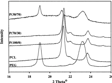
Figure 2.1 WAXD curves of films of PEG-containing copolymers 52 Figure 2.2 Water absorption and mass loss of P( 100/0), P(70/30), and P(30/70)
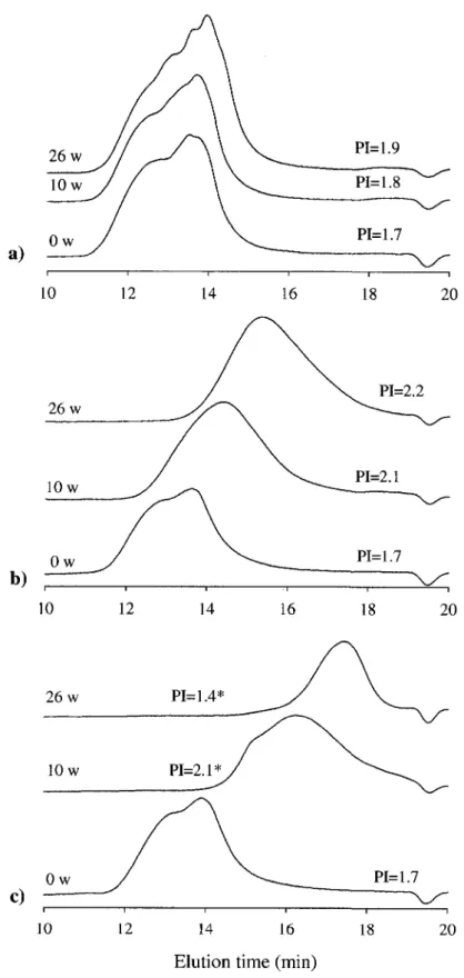
during degradation in PBS (37°C, pH 7,4) for up to 26 weeks 54 Figure 2.3 Gel permeation chromatograms showing the effect of degradation on the
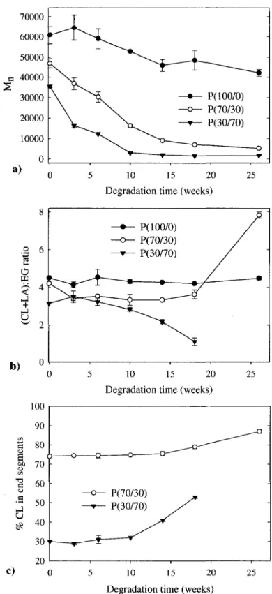
polydispersity and molecular weights 55 Figure 2.4 The effect of degradation on the average molecular weight, Mn, as
determined by GPC and the effect of degradation on monomer composition
as calculated from 'H-NMR spectra 57
Chapter 3
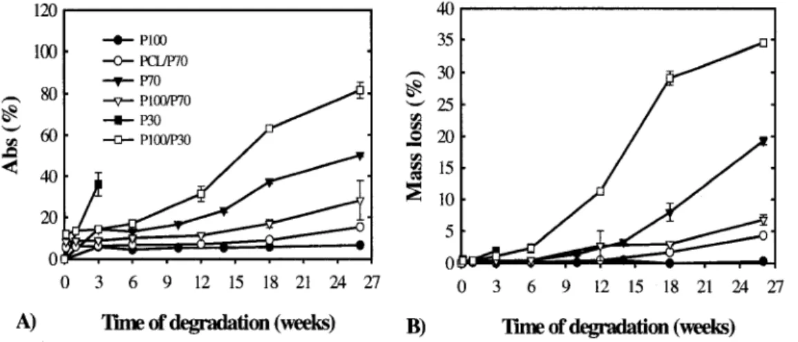
Figure 3.1 Mass loss (% of initial weight) and water absorption (% of dry weight)
presented as a function of degradation time over 26 weeks 71 Figure 3.2 Effect of sample thickness and blend composition on the pH of the
degradation media. 72 Figure 3.3 Scanning electron micrographs of partially degraded P100/P30 copolymer
blends. 73 Figure 3.4 Scanning electron micrographs of cross-sections of partially degraded
PCL/P70, P100/P70, and P100/P30 copolymer blends. 74
Chapter 4
Figure 4.1 MTT mitochondrial activity assay of the cytocompatibility of degradation
medium. 86 Figure 4.2 Absorbance at 570nm of aminolyzed copolymer films. 88
gure 4.3 Cell adhesion and proliferation on aminolyzed and protein-immobilized
P( 100/0) films. 90 Figure 4.4 Cell adhesion and proliferation on aminolyzed and protein-immobilized
P(70/30) films. 92 Figure 4.5 Scanning electron micrographs of SMC on modified P( 100/0), 8h and 48h
after seeding. 93 Figure 4.6 a-actin staining of SMC on modified P(100/0), 8h and 48h after seeding. 94
Figure 4.7 a-actin staining of SMC on modified P(70/30), 8h and 48h after seeding. 95
Chapter 5
Figure 5.1 Schematic drawing of cell-seeding device. 108 Figure 5.2 SEM images confirming continuous porosity in non-annealed samples of
P(100/0), P(70/30), and polystyrene. 110 Figure 5.3 Porous P(100/0): effect of annealing on pore size. 112
Figure 5.4 Porous P(70/30): effect of annealing on pore size. 112 Figure 5.5 Representative stress-strain curves for porous P( 100/0) and P(70/30). 114
Figure 5.6 Compressive Young's moduli and stress at 10% strain of porous P( 100/0)
and P(70/30) for different times of annealing. 114 Figure 5.7 A log-linear plot of the permeability constant K (m2) vs. annealing time
for porous P( 100/0) and P(70/30). 115 Figure 5.8 Aminated copolymer scaffold cross-sections stained blue by ninhydrin
reactant. 117 Figure 5.9 Scanning electron micrographs of SMC adhering in a P( 100/0) scaffold,
8h after cell seeding. 118 Figure 5.10 Number of SMC adhering on porous P( 100/0) scaffolds 7h after cell seeding. 119
Appendix
Figure A. 1 Setup for copolymer synthesis. 134 Figure A.2 Standard curve established for ninhydrin assay in Chapter 4. 136
L I S T OF TABLES
Chapter 1
Table 1.1 Some angiogenic factors used to promote vascularization of biomaterials. 14
Chapter 2
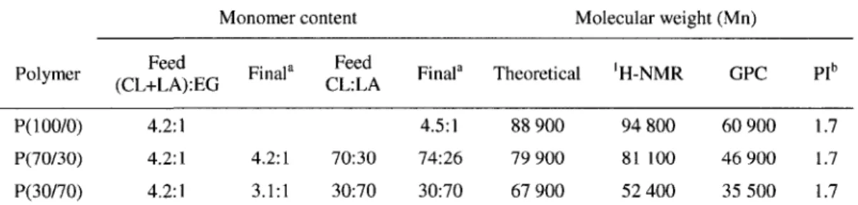
Table 2.1 Characterization of the three copolymers synthesized by
ring-opening polymerization. 50 Table 2.2 Thermodynamic transitions, fusion energy, and % of crystallinity
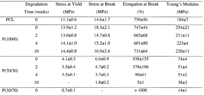
of P(100/0), P(70/30), and P(30/70) films. 51 Table 2.3 Changes in mechanical properties with time of degradation. 59
Chapter 5
Table 5.1 Pore size and approximate porosity as a function of annealing time
LIST OF ABBREVIATIONS bFGF DMA DMP DSC EC ECM EDTA ePTFE FAP Fgn FITC Fn GAG GPC HA HMVEC HUVEC MMP MTT NMR PBS PCL PDGF
basic fibroblast growth factor dynamic-mechanical analysis dimethyl pimelimidate
differential scanning calorimetry endothelial cells
extracellular matrix
ethylenediamine tetraacetic acid, expanded polytetrafluoroethylene focal adhesion plaques
fibrinogen
fluorescein isothiocyanate fibronectin
glucosaminoglycan
gel permeation chromatography hyaluronic acid
human microvascular endothelial cells human umbilical vein endothelial cells
matrix metalloproteinase
3-(4,5-dimethylthiazol-2-yl)-2,5-diphenyltetrazolium bromide nuclear magnetic resonance phosphate buffered saline polycaprolactone
platelet-derived growth factor
PDLLA PEG PEO PGA PHEMA PI PLA PLGA PLLA PMA PS PTFE PVA RGD SEM SMC SnOct Tc, Tg, Tm TCPS TE TGF-p VEGF WAXD poly(D,L-lactic acid) poly(ethylene glycol) poly(ethylene oxide) poly(glycolic acid) poly(2-hydroxyethyl methacrylate) polydispersity index poly(lactic acid) poly(lactic-co-glycolic acid) poly(L-lactic acid) 4P-phorbol 12-myristate 13-acetate polystyrene polytetrafluoroethylene poly(vinyl alcohol) arginine-glycine-aspartic acid peptide
scanning electron microscopy smooth muscle cells
stannous octoate
temperatures of crystallization, glass transition, and melting tissue culture polystyrene tissue engineering
Transforming growth factor-P vascular endothelial growth factor wide-angle x-ray diffraction
CHAPTER 1:
SCAFFOLD VASCULARIZATION: A CHALLENGE FOR THREE-DIMENSIONAL TISSUE ENGINEERING
1.1 Abstract
The prevalent challenge facing tissue engineering today is the lack of adequate vascularization to support the growth, function, and viability of tissues. Researchers rely on the increasing knowledge of angiogenic and vasculogenic processes to stimulate vascular network formation within three-dimensional tissue constructs. These processes are mainly endothelial cell-regulated, although in the context of tissue engineering, specific interactions with scaffold materials, growth factors, and other cell types may require in vitro vascularization schemes to be altered accordingly. In order to better mimic the complete in vivo environment, increasing attention is given to the integration of co-cultures and mechanical conditioning in bioreactors. Such approaches show great promise for the enhancement of the functionality and clinical applicability of tissue engineering constructs.
1.2 Introduction
Microvessel beds constitute the infrastructure that provides nutrient delivery and waste removal in living tissues. In the past few decades many an effort has been made to engineer functional vascular networks in vitro. Paralleled by progress in tissue-engineered blood vessels and in the decoding of the complex mechanisms of blood vessel formation, this work has also generated insight valuable for the repair and reconstruction of three-dimensional human tissue. There are two principal reasons for promoting the formation of vascular networks within three-dimensional cell/scaffold constructs. Firstly, in vitro angiogenesis assays provide useful tools for detailed study of this process by identifying and evaluating the role of specific biochemical pathways]!]. Secondly, in the absence of a vascular network that can ensure efficient exchange of metabolites in a growing cell mass, artificial tissue constructs will remain severely limited in obtainable size, function, and applicability in vivo[2\.
Vascularization of newly formed tissue in vivo takes place by angiogenesis; the process of sprouting of new capillaries from pre-existing vasculature[3]. Angiogenesis is essential for wound healing, tissue repair, and the growth of newly formed tissue. Tissue engineering (TE) constructs may also become vascularized via a process known as vasculogenesis; the in situ reassembly of undifferentiated endothelial cells into capillaries and networks[4]. In vivo, this process normally takes place in developing organs in the embryo[5]. Specific molecules, called angiogenic factors, that control blood vessel formation have been identified[3] and some of these are frequently used to enhance vascularization processes. Following the creation of vascular networks within a TE construct, whether established in vitro or in vivo, these must ultimately connect to the host vasculature after implantation to fulfill their intended function. This event is called inosculation and is the process by which the vasculature is rendered continuous. Finally, a tissue repaired or regenerated with the help of an implanted material, like any other tissue, will be subject to vascular remodeling with time.
At present, regeneration/repair of cartilage[6], bone[7], and skin[8] - although still dealing with issues of predictability and long-term stability - can be achieved with available techniques. However, larger and/or more metabolically demanding tissues such as the pancreas, liver, heart, and kidney require intrinsic blood supply for long-term survival and development^]. For example, it has been suggested that hepatocytes may not be grown further away than 200-300um from nutrient supply in order to survive[10]. Vascular networks are therefore critical for the efficient distribution of oxygen and nutrients to all tissues, as well as for the removal of waste products. In fact, for any cell mass large enough that it cannot rely on nutrient supply solely by diffusion, growth, function, and survival post-implantation are improved by and dependent on ingrowth of blood vessels from the host or, alternatively, by inosculation of an already existing network[ 11-14]. The idea of prevascularized TE scaffolds, as brought forward by Mikos et al.[15], was shown to enhance the performance of forming tissues compared to non-vascularized constructs [11,16]. However, most attempts to promote
vascularization of scaffolds have been pursued in vivo, by allowing for ingrowth of
vascularized fibrous tissue, which then comes to occupy an important fraction of the available void space, thus limiting the space available for engraftment of tissue-specific cells [15]. At present, the development of functional vascular beds in three-dimensional constructs presents
a great challenge to researchers, but is no less central to the successful therapeutic application of tissue engineering principles.
This review treats the background and recent advancements in the vascularization of porous scaffolds for three-dimensional tissue engineering, with an emphasis on in vitro tissue culture. Moreover, the effects of co-culture techniques as well as mechanical conditioning of tissue constructs within the confines of a bioreactor are discussed.
1.3 Scaffold materials
The requirements for three-dimensional TE scaffolds are generally defined in terms of biocompatibility, degradability, porosity (void volume), pore size, and mechanical properties. In addition, scaffolds should promote cell-material interactions suitable for a particular application, either inherently or as a result of surface modification. The choice of a suitable biomaterial is the first step in the scaffold design process. Polymeric biomaterials can be of either biological or synthetic origin, and they offer tremendous variation as pure preparations or as co-polymeric devices. Control over scaffold properties is crucial in the design of successful scaffolds. Ideally, a scaffold should be highly porous, permeable to flow, and possess mechanical strength to match the requirements of the tissue to be replaced. On the same note, it is important that the rate of degradation match an appropriate rate of tissue growth as it proceeds within the scaffold. Adequate pore size, pore morphology, and a high degree of interconnectivity are important to achieve efficient cell seeding and growth. Furthermore, scaffolds should be favorable to fluid flow, ensuring sufficient delivery of nutrients and removal of waste and scaffold degradation products [17].
1.3.1 Biologically derived polymers
ECM-DERIVED POLYMERS, such as collagens, glycoseaminoglycans (GAGs), and fibrin gels, are attractive scaffold materials in that they originate from the very tissues that need to be regenerated. As components of the extracellular matrix (ECM) they offer natural sites for cell attachment, and signaling that regulate cell phenotype. Traditionally, this group of ECM-based biomaterials has been the most popular choice for angiogenesis assays and vascularization studies. They are also used as coating materials to improve cell-material interactions on synthetic polymeric devices.
Collagen is the most abundant protein in mammalian tissues, and is therefore an interesting scaffold material. Chemical or physical cross-linking may be employed to improve the mechanical performance of collagen fibers and scaffolds and to moderate enzymatic degradation of these scaffolds, although cross-link density and porous microstructure are known to influence cellular interactions with these templates[18,19]. Immunogenic issues, especially with animal collagen, have been a concern for the application of collagen-based biomaterials. This effect is primarily due to telopeptides, which may be efficiently removed enzymatically[20]. However, the full immunogenic potential may not be completely removable from non-human proteins, and methods for production of recombinant collagen I and in have been proposed as an alternative source[21].
In response to injury, a fibrin clot forms spontaneously from fibrinogen and thrombin, and are further cross-linked by factor Xffla. The implication of fibrin in blood vessel repair makes it a natural candidate for angiogenic assays[22,23], and has also been used for controlled release of growth factors in vascular graft engineering and bone regeneration[24-26].
GAGs occur naturally as linear polysaccharide branches of certain protein structures called proteoglycans. Hyaluronic acid is the simplest of the GAGs, and is present in nearly all mammalian tissues. Its high molecular weight (50-500kDa) confers mechanical properties that make it interesting as a scaffold material, and it has been used mainly in ophthalmology and experimental treatments of joint dysfunction[27]. Pioneering work using collagen-based materials was carried out by Yannas et al.[28], who developed templates for regeneration of skin and nerve from co-polymers of collagen and another type of GAG, namely chondroitin 6-sulfate. Fibrous materials based on fibronectin have also been investigated for similar applications[29,30].
POLYSACCHARIDES, such as chitosan and alginate, that have been shown to elicit a
relatively mild immunogenic response, have also been investigated for various TE applications. Chitosan has a molecular structure similar to GAGs, and is susceptible to enzymatic degradation by the human enzyme lysozyme. The rate of degradation depends on the degree of crystallinity, which can be easily manipulated by varying the acetyl content[19,31]. Chitosan has been investigated for such applications as encapsulation, membrane barriers, and contact lens material[31]. Moreover, DNA complex formation is favored by its cationic nature, making it an ideal candidate for gene delivery[32]. More
recently, hybrid scaffolds containing a synthetic component have also been proposed for drug release and engineering of cartilage and bone[33,34]. Alginate gels are fabricated under relatively mild conditions, making it suitable for application in drug delivery and cell transplantation (e.g. chondrocytes and insulin-producing cells)[19,35]. In particular, growth factors incorporated into alginate beads have been shown to retain >90% of their bioactivity[36]. Disadvantages include issues of poorly regulated degradation and the lack of natural cell adhesion motifs [37].
1.3.2 Synthetic biomaterials
Despite the obvious advantages of using biologically derived materials, important drawbacks limit their use as scaffolds. These include poor mechanical properties, and inherent batch-to-batch variability. Synthetic polymers therefore represent an attractive alternative, especially for the fabrication of large and/or load-bearing scaffolds. These materials offer a high level of control, as well as seemingly endless variations, through the manipulation of monomer composition, molecular weight, degree of cross-linking and/or branching, co-polymerization, blending, etc. These and other polymer processing techniques may be used as tools to manipulate such material properties as degradability, mechanical strength, and surface chemistry.
Many classes of resorbable synthetic polymers are being studied for use in tissue engineering[38]. Degradable polyesters, such as poly(lactic acid) (PLA), poly(glycolic acid) (PGA), poly(8-caprolactone) (PCL), and their co-polymers have been approved by the FDA for a number of medical and drug delivery devices, and are thus very attractive candidates for scaffold fabrication. These biocompatible polymers exhibit interesting and tunable mechanical properties and predictable degradation kinetics. PLA, PGA and their copolymers (PLGA) were utilized in early applications for sutures and orthopedic fixations[39]. PGA is an inelastic and highly crystalline material. Poly(lactic acid) exists in two practically employed isoforms, namely the highly crystalline poly(L-lactic acid) (PLLA) and the amorphous poly(D,L-lactic acid) (PDLLA). Although less crystalline than PGA, PLA is more hydrophobic, and less susceptible to hydrolysis [2]. PCL possesses properties that set it apart somewhat from the
other polyesters mentioned. It has a very low glass transition temperature (Tg = -60°C), low
melting temperature (Tra = 55-60°C), and shows high thermal stability. Its decomposition
better suited for some processing techniques. Due to its low Tg, PCL is rubbery at room and
physiological temperatures, a temperature that contributes to its high permeability for many drugs[31]. PCL is furthermore a flexible plastic that degrades at a much slower rate than either PLA or PGA. a) b) c) 0 0 O II II II H - E O — CH2— C H O H H - f O — CH— C H OH H - f O —(CH9)—C -j- OH n | n z 5 n CH3
Figure 1.1: Polymeric structure of a) poly(glycolic acid), b) poly(lactic acid), and c) poly(e-caprolactone).
Polyesters can be easily co-polymerized, to achieve materials with intermediate properties. For example, block co-polymers of PCL and PLA combines the rapid degradation of PLA with the flexibility of PCL [40]. Incorporation of poly(ethylene glycol) (PEG) has also been shown to improve water uptake, and speed up degradation of PCL[41]. Vert and coworkers synthesized a series of block co-polymers containing different combinations of PLA/PCL/PEG to achieve materials exhibiting a wide range of mechanical properties and degradation rates[42-45]. This work clearly shows that such properties may be efficiently tailored to meet the demands of particular applications. In addition, PLA-PGA co-polymers (PLGA) are commonly occurring biomaterials, currently in clinical use for surgical sutures, orthopedic implants, and as drug delivery systems [46].
The main concern of these materials is related to the hydrolytic degradation mechanism, which propagates by ester bond cleavage and generates acidic by-products. These lower the local pH, increasing the rate of bulk degradation via autocatalysis, limiting cell viability in the proximity of the implant, and in some cases eliciting inflammatory reactions [47]. Furthermore, PGA and PLA have a tendency to crumble during degradation, creating large particles that may remain in the site for years. Such uncontrolled breaking of implants may also cause damage to nascent tissue[48].
Beside the classic group of aliphatic polyesters, a large quantity of biodegradable polymer and co-polymer systems have emerged for biomaterial systems. These include
polyanhydrides, polyorthoesters, polyphosphazenes, and polyurethanes. Polyanhydrides have been extensively investigated for use in drug delivery for over 25 years[49-51]. Initial limitation due to extensive degradation was addressed by researchers and resulted in materials with degradation rates ranging from weeks to years [7]. Vascularization of polyanhydride implants was observed after 4 weeks in vivo [52]. Polyorthoesters are hydrophobic polymers that are stable at physiological pH, but degrade rapidly at pH 5.5 via hydrolysis[38,53]. These two families of biodegradable polymers have found principal use in drug-delivery systems, as they are mechanically unsuited for scaffolding purposes[48,54]. Polyphosphazene-based biomaterials range in stiffness from soft to hard, show great backbone flexibility, and are being investigated for their applicability in engineering tissue[38]. Laurencin and coworkers studied the applicability of these materials for bone regeneration, and reported osteocompatibility as well as non-toxicity of the degradation products[55]. Composite materials containing hydroxyapatite - a natural component of bone - were investigated for the same application[56]. Furthermore, blending polyphosphazenes with PLGA was shown to moderate PLGA degradation, even as the polyphophazene degradation products had a buffering effect on the degradation milieu[57].
Polyurethanes constitute yet another class of polymers that have long been in use as implant materials[58]. They consist of alternating "soft" segments (polyether or polyesters) and "hard" segments (diisocyanate), and chain extenders that may be either diols or diamines. Great flexibility in chemical composition is possible through the choice of the soft and hard segments, and by varying the ratios of these components a wide range of mechanical properties can be obtained. Earlier concerns regarding the putative carcinogenity of the degradation products have been attended to mainly by replacing diisocyanate by e.g. 1,4-diisocyanatobutane[59] and lysine diisocyanate[60], which degrade into biocompatible components. The latter was polymerized with glucose, which produced glucose and lysine as the principal degradation products [60].
Synthetic polymers may also be processed into hydrogels, which by definition have high
water content, ranging from 10-20 wt% and up. Synthetic hydrogels structurally resemble the ECM of many tissues [19] and present inherently good mass transport properties [61]. The structural stability of synthetic hydrogels can be controlled by the degree of cross-linking between polymer chains. Typically, hydrogels used for scaffold applications can be processed
under relatively mild conditions, allowing for the incorporation of bioactive molecules or even cells. The disadvantages include issues of sterilization and low mechanical strength[61,62]. Synthetic polymers used to make hydrogels include poly(ethylene oxide) (PEO) and the chemically similar poly(ethylene glycol) (PEG), poly(vinyl alcohol) (PVA), poly(acrylic acid), and polypeptides[19]. Photo-polymerisable PVA hydrogels are injectible and found to be elastic and strong. Methods to improve the inherently poor cell adherence in PVA and PEG gels by modification with an RGD cell adhesion peptide have been developed[63,64]. Hydrogels have also been fabricated from PEO-containing block co-polymers with PLLA[65] and poly(propylene fumarate)[66], although the latter exhibited certain cytotoxicity in vitro .
Finally, newly synthesized materials have also been proposed for use in TE applications. Matrices based on PCL copolymerized with hydrophilic poly(ether ester amide)s have been investigated for drug deli very [67], and polypyrroles, possessing modifiable electrical properties have been suggested for nerve regeneration[68].
1.3.3 Scaffold porosity
The optimal pore size of a scaffold varies with application[46], but needs to be large enough that cells can easily penetrate into the scaffold bulk, while at the same time optimizing surface area, which usually increases with decreasing pore size. Highly porous scaffolds are usually preferred, as the surface area and the capacity for large cell mass increases with void volume. On the other hand, excessively porous structures become fragile and may not fulfill the mechanical requirements. For the regeneration of bone, pore sizes of 100-400um are usually recommended[69,70], whereas they should be 20-150um for skin regeneration[71], and approximately 20um for ingrowth of hepatocytes and fibroblasts [46]. In the case of neovascularization, data must be interpreted in the context they are generated. For example, independent microvessel invasion of membranes or thin porous scaffolds have been reported for pore sizes of 5-15um[72,73]. However, neovascularization of a TE scaffold (especially for larger structures) may also take place in conjunction with tissue ingrowth, and would in such a case be more dependent on a pore size that promotes successful invasion of the tissue in question. For example, van Tienen and coworkers [74] observed ingrowth of vascularized fibrous tissue in pores that measured at least 30um. It should be noted that reported results often depend on scaffold design and on the material chemistry that often differs from one study to another. Induction of bone regeneration has been reported in biodegradable scaffolds
with pore sizes as small as 16-32um[73], while in a general manner, much larger pore sizes are found in the literature, as mentioned.
Pore morphology and pore interconnectivity also play a significant role in regulating cell attachment, proliferation, and matrix deposition - e.g. for bone ingrowth[70] and in the culture of smooth muscle cells[75,76]. Subcutaneous PVA sponges, with well defined pores were found to induce better vascularized tissue ingrowth than did PTFE implants of similar pore size[77]. Furthermore, pore microstructure and interconnectivity strongly influence mass transport (permeability and diffusion) and mechanical properties, as well as degradation profiles[46,78-80]. Numerous techniques exist for the fabrication of porous scaffolds, and these have been reviewed elsewhere[81].
1.4 Vascularization of biomaterials
A variety of in vitro assays for angiogenesis exists. Classical testing schemes such as the chicken chorioallantoic membrane (CAM) assay, pouch assays or the study of neovascularization of the avascular cornea are still in use, but have to some extent been replaced by reconstituted biological materials (for reviews, see e.g. Cockerill et al.[82,83]). These assays provide a simplified but controlled environment in which the processes and mechanisms of tubular network formation can be studied in great detail. Given the proper 3D environment, endothelial cells will migrate into the matrix bulk, where they will rearrange and form a network of capillaries, which may or may not contain lumina[84]. Developments within the field tend to consist of substrates, growth factor delivery, and culture techniques that increasingly mimic the complex physiological environment of the cells. However, obtaining patent networks (i.e. that are capable of withstanding the strain of a circulating medium) still presents a challenge for researchers. Encouragingly enough, it was recently shown that engineered endothelial networks of tube-like structures indeed have the potential to evolve to functional blood vessels in v/vo[85]. The presence of such patent networks within TE constructs would greatly facilitate the invasion of tissue-specific cells and enhance the viability and function of the forming tissue.
1.4.1 Blood vessels and vascularization in vivo
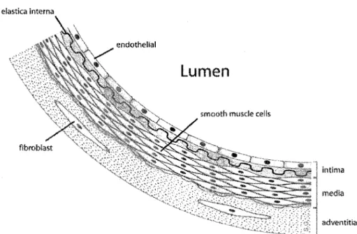
The structure of all veins and arteries in the body consist of three distinct layers: the intima, the media, and the adventitia[86] (see Figure 1.2). The adventitia is the outermost
layer of the blood vessels, and consists primarily of connective tissue and collagen fibers, which allows the vessel wall to return to its initial shape following dilatation or contraction. The media is composed mainly of elastic fibers and mural cells. Mural cells is the common name referring to either smooth muscle cells, as in the case of larger vessels, or pericytes, which are rather undifferentiated and present in newly formed and small blood vessels. The role of these cells is to stabilize the vessel structure. Smooth muscle cells also provide the mechanically dynamic properties of larger blood vessels. Finally, the intima makes up the part of the blood vessel which is in actual contact with the blood stream. This layer is composed of a monolayer of endothelial cells, supported by the basal lamina , and thin layer of connective tissue. Capillaries are much smaller, and consist only of a layer of endothelium, lacking both the media and adventitia. The cells on which the focus will be placed in this text are endothelial cells (EC) and smooth muscle cells (SMC), due to their implication in mature vessel structures in vivo.
eiastica interna
*~*-* adventitia
Figure 1.2: The structure of a large blood vessel, depicting the intima, the media, and the adventitia. (From original image http://en.wikipedia.0rg/wiki/Image:Anat0my_artery.png#file)
New blood vessels may form by one of the two following processes: 1) by angiogenesis, which is the sprouting of new blood vessels from preexisting ones, or 2) by vasculogenesis, which is the assembly of undifferentiated EC in situ.
Vasculogenesis proceeds through five basic steps [4]: 1) generation of differentiated EC from bone marrow-derived precursor cells; 2) EC form aggregates and establish cell-cell contacts; 3) EC polarize and rearrange into nascent lumen-containing tubes; 4) an immature vessel network is formed; and 5) pericytes and SMC are recruited, which stabilizes the newly formed vessels.
Angiogenesis involves a growth phase and a stabilization phase[87]. Four steps define the growth phase, which is initiated by 1) cell-mediated, proteolytic degradation of the basement membrane, allowing for endothelial cells to invade the surrounding ECM; 2) Activated EC then proliferate and migrate into the created space, where they 3) organize into aligned, lumen-containing cords. 4) Lastly, newly formed sprouts connect to form loops of immature vasculature. The immature vasculature is subsequently stabilized through the recruitment of smooth muscle cells or pericytes, accompanied by a halt in EC proliferation, and the deposition of a basement membrane (reinforced basal lamina) around the cords.
The first reports of in vitro tube formation by capillary endothelial cells was made in 1980 by Folkman and Haudenschild[84], who observed capillary networks developing on a gelatine substrate after several weeks of culture. Since then, methods have been refined, as the understanding of angiogenic processes has evolved. Today it is largely recognized that the interplay between EC and insoluble molecules in the ECM is critical in many events during angiogenesis and vasculogenesis, including the regulation of cell growth, migration, differentiation and tube formation[88]. Vessel growth is also influenced by cell-cell interactions and soluble species, such as growth factors. In addition to the choice of substrate, the source of cells will influence the outcome of a study. To some extent, endothelial cells from rat or bovine aorta behave differently than ECs from human tissue. The latter have higher requirements of growth factors, and may degenerate in some basic culture conditions where animal cells spontaneously differentiate into capillary structures [89]. Further distinctions can be made between human ECs of different origin. The commonly used human umbilical vein ECs (HUVEC) are of macrovascular origin, whereas the cells most involved in inflammation, wound healing and vascularization in vivo are microvascular ECs. However,
any difference in the capacities of these two cell types to form tube-like structures is not readily apparent[90]. Some differences may also be found in the ability to exert contractive forces on malleable substrates, where e.g. human blood outgrowth ECs are stronger compared to HUVEC[91]. In addition, recent publications have demonstrated capillary formation using endothelial progenitor cells, who are implicated in post-natal neovascularization[92,93].
1.4.2 Angiogenic Factors
A number of growth factors and other angiogenic promoters are used to induce and
enhance EC sprouting and capillary network formation in cell/polymer constructs. These factors may act directly on EC or indirectly, inducing an angiogenic response in vivo, without eliciting a direct response in cultured EC, such as an altered migration or proliferation^]. Vascular endothelial growth factor (VEGF) and basic fibroblast growth factor (bFGF) are examples of highly potent EC mitogens, stimulating angiogenesis via direct interactions with EC. Both growth factors are widely distributed in various tissues in the body, but while bFGF is known to act on various cell-types, VEGF is EC-specific[4]. Other angiogenic factors, such as platelet-derived growth factor (PDGF) and transforming growth factor-beta (TGF-beta), act indirectly to induce blood vessel formation and/or stabilization[4,94]. Table 1.1 lists these molecules and others, their roles in angiogenesis in vivo, and examples of their use to induce angiogenesis in biomaterials.
Many groups have explored the controlled delivery of angiogenic growth factors as a means to improve the local vessel recruitment in implants and cell transplants[ 11,16,103]. The modulation of growth factor delivery is highly important since growth factors such as VEGF and bFGF are rapidly cleared and inactivated following administration in an unprotected formulation! 16]. Another aspect is that bolus delivery may cause a toxic or otherwise unwanted response in the studied (or surrounding) cell population, as such administration does not correspond to any normal situation in v/vo[101,lll]. Moreover, if too high concentrations of e.g. VEGF are sustained within a tissue, the developing vasculature may become deformed and nonfunctional! 1.12]. Control of the release rate is therefore of utmost importance. To deal with these concerns, the encapsulation of drugs within degradable polymers from which they can be liberated in a controlled manner has come forth as a successful method. Biologically derived hydrogels, including fibrin, alginate, hyaluronic acid, chitosan, and gelatine have proved very useful as encapsulation vehicles due to their biocompatibility and inertness
towards bioactive molecules[16,19,25]. Moreover, encouraging results have been reported from trials with synthetic materials such as PLGA[11,36,102,103] and PEG hydrogels[98]. Growth factors may be released from encapsulating matrices via diffusion, mechanical stimulation, or matrix degradation[19]. Control of the release rate may be obtained by varying the rate of degradation of the capsule material, and this may in turn be achieved by altering the molecular weights or degree of polymer cross-linking[102]. Another very interesting approach consists of pacing the release of growth factor from the matrix to the demand of the growing cell popluation[98,101,H3]. Such designs aim at more closely mimicking the way in which naturally sequestered growth factor is released from the ECM by proteolytic activity, and have been shown to induce a more controlled growth of capillary networks in fibrin[101].
1.4.3 Tube formation in biological scaffolds
A few methods dominate the three-dimensional in vitro angiogenesis assays in current use. In so-called in-gel seeding cells are mixed with the matrix solution prior to gelation. Cell-seeded collagen type I gels prepared in this manner were not able to induce EC tube formation in the absence of endothelial mitogen[114]. Alternatively, ECs can be cultured to confluence
or near-confluence on top of a gel (e.g. fibrin, collagen, Matrigel3), at which time a second
layer is placed on top of the cells, creating a "sandwich" consisting of two layers of gel matrix. Trapped within the sandwich, the cells may subsequently differentiate, migrate and form tubes, an event which is largely dependent on the interaction between the overlaying matrix layer and the apical cell surface[116, 117]. A third approach involves the use of intact ring sections of aortas that are embedded in collagen, fibrin, or Matrigel gels, and the angiogenic activity is observed as radial sprouting of endothelial cells[118]. This so-called aortic explant model has traditionally employed segments of rat aorta[119], but can be extended to include tissues from other animals [120]. The advantage of this method compared to cell-based assays is the realistic representation of in vivo structural cell arrangements [120].
In addition to the models mentioned, in vivo experiments are conducted in which seeded or acellular matrices are implanted subcutaneously in mice, where a capillary network starts to form. Subcutaneous implantation of fibrin and Matrigel lead to blood vessel ingrowth[22,
121], while collagen type I did not[22,121]. Vascularization of the implant may be evaluated after surgical recovery[122] or, alternatively, in vivo through transparent "windows" in the
skin of the animal[85]. (Note: Several approaches are currently being used to quantify growing and established 3D tubular networks within a biomaterial. The lack of a standardized method for quantification makes the direct comparison between different assays and results complicated.)
Table 1.1: Some angiogenic factors used to promote vascularization of biomaterials.
Angiogenic agent Roles in vivo Examples of use in biomaterials in vitro*
Vascular endothelial growth factor (VEGF)
Potent angiogen, required for the formation of immature vasculature[95], promotes ECM
degradation[94]. Exists in five isoforms of different potency. Must be well regulated. Reinforces the angiogenic potential of bFGF[96].
Coll I[ 1,97], fibrin[98-100],
fibrin and CAM[ 101], PLGA[ 102,103], PEG[98]
Basic fibroblast growth factor (bFGF)
Stimulates expression of VEGF and its receptors in EC. Promotes ECM degradation[94], EC migration and pericyte attraction[104]. Reinforces the angiogenic potential of VEGF[96].
Coll I[ 1,97,105] Fibrin[99,100] PLGAinalginate[ll]
Tissue growth factor-beta (TGF- beta)
Induces mural cell differentiation^], inhibits ECM degradation[94]. Dose-dependent stimulation of tube formation in vitro [106]. EC proliferation and migration are inhibited at higher
concentrations [ 107].
Matrigel[106]
Platelet-derived growth
factor (PDGF) Recruitment of pericytes/SMC[83]. PLGA[102]
Angiopoietins (Ang-l,Ang-2)
Ang-1 stabilizes vessels, while its antagonist Ang-2 acts as a destabilizer, promoting EC migration and angiogenesis in presence of other factors, such as VEGF[108,109]. Co-culture in Coll I[109] 4 beta-phorbol 12-myristate 13-acetate (PMA)
Known tumor promoter. Coll I[ 1,97,110]
*Coll I: collagen type I; CAM: chorioallantoic membrane; PLGA: poly(lactic-co-glycolic acid).
The various matrices used for endothelial cell culture each represent different characteristics of the basement membrane. Therefore it is not surprising that the mechanism of tubulogenesis in vitro in some aspects is matrix-specific. It has even been suggested that the nature of the supporting material influences the angiogenic potential of growth factors[123]. The specific interactions between EC and the matrix are mediated by integrin receptors on the cell surface[124]. Dallabrida et al.[125] observed matrix-specific tubulogenesis in a publication where down-regulation of the expression of the
alpha v beta 3 integrin (a receptor expressed by angiogenic EC, binding both VEGF[126] and PDGF-B[127]) in ECs reduced tubulogenesis in fibrin gels, whereas no such effect was seen on Matrigel. Moreover, Hall et al. were able to induce angiogenesis on fibrin substrates, covalently modified with synthetic binding sites for the alpha v beta 3 integrin[124]. Chalupowicz et al.[117] identified the interaction between HUVEC and the betal5-41 sequence of fibrinopeptide B as necessary for tube formation in fibrin sandwich.
Soluble species and matrix constituents work in concert with many cell membrane-associated adhesion molecules during the angiogenesis process. Among these are the cell-cell junction proteins VE-cadherin (CD144) and PECAM-1 (CD31), which have been demonstrated to regulate lumen formation and intercellular association during EC network formation, respectively[128]. In addition, there is evidence to suggest that VE-cadherin is necessary for the maintenance of tubular networks in fibrin and collagen type I gels[l, 129]. Antibodies against VE-cadherin not only inhibited tubulogenesis but also disrupted already developed networks [129].
The controlled and localized release of the growth factor bFGF has been shown to significantly enhance vascularization of injured sites[16], and is furthermore suggested to increase the size of growing vessels[ll]. Sakakibara and coworkers[16] reported that slow release of bFGF from gelatin microspheres in infarction areas improved cardiac function and survival of transplanted myocytes in rats. Endothelial cells grown on gelatin-coated micro-carriers dispersed in a fibrin gel exhibited important increases in their capacity to sprout when bFGF or VEGF was added to the gel[100]. Ehrbar and coworkers[101] used an engineered version of VEGF121 that was covalently incorporated into fibrin gels and subsequently evaluated in the CAM assay. Grafted VEGF was gradually released as the matrix was enzymatically degraded by EC, and enhanced arterial and venous network development was
observed. In a control experiment, non-grafted soluble VEGF121 was released rapidly and triggering chaotic vessel growth. Other examples of VEGF encapsulation include the use of alginate[19,36] or combined alginate/PLGA matrices[102,103]. Furthermore, attempts have been made to coordinate the controlled delivery of multiple growth factors [102]. In an approach combining bFGF and VEGF delivery, Nillesen et al.[130] used collagen-heparin sponges with 100|im pores to stimulate vascularization subcutaneously in rats. (Heparin increases the amount of bFGF that may be incorporated into cross-linked collagen[105] and alginate[131] gels.) They found that while the two growth factors alone supported vessel formation, the combined delivery of bFGF and VEGF was superior in promoting the formation of mature, pericyte-lined, vasculature within the scaffolds. In addition, there was a dramatic decrease in the number of hypoxic cells in the scaffold centre when both growth factors were present[130].
INFLUENCE OF SUBSTRATE STIFFNESS
It has long been claimed that endothelial migration and elongation take place along lines of reorganized matrix - a result of cell forces acting on the matrix components [132]. These forces are communicated via cell adhesion sites called focal adhesion plaques (FAP) that consist of assemblies of structural and signaling proteins, such as integrins[133]. These sites enable mechanical tension from the ECM to be transferred into the cell, and conversely from the cytoskeleton to the ECM[134]. Relatively rigid substrates have been found to better support the formation of stable focal contacts than more compliant substrates, by promoting stronger integrin-cytoskeletal linkages[135]. However, they also bring on a more spread cell morphology, which in turn leads to decreased cell elongation and cells organized in isolated clusters or monolayers rather than network formations [91,132,136,137]. More malleable substrates promote a rounded morphology that is associated with a more differentiated phenotype in endothelial cells, although fully rounded cells will not form capillary tubes[136].
Appropriate matrix rigidity is not associated with any absolute magnitude of "stiffness", but needs to be put into a more elaborate picture, incorporating matrix type, matrix geometry, conditions of constraint, and the cell type involved. The force generated by cells cultured in or on a gel construct depends both on the cell type and on the flexibility of the matrix[91]. However, it has been clearly established that matrix contraction and tube formation within
reconstituted gels require an active cytoskeleton[ 137-139]. During gel contraction the number of stress fibers (actin filaments) present in cells can be related to the stress being imposed on the cytoskeleton, which in turn is a reflection of the matrix resistance to deformation[91,137]. Moreover, stress fibers become oriented along the tubule axis following EC rearrangement into capillaries [137]. Analysis of the concentration and organization of actin filaments and proteins associated in focal adhesion plaques reveals EC-specific morphology changes in response to changes in substrate flexibility[140].
Substrate stiffness can be altered by changing the polymer concentration, degree of cross-linking or polymerization conditions. Ingber and Folkman[136] reported on the role of ECM concentration on rigid surfaces, and found that intermediate coating densities supported EC rearrangement into tubular structures. Several groups have later repeated these results in thick substrates concluding that the onset of tubulogenesis can be controlled by altering the stiffness of the cell support[132,137]. For example, on Matrigel or polyacrylamide gels that were prepared with varying degree of cross-linking, HUVEC reorganized more quickly into tube-like structures on more compliant substrates [137]. Similar results were obtained by decreasing the density of collagen gels[141]. Furthermore, in a recent publication Sieminski and coworkers [91] demonstrated the possibility of controlling lumen size by varying matrix concentration. By doubling the concentration of type I collagen from 1.5mg/ml to 3mg/ml lumen size was increased by 57% and 40% in floating and constrained gels, respectively[91]. This increase was attributed to a resistance to contraction (increasing with the density of the substrate), bringing on a more spread cell morphology.
In order to study the role of cellular adhesion in tube formation, the inherent ability of cells to adhere to the support material can be manipulated. Many cell types, including endothelial cells, are dependent on the activity of the beta 1 integrin unit for adhesion on a variety of ECM-derived substrates. Tubule formation in Matrigel is, for instance, inhibited when the beta 1 unit is blocked by antibodies [125]. Indeed, the alpha 2 beta 1-integrin is the
main laminin and collagen receptor in EC [137], and varying the adhesion strength using anti
alpha 2 beta 1-antibodies, influences both the number and dimension of forming capillary tubes[110]. Deroanne et al.[137] found that as the rigidity of their collagen type I substrate decreased, the expression of the alpha 2 subunit was reduced, and EC more easily switched to a tube-like pattern. Furthermore, they established that parallel to these effects, increased
substrate malleability also resulted in reduced expression of actin and focal adhesion plaques proteins, as determined by Western blot[137]. Thus, there seems to be a "window" of favorable adhesion strength for tube formation, where cells are allowed to adhere to the substrate without being hindered to differentiate, migrate and rearrange into network patterns.
1.4.4 Tube formation in synthetic scaffolds
Cell scaffolds fabricated from synthetic polymers present a different set of challenges for vascularization compared to reconstituted gels. The identification of parameters crucial to capillary formation within synthetic materials is still a work in progress, and these may prove difficult to ascertain in general terms, given the variation in material properties. For "dry" synthetic polymer scaffolds, the concensus is that sufficient porosity is a prerequisite for uniform cellular invasion and long-term survival. Pore morphology may also be crucial to the ability of synthetic scaffolds to induce cellular invasion[142]. Related to more efficient cell invasion is the observation that porous implanted matrices become surrounded by loose and highly vascularized connective tissue, whereas non-porous scaffolds become enveloped by a dense fibrous capsule[143]. Vascularization around and into the implant has been observed to be increasingly facilitated with increasing pore size, reaching a maximum at pore sizes on the order of cellular dimensions[143,144]. Moreover, it is believed that in the absence of natural ECM components, EC must first secrete and organize their own basement membrane in order to trigger tube formation[84]. Alternatively, surface modification of synthetic polymers may improve cell-material interactions and facilitate the process. However, as in the case of biological ECM-based scaffolds, it is believed that intermediate cell adhesion is necessary for implanted EC to be able to reorganize and actively participate in the vascularization process[145].
Expanded polytetrafluoroethylene (ePTFE), although not a degradable biomaterial, has been extensively studied for use in vascular grafts. Investigations of implanted ePTFE arterial grafts, carried out by Clowes et al.[146], showed that internodal distances of 60u,m provoked the invasion of microvessels through the graft, ultimately leading to complete endothelial cell coverage of the luminal side of the implants. Internodal distances of 30(im did not allow for vessel invasion to the same extent, and endothelial coverage was significantly lower. Supporting results were reported by Saltzmann et al.[147], again indicating an optimum pore size of 60|im. Interestingly, by modifying the surface with ECM components prior to