Expression profile of plasticity-related mRNAs in the
cortex and hippocampus of young and aged rats and of
3xTg and wild type mice
par Mireille Moreau
Biochimie, Université de Montréal Médecine
Mémoire présenté à la Faculté de Médecine en vue de l’obtention du grade de Maîtrise en biochimie
option génétique moléculaire
décembre, 2011
Faculté des études supérieures et postdoctorales
Ce mémoire intitulé:
Expression profile of plasticity-related mRNAs in the cortex and hippocampus of aged and young rats and of 3xTg and wild type mice
Présentée par : Mireille Moreau
a été évaluée par un jury composé des personnes suivantes :
James G. Omichinski, président-rapporteur Luc DesGroseillers, directeur de recherche
Résumé
De récents travaux ont mis en évidence que des dysfonctionnements dans l’expression de gènes impliqués dans la plasticité synaptique contribuent aux déclins cognitifs qu’on observe chez les gens âgés et à la progression de la maladie d’Alzheimer. Notre étude avait comme objectif d’étudier le profil d’expression d’ARNm spécifiques impliqués dans la plasticité synaptique chez des rats jeunes et âgés et chez des souris transgéniques 3xTg et WT.
Des expériences en qRT-PCR ont été effectuées dans des extraits de cortex et d’hippocampe de rats jeunes et âgés et de souris 3xTg et WT, respectivement. Les résultats ont démontré une augmentation significative de l’expression d’ARNm MAP1B, Stau2, BDNF, CREB et AGO2 principalement dans l’hippocampe (régions CA1-CA3) des souris 3xTg comparé aux souris WT. Une diminution significative a également été observée pour l’ARNm αCaMKII dans le cortex des souris 3xTg comparé aux souris WT. Contrairement à ces observations, aucun changement n’a été observé pour l’expression de gènes impliqués dans la plasticité synaptique chez les rats âgés comparé aux rats jeunes.
Ces résultats démontrent qu’un dysfonctionnement existe réellement au début de la maladie d’Alzheimer dans l’expression de gènes spécifiques impliqués dans la plasticité synaptique et contribue potentiellement à la progression de la maladie en engendrant un déséquilibre entre la LTP et la LTD. De plus, les différences d’expressions sont particulièrement observées dans l’hippocampe (régions CA1-CA3) ce qui est consistant avec les études sur la progression de la maladie d’Alzheimer puisqu’il est connu que la région CA1 de l’hippocampe est la plus vulnérable à l’apparition de la maladie. Ces résultats permettent une meilleure compréhension des événements moléculaires qui deviennent dérégulés à l’apparition de la maladie d’Alzheimer.
Mots-clés : Plasticité synaptique, ARNm, déclins cognitifs, maladie d’Alzheimer, rats jeunes, rats âgés, souris 3xTg, souris WT, cortex, hippocampe
Abstract
Recent work has demonstrated that dysregulations in the expression profile of plasticity-related genes in specific brain regions contribute to age-plasticity-related cognitive decline and Alzheimer’s disease. The aim of this study was to determine the expression profile of a subset of plasticity-related mRNAs in different regions of the brain of young and aged rats as well as 3xTg and wild type (WT) mice.
qRT-PCR experiments were performed in extracts of cortex and hippocampus of young and aged rats and of 3xTg and WT mice, respectively. Results demonstrated significant increases in the expression of MAP1B, Stau2, CREB, BDNF, and AGO2 mRNAs, especially in the hippocampus (CA1-CA3 fields) of 3xTg mice compared to WT mice. A significant decrease was also observed in the expression of αCaMKII mRNA in the cortex of 3xTg mice compared to WT mice. On the other hand, no significant changes were observed in the expression of plasticity-related genes in the hippocampus of aged rats compared to young rats.
These results confirm that alterations in gene expression occur at the onset of AD and possibly contribute to the progression of the disease by causing an imbalance between long-term potentiation and long-long-term depression. In addition, patterns of significant altered gene expression, especially in the hippocampus (CA1-CA3 fields) of 3xTg mice are consistent with the progression of AD whereby the hippocampus (CA1 region) is most vulnerable at the onset of the disease. These results provide a better understanding of the molecular events that first become disturbed in AD.
Keywords : Synaptic plasticity, mRNAs, cognitive decline, Alzheimer disease, young rats, aged rats, 3xTg mice, WT mice, cortex, hippocampus
Contents
Résumé ... i Abstract ... ii Contents ... iii List of tables ... v List of figures ... vi Remerciements ... viii 1 Introduction ... 1 1.1 Memory ... 11.2 Memory and the brain ... 2
1.3 Brain structures involved in different types of memories ... 3
1.3.1 Structural organization of the medial temporal lobe ... 3
1.4 Synaptic plasticity ... 5
1.4.1 Structure of a neuron ... 5
1.4.2 Neuronal network in the hippocampus ... 7
1.4.3 Long-term potentiation and long-term depression ... 7
1.4.4 Transcriptional and translational regulations of plasticity related mRNAs and proteins ... 15
1.5 Aging and cognitive decline ... 17
1.6 Alzheimer’s disease ... 21
1.7 Hypothesis, aims, and rationale ... 24
1.7.1 Hypothesis ... 24
1.7.2 Aims ... 25
1.7.3 Rationale ... 25
2 Experimental procedures ... 28
2.1 Animals ... 28
2.1.1 Young and aged rats ... 28
2.2 RNA isolation ... 29
2.3 Quantitative PCR ... 29
3 Results ... 34
3.1 Expression profile of plasticity-related genes in different brain regions of 3xTg mice and WT mice ... 35
3.1.1 Hippocampus: CA1-CA3 fields ... 35
3.1.2 Hippocampus: Dentate gyrus ... 41
3.1.3 Cortex ... 44
3.2 Expression profile of plasticity-related genes in the cortex and hippocampus of aged 24-month old rat and young 3-month old Fisher 344 rats ... 49
3.2.1 Hippocampus ... 49
3.2.2 Cortex ... 50
4 Discussion ... 53
4.1 Alzheimer’s disease ... 53
4.1.1 LTD and plasticity-related mRNAs ... 54
4.1.2 LTP and plasticity-related mRNAs ... 57
4.2 Aging ... 62
5 Conclusion ... 64
5.1 Perspectives ... 65
List of tables
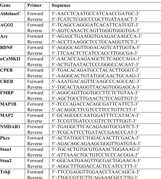
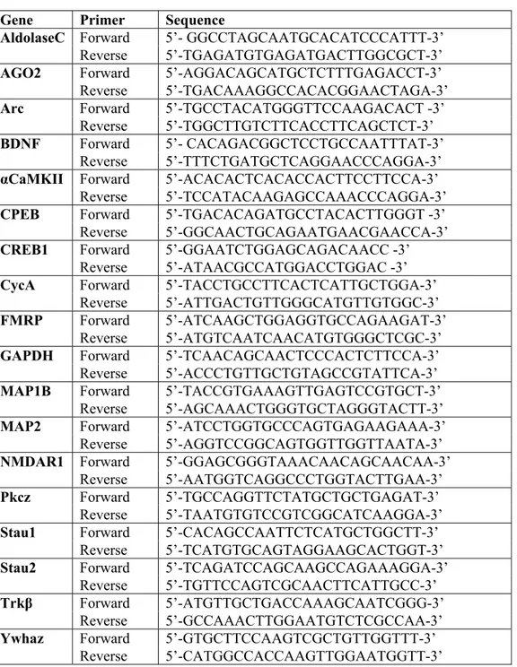
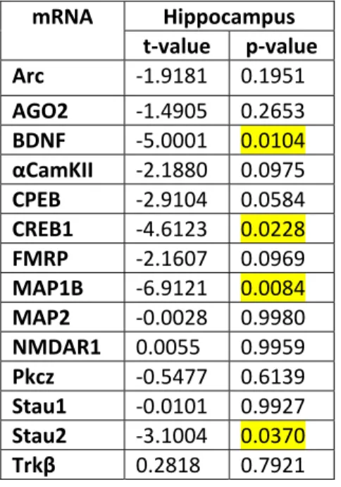
Table I Sequence information of the rattus norvegicus primer pairs for use in quantitative real-time PCR ... 31 Table II Sequence information of the mus musculus primer pairs for use in quantitative real-time PCR ... 32 Table III p-value: Statistical significance in the hippocampus (CA1-CA3 fields) of 3xTg
and WT mice relative to aldolase C ... 36 Table IV p-value: Statistical significance in the hippocampus (CA1-CA3 fields) of 3xTg
and WT mice relative to Ywhaz... 38 Table V p-value: Statistical significance in the hippocampus (CA1-CA3 fields) of 3xTg and
WT mice relative to GAPDH ... 40 Table VI p-value: Statistical significance in the dentate gyrus of 3xTg and WT mice
relative to aldolase C ... 42 Table VII p-value: Statistical significance in the cortex of 3xTg and WT mice relative to
aldolase C ... 45 Table VIII p-value: Statistical significance in the cortex of 3xTg and WT mice relative to
Ywhaz ... 47 Table IX p-value: Statistical significance in the hippocampus of young and aged Fisher 344
rats relative to aldolase C ... 49 Table X p-value: Statistical significance in the cortex of young and aged Fisher 344 rats
List of figures
Figure 1 Types of memories ... 2
Figure 2 Anatomical representation of the hippocampus ... 4
Figure 3 Structure of a neuron ... 6
Figure 4 Pyramidal neurons of CA1-CA3 fields ... 7
Figure 5 E-LTP, L-LTP, and LTD ... 8
Figure 6 Lesions in Alzheimer’s disease ... 22
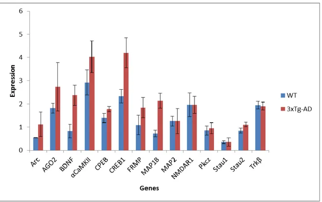
Figure 7 Normalized expression level of mRNAs in the hippocampus (CA1-CA3 fields) of 3xTg and WT mice (reference gene: Aldolase C) ... 37
Figure 8 Normalized expression level of mRNAs in the hippocampus (CA1-CA3 fields) of 3xTg and WT mice (reference gene: Ywhaz) ... 39
Figure 9 Normalized expression level of mRNAs in the hippocampus (CA1-CA3 fields) of 3xTg and WT mice (reference gene: GAPDH)... 41
Figure 10 Normalized expression level of mRNAs in the dentate gyrus of 3xTg and WT mice (reference gene: Aldolase C) ... 43
Figure 11 Normalized expression level of mRNAs in the cortex of 3xTg and WT mice (reference gene: Aldolase C) ... 46
Figure 12 Normalized expression level of mRNAs in the cortex of 3xTg and WT mice (reference gene: Ywhaz) ... 48
Figure 13 Normalized expression of mRNAs in the hippocampus of young and aged rats (reference gene: Aldolase C) ... 50
Figure 14 Normalized expression of mRNAs in the cortex of young and aged rats (reference gene: Aldolase C) ... 52
Je dédie ce mémoire à mes parents qui m’ont encouragé et qui m’ont porté conseil tout au long de mes études
Remerciements
Premièrement, j’aimerais remercier mon directeur de recherche Dr. Luc DesGroseillers pour m’avoir appuyé et m’avoir porté conseil tout au long de ma maîtrise au cours de laquelle j’ai pu acquérir une excellente expérience scientifique.
De plus, j’aimerais remercier tous ceux qui m’ont aidé au cours de ma maîtrise en m’encouragent et en m’offrant leur expertise en laboratoire. Merci à Sacha Blain pour les nombreuses heures qu’elle m’a consacré pour m’aider à m’adapter dans un tout nouveau domaine dans lequel j’avais très peu de connaissances avant de commencer. Merci également pour son aide avec les techniques de laboratoire et surtout pour la dissection de cerveaux de rats et de souris nécessaire pour mes expériences. Merci à Samuel Gatien pour son aide avec l’utilisation du polytron et surtout pour son support, encouragement, et sens d’humour. Merci à Véronique Trépanier pour son aide avec les techniques d’isolation d’ARN et de RT-qPCR ainsi que pour sa compagnie en laboratoire. Merci à Karine Boulay pour le temps qu’elle m’a consacré pour m’expliquer des notions scientifiques ainsi que pour m’encourager et me faire rire. Merci à Jean-François Denis pour son aide avec l’analyse statistique de mes données. Merci à tout le monde dans le laboratoire de Stéphane Roy (Stéphane Roy, Éric Villiard, Étienne Vincent, Jean-François Denis, Samuel Gatien) pour m’avoir laissé utiliser leurs machines et pour m’avoir enduré dans leur laboratoire et pendant nos sorties durant le dîner ou en soirée. Merci à Jojo (José Mario Capo-Chichi) pour son encouragement et tous ses mots de sagesse mais surtout pour son amitié. Merci à tous ceux que j’ai croisé au cours de ma maîtrise (dans notre bureau, dans les laboratoires, dans mes cours, dans le café du département de chimie) et qui m’ont aidé d’une façon ou d’une autre.
Finalement j’aimerais remercier ma famille, mes ami(e)s, et mon copain Raj qui m’ont tous appuyé durant ma maîtrise et qui m’ont aidé à maintenir un bon équilibre entre les heures de laboratoire et les sorties sociales pour se défouler et rire entre ami(e)s.
Abbreviation list
% Percentage
oC Degree Celsius
µl Microliter
3xTg Triple transgenic model of Alzheimer disease
AD Alzheimer’s disease
AGO2 Argonaute 2
Akt Protein kinase B
AMPA 2-amino-3-(5-methyl-3-oxo-1,2-oxazol-4-yl)propanoic acid AMPAR 2-amino-3-(5-methyl-3-oxo-1,2-oxazol-4-yl)propanoic acid
receptor
APP Amyloid precursor protein
APPswe APP swedish mutation
APT1 Acyl-protein thioesterase 1
Arc Activity-regulated cytoskeleton-associated
Aβ Amyloid-beta
Bax Bcl-2–associated X protein
Bcl-2 B-cell lymphoma 2
BDNF Brain derived neurotrophic factor
BIRC3 and 4 Baculoviral IAP Repeat-Containing 3 and 4 CA1/CA2/CA3 Cornu Ammonis 1/2/3
Ca2+ Calcium ions 2+
CaMKII Calcium/calmodulin protein kinase II αCaMKII CaMKII-alpha
cDNA Complementary DNA
CO2 Carbon dioxide
CPEB Cytoplasmic polyadenylation element binding protein
CRE cAMP responsive element
CREB cAMP responsive transcription factor
CycA Cyclin A
DEPC Diethylpyrocarbonate
DNA Deoxyribonucleic acid
dsRNA Double stranded ribonucleic acid E-LTP Early long-term potentiation
EPSP Excitatory postsynaptic potential
F-actin Filamentous-actin
FAD Familial Alzheimer disease
FMRP Fragile X mental retardation protein
GAPDH Glyceraldehyde 3-phosphate dehydrogenase GluR2 Metabotropic glutamate receptor 2
HBSS Hank’s balanced salt solution
HFS High frequency stimulation
hnRNP A2 Heterogeneous nuclear ribonucleoprotein A2 Hz Hertz
IEG Immediate early gene
KO Knock-out
LFS Low frequency stimulation
LIMK1 LIM domaine kinase 1
L-LTP Late long-term potentiation
LTD Long-term depression
LTP Long-term potentiation
MAP1B Microtubule associated protein 1b MAP2 Microtubule associate protein 2 MAPK Mitogen-activated protein kinase
mM Millimolar miRISC miRNA-induced silencing complex
miRNA micro-ribonucleic acid
mRNA Messenger ribonucleic acid
NCBI National Center for Biotechnology Information
NFT Neurofibrillary tangles
Ng Nanogram Nm Nanometer
NMDA N-Methyl-D-aspartic acid
NMDAR N-Methyl-D-aspartic acid receptor
NR1/NR2 N-Methyl-D-aspartic acid receptor subunit ½
PKA Protein kinase A
Pkcz Atypical protein kinase C
Poly(A) Polyadenylation
PP1 Protein phosphatase 1
Pri-miRNA Primary-miRNA
PRP Plasticity related protein PS1M146V Presenilin-1 mutation : M146V
PSD95 Postsynaptic density protein 95
RISC RNA-induced silencing complex
RNA Ribonucleic acid
RNase Ribonuclease
RNP Ribonucleoprotein
RT-qPCR Real-time reverse transcription polymerase chain reaction S Seconds
SNAP25 Synaptosomal-associated protein 25
Stau1 Staufen 1
Stau2 Staufen 2
TauP301L Tau mutation : P301L
Thy1.2 Thymocyte antigen 1.2
Trkβ Tropomyosin receptor kinase Beta
UTR Untranslated region
VAMP2 Vesicle-associated membrane protein 2 VDCC Voltage-dependent calcium channels WT Wild-type
Ywhaz Tyrosine 3-monooxygenase/tryptophan 5-monooxygenase activation protein, zeta polypeptide
1
Introduction
1.1 Memory
Memory, the process of encoding, consolidating, storing, and retrieving knowledge, is achieved through the plasticity of the human brain: the ability of neurons to modify their connections to make certain neural circuits more efficient (1). With aging, as much as 60% of the population is affected by learning and memory impairments of varying severity (2). These can range from mild cognitive decline to more severe cases such as Alzheimer’s disease (AD). Consequently, the aging population affected suffers from diminished quality of life and independence and inadvertently, imposes costly and time consuming demands on society and the health care system.
At the onset of cognitive impairment, specific regions of the brain are especially vulnerable and as a result a specific type of memory, termed episodic memory, is impaired (3). This type of hippocampus dependent long-term memory enables the consolidation and recollection of information in a determined temporal and spatial context. Initially, various areas of the neocortex process visual, auditory and somatic characteristics of information which are then integrated by the hippocampus (4,5). Repetition via the hippocampal-neocortical loop allows strengthening of associations among new elements. Eventually the neocortex associates these various properties itself to reconstruct a memory.
At the molecular level, the ability to strengthen or weaken neuronal associations is referred to as synaptic plasticity (1). The synaptic transmission between neurons can either be enhanced by activity via long-term potentiation or depressed by activity via long-term depression. Morphological and cellular modifications ensue due to the actions of several plasticity-related mRNAs and proteins and can last from milliseconds to long-lasting alterations. The precise transcriptional and translational regulations as well as the localization of these mRNAs and proteins are crucial in maintaining an optimal synaptic transmission between neurons. Throughout the lifespan, apparent age-related changes occur in the expression profile of these mRNAs and proteins and can contribute to cognitive decline by engendering a dysregulation in synaptic transmission (2,6,7).
1.2 Memory and the brain
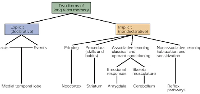
Memory is a broad term that encompasses both short-term memory, which can last several seconds to several minutes, and long-term memory which may be stored for an unlimited duration (8). Long-term memory is divided into explicit memory (declarative) and implicit memory (nondeclarative) – (figure 1) (9,10). The fundamental difference between these types of memories lies in the conscious effort required in remembering things and facts (explicit) and the unconscious effort required in remembering procedural skills or emotional responses (implicit). Explicit memory is further subdivided into semantic and episodic memories which include the knowledge of facts as well as the spatial and temporal context of these facts, respectively.
Figure 1 Types of memories. Various forms of memory can be classified as explicit or implicit memory. Adapted from (1).
1.3 Brain structures involved in different types of memories
Various brain structures are involved in the acquisition, consolidation, and recollection of the different memory subtypes. Whereas non-declarative memory relies mostly on the neostriatum and cerebellum, declarative memory is processed through the medial temporal lobe (11-13). This region contains the hippocampus and connected areas such as the entorhinal, perirhinal and parahippocampal cortex. The importance of the medial temporal lobe in declarative memory consolidation is best described by Nadel and O’Keefe’s cognitive map theory which suggests that episodic memory, a subtype of declarative memory, is dependent on the hippocampus (14).
The first and best studied case exemplifying Nadel and O’Keefe’s theory is the case of patient H.M. Due to a severe case of seizures, patient H.M. underwent bilateral surgery to remove portions of his temporal lobe including the hippocampus. After surgery, H.M. retained intact reasoning, motor skills, short-term memory as well as long-term memory for events that occurred prior to surgery, but he could no longer create new long-term memories (15). A similar phenomenon is observed in patients with damage to the hippocampus and in elderly suffering from cognitive impairments (16-18). In support of these observations, the discovery of hippocampal place cells in animals further validates the cognitive map theory. The firing of these neurons helps orient animals in their environment by providing them with a so-called “cognitive map” (19).
1.3.1 Structural organization of the medial temporal lobe
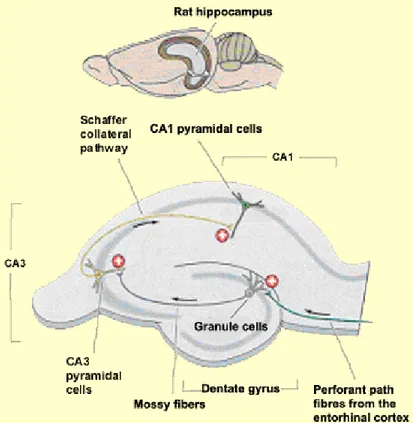
At the structural level, the anatomical organization of the medial temporal lobe corroborates its importance in the encoding and consolidation of episodic memory. A unidirectional path of various linked structures enables the processing of information in an efficient and logical manner. Essentially, neocortical inputs that process unimodal sensory information about object features converge in the perirhinal cortex and lateral entorhinal area (20). Alternatively, polymodal spatial information converges in the parahippocampal
cortex and medial entorhinal area. Subsequently, this information reaches the dentate gyrus and converges mainly in the hippocampus. In the hippocampus, the mossy fibre pathway projects the information to the CA3 field which relays the information to the CA1 field via the Schaffer collateral pathway. Ultimately, the information enters the subiculum and is sent back to the deep layers of the entorhinal cortex where the information was originally processed (figure 2) (20,21).
Figure 2 Anatomical representation of the hippocampus. Adapted from (22).
Under this system-level consolidation model, the hippocampus is depicted as part of a retrieval network for recently acquired memories (5). As associations amongst the different elements of a memory are formed and strengthened via repetition, recent memories become gradually transferred to neocortical circuits for long-term storage. Consequently, the
reorganized information can be retrieved independently of the hippocampus upon recall (4,5).
1.4 Synaptic plasticity
It is important to understand memory acquisition at the level of individual cells. Information flows through the different structures of the brain and through the subfields within these structures via neurons that communicate by means of chemical synapses. The chemical messages conveyed between neurons result in functional alterations at existing excitatory synapses (1). This creates changes in the efficacy of communication or in other words, in the excitability of a neuron, for a short period of time. For persistent changes to occur, anatomical alterations consisting of the loss or growth of new synaptic connections, must take place. The process of strengthening particular neural circuits through the modification of synapses is referred to as synaptic plasticity. Donald Hebb best describes this process by postulating that “if neuron A connects to neuron B and repeatedly or persistently takes part in firing it, some growth process or metabolic change takes place in one or both cells such that A’s efficiency, as one of the cells firing B, is increased” (23).
1.4.1 Structure of a neuron
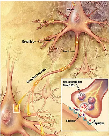
As aforementioned, the hippocampus is compartmentalized in a way that allows for the laminar organization of neurons which provides an optimal layout for a presynaptic ‘neuron A’ to persistently fire a postsynaptic ‘neuron B’. An efficient synaptic transmission between neurons is possible due to the highly specialized structure and function of a neuron.
A neuron contains four distinct regions consisting of a cell body, dendrites, an axon, and axon terminals (figure 3) (24). Synaptic signals are received as neurotransmitters from synaptic boutons on the axon terminals of a presynaptic ‘neuron A’ and form a synaptic connection with the cell body and dendrites of a postsynaptic ‘neuron B’. Chemical signals are rapidly propagated via the axon of the postsynaptic neuron and if they create a
sufficiently large excitation within the neuron, the signal is transmitted to other neurons in a similar manner.
As for the multiple dendrites found on most neurons, they are replete with synaptic spines that are specialized in receiving chemical signals owing to particular receptors embedded in the cell membrane. Activation of these receptors causes a series of molecular reactions that lead to the transcription and synthesis of mRNAs and proteins, respectively. The cell body is the main site for protein synthesis but local translation of mRNAs can also take place in dendrites at synapses. The cell body also contains the nucleus where gene activation and transcription take place. Newly synthesized proteins and transcribed mRNAs can be assembled and transported to active synapses in vesicles or multiprotein particles in dendrites along tracks known as microtubules.
1.4.2 Neuronal network in the hippocampus
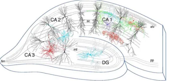
In the hippocampus, the chemical messages are transmitted by pyramidal neurons situated in a single continuous layer in the CA1 and CA3 subfields (figure 4) (1,26). About five thousand CA3 pyramidal cell axons converge onto the dendritic spines of a single CA1 cell (1). Unlike many parts of the brain where both excitatory and inhibitory synapses are present on a single spine of a cell, the CA1 region of the hippocampus is solely comprised of cells with one excitatory synapse per spine. The shape of the spine enables the confinement of the molecular reactions that occur during a synaptic transmission thereby, allowing each spine to function as a distinct biochemical region. Therefore, each spine can participate in strengthening a specific neural circuit during learning and memory.
Figure 4 Pyramidal neurons of CA1-CA3 fields. Adapted from (27).
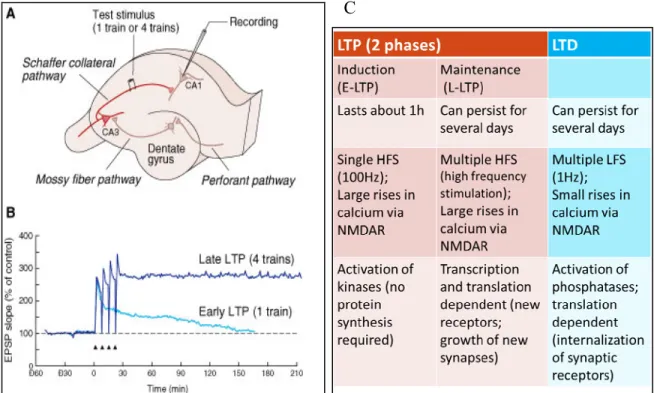
1.4.3 Long-term potentiation and long-term depression
The strengthening of a neural circuit for learning and memory is dependent on the different forms of synaptic plasticity that exist. For one, long-term potentiation (LTP) is described as the enhancement in synaptic transmission between neurons and is broken down into an early (E) and a late (L) component (1). On the other hand, long-term
depression is defined as the diminution of a synaptic transmission by activity. The regional anatomy of the hippocampus is ideal for the physiological recordings of LTP and LTD. By stimulating afferent axons of presynaptic neurons in the CA3 region, researchers can record excitatory postsynaptic potentials from the cell bodies of postsynaptic neurons in the CA1 region. By doing so, the physiological and temporal activities of E-LTP, L-LTP and LTD can be assessed, respectively. From these recordings, it was established that LTP involves an induction phase (E-LTP) that lasts about one hour after stimulation and a maintenance phase (L-LTP) that can persist for several days as can LTD (figure 5).
Figure 5 E-LTP, L-LTP, and LTD. A) Experimental setup for studying LTP in the CA1 region of the hippocampus. The Schaffer collateral pathway is stimulated electrically and the response of the pyramidal population of neurons is recorded. B) Comparison of early and late LTP in a pyramidal neuron in the CA1 region of the hippocampus. The graph is a plot of the slope (measure of synaptic efficacy) of the excitatory postsynaptic potential in the cell as a function of time. Apdated from (1). C) Characteristics of E-LTP, L-LTP, and LTD, respectively.
Multiple molecular mechanisms can explain the electrophysiological phenomena of LTP and LTD. Firstly, E-LTP does not require protein synthesis whereas L-LTP requires both mRNA transcription and translation (28-32). In fact, studies have demonstrated that if either RNA or protein synthesis is blocked in the CA1 area of the hippocampus, long-term memory is disrupted but not short-term memory (33-35). As for LTD, it solely depends on translation. Despite these differences, LTP and LTD share a common feature in that they both require specific glutamate receptors in order to take place. The main receptors are known as the NMDA (N-Methyl-D-aspartic acid) and AMPA (2-amino-3-(5-methyl-3-oxo-1,2- oxazol-4-yl)propanoic acid) receptors which often co-localize on individual synaptic spines (1).
1.4.3.1 Early long-term potentiation
E-LTP, is initiated upon a single strong stimulation that causes the release of the neurotransmitter glutamate from a presynaptic bouton into the synapse (1). The release of glutamate can be increased by brain derived neurotrophic factor (BDNF) and conversely, glutamate increases the transcription and secretion of BDNF (36). Glutamate crosses the synapse and binds to receptors on the postsynaptic spine (1). At first, only the AMPA receptors open their channels to allow a flow of ions into the postsynaptic neuron. NMDA receptors remain closed due to their unique characteristic of being both ligand-gated and voltage-dependent, a result of ion channel block by extracellular Mg2+ ions. Once the voltage change induced by the flow of ions through AMPA receptors is large enough, Mg2+ ions dissociate from their binding sites on the NMDA receptor channels. Subsequently, co-activation by two ligands, glutamate and glycine, triggers the opening of NMDA receptors resulting in a large calcium influx in the postsynaptic neuron.
When calcium enters the postsynaptic cell a series of chemical reactions occur that eventually lead to the activation of plasticity-related target genes. The cascade of events includes the activation of protein kinases such as calcium/calmodulin protein kinase II (CaMKII) and atypical protein kinase C (Pkcz). The protein kinases work in synergy to
phosphorylate AMPA receptors thus, increasing their conductance to ions and their responsiveness to glutamate (29,37-40).
When CaMKII is activated, it is capable of transiently autophosphorylating and its activity becomes independent of Ca2+/calmodulin (41). If this Ca2+/calmodulin independent postsynaptic activity is inhibited, LTP induction is blocked, but not LTP maintenance (42). Furthermore, similar to the actions of CaMKII and Pkcz, BDNF participates in increasing NMDA single-channel open probability (43,44) and in regulating the expression of NMDA receptor subunits in hippocampal neurons (45,46). This is achieved by propagation of an intracellular signalling pathway mediated by the binding of BDNF to its receptor Trkβ tyrosine kinase (47). As a result, the excitability of the synapse is temporarily increased during a subsequent synaptic transmission. The changes that occur during E-LTP are short-lived and within one hour, basal conditions are restored.
1.4.3.2 Late long-term potentiation
For persistent changes to occur, repeated strong synaptic stimulations (32) must take place for E-LTP to translate into L-LTP. L-LTP can also be induced at a “tagged” synapse if “tagging” is accompanied by the capture of “plasticity related proteins” (PRPs) (48). The concept of “synaptic tagging” was suggested by Frey and Morris, based on their observation that the induction of L-LTP at a synapse stimulates the synthesis of PRPs. PRPs can be captured at a second weakly stimulated synapse, termed tagged synapse, to convert a weak short-lived signal into a signal strong enough to generate L-LTP providing that the capture occurs within a specific time frame. Synaptic tagging also ensures that only activated synapses become modified via PRPs.
The best PRP candidate currently known is BDNF (49). Induction of a weak synaptic stimulation accompanied by elevated BDNF expression is sufficient to generate L-LTP (50,51). However, this is not the case if BDNF is delivered 70 min after stimulation (52) since the lifetime of a tag is about 1 to 2 hours (53). “Tags” must meet several criteria that include being activated by weak stimulation that induces E-LTP only, being activated
independent of protein synthesis, and being capable of interacting with PRPs for L-LTP. Under this definition, a great potential “tag” is Trkβ; the receptor for BDNF.
Regardless of the mechanism by which L-LTP is induced, it lasts about 1 to 3 hours and requires the translation of dendritically localized mRNAs independent of transcription (53,54). The transcription independent induction phase of L-LTP is followed by the transcription dependent maintenance phase which occurs about >4 hours after L-LTP induction (55). The time lag between the induction and maintenance phase results from the time required to activate the nuclear transcription of plasticity-related mRNAs, and their assembly and transport to the active synapse for local translation. These events must be tightly regulated by various proteins.
The first regulatory proteins involved in propagating the signal for the onset of the maintenance phase of L-LTP include a wide range of protein kinases such as Pkcz, CaMKII, and protein kinase A (PKA). Activation of these kinases initiates a molecular pathway that leads to the phosphorylation and thereby, the activation of cAMP responsive transcription factor CREB (56,57). The first kinase identified for CREB activation is PKA. Upon synaptic stimulation, PKA becomes activated through dissociation of its subunits (57). The released catalytic subunits can translocate to the nucleus where they can phosphorylate CREB (56,57). CREB is responsible for the activation of genes implicated in synaptic structural and functional remodelling as well as in modulation of intrinsic excitability (58). When specific CREB isoforms (CREBα/Δ) are knocked out in mice, spatial and contextual memories are impaired (59). These findings are further supported by numerous other studies demonstrating the role of CREB during memory formation (60-63). 1.4.3.2.1 Transcription
During synaptic plasticity, transcription is mediated by the concerted action of a multitude of transcription factors, such as CREB, that mediate the transcription of specific genes upon L-LTP induction. The immediate early gene Arc (activity-regulated cytoskeleton associated protein) has been extensively studied for its role in synaptic plasticity. It is one of the first genes activated at the onset of synaptic transmission and its
RNA and protein rapidly localize to dendrites and spines after activition (64,65). It is required for L-LTP and memory consolidation (66,67) as demonstrated in a study in which Arc KO mice exhibited enhanced E-LTP while L-LTP was blocked in both the dentate gyrus in vivo and in the CA1 region of acute hippocampal slices (68). Rapid Arc expression is necessary for the modulation of postsynaptic density expansion as well as for growth of postsynaptic dendritic spines by promoting F-actin stabilization through major regulators of F-actin dynamics (69).
1.4.3.2.2 Assembly and transport
Newly transcribed mRNAs must be assembled into ribonucleoprotein (RNP) transport granules in the nucleus before being transported to distal active synapses. Mechanisms must be in place to ensure that the correct mRNAs are assembled and transported in specific particles for their precise trafficking to active synapses. The particles in which the proteins and their cognate mRNAs are packaged include other proteins; some involved in the translational repression of the mRNAs during transport, others implicated in associating with microtubules (70,71), and more as components of the translational machinery (72).
Transport of mRNAs from the cell body to dendrites requires intact microtubules (70). The regulation of microtubule assembly into a functional cytoskeletal network is highly dependent on the functions of microtubule associated proteins such as microtubule associated protein 1B and 2 ( MAP1B and MAP2) (73). A disruption in the functions of MAP1B and/or MAP2 affects the trafficking of new mRNAs and proteins critical for the maintenance of LTP (74) .
Additionally, various proteins are involved in selecting their respective mRNAs by recognizing cis-acting sequences in the 3′-untranslated region (UTR) of the mRNAs (75) and enabling the transport of these mRNAs to their final destination (76). Some of these mRNA-binding proteins include hnRNP A2 in cortical and hippocampal rat neurons (77),
zipcode-binding protein involved in the transport and translation of β-actin mRNA in rat dendrites (78) as well as Staufen proteins.
The double stranded RNA (dsRNA)-binding proteins Staufen 1(Stau1) and Staufen 2 (Stau2) are found in distinct RNPs in the cell body and dendrites of mammalian neurons (79-82) where they each regulate the microtubule mediated transport of a variety of mRNAs (79,83-86). In fact, down-regulation of Stau1 and Stau2 by siRNA reduces the amounts of RNA in dendrites of neurons. For example, down-regulation of Stau1 reduces the amount of αCaMKII at synapses (81) while down-regulation of Stau2 reduces the amount of β-actin (87) and of a reporter with the 3′-UTR at synapses (88).
1.4.3.2.3 Translation
Once at the active synapse, the silenced mRNAs are de-repressed and translated into proteins that participate in the growth of new synapses and in the formation of new receptors (BDNF and its receptor Trkβ, αCaMKII, Arc, etc – see previous paragraphs). CPEB (see section 1.3: post-transcriptional modifications) and possibly Stau1 are some of the proteins responsible in regulating the local translation of a subset of mRNAs in an activity-dependent manner (89-92). For Stau1, it was demonstrated that it increases the translation of a specific subset of mRNAs by binding to their 5’ end (89).
It can be inferred from the above information that LTP is a complex phenomenon. It includes a variety of steps in which specific regulatory proteins participate in ensuring that an effective synaptic transmission can take place between neurons. However, it is the determination of the interaction between LTP and LTD in a neuron that will ensure that a distinctive neural circuit will be reinforced for enhanced memory and learning.
1.4.3.3 Long-term depression
Similarly to LTP, LTD is a complex molecular event but unlike LTP, transcription is not required since pre-localized mRNAs are translationally activated upon LTD induction (93). LTD is initiated upon multiple low frequency stimulations that cause small
rises in calcium via NMDA receptors (94). The low calcium concentration is responsible for the distinctive cascade of events that prompts the activation of protein phosphatases during LTD compared to protein kinases activated during LTP (95). A few advances have been made regarding the precise phosphatases that are activated during LTD. It has been demonstrated that postsynaptic addition of phosphatase inhibitors that primarily target calcineurin or protein phosphatase 1 (PP1) prevent LTD (96). Protein phosphatases are involved in the de-phosphorylation of AMPA receptors thus reducing their open channel probability (97). In addition, the change in phosphorylation is accompanied by the internalization of these receptors (98-100).
Besides its role in LTP, Arc was found to be crucial for LTD (101,102) by facilitating endocytosis of AMPA receptors through its interaction with endocytic proteins (66,103). This event is inhibited upon LTD induction by acute blockade of new Arc synthesis (101). In reference to LTD, Arc mRNA translation is believed to be regulated by the fragile X mental retardation protein (FMRP) (104) known to be a key player in LTD (105). Indeed, activation of Arc is absent in Fmr1 KO mice (102). FMRP associates with a subset of mRNAs, including Arc, and possibly acts as a negative regulator of translation. This theory is based on the observation that in the absence of FMRP, there is excess protein synthesis that leads to the increased internalization of AMPA receptors and exaggerated LTD (106).
Furthermore, other candidates, including MAP1B that co-localizes with FMRP in dendrites of cultured neurons (107), are thought to play equally important roles in the internalization of AMPA receptors during LTD (104,108). It was shown that the rapid synthesis of MAP1B is linked to the internal trafficking of AMPA receptors (109) and that the basal expression of MAP1B is augmented in hippocampal slices of Fmr1 KO (110). Conversely, the expression of MAP1B in dendrites is reduced in the absence of Stau2. Specifically, Stau2 knockdown, but not Stau1 knockdown, with siRNA in primary hippocampal neurons blocks LTD and decreases the endogenous expression of MAP1B protein as well as the level of a reporter with the MAP1B 3’UTR that is transported to dendrites (88). It also prevents the dissociation of this reporter from Stau2 mRNA transport
granules. Consequently, translational activation of MAP1B is inhibited thus, preventing MAP1B dependent AMPAR internalization required for efficient LTD. Therefore, Stau2 is thought to be required for the loading of MAP1B mRNA into transport granules, for its transport within these granules to active synapses, and for its local translational activation by way of dissociation with MAP1B mRNA.
All of the events described for LTD and LTP rely on the precise and accurate function of plasticity related proteins. Any disruptions in the expression and activity of a protein can hinder the efficacy of a synaptic transmission. For that reason, strict transcriptional and translational regulations of plasticity-related mRNAs and proteins exist.
1.4.4 Transcriptional and translational regulations of plasticity related
mRNAs and proteins
Transcriptional and translational regulations are crucial in preventing ectopic and/or altered expression of synaptic plasticity-related mRNAs and proteins which could otherwise result in various defects such as fragile X syndrome (caused by the absence of Fmr1 gene expression) (111), Alzheimer’s disease (caused by accumulation of Aβ peptides due to disruption of APP metabolism) (112), and more. Translational repression via RNA-binding proteins and microRNAs (miRNA) as well as polyadenylation of mRNAs has emerged as important transcriptional regulations (113,114). Similarly, post-translational modifications such as phosphorylation, adenylation, and methylation are crucial in altering the activity of a protein (115-117).
1.4.4.1 Post-transcriptional modifications
Transcription is the first regulatory step in gene expression. Following transcription, mRNAs are assembled in ribonucleoprotein transport granules for their export from the nucleus to the cytosol. During transport, mRNA translation is silenced by repressors associating to specific recognition sequences in the 3’UTR of a mRNA (118-122) or by the presence of an inhibitory RNA structure in the 5′-UTR to block translation initiation (123,124).
A great example is the cytoplasmic polyadenylation element protein (CPEB) that regulates the polyadenylation (poly(A)) of the 3’ end of a subset of mRNAs. These mRNAs contain CPE recognition sequences in their 3’UTR that bind to CPEB (125). Translation can only be initiated when CPEB becomes phosphorylated by a protein kinase Aurora (116). This induces CPEB to interact with proteins involved in the recruitment of a poly(A) polymerase for the process of polyadenylation (117). This step is thought to increase translation (126). A likely target of polyadenylation during synaptic plasticity is the 3’UTR CPE-containing N-actin mRNA in Aplysia (127). Polyadenylation of this mRNA was shown to occur in response to stimulation with serotonin (128,129). Furthermore, it was demonstrated in mice that αCaMKII contains two CPEs in its 3′UTR (130) thereby, making it another potential candidate to be regulated by CPEB via the addition of a poly(A) tail.
miRNAs also take part in the tight spatial regulation of mRNA translation in neuronal dendrites (131). miRNAs represent an extensive class of small non-coding RNAs that act as post-transcriptional regulators of gene expression. They have been discovered in a plethora of tissues, including the nervous system and fittingly, have a widespread role at various stages of synaptic development and during synapse function and plasticity. In post-mitotic neurons, many miRNAs are associated with translation regulatory complexes (132) where one miRNA can control the fate of a few hundred different synapse-relevant mRNAs simultaneously (133).
miRNAs result from a multi-step maturation process starting with the transcription of miRNAs into primary miRNAs (pri-miRNAs) (134,135). Cleavage of pri-miRNAs produces precursor hairpin miRNAs which are exported to the cytoplasm to be further processed by the RNase Dicer. The end product is a mature single-stranded miRNA loaded into a multi-protein miRNA-induced silencing complex (miRISC). An important component of this complex with regard to synaptic function is the Argonaute protein. The miRNA within the complex is guided to target mRNAs in order to extensively bind, albeit imperfectly, to sequences usually present within the 3’UTR of these mRNAs. This results in the translational repression and/or degradation of the mRNAs in question.
In particular, miR-134 is known for its role in suppressing the translation of LIMK1 mRNA in dendrites of mature hippocampal neurons. LIMK1 is a kinase that promotes actin polymerization and spine growth (136). The miR-134-LIMK1 association seems to be regulated by synaptic activity whereby increased levels of BDNF release LIMK1 from the inhibitory effects of miR-134. miR-138 is another miRNA with a role in spine morphogenesis. miR-138 acts on the depalmitoylation catalytic enzyme APT1 which participates in modulating the actin cytoskeleton of dendritic spines (137).
1.4.4.2 Post-translational modifications
Phosphorylation is a major post-translational modification that, in most cases, induces the activation of a protein. The constitutively expressed CREB protein is well known for being subjected to activation through phosphorylation (115). In the context of synaptic transmission, a signalling pathway originating from activation of NMDA receptors, leads to an increase in cAMP. In turn, cAMP activates PKA and the mitogen-activated protein kinase (MAPK) and both proteins translocate to the nucleus where they phosphorylate an activator form of CREB (138,139). This activator form is responsible for the stimulation of gene expression for target genes containing a cAMP responsive element (CRE) (140).
The regulatory mechanisms described must remain functional for the maintenance of a healthy cognitive status. Cognitive decline associated with aging and neurodegenerative diseases arises when disturbances occur in the expression and/or function of plasticity-related mRNAs and proteins that bring about changes in the efficacy of synaptic transmission.
1.5 Aging and cognitive decline
Aging is associated with learning and memory impairments, especially hippocampus-dependent spatial memories. Unfortunately, due to the limited knowledge about the etiology of cognitive impairment associated with aging, few pharmaceutical
interventions have proven to be effective in preventing and/or treating age-related memory loss. Currently, it is established that during normal aging, various other structural modifications are attributed to cognitive decline despite the lack of significant neurodegeneration. Neurons in the cerebral cortex and especially in the medial temporal structures undergo attrition of dendritic branches, loss of dendritic spines and loss of synapses (141-143). These changes contribute to the decreased efficiency in relaying information through the perforant, mossy fibre, and Shaffer collateral pathways in aged rodents not to mention that they are likely a result of dysregulations in the transcription and translation of plasticity-related mRNAs and proteins. Such disruptions lead to defects in cytoskeletal integrity, microtubule-dependent transport, synaptic functions, and neurotransmission (6,7,144-146).
The effects of these modifications can be observed in experimental settings. Importantly, with regards to hippocampus-dependent spatial memory, there is ample evidence that aged rats that undergo the Morris water maze test show deficits in recalling the path to the hidden platform after several trials (when the elapsed time between trials is long) when compared to young rats (147-155). Additional electrophysiological studies substantiate these findings by providing LTP and LTD recordings in rodent models of healthy aging that suggest perturbations in hippocampal plasticity during aging. These recordings display decreased basal synaptic transmission (156), enhanced LTP threshold (157), decreased LTP maintenance (158), altered LTP mechanisms (159), and increased chances of LTP reversal (160) in aged rodents compared to young ones. There also appears to be facilitated LTD and hence, not surprisingly, an imbalance in synaptic transmission whereby LTD prevails over LTP (157,161,162).
Each one of the altered electrophysiological recordings observed in aged rodents is associated to defects that occur at the molecular level. For example, significant decreases in proteins such as SNAP25, syntaxin 1, VAMP2, and synaptophysin (2,163) critical in the docking and fusion of presynaptic vesicles, and in the scaffold protein PSD95 (7,164,165), have been observed in aged rodents. These proteins are essential for the formation of a functional synaptic contact. Causally, a decrease in their expression can help explain the
diminution in basal synaptic transmission and in LTP maintenance in aged rodents. More specifically, it may provoke slowed endocytic replenishment and/or abolished exocytosis of synaptic vesicle pools as well as impaired receptor aggregation (PSD95) upon prolonged or intense stimulation of synaptic transmission (166,167).
Furthermore, perturbations in LTP maintenance can arise from defects in the expression or function of individual mRNAs and/or proteins. Immediate early genes (IEG) such as Arc represent an important class of transcription factors that are transiently and rapidly activated to regulate the expression of target genes involved in synaptic plasticity (168,169). One such IEG is zif268. Normally, Zif268 expression increases during hippocampus-dependent spatial orientation tasks (170) but in aged rodents that express spatial memory impairments, resting levels of zif268 mRNAs are decreased in the CA1 and CA2 areas of the hippocampus and in the neocortex (171). These results demonstrate that zif268 expression may play an important role in the maintenance of intact spatial memory of aged rodents.
A dysfunction in the activity of an individual protein can also impair synaptic transmission. The previously discussed CREB protein (sections 1.2 and 1.3) shows phosphorylation dependent activity upon LTP induction. There is evidence for the absence of change in the resting expression of CREB in the hippocampal CA1 region and dentate gyrus of aged rodents with cognitive decline when compared to young rodents (172,173). In contrast, there is a reduced amount of phosphorylated CREB activity that is noticeable in the same aged rodents. Over-expression of CREB in aged rodents can partly rescue age-related memory changes (174). This demonstrates the great importance in the activation status of CREB for its effective role in synaptic plasticity. The decreased activity of CREB may be partially responsible for the increased LTP reversal observed in aged rats due to decreased activation of target genes important in maintaining LTP.
Another salient study offered enlightenment for the facilitated LTD observed in aged rodents suffering from cognitive impairments. VanGuilder H.D., et al. demonstrated, in hippocampal slices of aged rats, an increased expression of hippocalcin, a protein involved in facilitating LTD through calcium sensitive dynamics (2). Moreover, alterations
in the subunit composition of AMPA and NMDA receptors may exacerbate LTD prevalence. The subunit subtypes of NMDA and AMPA receptors have differing kinetics that dictate whether LTP or LTD will take place (175-178). For example, the GluR2 AMPA receptor subunit prevents calcium permeability through the receptor channel. Aged cognitively-impaired rodents show a decrease in the GluR2 subunit which renders AMPA receptors more permeable to calcium. Increased calcium permeability augments susceptibility to excitotoxicity and postsynaptic calcium concentrations determine the strength and direction of synaptic transmission (179-182). Therefore, altered calcium homeostasis can result in a wider range of low-frequency activity patterns thereby, triggering excessive LTD (183-185).
As for NMDA receptors, they are made up of an obligatory NR1 subunit and two additional NR2 subunits: NR2A and NR2B (186). The NR2A or NR2B subunit governs at different developmental stages (187,188). One study determined that the expression profiles for both types of NR2 subunits shift in a similar manner in both aged rodents suffering from cognitive impairment and young rodents (7). However, they found an increase in NR1 subunits in aged rodents. These researchers speculated that the additional NR1 subunits may represent non-functional subunits that could bring about the internalization of NMDA receptors. Conversely, another study observed an increased NR2A/NR2B ratio in the hippocampus of aged memory-impaired mice as compared to young adults (189). This led to changes in the induction thresholds for LTP. Despite the differences between studies, the outcome remains the same. Altogether, these findings provide insights into possible mechanisms underlying the predominance of LTD over LTP as well as the enhanced LTP threshold observed in aged cognitively- impaired rodents.
NMDA receptors in the hippocampus are also susceptible to altered calcium homeostasis; (162,190-192) a process that is regulated by two different calcium sources during synaptic activity. Voltage-dependent calcium channels (VDCCs) as well as intracellular calcium stores release calcium during synaptic transmissions and their activity is altered in aged hippocampal neurons (193,194). For instance, aged rodents display greater numbers of VDCCs in the CA1 region of their hippocampus (195) and thus, cause
an imbalance towards an increased dependence on a form of LTP that relies on VDCCs rather than NMDA receptors (196-198). Consistent with this, the activity of NMDA receptors is reduced in the hippocampus of aged rats (199). Such disturbances have important implications for LTP induction which henceforth, requires a greater stimulus to be triggered via the NMDA receptors. This represents an altered mechanism by which LTP can take place and by which its threshold can be increased.
All of the aforementioned alterations observed in cognitively-impaired aged rodents occur in the absence of significant neural loss. It is difficult to imagine the increased detrimental effects that neurodegeneration can have on the structural, cellular, and molecular processes that take place during synaptic transmission. These processes will be reviewed for a more severe form of cognitive decline exhibiting neurodegeneration that is, Alzheimer’s disease (AD).
1.6 Alzheimer’s disease
Alzheimer’s disease is a fatal neurodegenerative disease afflicting over 35 million people worldwide and representing 70% of dementia cases in the world. Its prevalence is constantly rising as a consequence of the general increase in the aging population which is expected to quadruple by 2050 in the United States (200). Despite the ongoing studies and our better understanding of the disease, there are currently no effective treatments available to cure Alzheimer’s disease. Its emergence is most often sporadic and in a population aged over 65 years. However, about 5% of cases present with a less common form known as familial Alzheimer disease (FAD) which usually strikes sooner (201). FAD is the result of mutations in specific genes including the amyloid precursor protein (APP), presenilin 1, and presenilin 2 genes. Regardless of its cause of onset, Alzheimer disease has detrimental effects on the quality of life of those affected and imparts costly burdens to the public health sector.
At the neuropathological level, Alzheimer’s disease is characterized by cerebral atrophy which is especially apparent in the hippocampus but also affects the neocortex
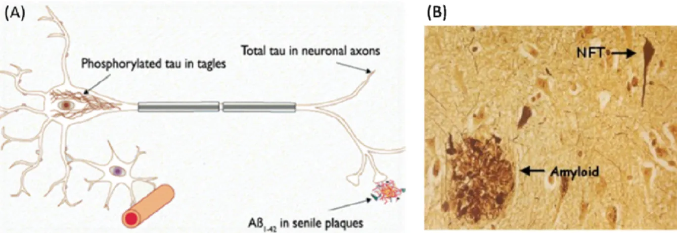
(202). In addition there are manifestations of neuronal cell death, neuroinflammation, synapse loss, and the accumulation of two specific types of lesions. These lesions first appear in the medial temporal lobe (203,204) and consist of extracellular senile plaques made up of aggregated amyloid-beta (Aβ) peptides (205) and intracellular neurofibrillary tangles (NFT) (206) composed of hyper-phosphorylated forms of the tau protein (figure 6) (207).
Figure 6 Lesions in Alzheimer’s disease. A) Schematic representation of senile plaques and neurofibrillary tangles. Adapted from (208). (B) Histological representation of senile plaques (Amyloid) and neurofibrillary tangles (NFT). Adapted from (209).
NFT arise from a dysregulation in the phosphorylation state of tau (210). Tau is a microtubule-associated protein that participates in the stabilization and regulation of microtubule dynamics necessary for neurite outgrowth, morphogenesis and axonal microtubule-dependent transport. In its de-phosphorylated state, tau’s affinity for microtubules is reduced thereby, promoting their depolymerisation. In contrast, in its phosphorylated state, tau binds to microtubules and enhances their stability (211,212). Hyperphosphorylation of tau in Alzheimer’s disease hinders the microtubule network and promotes self-assembly of tau into paired helical filaments (212,213).
On the other hand, Aβ peptides present in senile plaques are cleavage products of APP (214). A dysfunction in the metabolism of APP processing results in excessive Aβ fibril accumulation of 39-42 amino acids long. The 40 (Aβ40) and 42 amino acid residues (Aβ42) are the most prevalent while the Aβ42 fibril has a higher propensity to form aggregates. The translational regulation of APP is carried out by FMRP which when bound to APP mRNA, represses its translation (215).FMRP may therefore play an important role in the pathogenesis of Alzheimer’s disease if its expression is disrupted as seen in fragile X mental disorder.
Similar to the electrophysiological recordings described for cognitive decline (see section 1.4) the recordings observed for Alzheimer’s disease display inhibited LTP (216) and facilitated LTD in the presence of oligomeric Aβ in hippocampal synapses (217). The mechanisms underlying the disruptions in LTP and LTD include partial inhibition of NMDA receptor activity due to Aβ deposition (218) which provokes decreased calcium influx through the NMDA channels. The resultant disruption of calcium homeostasis causes the activation of calcineurin; a key player involved in the internalization of NMDA and AMPA receptors (219,220). Internalization of NMDA receptors also occurs via dephosphorylation of NR2B subunits by oligomeric Aβ induced-phosphatases (221).
Furthermore, reduced calcium concentration limits downstream events such as activation of plasticity related proteins including CaMKII, MAPK, and Akt/protein kinase B (222). All of these events lead to Aβ mediated dendritic spine loss and altered LTP and LTD mechanisms.
Other plasticity-related proteins downstream of receptor activation are dysregulated in Alzheimer’s disease. The previously discussed CREB protein (section 1.2: late long-term potentiation) is downregulated in the brains of Alzheimer mice (223). As a result, its mRNA and protein levels are lowered thereby, decreasing the phosphorylation-dependent activation of the protein. The underlying cause appears to be the oxidative stress caused by the accumulation of Aβ fibrils. In the presence of antioxidants, the adverse effects of oxidative stress on CREB are prevented and so CREB can resume its normal function as an activator of target genes. Under stress conditions, the target genes under the control of
CREB are also affected. CREB target proteins such as BDNF, and anti-apoptotic proteins Bcl-2, BIRC3, and BIRC4 are downregulated while the pro-apoptotic protein Bax is increased in Alzheimer mice. Therefore, decreased CREB activity ultimately leads to neuronal apoptosis, a major hallmark of Alzheimer’s disease.
In addition, miRNAs may also play an important role in the translational regulation of APP in Alzheimer’s disease. Various miRNAs have been recognized to bind the 3’UTR of APP mRNA (224-226). In human brains with Alzheimer, the level of miR-106b is significantly reduced (225). However, there is no convincing evidence of a correlation between miRNA levels and APP in Alzheimer tissues. However, regulation of APP by miRNAs has not been ruled out. In rat hippocampal neurons, silencing of AGO2, a RISC protein involved in the miRNA pathway (see section 1.3) was shown to increase APP protein levels (226). Further investigations demonstrated the effects of the brain-enriched miR-10 on APP levels. Inhibition of endogenous miR-101 increased APP levels whereas overexpression of miR-101 significantly reduced APP levels and consequently Aβ aggregates decreased as well. In agreement with these observations, miR-101 is downregulated in the cerebral cortex of patients afflicted with Alzheimer’s disease (227,228).
As can be deduced from the above information, Alzheimer’s disease exhibits similar dysfunctions in mRNA and protein expression, in protein activity and in transcriptional and translational regulations as observed in age-related cognitive decline. The main differences are the presence of senile plaques and neurodegeneration in Alzheimer’s disease compared to memory impairments in elderly. Despite these differences, the key players involved in synaptic plasticity are equally affected in both cases.
1.7 Hypothesis, aims, and rationale
1.7.1 Hypothesis
The expression of plasticity-related mRNAs will be altered in aged rodents and in transgenic rodent models showing early stages of AD. More specifically, the expression of
mRNAs mainly involved in LTD will increase whereas the expression of mRNAs mainly involved in LTP will decrease and in this manner, cause an imbalance between LTP and LTD whereby LTD will prevail over LTP. The disturbances in mRNA expression will be most prevalent in the hippocampus (CA1-CA3 fields) than in other regions of the brain such as the dentate gyrus or the cortex. In addition, the genes affected will differ from one brain region to the other.
1.7.2 Aims
1) To study the expression of plasticity-related mRNAs in the hippocampus (CA1-CA3 fields), dentate gyrus, and cortex of 3xTg mice showing early stages of AD and their wild-type counterparts by means of RT-qPCR experiments.
2) To study the expression of plasticity-related mRNAs in the hippocampus and cortex of young (3 months old) and aged (24 months old) Fisher 344 rats by means of RT-qPCR experiments.
1.7.3 Rationale
The morphological modifications of neurons that occur in the cerebral cortex and more notably in the medial temporal structures where the hippocampus is located (141-143), are the outcome of disturbances in the transcription and translation of plasticity-related mRNAs and proteins which together, results in a less efficient synaptic plasticity within these structures (6,7,144,145,229). During aging (157,161,162) and AD (216,217), these defects cause an imbalance between LTP and LTD whereby LTD starts prevailing over LTP. Therefore, the expression of mRNAs mainly involved in LTD should increase to help explain the increase observed in LTD whereas the expression of mRNAs mainly involved in LTP should decrease to help explain the inhibition of LTP observed in these pathologies.
Additionally, during age-related cognitive decline and AD, progressive cognitive decline and neurodegeneration (in the case of AD) occur in a sequential process across
brain tissues, with the temporal lobe being affected earlier than the frontal part of the cortex (3,203). Thus, changes in the expression of plasticity-related genes most likely appear at a later stage in the cortex than in the hippocampus. Furthermore, Kaiwen He et al. (230) demonstrated that the mechanisms for synaptic plasticity might diverge depending on the brain region. Therefore, the genes that become dysregulated in age-related cognitive decline and AD might differ across brain tissues and for different sub-regions within these tissues.
For example, the hippocampus contains distinctive anatomical regions consisting of the CA1 field, CA3 field, and the dentate gyrus all of which possess unique molecular and biophysical properties (231). Whereas the CA1-CA3 sub-regions are made up of four layers of pyramidal cells, the dentate gyrus is made up of three layers of cells with granule cells representing the major cell type. These cells have the ability to proliferate throughout the lifespan by means of neurogenesis. This process is thought to be important for learning and memory (232). Neurogenesis declines with age (233-235) and therefore, might be associated to cognitive impairments as demonstrated in various studies (236-238). The defects in neurogenesis observed in these studies likely reflect changes in gene expression. The differences between the sub-regions within the hippocampus result in characteristic region-specific electrophysiological properties. These arise from the fact that each field exhibits a specific synaptic response to plasticity-related genes normally expressed in all regions of the hippocampus (239,240).
At the molecular level, each hippocampal sub-region has been shown to display unique transcription and protein expression patterns (241,242). Additionally, analysis of the hippocampal regions in aged cognitively impaired animals compared to young and aged cognitively unimpaired animals revealed that changes in gene expression were more pronounced in the CA3 field than in the CA1 field and dentate gyrus of the hippocampus (243). Similarly, in transgenic AD mice, the Schaffer collateral pathway (pathway from CA1 to CA3 field) of the hippocampus exhibits diminished LTP whereas the Mossy fibre pathway (from the dentate gyrus to the CA3 field) is slightly enhanced (244). This supports a unique role for each sub-region within the hippocampus in learning and memory.
For all of the aforementioned reasons, qRT-PCR analyses must be performed separately in the cortex and sub-regions (dentate gyrus and CA1-CA3 fields) of the hippocampus in 3xTg and WT mice and in the cortex and hippocampus of young and aged rats.
2 Experimental procedures
2.1 Animals
Experiments were conducted in accordance with the guidelines of the Canadian Council of Animal care and were approved by the “Comité de déontologie de l’expérimentation sur les animaux (CDEA)” of “Université de Montréal”.
2.1.1 Young and aged rats
Four young (3 months) and four aged (24 months) Fisher 344 rats obtained from Harlan Laboratory were used for qPCR analysis. Animals were euthanized in a CO2
chamber one at a time and cortices and hippocampi were dissected within thirty minutes after death. Dissection was performed under RNAse free conditions and tissues were maintained in cold Hank`s balanced salt solution (HBSS: Potassium chloride 5.33 mM, potassium phosphate monobasic 0.441 mM, sodium chloride 137.93 mM, sodium phosphate dibasic anhydrous 0.338 mM, D-Glucose 5.56 mM, Phenol Red 0.0266 mM, sodium pyruvate 1 mM, Hepes 10 mM). Once dissected, tissues were immediately placed in Trizol Reagent (Invitrogen) and kept on ice. Tissues were then homogenized using a polytron and immediately subjected to RNA isolation or stored at -800C until use.
2.1.2 3xTgmice and WT mice
The hippocampi, dentate gyrus, and cortices of the right brain hemisphere ofthree transgenic Alzheimer (3xTg) mice and three control wild-type (WT) mice were obtained from Karl Fernandes laboratory in the “départment de pathologie et biologie cellulaire à l’Université de Montréal”. The generation of 3xTg mice has been described previously (245). Briefly, 3xTg mice were produced from co-microinjection of two independent transgenes encoding human APPswe and human tauP301L into single-cell embryos harvested
from homozygous mutant PS1M146V knock-in mice. The APPswe and tauP301L transgenes are
both under the control of the neuron-specific mouse Thy1.2 regulatory element. The non-transgenic WT mice used have the same background as the 3xTg mice (C57BL6/129SVJ)
and were littermates of the original PS1M146V knock-in mice. Housing conditions were
identical for 3xTg and WT mice.
The brain tissues (Hippocampus (CA1-CA3 fields), dentate gyrus, and cortex) of each of the 3xTg and WT mice were dissected from the brains of the mice once they had reached seven months of age. At this age, cognitive impairments started to be apparent in the 3xTg mice. Immediately after dissection, the brain tissues were stored at -800C. Upon reception, the frozen tissues were deposited in Trizol Reagent (Invitrogen) and homogenized using a polytron. RNA isolation was performed immediately afterwards or stored at -800C until use.
2.2 RNA isolation
All tissue samples were homogenized with a polytron in Trizol Reagent (Invitrogen), according to the protocol of the manufacturers. RNA concentration was determined using DO readings at 260 nm and 280 nm in a spectrophotometer (Thermo Fisher Scientific). RNA samples were diluted in DEPC water (RNAse free water) to obtain a uniform concentration of 250 ng/µL across samples. The quality of the RNA dilutions was determined with 1% agarose gel electrophoresis. Samples were subjected to reverse transcription (RT) or kept at -800C until additional analysis.
2.3 Quantitative PCR
Prior to qPCR reactions, synthesis of cDNA was achieved using the GeneAmp RNA PCR kit (Applied Biosystems) following the guidelines of the manufacturer. For the cortex and hippocampus samples of young and aged rats and for the cortex samples of WT and 3xTg mice, the starting concentration of RNA used for cDNA synthesis was 750 ng/µL and for the dentage gyrus and hippocampus samples of WT and 3xTg mice the starting concentration of RNA was 200 ng/µL. Subsequently, qPCR reactions were performed
using the LightCycler 480 SYBR Green I Master. For each reaction, 3 µL of cDNA was
added to a mix containing 3 µL PCR-grade water, 2 µL of 10X PCR primers, and 10 µL of 2X SYBR Green Master Mix (Roche) for a total of 20 µL to obtain a final concentration of 1X for the PCR primers and SYBR Green Master Mix. Primers were designed using Integrated DNA Technology Primer Quest
(http://www.idtdna.com/Scitools/Applications/Primerquest/), blasted using NCBI
Primer-Blast (http://www.ncbi.nlm.nih.gov/tools/primer-blast/) and synthesized at Integrated DNA Technology. Primer sequences are listed in Table I and Table II. Cycling conditions were 5 min at 950C, followed by 50 cycles of 10 s at 950C, 20 s at 600C, and 30 s at 720C. A melting curve followed the cycling conditions for identification of the specificity of the amplified products by fluorescence measurement every 10C from 720C to 950C.