UNIVERSITÉ DE MONTRÉAL
RPA exhaustion as a major cause of genomic instability in polymerase eta-deficient cells Par
Abdelhamid Elsherbiny
Programmes de biologie moléculaire Faculté de médecine
Mémoire présenté à la Faculté de médecine en vue de l’obtention du grade de M.Sc.
en biologie moléculaire Mars 2019
Résumé
Les agents génotoxiques présents dans l’environnement représentent un danger omniprésent pour l’intégrité de l’ADN. Par exemple, les êtres humains sont constamment exposés au rayonnement ultraviolet (UV) solaire, ce qui induit la formation de lésions hautement génotoxiques à l'ADN. Ces lésions bloquent la réplication de l’ADN dans les cellules de la peau et sont la principale cause du développement de tumeurs cutanées. Heureusement, les cellules sont équipées de mécanismes qui atténuent les effets néfastes des UV : i) la réparation par excision de nucléotide (NER) qui permet l'élimination sans erreur des adducts de dipyrimidine induits par les UV (à savoir les photoproduits de 6-4 pyrimidine pyrimidone, 6-4PP et cyclobutane pyrimidine) les dimères; CPD), et ii) la synthèse translésion (TLS) via l'ADN polymérase eta (polη) pour un contournement précis, spécifiquement des CPD. Les mutations germinales dans les gènes de la voie NER conduisent au syndrome autosomique récessif Xeroderma pigmentosum (XP), caractérisé par une sensibilité accrue au soleil et un risque extrêmement élevé de cancer de la peau. D'autre part, l'inactivation germinale de polη est à l'origine du syndrome variant de Xeroderma pigmentosum (XPV), également caractérisé par une sensibilité aux rayons solaires et une incidence élevée de cancers de la peau. En comparaison aux cellules normales, les cellules XPV exposées aux UV présentent i) une hypermutabilité ii) une mort cellulaire accrue iii) un stress de réplication de l'ADN élevé et iv) un NER défectueux exclusivement pendant la phase S. L'objectif principal de cette thèse est de mieux comprendre la base moléculaire de ces phénotypes. Des études récentes ont mis en évidence le rôle du stress réplicatif dans l'épuisement des stocks cellulaires du complexe trimérique RPA (Replication Protein A), ce qui cause subséquemment une augmentation des dommages à l'ADN
émis l’hypothèse que ce stress réplicatif pourrait entraîner une réduction de la disponibilité de RPA et, conséquemment, des niveaux plus élevés d’instabilité génomique. Notre objectif était d'évaluer la dynamique du complexe RPA sur la chromatine post-UV et de vérifier si un excès de RPA pouvait réduire la génotoxicité des UV. Fait intéressant, les cellules dépourvues de pol eta présentent une charge persistante de RPA sur la chromatine, qui est associée à une phosphorylation élevée de l’histone H2AX et à la mort cellulaire. De plus, la surexpression de la RPA dans des cellules XPV atténue la génotoxicité induite par les UV, y compris un défaut de progression du cycle cellulaire, une sensibilité accrue aux UV et la formation de cassures double brin; cependant, la surexpression de RPA n’a aucun effet sur mutagenèse causée par l’absence de pol eta. Nos données indiquent pour la première fois que la disponibilité de la RPA est un facteur déterminant de la réponse cellulaire aux UV dans les cellules déficientes en polη.
Abstract
Genotoxic agents from the surrounding environment represent an omnipresent danger to the integrity of DNA. For example, humans are constantly exposed to solar ultraviolet (UV) radiation, which induces the formation of highly-genotoxic replication-blocking DNA lesions in skin cells that are the major cause of cutaneous tumor development. Fortunately, cells are equipped with mechanisms that mitigate the deleterious effects of UV, particularly i) nucleotide excision repair (NER) for error-free removal of UV-induced dipyrimidine adducts (i.e. 6-4 pyrimidine pyrimidone photoproducts; 6-4PP and cyclobutane pyrimidine dimers; CPD), and ii) translesion DNA synthesis (TLS) via DNA polymerase eta (polη) for accurate bypass specifically of CPD. Germline mutations in NER pathway genes lead to the autosomal recessive syndrome Xeroderma pigmentosum (XP), characterized by increased sun sensitivity and extremely high risk of developing skin cancer. On the other hand, inactivating germline mutations in polη cause Xeroderma pigmentosum variant (XPV) also characterized by sun sensitivity and high incidence of skin cancer. Upon UV exposure, compared to normal counterparts, XPV cells exhibit i) exquisite hypermutability ii) increased cell death iii) elevated DNA replication stress, and iv) defective NER exclusively during S phase. The major goal of this thesis is to better understand the molecular basis for these deleterious phenotypes. Recent studies have highlighted the role of uncontrolled replication stress in exhaustion of the trimeric replication protein A (RPA) and subsequent increase in DNA damage/repair defects. Since the deficiency of polη leads to increased levels of replication stress in UV-exposed cells, we hypothesized that such replication stress can lead to reduced availability of RPA and subsequent genomic instability. Our aim was to evaluate
which was associated with elevated H2AX phosphorylation and cell death. In addition, RPA overexpression mitigates the UV-induced genotoxicity including cell cycle progression defect, increased UV sensitivity, and double strand break formation; however, RPA overexpression did not rescue the increased mutagenesis. Our data indicate for the first time that RPA availability is a major determinant of the cellular response to UV in polη-deficient cells.
Contents
Résumé ... i
Abstract ... iii
List of abbreviations ... x
List of Figures ... xiv
List of Tables ... xvi
Introduction ... 1
Cell cycle and replication control ... 1
The cell cycle ... 1
Fidelity of DNA replication ... 3
Origins of replication control ... 4
DNA replication stress ... 5
I. Endogenous sources of replication stress ... 5
II. Exogenous sources ... 6
UV radiation... 7
UV physical properties ... 7
II. Activation of translesion DNA synthesis ... 10
Consequences of inadequate replication stress response ... 17
DNA damage and repair ... 19
I. Intrinsic DNA damage ... 19
II. Extrinsic DNA damage ... 20
DNA repair deficiency and cancer ... 22
Skin cancer ... 22
Mechanism of UV induced mutagenesis ... 23
Xeroderma pigmentosum (XP) disease ... 25
UV-induced genotoxicity in XPV patient-derived cells ... 26
Rational ... 28
Hypothesis and specific aims... 29
Materials and methods ... 30
Cell culture ... 30
Clonogenic survival ... 30
Ectopic RPA expression and siRNA treatment. ... 30
Immunoblotting... 31
Dual detection of γ-H2AX and RPA following UV treatment by flow cytometry. ... 31
HPRT mutation assay ... 33
Results ... 34
Validation of the experimental model system ... 34
XP30ROsv cell sensitivity to UV radiation ... 34
The absence of Polη TLS activity in XP30RO does not alter Replication fork progression 36 XP30RO cell exhibit cycle delay Upon UVC radiation ... 37
XP30RO cells exhibit an increased UV mutation rate ... 38
Polη-deficient cells manifest a high level of RPA loading on chromatin and induction of γ-H2AX post-UV. ... 40
Assessment of RPA overexpression on UV-induced genotoxicity in polη-deficient cells .. 44
RPA overexpression protects ssDNA and reduces UV-induced DNA damage in Polη-deficient cells. ... 44
UV sensitivity in Polη-deficient cells is rescued by RPA overexpression. ... 48
RPA overexpression mitigates the cell cycle progression defect in Polη-deficient cells. ... 50
RPA overexpression does not affect mutation rate in polη depleted cells. ... 53
Conclusion and future perspectives ... 59
Acknowledgment
I am very grateful to Dr. Elliot Drobetsky for his support and mentorship throughout my MSc. Elliot has guided me on how to conduct experiments properly, develop logical thinking, and deliver a clear presentation both written and oral.
I am also deeply grateful to my co-supervisor Dr. Hugo Wurtele for his expert advice. Hugo helped progress this project with his brilliant ideas. He always pushed me to improve myself and develop critical thinking. His outstanding communication and “people” skills have provided me a basis on how to become a better scientist.
Finally, I want to thank all the lab members; Emile Fortier, Jean-Francois Lemay, Francois Belanger, Mathieu Neault, and Jiana Li for their continuous help and support.
Dedication
To my family
for your endless support
List of abbreviations
DNA Deoxyribonucleic acid G1 gap 1 phase S synthesis phase G2 gap 2 phase M Mitosis phase dNTP Deoxynucleotide TLS Translesion synthesis
RPA Replication protein a
CPD cyclobutane pyrimidine dimer
6-4PPs 6-4 photoproducts
ssDNA single-stranded DNA
NER Nucleotide excision repair
TC-NER Transcription-coupled nucleotide excision repair
GG-NER Global genome nucleotide excision repair
DSBs DNA double-strand breaks
HR Homologous recombination
NHEJ Non-homologous end joining
DDR DNA damage response
ATM Ataxia telangiectasia mutated
ATR Ataxia telangiectasia and Rad3-related protein
HPRT Hypoxanthine-guanine phosphoribosyltransferase
ICL Interstrand DNA cross-link
IR Ionizing radiation
PARP-1 Poly (ADP-ribose) polymerase 1
PBS Phosphate buffered saline
PBS-TB Phosphate buffered saline/tween-BSA
PCNA Proliferating cell nuclear antigen
DAPI 4′,6-diamidino-2-phenylindole
ROS Reactive oxygen species
SSBs DNA single-strand breaks
siRNA Small interfering RNA
CDK cyclin-dependent kinase
TFIIH transcription initiation factor IIH
MMR the mismatch repair
DRC DNA replication checkpoint
DDC DNA damage checkpoint
γH2AX phosphorylated H2AX
RS replication stress
TRCs transcription-replication conflicts
HO head-on
CD co-directional
DDK DBF4-dependent kinase
RIR Rev1 interacting region
BCC Basal cell carcinoma
NMSC non-melanoma skin cancer
ml milliliter
μl microliter
mg milligram
μg microgram
J/m2 joule per square metre
List of Figures
Figure 1 DNA replication fork ... 2
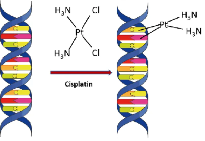
Figure 2 Cisplatin induced DNA adducts ... 6
Figure 3 Thymidine dimer induced by UV light... 8
Figure 4 UV-induced activation of S phase checkpoint ... 10
Figure 5 Drawing describe the mechanism of translesion DNA synthesis ... 12
Figure 6 Drawing describe the mechanism of UV adducts bypass by polη ... 14
Figure 7 Schematic drawing describes the sequential events of GG-NER pathway ... 16
Figure 8 Model of replication stress associated RPA exhaustion ... 18
Figure 9 Mechanism of UV-induced DNA mutations ... 24
Figure 10 Increased replication stress in polη-deficient cells ... 28
Figure 11 Clonogenic survival after UV irradiation in XPRO30 and Cl6 ... 35
Figure 12 DNA fiber assay post UV irradiation ... 36
Figure 13 Cell Cycle progression in XP30RO post UV ... 37
Figure 14 HPRT mutation assay in XP30RO post UV irradiation ... 39
Figure 16 Dual detection of RPA and γ-H2AX in U2OS cells post UV irradiation ... 47
Figure 17 Clonogenic survival post UV in U2OS-RPA and U2OS-GFP ... 49
Figure 18 Cell cycle progression in U2OS-RPA and the control U2OS-GFP... 52
Figure 19 HPRT mutation assay in U2OS post UV irradiation ... 54 Figure 20 Possible mechanism by which RPA rescue UV genotoxicity in Polη-deficient cells . 58
List of Tables
Introduction
Cell cycle and replication control
DNA constitutes the genetic material in all living organisms responsible for producing mRNA and its translation into proteins (Kültz 2005). The double-helical structure of DNA facilitates its interaction with a versatile replication machinery to faithfully copy the genetic material of each cell for accurate transmission to daughter cells during mitosis (Brown 2002).
The human genome contains about 3.2 billion bases that must to be copied accurately in a timely manner (Chatterjee and Walker 2017). For this reason, the cell has evolved a plethora of mechanisms to ensure faithful replication and transmission of the entire genome in the face of a multitude of challenges (Branzei and Foiani 2010).
The cell cycle
The cell cycle is a complex process that is divided into multiple stages: gap 1 (G1), synthesis (S) phase, gap 2 (G2) and mitosis (M) phase. During G1 phase the cell synthesizes mRNA and proteins necessary for DNA replication. Replication is initiated at specific sequences known as origins which are licensed in late M and early G1 (Nishitani and Lygerou 2002). The origin recognition complex (ORC) binds at replication origins and triggers the assembly of the pre-replication complex (pre-RC) (Bell and Stillman 1992). After ORC binding, Cdc6 is recruited which in turn recruits the licensing factor Cdt1. Cdt1 then triggers the recruitment and loading of MCM2-7 helicase on the DNA (Bell and Dutta 2002).
forming the replication fork which generates single-stranded DNA (ssDNA) that is bound by the heterotrimeric complex; replication protein A (RPA) (Nguyen et al. 2014), Pol-α then synthesize primer to initiate new DNA strands synthesis (Fig 1). Replication factor C (RFC) load proliferating cell nuclear antigen (PCNA) at the nascent DNA strand with the subsequent recruitment of processive DNA polymerases; Polδ and Polε. Replication fork moves bidirectionally until the replisome from nearby origins converge (Dewar, Budzowska, and Walter 2015). During replication fork progression, RPA binds the ssDNA formed ahead of the replicative polymerases to protect it from attack by endonucleases and to prevent the formation of secondary structures, thus allowing unperturbed and accurate copying of the genetic information (Nguyen et al. 2014). After completion of replication, the cell enters G2 whereupon it continues to grow, synthesize protein, and check the duplicated genome for errors. Finally, in M phase, sister chromatids are separated and distributed to the new daughter cells (Harashima, Dissmeyer, and Schnittger 2013).
The master regulator of the cell cycle, i.e., the cyclin-dependent kinases (CDKs), become active only when associated with cyclin (Hunt 1991). Cyclin-CDK complexes regulate hundreds of proteins involved in cell cycle progression through phosphorylation (Nasmyth 1996). While CDK levels remain constant during the different stages of the cell cycle, cyclin levels oscillate to allow activation of specific CDKs allowing cell to transit form one stage to another (Schafer 1998). The activity of cyclin-CDKs is regulated through phosphorylation; Wee1 protein kinase inhibits cyclin-CDK activity by phosphorylation at the roof of the kinase active site, while CDC25 activates cyclin-CDKs by dephosphorylation of these sites (Nilsson and Hoffmann 2000). This mechanism plays an important role in halting cell progression in case of DNA damage.
Fidelity of DNA replication
The replication process is carried out by high fidelity B family DNA polymerases (Pol δ and Pol ε) that has an error rate of only one misincorporated nucleotide per 105–107 nucleotides
polymerized (Bębenek and Ziuzia-Graczyk 2018). The accuracy of B family polymerases is guaranteed by their unique structure which forms a stable complex with the correct deoxyribonucleotides (dNTP) that facilitates the formation of the phosphodiester bond, while the binding of incorrect dNTPs destabilize the complex such that the wrong nucleotide get repelled outside the active site before incorporated into the DNA polymer (Doublié et al. 1998). B family DNA polymerases have also proofreading capabilities meaning that they check every nucleotide incorporated in the newly synthesized strand and if a mistake is made the nucleotide is removed by 3 → 5 exonuclease activity which further increases the fidelity of replication (Brutlag and Kornberg 1972).
Origins of replication control
Although the human genome contains between 30,000 and 50,000 origins, only a few thousand are ever activated simultaneously while the rest remain dormant and get activated during different stages of replication (CVETIC and WALTER 2005). Dormant origins play an essential role in completing genome duplication in case of replication perturbation, where they can fire to complete replication as in case of blocked forks (McIntosh and Blow 2012).
The control of the number of origins activated at the same time is critical for proper duplication of the genome, since uncontrolled origin firing may cause a shortage of factors needed for DNA synthesis and protection, i.e., dNTPs and RPA. Moreover, proper dNTP levels are crucial for the accuracy of DNA copying. Studies have shown that dNTP pool imbalance can cause nucleotide misincorporation by DNA polymerase which increases mutation rate(Kumar et al. 2011). The upregulation of dNTP production may also cause increased mutation through the involvement of the less accurate Y family DNA polymerases, since the latter have a higher Km for dNTPs than Pol δ and Pol ε (Lis et al. 2008).
The control of the replication program, the proper balance of replication factors, and the high fidelity of replicative polymerases ensure the maximum accuracy of genetic material transfer.
DNA replication stress
The replication process is a challenging task, during which replication forks meet various obstacles that could be endogenous, e.g. oncogene activation and conflicts with transcription machinery, or exogenous e.g., chemicals that intercalate DNA, UV radiation or chemotherapeutic agents (Faddeeva and Beliaeva 1991) (Zhao, Traganos, and Darzynkiewicz 2010). Replication obstacles cause forks to slow down or stall, a phenomenon known as replication stress (Zeman and Cimprich 2014). Replication stress has gained particular interest in the last few years as evidence are emerging that it promote genomic instability and carcinogenesis (Macheret and Halazonetis 2015).
I. Endogenous sources of replication stress Oncogene activation
Mutation in normal genes (proto-oncogenes) that control cell growth leads to oncogene activation which promote transformation of normal cells into cancer cells (Graziano and Gonzalo 2017). Oncogene activation, e.g., Myc, Cyclin E, Ras disrupt replication control by inducing cell cycle entry and origin hyper-activation (Rohban and Campaner 2015). The uncontrolled activation and re-licensing of origins lead to elevated replication stress as replication elements i.e. dNTPs and RPA become limiting and the incidence of conflict between replication and transcription machinery is increased (Jones et al. 2013) .
Transcription replication conflicts
Transcription-replication conflicts (TRCs) represent one of the primary sources of genomic instability and replication stress (Dutta et al. 2011). As DNA replication forks move bidirectionally while transcription proceeds only in one direction, two types of collision
fork stalling and higher replication stress than co-directional clash (Hamperl and Cimprich 2016). To prevent such a catastrophic event, DNA replication, and transcription are coordinated spatially and temporally, i.e., proceed in different areas of the genome at a unique time (Wei et al. 1998).
II. Exogenous sources Chemotherapeutic agents
Chemotherapeutic drugs like platinum compounds, e.g., cisplatin, and oxaliplatin crosslink DNA by forming platinum-DNA adducts (Huang and Li 2013). There are two types of DNA crosslinking; Interstrand crosslinks (ICLs) and Intrastrand crosslinks. ICLs occur between DNA bases on opposite strands and are considered highly toxic as they prevent strand separation and consequently block DNA replication and transcription resulting in cell death (Bignon et al. 2017). On the other hand , intrastrand crosslinks occur between bases on the same strand and are less toxic as other DNA damage pathway i.e. translesion DNA synthesis (TLS) can mitigate their genotoxic effect (Albertella et al. 2005).
UV radiation
Although solar radiation provides us with beneficial health outcomes e.g., providing a source of vitamin D, improved mood, and better immunity, it is also highly genotoxic (Elwood and Jopson 1997). The energy of the UV range that reaches the earth can induce chemical changes in the DNA which induce high level of replication stress and pose a severe risk to the integrity of the genetic information (Modenese et al. 2018).
UV physical properties
Sunlight is composed of three wavelength regions: UV, visible and infrared, among which the solar UV is responsible for skin aging and cancer (Modenese et al. 2018). The UV radiation is subdivided into ultraviolet A [UVA (315–400 nm)], ultraviolet B [UVB (280–315 nm)] and ultraviolet C [UVC (100–280 nm)](Modenese et al. 2018). The ozone layer of the atmosphere is a vital shield that prevents harmful solar radiations from reaching the surface of the earth. Ozone filters out UVC completely, while it filters 95% of UVB and 5% UVA. The International Agency for the Research on Cancer classifies both UVA and UVB as class I carcinogen (El Ghissassi et al. 2009). UVA radiation has a longer wavelength and can penetrate deeper into the skin which contributes to its role in skin aging. Recent studies suggest that UVA can induce the formation of DNA photoproducts and cause excessive DNA damage than thought before (Guerra and Crane 2018). UVB penetrate the epidermal layer of the skin and is the key cause of skin reddening and sunburn and contribute to photoaging. UVB is absorbed by the DNA thereby inducing DNA photoproducts (de Laat, van der Leun, and de Gruijl 1997).
UV-induced DNA damage
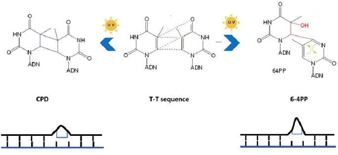
The UV energy can induce two distinctive types of cellular damage (i) oxidative damage to DNA bases (Meyskens, Farmer, and Fruehauf 2001) and (ii) DNA adducts formed when the energy absorbed by the DNA breaks the internal 5–6 double bond of pyrimidines, inducing the formation of a new bond between two adjacent pyrimidines on the same strand (Silvia Tornaletti and Pfeifer 1996). The two types of UV DNA adducts formed are; cyclobutane pyrimidine dimers (CPD) and 6-4 photoproducts (6-4PP) which introduce distortion in the double helix and represent a source of replication stress as it impede replication fork progression (S Tornaletti and Pfeifer 1996).
Cellular response to UV-induced DNA damage I. Activation of the ATR checkpoint
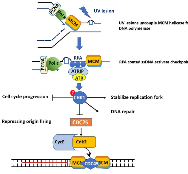
The replicative polymerases cannot accommodate UV adducts which results in complete blockage of the replicative polymerases (Vallerga et al. 2015). The MCM DNA helicase is uncoupled from the blocked DNA polymerase (Byun et al. 2005) and continues to unwind the DNA creating vast stretches of ssDNA that are bound by RPA which in turn triggers activation of ataxia telangiectasia and rad3 related (ATR) kinase (Zou and Elledge 2003). ATR then goes on to phosphorylate multiple downstream substrates including CHK1 kinase (Liu et al. 2000) that reduce replication stress and facilitate DNA synthesis restart by (i) inhibiting late origin firing, (ii) stabilizing stalled replication forks iii) activation of DNA repair (Fig.4) (Feijoo et al. 2001).
ATR regulates cell cycle progression in response to DNA damage, through Chk1 which phosphorylates and inactivates the phosphatase Cdc25, results in Cdk2 inactivation and cell cycle arrest (Peng et al. 1997). ATR also phosphorylates H2AX (γH2AX) at blocked forks which enforces the assembly of repair factors at the damaged site (Ward and Chen 2001). The outcome of checkpoint activation is slowing down of replication and cell cycle arrest to ensure the repair of the genome before further progression in the cell cycle.
II. Activation of translesion DNA synthesis
Another major pathway for dealing with UV-induced lesions is the DNA damage tolerance pathway that is carried out by Y family DNA polymerases (Polη, Pol ι, Pol κ , and Rev1) (Sale 2013). Y family DNA polymerases are translesion (TLS) polymerases, and each of them works on specific DNA lesions (Makridakis and Reichardt 2012). Rev1 interact with other TLS pols through Rev1 interacting region (RIR) and function as a scaffold that direct TLS polymerases to the site of DNA damage (Guo et al. 2003). The fidelity of Y family polymerase on undamaged
DNA is generally low than replicative polymerases (Table 1), while they show variable accuracy when performing TLS depending on the type of DNA lesions (Makridakis and Reichardt 2012). While Pol κ support accurate bypass of bulky DNA adducts like benzopyrene-N2-dGuanine adducts (BP-dG) (Suzuki et al. 2002) , in vitro and in vivo studies have shown that it cannot mediate TLS across UV lesions (Ohashi et al. 2000). On the other hand, Pol ι can bypass a wide variety of DNA lesions, such as pyrimidine dimers, and oxidative damage adducts. While Pol ι support UV lesion bypass, it produce errors by incorporating the wrong base more frequently than the correct one (Y. Wang et al. 2007). Polη is the only Y family DNA polymerase which can bypass UV lesions with high accuracy (Stary et al. 2003).
The ability of TLS polymerases to bypass DNA adducts is attributed to their unique structural features (i) lack of proofreading capabilities (Goodman and Woodgate 2013), (ii) accommodation of modified base on the damaged strand to direct nucleotide incorporation (Sale, Lehmann, and Woodgate 2012). These features allow TLS pols to perform lesion bypass efficiently but with an increased risk of mutation (Waters et al. 2009).
Mechanism of translesion DNA synthesis
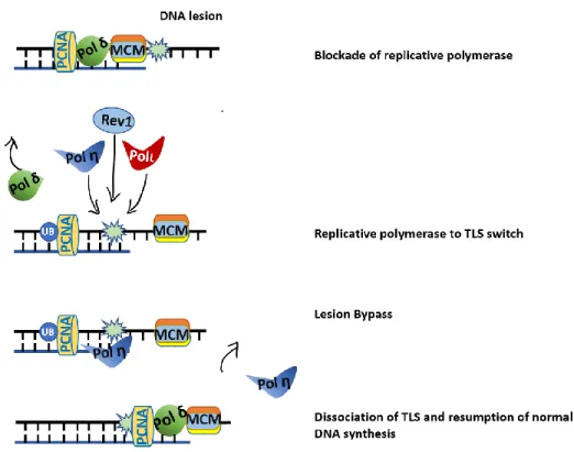
PCNA act as a processivity factor for DNA polymerase δ, moreover, it is a scaffold for the recruitment of other protein involved in DNA repair (Moldovan, Pfander, and Jentsch 2007). TLS polymerase Polι and Rev1 have ubiquitin binding motifs (UBM) , and Polη and Polκ have ubiquitin-binding zinc finger motifs (UBZ) which can interact with PCNA through PCNA-binding motifs (PIP-box) (Pustovalova et al. 2016). When replicative polymerases are stalled by UV adducts, the ssDNA formed activates the E3 ubiquitin ligase Rad18 which forms a complex with Rad6 that in turn mediates monoubiquitination of PCNA (Kannouche, Wing, and Lehmann 2004). Monoubiquitinated PCNA has increased affinity for TLS polymerases mediating the switch from replicative to TLS polymerases (Fig.7). The nature of the DNA lesion determine which TLS polymerase will replace the replicative polymerase. After the TLS polymerases perform lesion bypass, they dissociate from the DNA.
Polη accurate bypass of UV lesions
The Y family TLS polymerase Polη exerts an important role in protecting the cell against UV genotoxicity. Polη can replicate past different types of DNA lesions including UV and cisplatin adducts. Although Polη has a low fidelity on undamaged DNA (error frequency of 10−2 to 10−3)
compared to replicative polymerases, it can accurately bypass CPD adducts (Johnson et al. 2000). The accuracy of Polη-mediated TLS is attributed to the active site pocket which can perfectly accommodates the thymine dimer then insert two As opposite this lesion (Biertümpfel et al. 2010) (Washington et al. 2001).
The translesion of CPD requires the activity of Polη alone, which it inserts nucleotides opposite the dimer and then extends it (Fig.8) (Quinet et al. 2016). On the other hand, the bypass mechanism of 6-4PP requires the cooperation of more than one TLS polymerase, where Polη inserts the correct nucleotide opposite the lesion, then Rev3L the catalytic subunit of pol zeta extend and fill the gap (Quinet et al. 2016).
it has been shown that Polη is overexpressed in cancer stem cells which cause chemoresistance in those patients and failure in therapy, as Polη support the TLS activity across cisplatin adducts (Srivastava et al. 2015).
Figure 6 Drawing describe the mechanism of UV adducts bypass by polη III. NER-dependent photolesion removal
UV adducts constitute a risk to genome integrity and a source of replication stress which can be detect and removed by nucleotide excision repair (NER) in error-free manner. CPD constitute 75% of the UV lesion burden and cause only slight distortion in the DNA helix; therefore, it is detected and repaired slowly; 60% of lesion repair in 24 hours (S Tornaletti and Pfeifer 1996). On the other hand, 6-4PP constitute only 25% of the lesions and cause a much more profound distortion in the DNA helix(PERDIZ 2000), allowing much faster detection and repair; 90 % repaired within 6 hours (Bohr et al. 1985).
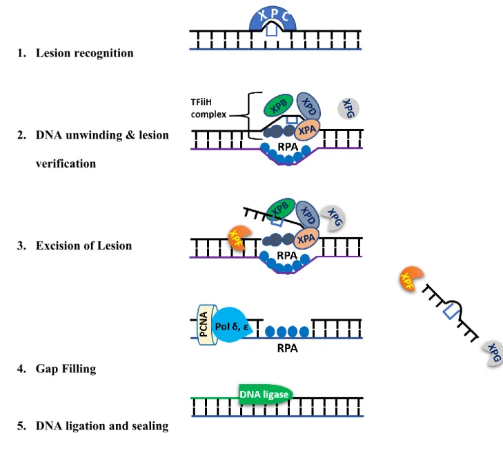
NER constitutes a robust repair mechanism that can identify and repair a plethora of different helix-distorting DNA lesions such as CPD and 6-4PP, and also represents the only pathway in humans for the error-free removal of such lesions (Spivak 2015). NER consist of two subpathways; Global genomic NER (GG-NER) which removes UV lesions from the entire genome and transcriptional coupled NER (TC-NER) which eliminates photolesions from the transcribed strand of active genes (Hanawalt 2002). NER works by sequential recruitment of repair factors to the
damage site. Its steps include lesion recognition, DNA unwinding, lesion verification, lesion excision and finally restoration of the original DNA sequence (Fig.6). GG-NER and TC-NER are different only in the lesion recognition step. In the case of GG-NER, helix distortion triggers the recruitment of the DNA damage sensor XPC protein to lesion sites (Fitch et al. 2003). XPC can identify a different range of structurally unrelated DNA adducts (CPD, 6 -4 PP, cisplatin adducts).
On the other hand, the blockage of the RNA polymerase II triggers the activation of the TC-NER pathway (Schärer 2013). Following lesion recognition in the case ot either GGNER or TCNER, transcription initiation factor IIH (TFIIH) complex is recruited to the damages site. TFIIH includes two DNA helicase subunits (XPB and XPD helicases) that unwind the DNA helix around the damage site creating a 20 to 30-nucleotide bubble which is stabilized by XPA and RPA(Volker et al. 2001). The dual incision on both sites of the lesion is carried out by XPG and ERCC1-XPF nucleases creating a gap of ≈ 30 bp which is filled in by normal DNA replication factors using the intact nondamaged strand as template (Ogi et al. 2010). The final step is a ligation to seal the DNA backbone (Moser et al. 2007).
Figure 7 Schematic drawing describes the sequential events of GG-NER pathway 1. Lesion recognition
2. DNA unwinding & lesion verification
3. Excision of Lesion
4. Gap Filling
Consequences of inadequate replication stress response
Recent studies have revealed that checkpoint-defective mutants when treated with hydroxyurea (HU) which induce replication stress through the inactivation of ribonucleotide reductase (RNR) leading to depletion of dNTPs levels, manifest large stretches of ssDNA at stalled forks (Sogo, Lopes, and Foiani 2002). Further studies have highlighted the role of ssDNA formed during replication stress in exhaustion of the RPA pool which in turn make it unavailable to protect the ssDNA and result in their breakage (L. I. Toledo et al. 2013).
While the cell depends on the ATR checkpoint and polη TLS to relive the UV-induced replication stress, any deficit in either pathway cause high levels of replication stress through different mechanisms that allow completion of genome duplication i.e. firing of dormant origins or by lesion skipping mechanism. Such events cause accumulation of ssDNA that lead reduced availability of RPA making ssDNA prone to breakage with subsequent cell death.
Exhaustion of RPA pool
RPA is the major ssDNA-binding protein and constitute one of the limiting factors which participate in DNA metabolism in eukaryotic cells (Lavrik et al. 2016). Beside participating in DNA replication, RPA interacts with multiple proteins involved in different DNA repair processes like NER, MMR (Li 2008), BER and DSBs repair (Eggler, Inman, and Cox 2002).
A recent study by Toledo et al. showed that during replication stress, ATR activity prevents the exhaustion of RPA by inhibiting late origin firing (L. I. Toledo et al. 2013). The activity of ATR
increasing origin firing by inhibiting ATR activity during replication stress results in sustained RPA levels on chromatin, which was associated with increased fork collapse (Fig.8). Moreover, when they provided an excess of RPA to the cell, the threshold at which forks break was increased.
The RPA exhaustion model in ATR inhibited cell agrees with previous finding of our lab where we highlighted the role of ATR in NER in S phase (hereafter S phase NER is referred as SPR). Inhibition of ATR activity by either caffeine treatment or si-RNA knockdown leads to a striking defect in NER exclusively during S (Y. Auclair et al. 2008). As RPA participate in NER pathway, the inhibition of ATR would result in RPA being sequestrated at ssDNA regions and become unavailable to carry out its role in NER which result in defective SPR.
DNA damage and repair
The accuracy of transferring the genetic information does not depend only on high fidelity DNA polymerases and control of the replication process; indeed it also depends on the ability of the cell to accurately repair DNA damage (Chatterjee and Walker 2017). Every day the human cell is exposed to more than 50,000 DNA damage events, either intrinsic e.g., oxygen free radical damage from oxidative respiration, or extrinsic e.g., UV radiation, and ionizing radiation (IR) (Maynard et al. 2015), which can introduce conformational changes in DNA and therefore in Watson-crick base pairing (Guengerich 2006). Human cells have evolved different mechanisms that can detect specific types of DNA damage and elicit the proper repair pathway to restore the DNA to its original state.
I. Intrinsic DNA damage Mismatched base
Even though replicative polymerases possess high fidelity properties, they can make mistakes by introducing a mismatched base. If such errors are not corrected, they will become fixed as mutations after the next round of DNA replication (Loeb and Monnat 2008). The mismatch repair (MMR) pathway can effectively detect and repair mismatched bases, which increases the fidelity of replication 50-1000 fold (McHugh, Sones, and Hartley 2000). The MMR pathway has a vital role in reducing mutations and hence protecting against carcinogenesis since defects in the MMR pathway are associated with elevated incidence of colon cancer;70% of the cases of hereditary non-polyposis colon cancer (HNPCC) are attributed to mutation in MMR pathway genes
Oxidative DNA damage
Mitochondrial respiration produces reactive oxygen species (ROS), e.g., superoxide radicals (•O−
2), hydrogen peroxide (H2O2), and the hydroxyl radical (•OH), (Tropp 2012). ROS can damage
DNA by forming an oxidative base, e.g., oxidation of guanine into oxoguanine (oxoG). 8-oxoG can cause mutation in the DNA as it pairs with A instead of C resulting in G:C to T:A transversions (Aguiar et al. 2013). The base excision repair (BER) pathway can effectively detect and repair the oxidized bases (Demple and Harrison 1994). BER steps include detecting the damaged base by DNA glycosylases which subsequently cleave the bond between the oxidized base and the deoxyribose sugar which results in an abasic site (AP-site)(Hegde, Hazra, and Mitra 2010). The AP-endonuclease create a nick in the phosphodiester backbone of the AP site followed by base excision and release, then DNA polymerase fills the gap which is later sealed by a ligase (Wallace 2014).
II. Extrinsic DNA damage Ionizing radiation
Ionizing radiation (IR) is abundant in the environment, and can damage DNA through two different mechanisms; (i) indirectly by production of free radicals (WARDMAN 2009), and (ii) directly through the formation of DNA double-strand breaks (DSBs) (Hutchinson 1985). DSBs are detected and repaired by either homologous recombination (HR) or non-homologous end-joining (NHEJ) pathway (Jackson 2001). NHEJ is an error-prone mechanism as it catalyzes direct religation between non-allelic broken DNA ends (Davis and Chen 2013). On the other hand, HR is error-free as it uses the information in the intact homologous chromosome to repair the DSB. The first step in HR is the resection of the DNA DSB in the 5‘ - 3’ direction by nucleases with the
association of Rad50, Mrell and Xrs2 complex (Della-Maria et al. 2011). Rad51 then binds to the 3’ single-stranded DNA tails forming a nucleoprotein filament that searches for homologous sequences on the intact chromosome (Baumann and West 1998). Once the target sequence is found, the damaged strand invades the intact DNA in a strand exchange reaction. The 3’ terminus of the damaged molecule is then extended by a DNA polymerase that copies information from the undamaged strand.
DNA repair deficiency and cancer
Certain diseases like xeroderma pigmentosum and Fanconi anemia are defective in DNA repair (Knoch et al. 2012), which results in a hypermutable phenotype leading to mutations in oncogenes and tumor suppressor genes that in turn promote cancer development (Sale, Lehmann, and Woodgate 2012). The consequence is uncontrolled growth of cells and the formation of a malignant tumor, which in most cases requires surgical removal followed by chemotherapy to eradicate the remaining cancerous cells. The outcome of cancer development and chemotherapy is a reduced quality of life, high health care expenses, and high mortality rate.
Skin cancer
Skin is the largest organ in the human body providing us with vital functions like sensation, temperature regulation, and protection against the external environment. The number of patients diagnosed with skin cancer increases every year in the world reaching more than a million persons annually (D’Orazio et al. 2013). The depletion of the ozone layer has been of great concern as it contributes to more accumulation of UV radiation that reaches the earth (Slaper et al. 1996). The risk factors for developing skin cancer are fair skin, long sun exposure, older ages and heredity disorders, i.e., Xeroderma pigmentosum (XP).
Skin cancer is divided into melanoma and non-melanoma skin cancer (NMSC). Basal cell carcinoma (BCC) affects the epidermis of the skin and constitutes the majority of NMSC ≈75% while squamous cell carcinoma (SCC) represents the rest of cases. Melanoma is cancer of the melanocyte, i.e. the pigment-producing cells, and characterized by resistance to chemotherapy, aggressive metastasis to other organs and dismal prognosis if not diagnosed very early (Marks 2000).
Mechanism of UV induced mutagenesis
Failure in NER dependent repair of photolesions can cause replication fork stalling that will eventually collapse into DSB at the damaged site and result in cell death (Elvers et al. 2011). However, cells can use one of the TLS polymerases to restart DNA synthesis at blocked forks and prevent fork collapse (Vaisman and Woodgate 2017). While TLS polymerase effectively bypass DNA lesion, they retain high errors and could introduce mutations to the cellular genome at a high frequency. Among all the TLS polymerase, polη suppresses the induction of mutations efficiently after UV irradiation by performing an essentially error-free bypass of T-T dimers (Yoon, Prakash, and Prakash 2009). In the absence of polη, the involvement of polι in lesion bypass is the primary cause of mutation induction as it make errors much more frequently during UV lesion bypass (Fig9) (Y. Wang et al. 2007).
Another mechanism of mutation caused by UV is deamination of DNA bases (Barak, Cohen-Fix, and Livneh 1995). In contrast to the intact cytosine base, the cytosine in the CPD is less stable and can be converted to uracil through deamination. Polη then mediates error-free bypass of the T-U dimer by adding A opposite to U, which will lead to C → T transition (Choi et al. 2006). The induction of UV mutations depends on a balance between the relative proficiency of NER and polη-mediated TLS.
Xeroderma pigmentosum (XP) disease
Xeroderma pigmentosum (XP) is a rare autosomal recessive disorder affecting areas exposed to the sun, i.e., the skin and the eyes. It was first defined in 1870 as "xeroderma or parchment skin" by Moriz Kaposi (Kenneth H. Kraemer, Lee, and Scotto 1987). In 1882, the term “Xeroderma pigmentosum” was coined, since the massive change in pigmentation pattern is one of the striking characteristics of the disease. XP patients have a varying degree of photosensitivity and skin cancer predisposition, ranging from slight to severe (Alan R Lehmann, McGibbon, and Stefanini 2011). In addition to skin cancer 25% of XP patients develop neurological symptoms, e.g., mental retardation and impaired skeletal reflexes (Anttinen et al. 2008).
Understanding the genetic basis of Xeroderma pigmentosum (XP) patients has highlighted the role of NER in preventing skin cancer development. The first scientist to report a DNA repair defect in XP patients was James cleaver in 1968 (Cleaver 1968). Cleaver showed that XP cells are deficient in UV damage repair (Cleaver 1969). It was subsequently revealed that XP can result from mutations in any among seven NER genes (XP-A, XP-B, XP-C, XP-D, XP-E, XP-F, and XP-G) which cause sensitivity to sunlight and at 1000 to 10,000-fold increase in skin cancer (K H Kraemer et al. 1994) (Bradford et al. 2011). On the other hand, xeroderma pigmentosum variant (XPV) patients have mutation in DNA polη and show similar UV sensitivity and skin cancer susceptibility compared with XP patients (DiGiovanna and Kraemer 2012).
The study of the effect of UV radiation on polη-deficient cell line, has been an active area of research in the past few years to reveal the molecular mechanism of skin cancer development in XPV patients. Studies have shown that XPV-derived cells are associated with the following
UV-(Stary et al. 2003) (Alan R Lehmann, McGibbon, and Stefanini 2011). However, the exact molecular mechanism by which XPV-derived cells manifest such phenotypes still unclear.
UV-induced genotoxicity in XPV patient-derived cells I. UV sensitivity
XPV patient-derived cells lacking polη shows high UV sensitivity due to replication fork stalling, which results in the formation of ssDNA regions along the entire genome that are prone to breakage (Meneghini 1976). When XPV cells are complemented with a plasmid carrying polη the sensitivity to UV was restored to the level of the normal fibroblast, affirming that the sensitivity is attributed to a deficiency of polη (Kaufmann et al. 2003). The sensitivity to UV is dramatically increased in the presence of caffeine which was attributed to the post-replication repair defect caused by ATR attenuation (Stary et al. 2003). ATR also plays an important role in reducing replication stress during DNA damage which further stabilize replication forks and prevents their collapse. caffeine treatment has no effect on polη-complemented cells, which indicates that lesion bypass by polη has a major rule in reducing replication stress when the ATR checkpoint is compromised.
II. Increased mutation rate
The high induction of UV mutation in polη-deficient cells is attributed to the involvement of the more error-prone polymerases, i.e., Pol ι in lesion bypass (Y. Wang et al. 2007). In vitro mutation assay has demonstrated that Pol ι is capable of replicating across T-T dimer by misinserting G or T more frequently than inserting the correct base (Tissier et al. 2000). Such error during replication generates T → A transversions which is characteristic of XPV cells.
III. Elevated replication stress
Previous studies have demonstrated that cells lacking polη show profound cell cycle delay compared to wild type cells (Imray et al. 1983). Kannouche et.al have revealed that the accumulation of cells in S phase is due to replication fork stalling and the strong activation of ATR checkpoint which reduce the number of active forks (Despras et al. 2010).
IV. Defective NER only in S phase
Previous studies suggested a normal level of NER in XPV cells. These studies used techniques that determine the NER capacity regardless of the cell cycle stage (A R Lehmann et al. 1975). Our lab has taken a further step towards studying excision repair as a function of the cell cycle using a flow cytometry assay (Rouget et al. 2008). For the first time, Dr Drobetsky’s group demonstrated that UV-irradiated XPV cells exhibit a profound defect in NER exclusively during S phase. (Yannick Auclair et al. 2010). This SPR defect is attributed to loss of polη activity, as ectopic expression of wild-type polη restores NER proficiency. SPR defect rescue by polη expression depends on nuclear localization and interaction with PCNA since the ectopic expression of a polη mutant defective in either motif does not recover SPR (Yannick Auclair et al. 2010).
The SPR defect is only manifested when the cell is actively replicating the genome, since inhibition of replication by HU rescues defective SPR. The treatment of cells with HU before UV irradiation prevents replicative polymerases to collide with UV adducts which may explain that blocked forks in XPV cell contribute to SPR defect (Alvino et al. 2007). Our lab and others have proposed RPA exhaustion at stalled forks as a cause of such defect (Tsaalbi-Shtylik et al. 2014)(Yannick Auclair
Rational
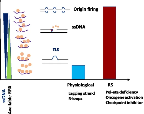
UV radiation cause DNA damage by inducing the formation of UV adducts i.e. 6-4PP and CPD, these lesions impede the progression of the replicative DNA polymerase while the DNA helicase continue to unwind the double helix creating stretches of ssDNA. The ssDNA is bound by RPA which result in the activation of two important pathways; the ATR checkpoint and the TLS by polη. ATR limit ssDNA formation to active replicon, stabilize stalled replication fork and enhance DNA repair by targeting multiple substrates including RPA, MCMs, and others (Yazinski and Zou 2016). Polη overcome replication block by replicating past UV lesions with high accuracy which help the cell to continue DNA synthesis and fill the ssDNA gaps without risk to the integrity of the genetic information (Alan R. Lehmann 2005). In the absence of polη ssDNA gaps accumulate and not efficiently filled, causing high level of replication stress and genomic instability (Fig 10).
Figure 10 Increased replication stress in polη-deficient cells
A model shows the accumulation of ssDNA in polη-deficient cells after UV irradiation, which negatively affect RPA dependent DNA repair and replication processes.
Hypothesis and specific aims
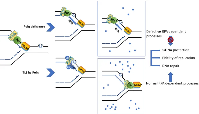
We hypothesize that in the absence of Polη, photolesions cause a high level of replication stress due to persistent fork blockage along with the generation of ssDNA which negatively affects RPA availability. Since RPA plays multiple roles in DNA replication, checkpoint activation, and DNA repair, such elevated replication stress causes exhaustion of RPA pool which compromise RPA dependent process, i.e., SPR, cell cycle progression, and UV sensitivity. We expect that the aforementioned defects can be resolved by overexpression of RPA.
Specific aims
1) To study RPA dynamics on chromatin in Polη-deficient cells upon UV exposure 2) Stably overexpress RPA in XPV or Polη-deficient U2OS cells
3) Re-evaluate the UV-induced genotoxicity in the presence of surplus RPA 4) Underpin the mechanism by which RPA could resolve UV genotoxicity
Materials and methods
Cell culture
The SV40-transformed XPV-skin fibroblast strain XP30RO, and its isogenic derivative ectopically expressing wild-type polη (XP30RO-polη/cl6), were kindly provided by Dr. A.R. Lehmann (University of Sussex). XP30RO cells were cultured in MEM medium (Wisent) supplemented with 15% FBS, L-glutamine, vitamins, and antibiotics (Thermo Fisher). U2OS osteosarcoma cells were purchased from ATCC and cultured in DMEM (Corning) supplemented with 10% FBS, L-glutamine, and antibiotics. U2OS cell lines were authenticated in 2018 using STR profiling by the McGill University Genome Center (Montreal, Canada). All cell lines were used within 25 passages after thawing.
Clonogenic survival
Cells were seeded on 100 mm dishes and treated with the indicated doses of UVC then incubated with either regular medium or medium containing 75 μg/ml caffeine for two weeks. Medium was removed, and colonies were fixed by staining with 50% v/v methanol + 0.5% w/v methylene blue. Colonies containing a minimum of 50 cells were counted.
Ectopic RPA expression and siRNA treatment.
The RPA expression plasmid (pAC-GFPRPA) is a generous gift of Dr. J. Lukas (University of Copenhagen). The plasmid expresses the three RPA subunits from the same promoter in stoichiometric manner where the three subunits are spaced by self-cleaving peptide PA2 (L. I. Toledo et al. 2013). The RPA-3 subunit is GFP tagged.
The RPA plasmid was transfected using Lipofectamine 2000 (ThermoFisher). Stable clones were sorted by FACS in 96-well plates and selected with 500 g/ml Geneticin (ThermoFisher). siRNA
smartpools for knockdown of polη were purchased from Dharmacon and have the following sequence: 5′–CAUUGAUGAGGCUUACGUA–3′ 5′–CAUAGAGAGGGAGACUGGU–3′ 5′–GAAUAAACCUUGUGCAGUU–3′ 5′–CCAAAUGCCCAUUCGCAAA–3′
The si-RNA were transfected using RNAiMax (ThermoFisher). Pools of non-targeting (NT) duplexes were used as controls.
Immunoblotting.
Whole cell extracts were fractionated by SDS-PAGE and transferred to a polyvinylidene difluoride membrane using a transfer apparatus according to the manufacturer’s protocols (Bio-Rad). After incubation with 5% BSA in TBST (10 mM Tris, pH 8.0, 150 mM NaCl, 0.5% Tween 20) for 60 min, the membrane was washed once with TBST and incubated with antibodies against anti-RPA2 (Calbiochem; NA18), rat anti- actin (Abcam; ab6161), rabbit anti-RPA1 (Abcam; ab79398), mouse anti-RPA3 (Abcam; ab6432), anti-γ-H2AX Ser139 (Millipore), and rabbit POL H Antibody (H-300) (Santa-Cruz, sc-5592) at 4 °C overnight. Membranes were washed three times for 10 min and incubated with a 1:1000 dilution of horseradish peroxidase-conjugated mouse or anti-rabbit antibodies for 3 h. Blots were washed with TBST three times and developed with the ECL system (Amersham Biosciences) according to the manufacturer’s protocols.
Dual detection of γ-H2AX and RPA following UV treatment by flow cytometry.
incubated with 50 μl of BD Perm/Wash buffer containing primary antibodies; anti-RPA1 (Abcam, ab79398) (1:100) and anti-γ-H2AX Ser139 (Millipore) (1:100) for one hour, followed by washing with BD Perm/Wash buffer. Cells were then incubated with 50 μl of BD Perm/Wash buffer containing 2ry antibodies; goat anti mouse Alexa 488, and goat anti rabbit Alexa Fluor 647 (ThermoFisher) (1;200) for 30 minutes at RT followed by washing. Cells were resuspended in analysis buffer ( 0.02% (w/v) sodium azide, 250 μg/ml RNase, 0.5 μg/ml DAPI in PBS-B) and stored at 4 C overnight then analyzed on LSR II flowcytometry machine (Forment and Jackson 2015).
DNA Fiber Analysis.
DNA fiber assays were performed as described (Ray Chaudhuri et al. 2016). Cells were labeled for 20 min with 5 μM IdU, washed, UV-irradiated with 20 J/m2, then labeled for 60 min with 25
μM CldU. Cells were trypsinzed, then washed with PBS and resuspend in PBS at concentration of (500 cells per ul). 2 μl of the cell suspension was lysed using 7 μl of lysis buffer (200 mM Tris– HCl, pH 7.5, 50 mM EDTA, 0.5% SDS) on microscopic slide. Slides were fixed by immersing in a freshly prepared 3:1 mix of methanol and acetic acid. Slides were Blocked by pipetting 200 μl 5% BSA in PBS on top of each slide. Each slide was incubated for two hours with 60 μl of the primary antibody solution; ab6326 anti-BrdU (cross-reacts with CldU) antibody (rat) 1:400 and BD Biosciences 347580 anti-BrdU (cross reacts with ldU) antibody (mouse) 1:25 in 5% BSA in PBS. Slides were washed with 1X PBS + 0.05% tween three times then incubated for one hour with 60 μl of the secondary antibody solution; goat anti-rat AIexa-594 1:100 and goat anti-mouse Alexa—488 1:100 in 5% BSA in PBS followed by washing as pervious and covering with coverslip. Imaging was performed using a DeltaVision Elite system (GE Healthcare) in conjunction with FIJI software (NIH). A minimum of 100 fibers was counted for each experiment.
HPRT mutation assay
U2OS cells were grown for 5 days in HAT media (Thermo Fischer #21060017) containing hypoxanthine, aminopterin, and thymidine to eliminate background HPRT mutations. Cells were grown in HT media for 1 day to recover (ThermoFisher #11067030). Cells were stably depleted of Polη with siRNA targets versus a non-silencing control. 2 days post-transfection cell were seeded at 2x10⁶ (14 million per cell line) then either irradiated with 2 J/m2 UVC or mock-irradiated.
Post-irradiation cells were allowed to recover to 6x10⁶ cells or with mock cells 6 population
doublings. For HPRT phenotypic expression, cells grown for a total of 11 days where they were sub-cultured every 2 days at a confluence of 2x10⁶/10 cm dish. To select for HPRT-inactivated
colonies, a million cells were seeded at 50*103 density in media containing 5 µg/ml 6-Thioguanine
(6-TG), at the same time 200 cells were also seeded in 6-TG-free media to determine colony-forming efficiency.
The frequency of inactivating mutations at the HPRT locus was calculated as the MF = a/ (cells seeded × [b]).
a = total number of 6-TG resistant colonies and b = plating efficiency (counted/plated)
Results
Validation of the experimental model system XP30ROsv cell sensitivity to UV radiation
As a model for cells deficient in Polη, we used XP30ROsv immortalized XPV fibroblasts and the isogenic counterpart complemented with Polη (cl6). The absence of Polη in XP30ROsv and the expression in cl6 was confirmed by western blot (Fig.11 A).
We have confirmed previous reports that indicate a higher sensitivity of Polη-deficient cells to UV irradiation (10.66% and 58% survival at 2J/m2 and 0.86% and 20% J/m2 at 5 in XP30ROsv
and cl6 respectively Fig.11 B) (Pope-Varsalona et al. 2014).
To test for the effect of caffeine on survival, we used a concentration of caffeine (75 µg/ml) that does not affect the colony-forming ability of XP30RO cells. We noticed a dramatic increase of UV sensitivity by the addition of caffeine to the media in XP30RO (0.47% and 0.04% at 2 and 5 J/m2 respectively), while in the complemented cl6 the sensitivity is not affected by caffeine
addition (Fig.11 B). Caffeine sensitivity is consistent with attenuation of ATR checkpoint that results in aggravating the replication stress already present in Polη-deficient cells.
0% 0% 1% 10% 100%
0
2
4
6
Sur
viv
al
UV dose
Cl6
Cl6-CAF
XPV
XPV-CAF
XP30RO+ Polη(cl6) XP30RO
Figure 11 Clonogenic survival after UV irradiation in XPRO30 and Cl6
A) Western blot for detection of PolH expression in XP30RO and the isogenic cell A
The absence of Polη TLS activity in XP30RO does not alter Replication fork progression
To study the role of Polη in replication fork progression after UV, we used DNA fiber assay which monitors replication fork dynamics through the incorporation of thymidine analogs into newly synthesized DNA (Quinet et al. 2017). As expected, we noticed a slower fork progression after UV in both cell lines (Fig.12) (ratio is≈2). We see no significant difference in fork speed between XP30RO and the complemented Cl6 which is consistent with recent reports that show the involvement of Primpol polymerase in restarting DNA synthesis by repriming past the lesion in UV-irradiated XPV cell line (Kobayashi et al. 2016). Re-priming behind the lesion allows DNA replication to continue but leaves a gap of ssDNA which require protection by RPA against endonucleases.
Figure 12 DNA fiber assay post UV irradiation
Replication fork progression after irradiation with 20 J/m2 UV. Cells were firstly labeled with 10 mM IdU for 20 minutes followed by washing then irradiation with 20 J/m2 UV and incubated with 250 mM CldU for 60 minutes. Cells were lysed then fixed on slide followed by immunostaining. results are blotted as CldU/IdU ratio. The graph was generated using GraphPad Prism 8.0. Statistics used mann whitney with multiple comparisons test (*P ≤ 0.05, **P ≤ 0.01, ***P ≤ 0.001, and P ≥0.05 ns).
XP30RO cell exhibit cycle delay Upon UVC radiation
Previous studies have shown that Polη-deficient cells exhibit a profound delay in cell cycle progression post-UV irradiation (Stary et al. 2003). To study the cells ability to progress through cell cycle we irradiated XP30RO with 2 j/m2 then incubated with media for 24 hours. We noticed
that UV-irradiated XP30RO cells accumulate in S phase while the isogenic cells complemented with Polη show standard cell cycle profile (Fig.13). This data is consistent with the generation of ssDNA upon fork collision with UV adducts which strongly activate the checkpoint leading to cell cycle arrest. However, in Cl6, Polη mediated bypass results in reduction of ssDNA and consequently the shutdown of the checkpoint and resumption of normal cell cycle progression. This result also demonstrates that Polη can effectively bypass UV lesions while the other TLS polymerase i.e. Polι and Polκ are less efficient.
UV
NO UV
DNA content (DAPI)
Count
XP30RO+Polη
XP30RO
(Cl6)
Figure 13 Cell Cycle progression in XP30RO post UV G1 S G2 G1 S G2
XP30RO cells exhibit an increased UV mutation rate
Previous studies have investigated the role of Polη in the error-free bypass of UV lesions (Y. C. Wang, Maher, and McCormick 1991). We sought to reproduce these results, using the HPRT forward mutation assay which has been a standard tool to look at UV mutagenesis in multiple cell lines. XP30RO and XP30RO/Cl6 cells were exposed to 2 J/cm2 of UVC light, then allowed to
grow in regular media for 11 days for mutant phenotypic expression. After this period, one total million cells were seeded at 50000 per 10 cm dish in 6-TG media to select for HPRT mutants. Our data show a statistically significant increase in HPRT mutation frequency post UV in XP30RO cells compared to mock treated controls (Fig.14). We also observed that the UV mutation frequency for XP30RO cell was higher than XP30RO/Cl6 (41.3 × 10⁻⁵ and 4.84 × 10⁻⁵ for XP30RO
and XP30RO/Cl6 cells, respectively; Fig. 14). The observed mutation frequencies after UV exposure agrees with previous UV mutagenesis studies in XPV cells (Y. Wang et al. 2007). These data also demonstrate that UV can induce mutagenesis but at a relatively low level when polη is present, and deficiency of polη greatly aggravates this phenotype.
Figure 14 HPRT mutation assay in XP30RO post UV irradiation
Mutation frequencies were calculated as described by Bassett et al.; mutation frequency = (number of colonies (number of cells plated× CFE). Mutation frequencies were calculated for each separate experiment, with a minimum of two independent experiments for each treatment. The averages of multiple trials are shown on the graph ± SD of mean. The graph was generated using GraphPad prism using unpaired t test Statistics (*P ≤ 0.05, **P ≤ 0.01, ***P ≤ 0.001, and P ≥0.05 ns).
2.62 n=2 1.82, 4.84 n=2 n=2 41.3 n=2
Polη-deficient cells manifest a high level of RPA loading on chromatin and induction of γ-H2AX post-UV.
As the ATR checkpoint plays an important role in reducing replication stress-associated RPA exhaustion by limiting the formation of ssDNA to active replicons, one would expect that the absence of Polη lesion bypass activity leading to the generation of ssDNA would also cause RPA recruitment to chromatin; moreover if such ssDNA regions are not resolved they will be converted into DSB (L. Toledo, Neelsen, and Lukas 2017).
To get a clear vision about the dynamics of RPA loading on chromatin along with induction of γ-H2AX in Polη-deficient upon UV irradiation, we used a flow cytometry-based assay that allowed us to simultaneously look at protein association with chromatin and γ-H2AX formation as a function of cell cycle (Forment and Jackson 2015). We found that XP30RO cells exhibit a high level of RPA loading on chromatin at 24 hours post-irradiation, which was further increased by addition of caffeine to the media, whereas wild-type cells show no difference compared with non-irradiated cells at these time points (Fig 15 A and B). We also noticed a significant induction of γ-H2AX which was magnified by caffeine treatment only in XP30RO cells (Fig 15 C and D). The induction of γ-H2AX is consistent with the conversion of ssDNA into DSBs. The relationship between RPA loading on chromatin and the induction γ-H2AX is obvious as only cells that have high levels of RPA on chromatin showed induction γ-H2AX (Fig 15 E and F). Moreover, those cells with the elevated level of RPA and γ-H2AX are exclusively limited to S and G2 (Fig 15 G).
XP30RO+Pol η XP30RO
(Cl6)
DNA content (DAPI)
RP
A70
A
B
Caffeine Normal mediaDNA content (DAPI)
γ-H2AX
XP30RO+Pol η
XP30RO
(Cl6)
C
D
Caffeine Normal media 1.56 6.21 n=2 n=2 1.52 11.2 n=2 n=2 1.17 8.88 n=2 n=2 1.11 6.27 n=2 n=2Count
DNA content (DAPI)
UV
UV/caffeine
E
F
G
XP30RO+Pol η
XP30RO
(Cl6)
γ-H2AX
35.2%
79%
RPA70
1%
0.5%
UV/Caf UV No UV G1 S G2 G1 S G2 0.5 35.2 n=2 n=2 1 n=2 79 n=2Figure 15 Dual detection of RPA and γ-H2AX in XP30RO cells post UV irradiation
Experiment was done twice independently, where cells were either irradiated with 2 J/m2 UV or
mock irradiated and incubated for 24 hours with normal media or media containing 75 μg/ml caffeine, then harvested and stained with antibody against RPA70, γ-H2AX and DNA content, then analyzed on flow cytometry machine.
The averages of multiple trials are shown on the graph ± SD of the mean. The graph was generated using GraphPad prism using unpaired t test Statistics ((*P ≤ 0.05, **P ≤ 0.01, ***P ≤ 0.001, and P ≥0.05 ns).
A) Graph representing the percentage of cell with the level of RPA on chromatin in XP30RO
cell compared to mock treated. B) Bar graph comparing RPA induction in XP30RO to the complemented Cl6. C) Graph representing the percentage of cell with the level of γ-H2AX in XP30RO cells compared to mock treated. D) Bar graph comparing γ-H2AX induction in XP30RO cells to the complemented Cl6. E) Graph representing the percentage of cell with the high level of RPA and γ-H2AX in XP30RO cell compared to mock treated. F) Bar graph comparing percentage of cell with the high level of RPA and γ-H2AX in XP30RO cells to the complemented Cl6. G) XP30RO cells with high level of RPA and γ-H2AX are mostly is S phase.
Assessment of RPA overexpression on UV-induced genotoxicity in polη-deficient cells RPA overexpression protects ssDNA and reduces UV-induced DNA damage in Polη-deficient cells.
Since Polη-deficient cells manifest high level of replication stress and accumulate ssDNA which exhausts RPA, an excess of RPA should protect forks against breakage. Due to technical difficulties in stably overexpressing RPA in XP30RO, we generated U2OS overexpressing the three RPA subunits from the same plasmid in a stoichiometric manner as previously described (L. I. Toledo et al. 2013). As a control, we generated U2OS overexpressing GFP from the same plasmid. The expression of trimeric RPA was confirmed by western blot (figure 16 A). U2OS Cell overexpressing either RPA or GFP as a control were depleted of polη using si-RNA with non-target (NT) si-RNA as control. The depletion was confirmed with western blot (fig16 B).
Strikingly, RPA overexpression significantly reduced the fraction of cells with elevated RPA bound chromatin in polη depleted cells compared to the GFP expressing control, while the non-targeted siRNA didn’t produce any effect (2% and 3.5 %, fold induction in RPA and GFP U2OS cells respectively; fig16 C and D). These data reflect the reduced level of ssDNA formed in RPA overexpressing cells which in turn leads to reduced RPA on chromatin.
We also noticed that RPA overexpression reduced the fraction of cells with high levels of γ-H2AX (2.8% and 3.5% in RPA and GFP U2OS respectively; fig16 D and E). The reduced level of RPA on chromatin is consistent with the role of RPA overexpression in stabilizing ssDNA and reducing DSB formation as reflected by reduced γ-H2AX induction, eventually resulting in filling the ssDNA gap.
RPA1
Plasmid born
Endogenous
U2OS
GFP RPA
Actin
Plasmid born
Endogenous
RPA3+GFP
RPA3 (Endogenous)
RPA2
A
B
POLH
Actin
GFP RPA
GFP RPA
-
-
+
+
Si-PolH
U2OS
C
D
RP
A7
0si-PolH
si-NT
U2OS
U2OS
GFP
RPA
3.37 1.86 n=2 n=20.67 0.35 n=2 n=2 61.13 28.1 n=2 n=2