Université de Montréal
Frontal-limbic brain processes in healthy individuals:
environmental, epigenetic and behavioral correlates
par Elmira Ismaylova
Sciences Biomédicales, option Psychiatrie Faculté de Médecine
Thèse présentée
en vue de l’obtention du grade de doctorat en Sciences Biomédicales
option Psychiatrie
Mai 2018
Résumé
Des altérations au niveau du cerveau ont été observées dans le circuit fronto-limbique (incluant le cortex préfrontal, le cortex cingulaire antérieur, hippocampe et amygdale), densément innervé avec la sérotonine, chez les individus souffrant de troubles affectifs. La relation entre les processus fronto-limbiques et le bien-être émotionnel peut être influencée par la génétique et l’environnement, ainsi que par leur interaction (GxE). Toutefois, les mécanismes spécifiques sous-tendant cette relation ne sont pas connus à ce jour. Un mécanisme physiologique sous-jacent l’effet GxE sur l’expression des gènes est la méthylation de l’ADN. Le but de cette thèse était, donc, d'étudier les effets environnementaux sur et la pertinence de la méthylation de l'ADN pour les processus fronto-limbiques chez des individus en bonne santé. Dans la première étude, l'association entre l'humeur au quotidien (évaluée à l’aide de la méthode de journal quotidien) et les processus cérébraux a été étudiée. Dans la deuxième étude, l'association entre la méthylation périphérique du gène du transporteur de la sérotonine [SLC6A4] (liée au fonctionnement émotionnel), provenant de différents tissus, et les processus cérébraux a été étudiée. Dans la troisième étude, nous avons examiné si l'association entre la méthylation périphérique du gène SLC6A4 et les processus cérébraux était indépendante de la variation génétique (en utilisant un échantillon de jumeaux homozygotes). Sommairement, l'humeur négative et positive était positivement associée à la connectivité fonctionnelle au repos entre le cortex cingulaire postérieur et antérieur. La méthylation du gène SLC6A4 était positivement associée au volume cortical préfrontal lorsqu'elle était dérivée du sang, de la salive et des cellules buccales. La méthylation du gène
SLC6A4 dérivée des cellules buccales était également positivement associée au volume
cortical antérieur cingulaire et à la connectivité fonctionnelle au repos entre les régions pariétales et le cortex cingulaire antérieur. Aussi, la méthylation périphérique du gène SLC6A4 était positivement associée à l'activité corticale orbitofrontale ainsi qu'à la connectivité fonctionnelle entre l’amygdale, le cortex orbitofrontal et le cortex cingulaire antérieur en réponse à des stimuli émotionnels négatifs, indépendamment de la séquence d'ADN des individus. Dans l'ensemble, les résultats actuels pourraient indiquer que la fonction et la structure cérébrale dans les régions fronto-limbiques, particulièrement dans le cortex cingulaire antérieur et préfrontal, sont positivement associées à l'humeur dans la vie
quotidienne et à la méthylation périphérique du gène SLC6A4 chez des individus en bonne santé. En outre, la relation entre ces processus fronto-limbiques et la méthylation périphérique du gène SLC6A4 serait largement sous influence de l’environnement. Aussi, les résultats actuels suggèrent que les cellules buccales constitueraient un tissu préférable pour étudier la méthylation du gène SLC6A4 et les processus neuraux apparentés. Des études supplémentaires sont nécessaires pour valider ces résultats auprès de populations cliniques ou chez les individus exposés à des conditions environnementales différentes.
Mots-clés: Cerveau, émotion, gène du transporteur de la sérotonine, méthylation de l’ADN,
Abstract
Neural alterations have been observed in the frontal-limbic circuitry (including the prefrontal cortex, the anterior cingulate cortex, the hippocampus and the amygdala), densely innervated with serotonin, in the individuals with affective disorders. Relationship between the frontal-limbic processes and emotional well-being is affected by the genetics and the environment as well as by their interaction (GxE). Yet, the specific mechanisms are not known to this day. The aim of the present thesis was to study the environmental effects on and the relevance of DNA methylation (a physiological mechanism underpinning the GxE influences on gene expression) for the frontal-limbic brain processes in healthy individuals. In the first study, association between the daily-life mood (assessed using a daily diary method) and the brain processes was studied. In the second study, association between the peripheral DNA methylation in the serotonin transporter [SLC6A4] gene (linked to emotional functioning), derived from different tissues, and the brain processes was examined. In the third study, we examined whether the association between the peripheral SLC6A4 gene methylation and the brain processes was independent of the genetic variation (using a monozygotic twin sample). Briefly, daily-life negative and positive mood was positively associated with the resting-state functional connectivity between posterior and anterior cingulate cortices. The SLC6A4 gene methylation was positively associated with the prefrontal cortical volume when derived from blood, saliva and buccal cells; buccal-derived SLC6A4 gene methylation was also found to be positively associated with the anterior cingulate cortical volume and the resting-state functional connectivity between parietal areas and the anterior cingulate cortex. The peripheral
SLC6A4 gene methylation was also positively associated with the orbitofrontal cortical
activity as well as with the functional connectivity between the amygdala, the orbitofrontal and the anterior cingulate cortices in response to negative emotional stimuli, regardless of individuals’ DNA sequence. Overall, current findings might indicate that brain function and structure in the frontal-limbic regions, particularly in the anterior cingulate and the prefrontal cortices, are positively associated with the daily-life mood and the peripheral SLC6A4 gene methylation in healthy individuals. Additionally, the relationship between these frontal-limbic processes and peripheral SLC6A4 gene methylation appear to be largely driven by the environmental influences. Also, current results suggest that buccal cells may be a suitable
peripheral tissue for studying the SLC6A4 gene methylation and its related neural processes. Future studies are necessary to validate these results in the clinical population as well as in the individuals exposed to differential environmental conditions.
Keywords: Brain, emotion, serotonin transporter gene, DNA methylation, peripheral tissue,
Table des matières
Introduction 1
Neural background of emotion processes 3
Role of serotonin in neural and emotion processes 6
Role of the early-life environment in brain processes 8
DNA methylation of the serotonin transporter gene 9
DNA sequence and DNA methylation 9
DNA methylation of SLC6A4 gene 12
Objective 14
Chapter 1 - Daily-life mood and brain processes 15
Foreword 15
Title page “Associations between daily mood states and brain 16
gray matter volume, resting-state functional connectivity and task-based activity in healthy adults” Abstract 17
Introduction 18
Materials and Methods 20
Participants 20
Image Acquisition 20
Daily Diary of Mood States 21
Statistical Analyses 22 Results 24 Descriptive Analyses 24 Imaging Analyses 25 Discussion 26 References 29 Table 1 37 Table 2 37 Table 3 38
Figure 1 39
Figure 2 40
Figure 3 41
Chapter 2 - Tissue specificity: serotonin transporter gene 42
methylation and brain processes Foreword 42
Title page “Serotonin transporter gene promoter 43
methylation in peripheral cells in healthy adults: neural correlates and tissue specificity” Abstract 44
Introduction 45
Experimental procedures 47
Participants 47
Procedures 47
DNA Methylation Analyses 48
Image Acquisition 49 Image Analyses 49 Results 51 Participants 51 Imaging Analyses 52 Discussion 53 References 59 Table 1 67 Table 2 67 Figure 1 68 Figure 2 69
Chapter 3 - Genes and environment: serotonin 70
transporter gene methylation and brain functioning Foreword 70
Title page “Serotonin transporter promoter 71 methylation in peripheral cells and neural
responses to negative stimuli: a study of adolescent monozygotic twins”
Abstract 72
Introduction 73
Subjects and Methods 74
Participants 75
DNA Methylation Protocol 75
Neuroimaging 76 Image Processing 77 Results 78 DNA methylation 79 fMRI results 79 Discussion 80 References 84 Table 1 90 Figure 1 91 Figure 2 92 Figure 3 93 Conclusion 94
Limitations and Strengths 100
Summary 105
Liste des tableaux
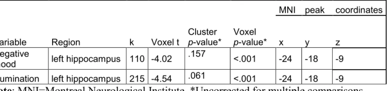
Chapter 1Table 1. Characteristics of the sample 37 Table 2. Exploratory gray matter findings of whole-brain voxel-based 37 morphometry analyses and their associations with daily mood states
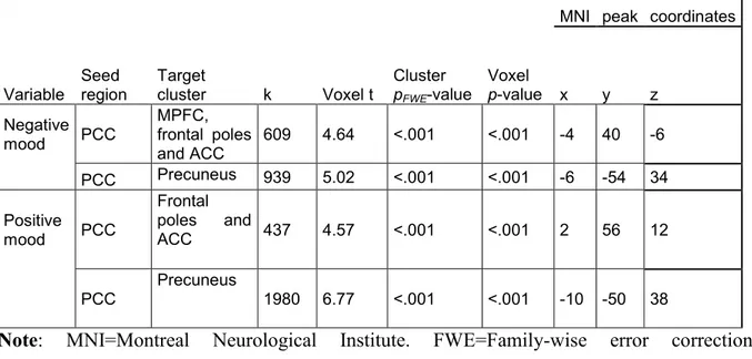
Table 3. Results of whole-brain seed-to-voxel resting-state functional 38 connectivity analyses and their associations with daily mood states
Chapter 2
Table 1. Characteristics of the sample 67 Table 2. Results of whole-brain VBM analyses and its association with 67 SLC6A4 methylation
Chapter 3
Table 1. Significant within-pair fMRI associations with SLC6A4 90 promoter methylation
Liste des figures
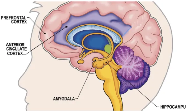
IntroductionFigure 1. Frontal-limbic neurocircuitry 3 Chapter 1
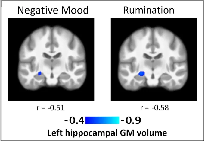
Figure 1. T-statistics maps of the negative association between regional 39 left hippocampal gray matter volume and daily negative mood and rumination
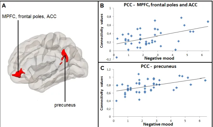
Figure 2. Positive correlation between daily negative mood and resting- 40 state connectivity between posterior cingulate cortex and medial
prefrontal cortex, anterior cingulate cortex, as well as between posterior cingulate cortex and precuneus
Figure 3. Positive correlation between daily positive mood and resting- 41 state connectivity between posterior cingulate cortex and frontal poles,
anterior cingulate cortex, as well as between posterior cingulate cortex and precuneus
Chapter 2
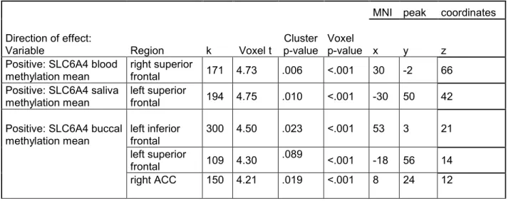
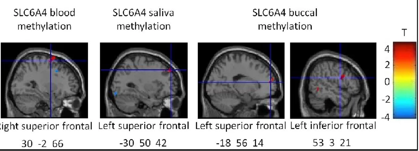
Figure 1. T-statistics maps of the positive association between regional 68 prefrontal gray matter volume and average DNA methylation level of
SLC6A4 promoter derived from whole-blood, saliva and buccal cells
Figure 2. Positive correlation between right lateral parietal area and 69 anterior cingulate cortex, right frontal pole and medial prefrontal cortex
seed-to-voxel resting-state functional connectivity and buccal average DNA methylation level of SLC6A4 promoter
Chapter 3
Figure 1. Greater within-pair difference in peripheral SLC6A4 91 promoter methylation was associated with greater responses to sad
stimuli in left orbitofrontal cortex
Figure 2. Connectome ring representation of ROI-to-ROI 92 connectivity between amygdala, anterior cingulate cortex,
orbitofrontal cortex and insula in the sad condition
connectivity between anterior cingulate cortex, left amygdala 93 and insula in the fearful condition
Liste des sigles
5-HT: Serotonin5-HTTLPR: Serotonin-transporter-linked polymorphic region AAL: Automated Anatomical Labeling
ACC: Anterior cingulate cortex ART: Artifact detection tools ATD: Acute Tryptophan Depletion BDI: Beck Depression Inventory
BDNF: Brain-derived neurotrophic factor BOLD: Blood Oxygen Level Dependent CBT: Cognitive-behavioral therapy CH3 +: Methyl cation
CpG: Cytosine-phosphate-guanine dinucleotide CSF: Cerebrospinal fluid
dlPFC: Dorsolateral prefrontal cortex dmPFC: Dorsomedial prefrontal cortex DMN: Default-Mode Network
DNA: Deoxyribonucleic acid
DSM: Diagnostic and Statistical Manual of Mental Disorders DZ: Dizygotic
EPI: Echo-planar imaging
EPQ: Eysenck Personality Questionnaire FA: Flip angle
fMRI: Functional magnetic resonance imaging FOV: Field of view
FWE: Family-wise error
FWHM: Full-Width Half-Maximum GLM: General linear model
GM: Gray matter
LLP: Left lateral parietal area LPFC: Lateral prefrontal cortex MAO-A: Monoamine oxidase A MDD: Major depressive disorder MNI: Montreal Neurological Institute mPFC: Medial prefrontal cortex MPFC: Medial prefrontal cortex
MPRAGE: Magnetization-prepared rapid gradient-echo sequence MRI: Magnetic resonance imaging
MZ: Monozygotic
OFC: Orbitofrontal cortex OXTR: Oxytocin receptor
PANAS: Positive and Negative Affect Schedule PET: Positron emission tomography
PFC: Prefrontal cortex
PCC: Posterior cingulate cortex PTSD: Post-traumatic stress disorder
QLSCD: Québec Longitudinal Study of Child Development QNTS: Quebec Newborn Twin Study
RLP: Right lateral parietal area ROI: Region of interest
RSQ: Response Styles Questionnaire rsFC: Resting-state functional connectivity
rsfMRI: Resting-state functional magnetic resonance imaging SCID: Structured Clinical Interview for DSM
SD: Standard deviation
SLC6A4: Serotonin transporter SPM: Statistical Parametric Mapping TE: Echo time
TPH1: Tryptophan hydroxylase 1 TPH2: Tryptophan hydroxylase 2
TR: Repetition time
VBM: Voxel-based morphometry vlPFC: Ventrolateral prefrontal cortex vmPFC: Ventromedial prefrontal cortex WM: White matter
Liste des abréviations
Min: MinuteRemerciements
It is with the deepest gratitude that I thank those who stood beside me, guiding me, and challenging me to do and to be my best:
Linda Booij, it is a delight and inspiration to work with you. Thank you for your gentle, yet firm guidance. Thanks to you and your inspiration, I became captivated with the serotonin system and brain imaging. You have an endless well of ideas and a wonderful way of keeping me on track and pushing me forward. A heartfelt thank you is but a small part of the gratitude and honor I feel in having you supervise me during all these years.
Florence B. Pomares, your feedback on my dissertation helped improved its quality and learn a lot about the brain imaging methods as well as the about the interpretation of the findings. Thank you for being there for me as a friend, as a colleague and even as a mentor and, for your thoughtful attention, help and encouragement throughout my PhD.
Jessica Di Sante and Naomi Azar, you have been my rock. You believed in me, encouraged me and helped me in my endeavours, whether it is by giving feedback on my studies or clearing my head with your radiant personalities.
I would also like to thank Melissa L. Levesque and Cherine Fahim as well as countless members of Montreal Neurological Institute, of GRIP unit of Sainte-Justine hospital research center and of Moshe Szyf's laboratory at McGill University (in particular, Zsofia Nemoda, Farida Vaisheva and Wei-Jo Yu), without whom these studies would not have been feasible.
Tatyana Ismaylova and Vahid Ismayilov, you are the safe harbor of love, wisdom and peace I return to over and over. You are the guardians of whom I have been, of whom I am now, and of whom I strive to be. Also, thank you, Simon Jutras, for keeping encouraging me to pursue my dreams all these years, no matter what.
Introduction
The burden of the mental health disorders measures up to that of the physical health diseases [1]. In fact, mental health disorders, affecting the school engagement, the employment, as well as the peer and personal relationships [2], were ranked as a leading cause of years lived with disability globally [3]. It was estimated that by the time Canadians reach forty years of age, half of them would have personally suffered from a mental health disorder [4]. Affective disorders, in particular depression, were found to be among the most prevalent mental health disorders in the general population [5]. A vast majority of studies on affective disorders has focused on such emotional and cognitive aspects as high negative affect and rumination [6-11]. However, these characteristics were also argued to be relevant for healthy individuals with and without subclinical depressive symptoms [12, 13], allowing a deeper understanding of the mental health spectrum, along which one might observe a variation between positive and negative affect potentially culminating at the psychopathological state of depression.
A meta-analysis of genetic epidemiological studies of major depressive disorder in the monozygotic twins showed that depression heritability is in the range of 31%-42% [14]. Perhaps the most widely studied genetic component in the context of depression is the 5-HTTLPR polymorphism of the serotonin transporter (SLC6A4) gene [15-23]. Briefly, the serotonin transporter is one of the fundamental elements of the serotonin system because it generates the protein transporting serotonin from the synaptic cleft to the presynaptic neuron, upholding its availability to the organism [21]. A large number of studies has demonstrated that animal and human carriers of the short allele of the 5-HTTLPR polymorphism were at higher risk of presenting anxiety and depressive symptoms, especially in the presence of early-life adversity such as child abuse and neglect [15, 18-20, 24]. However, there is also a number of results showing a lack of association between the short 5-HTTLPR variant and affective disorders [25-29].
In addition to the genotypic variation, various early-life environmental factors have been associated with the onset and the persistence of affective disorders [30-40]. For instance, such family characteristics as low parental education and early childbearing age have been
associated with an increased risk of belonging to a high depression-anxiety trajectory [30-33]. Early exposure to abusive, hostile and over-controlling parental attitudes/behaviors has been associated with depressive symptoms in late childhood, adolescence and adulthood [36-40]. However, some studies looked for but found no relationship of parental neglect or socioeconomic conditions with the offspring’s depressive symptoms later in life [41-43]. That being said, emotional development appears to be quite sensitive to the environmental conditions in early life.
Essentially, environmental signals are assumed to interact with the genetic blueprint to foster developmental trajectories in the central nervous system [44, 45]. In turn, these developmental trajectories were argued to regulate organism’s assessment of the environment and its subsequent reactivity to the latter [34]. DNA methylation of the SLC6A4 gene has been considered by multiple studies and reviews to potentially enact the way in which adverse environmental experiences mesh with the genetic functioning, shaping neural and emotional processes [21, 46-49].
As part of the present thesis, genetic and environmental influences on the relationship between the peripheral SLC6A4 gene methylation and brain features (e.g., the activity in response to emotional stimuli, the volume and the functional connectivity) are examined. The association between the brain features and the daily emotions is also studied. Here, these important methodological questions are studied in healthy members of the prospective longitudinal cohorts. Doing so allows a thorough consideration of individuals’ early- and daily-life environment as well as their history of emotional well-being.
In the following section, the neural bases of emotion processes will be covered. Next, the role of serotonin in brain development will be delineated. Then, the role of early-life environment in brain processes will be described. Finally, DNA methylation of the serotonin transporter gene will be discussed as a potential underlying mechanism linking these elements together.
Figure 1. Frontal-limbic neural circuitry. Adapted from
https://medium.com/@brainandspace/the-connection-point-brain-computer-interface-the-cerebral-cortex-89a80ebdcb3e
Neural background of emotion processes
At the heart of the neural models of affective (depressive, bipolar) disorders, as argued by Mayberg [50, 51] and Phillips [52], lies the frontal-limbic neurocircuitry (Figure 1), which includes various cortical and subcortical brain regions involved in emotional reactivity and emotion regulation. For instance, the posterior cingulate cortex [PCC], parietal cortex as well as prefrontal cortex – subdivided into medial prefrontal cortical regions (e.g., ventromedial prefrontal cortex [vmPFC], dorsomedial prefrontal cortex [dmPFC], orbitofrontal cortex [OFC], anterior cingulate cortex [ACC]) and the lateral prefrontal cortical regions (e.g., ventrolateral prefrontal cortex [vlPFC], dorsolateral prefrontal cortex [dlPFC]) – are considered to be the higher-order cortical regions the functioning of which has been linked to the (dys)regulation of cognitive-emotional processes in depressed individuals, such as rumination [53-56]. Additionally, the frontal-limbic neurocircuitry includes such regions as amygdala, hippocampus, and insula, located within the temporal lobe [50, 51]. These relatively archaic brain regions have been associated with the reactivity to emotional stimuli
and affective behavior. For instance, the amygdala has been associated with assessment, recognition of and responsivity to emotional stimuli [57-62]. The hippocampus and temporal cortex (in particular, medial temporal region) have been linked to encoding, consolidation and retrieval of the emotional memory [63-66]. The insula, PCC and precuneus have been implicated in the awareness of personal emotional and mental states as well as those of others [67-69], while the ACC has been associated with the dampening of emotional responsivity to the threatening stimuli [70-72]. What’s more, the medial PFC, PCC as well as parietal and medial temporal areas are also part of the Default-Mode Network [DMN]. The DMN represents a set of brain regions shown to simultaneously activate when the brain is at rest and deactivate when the brain is engaged by a specific task [73-78]. The major nodes of the DMN are the vmPFC, PCC and lateral inferior parietal areas [77, 78]. The DMN is typically more active – hence, presenting greater functional connectivity between its neural components – during rest or while pondering over one’s own attitudes, emotions and behaviors in the past, present and future [73, 78, 79].
Neuroimaging studies examining the frontal-limbic regional connections showed that the ventromedial PFC has connections with and receives input from such regions as the lateral prefrontal cortex, the (anterior and posterior) cingulate cortex, the insula, the hippocampus and the amygdala, which together have been associated with such meta-/cognitive processes as self-awareness, decision-making and the adaptive responses to emotional environmental stimuli [65, 80]. One way to examine the neural bases of emotion processes in humans consists in administering an emotion-eliciting task containing stimuli of emotional nature during the functional magnetic resonance imaging [fMRI]. Generally, individuals with a disorder characterized by altered emotion regulation (relative to healthy controls) were found to exhibit altered activity in the (orbital, dorsolateral, superior, inferior) frontal regions, ACC, insula and amygdala as well as in PCC, pre/cuneus and middle temporal regions in response to sad and fearful facial expressions [81-87]. Additionally, relative to healthy controls, affected individuals were found to display altered functional connectivity in response to the negative emotional facial expressions between such frontal-limbic brain regions as the (e.g., orbital, medial, superior) frontal regions, insula, hippocampus and amygdala [88, 89].
While maladaptive affect related to emotional disorders has been associated with altered frontal-limbic processes, transient changes in mood were also found to alter neural functioning in these regions. This has been demonstrated by experimental studies conducted in healthy individuals, combining the fMRI with experimental mood-induction techniques such as emotional music or videos, and autobiographical recall of emotional information. In healthy adults, experimental induction of negative mood has been associated with heightened brain activity in the amygdala, hippocampus, vlPFC, and ACC as well with lowered resting-state functional connectivity between PCC and mPFC, and between ACC and insula [90-93]. On the other hand, induction of positive mood in healthy adults has been associated with greater neural activity in the mPFC, dlPFC, and PCC [91, 93]. While the combination of the fMRI and a mood induction technique permits to temporarily prompt the emotion of interest and examine its neural substrates, the former does not allow examining neural correlates of the mood states occurring naturally in the everyday life. The latter can be achieved by coupling the fMRI technique with a daily diary of mood states. The daily diary, typically in the form of an online questionnaire, would assess the extent to which various emotions and social conflicts are experienced in the everyday life. A growing number of studies employ the combination of the fMRI with the daily diary, the majority of which are primarily focused on clinical or at-risk samples. For instance, Hooker and colleagues (2010; [94]) conducted an fMRI study in healthy young adult couples to examine the link between daily mood following an interpersonal conflict and neural activity in response to emotional stimuli. Results demonstrated that greater daily negative mood and rumination following an interpersonal conflict were associated with lowered vlPFC responses to negative emotional stimuli [94]. Quite similarly, greater daily negative mood following a distressing interpersonal conflict was associated with lowered dlPFC activity in individuals with schizophrenia, relative to their healthy counterparts, during cognitive control of negative emotional stimuli [95]. Additionally, Collip and colleagues (2013; [96]) combined a structural MRI with the daily diary method in order to examine the link between daily emotional reactivity and hippocampal gray matter [GM] volume in adult patients with schizophrenia, their at-risk first-degree relatives and healthy controls. As a result, greater daily emotional reactivity, in particular negative affect, was found to be associated with greater hippocampal volume in healthy controls and with smaller left hippocampal volume in affected and at-risk individuals [96].
Considering that the aforementioned frontal-limbic regions are densely innervated with serotonergic receptors [46], this neurocircuitry and related emotional functioning might be susceptible to any alterations occurring in the serotonin system [46, 97]. In the following section, the association between altered functioning of the serotonin system – in particular,
SLC6A4 – and the frontal-limbic processes will be discussed.
Role of serotonin in neural and emotion processes
Serotonin (5-hydroxytryptamine; 5-HT) is a neurotransmitter mainly synthesized from the amino acid tryptophan in the brain cells with the help of tryptophan hydroxylase (TPH1 and TPH2) enzymes [98, 99]. Serotonin is carried across the brain by the SLC6A4 and, eventually, degraded by the monoamine oxidase A (MAO-A) [98]. While the 5-HT neurons, emerging early during gestation, are scattered throughout the entire brain, the 5-HT population originates and is mostly contained in the raphe neurons, the ascending projections of which wind up in the hypothalamus as well as the PFC, the cingulate cortex and the limbic regions [46, 100, 101].
The implication of the 5-HT in the brain development has been delineated in several reviews [46, 102, 103]. In rodents, first 5-HT-containing raphe neurons are spawned within first two weeks of gestation in rodents, while the mature patterns – in terms of the density and innervation of the 5-HT fibers – are attained by the end of the third postnatal week [46, 102-105]. In humans, first 5-HT neurons are generated within the first two months of pregnancy [46, 103] and continue to proliferate throughout the first five postnatal years upon which they gradually wane until reaching mature levels at 14 years of age [46, 103]. During its expansion in the brain, the 5-HT system is involved in various developmental and lifelong adaptive brain processes including neuronal formation, migration, proliferation and differentiation, synaptic remodelling, axonal myelination as well as the effectiveness of neurotransmission [102, 106, 107].
Alterations in the 5-HT system, in particular during the early-life critical developmental periods, have been associated with the alterations in the brain processes. For instance, pharmacological studies examining the effect of lowered 5-HT levels in rats, by implementing a depletion of the 5-HT concentration or an inhibition of the serotonin
transporter levels during the early postnatal period, reported a long-lasting reduction in the hippocampal dendritic spine density [108] and an increase in the anxious-depressive behaviors in adult rodents [109]. One way to (transiently) lower 5-HT levels in humans is acute tryptophan depletion [ATD] [110]. Since the fMRI connectivity levels depend on the frequencies of the blood oxygen level dependent [BOLD] activation in the brain regions [111], the BOLD signal within the brain regions that are densely innervated with serotonergic receptors might be influenced by the serotonin functioning. Indeed, compared to placebo condition, healthy young adults displayed an ATD-induced lowered resting-state functional connectivity of precuneus and OFC [112]. Moreover, this resting-state functional connectivity, in particular within the middle orbital region, was negatively correlated with depressive mood [112]. In other words, orbital frontal region appears to be involved in serotonin depletion-induced neural and emotional alterations. Furthermore, findings of a systematic review of the fMRI ATD studies conducted in healthy individuals reported, among others, a decreased middle frontal cortical activation as well as an increased dorsomedial and inferior frontal cortical, middle temporal cortical, amygdala and angular activation during detection, processing and recognition of sad and fearful (relative to neutral) emotional stimuli [113]. In addition to altered brain activity, a transient diminution of the 5-HT levels in the brain was associated with a heightened bias towards identification and recognition of negative emotional stimuli [113]. Results of a more recent fMRI ATD study conducted in healthy adults indicated an association between lowered 5-HT neurotransmission in the brain and greater functional connectivity between the amygdala, the insula, the PCC, the precuneus and the superior temporal gyri at rest [114]. Together, these animal and human studies highlight the relationship of the 5-HT functioning with the frontal-limbic processes and the emotional processes.
Considering that the (dys)functions in the 5-HT system occurring early in life have been argued to modulate brain processes [46], the following section will cover the role of the early-life environment in neural processes, particularly in the 5-HT-containing brain regions.
Role of the early-life environment in brain processes
The early-life environment is crucial in calibrating the brain processes since the brain is undergoing significant development during that period of time [115-118]. In other words, the brain is particularly vulnerable to the early environmental context. For instance, the early-life adverse conditions such as childhood maltreatment and poverty has been associated with the short- and long-term alterations in the neural features in such regions as the prefrontal cortex [PFC], the ACC, the hippocampus and the amygdala [119-129]. For instance, pre-/adolescents (age range 8 to 19 years) exposed to childhood domestic violence displayed lower hippocampal volume and responses to threatening faces in a realistic (e.g., school playground) context and greater hippocampal functional connectivity with vlPFC, relative to their non-exposed peers [130]. Greater hippocampus-vlPFC functional connectivity was also associated with a poor context encoding in the presence of a threat [130]. These results may suggest that childhood exposure to violence might be related to a magnified attention to threat at the expense of contextual information [130]. The structural magnetic resonance imaging studies pondered on the association between childhood maltreatment and GM structure. For instance, exposure to childhood maltreatment in healthy individuals (age range 13 to 36 years) was found to be associated with smaller GM volume in the hippocampus and lowered cortical thickness in the medial and lateral PFC and in various temporal regions including the parahippocampal gyri [131, 132]. Moreover, the parahippocampal GM structure was found to mediate the association between childhood maltreatment and the subsequent antisocial behavior [131]. Results of another MRI study showed that the childhood maltreatment was associated with an impaired hippocampal growth later in life (age range 14 to 28 years), even after controlling for the emerging psychiatric symptoms [133]. Taken together, these findings suggest that early-life environment is related to the frontal-limbic brain processes; however, this relationship does not clearly translate into psychopathology.
In fact, not everyone who has been exposed to an early-life stress will develop psychopathology later in life. This is where the concept of resilience comes into play. Resilience can be used to describe the processes and characteristics enabling an individual to adapt well to environmental challenges [134]. For instance, among the individuals who have been exposed to inter-parental violence, those who have developed depressive symptoms were
found to display lower self-confidence and self-control, relative to those who remained healthy [43]. While concept of self-confidence refers to the trust in one’s own ability to succeed in various situations, self-control refers specifically to the ability to regulate one’s own emotional reactivity [135],which might be reflected in the altered frontal-limbic features. Indeed, as the vulnerability for psychopathology is not likely to be transmitted through one single gene, endophenotypic strategy – referring to the investigation of the characteristics that reflect the actions of (epi)genomes either predisposing the individual to or “buffering” him from the disorder – is typically used to examine the characteristics related to the risk and the resilience to the disorder. Indeed, results of a longitudinal fMRI study, investigating risk and resilience endophenotypes for major depressive disorder in adults indicated a distinct pattern of neural activity during self-regulatory task that included the superior frontal and anterior cingulate cortical regions as part of a brain-based resilience endophenotype [136]. Additionally, greater superior frontal GM volume has been associated with self-regulation in youth who experienced high levels of adversity but displayed positive adaptation [137].
Although the early-life environment is noticeably linked to the frontal-limbic brain processes, the DNA methylation should also be considered to play a role in shaping the neural and the emotional processes.
DNA methylation of the serotonin transporter gene
DNA sequence and DNA methylation. First, it is important to grasp the difference between the
genome and the epigenome. Essentially, the genomic (i.e. DNA) sequence is identical across the entire body and throughout the lifespan [138, 139]. The vast majority of the (f)MRI studies of 5-HT genes in the healthy and the depressed individuals outlined the importance of the short (versus long) allele of the 5-HTTLPR polymorphism of the SLC6A4 gene, suggestive of lowered serotonin transporter function, in the frontal-limbic brain features [22, 140-143]. Altered frontal-limbic features included but were not limited to smaller GM volume in the amygdala, hippocampus and ACC as well as greater amygdala activity and connectivity with dlPFC and ACC in response to emotional stimuli [22, 139-143].
In contrast to the genome, the epigenome varies across body tissues and its workings drive the expression of the genes [21, 138, 144, 145]. The epigenetic processes are involved in calibrating the gene expression in response to environmental stimuli, thereby adding plasticity to the rigid genome [138, 139, 145, 146]. Precisely, epigenetic processes are characterized by a biochemical modification to the DNA functioning without altering the structural composition of the DNA sequence [138].
One of the most widely studied epigenetic processes is the DNA methylation (see reviews [46, 138, 145, 147-149]. Essentially, DNA methylation entails the addition of a methyl group (CH3+) onto cytosine, one of the fundamental components of the DNA sequence located on a cytosine-phosphate-guanine [CpG] dinucleotide [138, 147, 150, 151]. This “annexation” is catalyzed by a class of enzymes called the DNA methyltransferase, which are crucial for detecting the genome’s regulatory (i.e. promoter) site [138, 147, 150, 151]. Once on site, the enzymes covalently bind with the genome, detach the cytosine, plant the methyl group onto the cytosine and breaks away while consolidating the now-methylated cytosine back in its place [138, 147, 150, 151]. DNA methylation is linked to the repression of the gene expression at the promoter site, by hindering the DNA transcription factors’ binding onto the altered promoter region of the gene [138, 147, 150, 151]. DNA methylation patterns are, for a large part, formed early during development and are, therefore, susceptible to the influence of the early-life environment [138, 147, 150-153]. Furthermore, DNA methylation patterns have been suggested to confer variation in the emotion processes and personality in humans.
There exists a pattern of mixed evidence that epigenetic modifications confer individual differences in personality in humans. Perhaps the most widely used model of personality traits is the five-factor model, also known as the Big Five [154]. The Big Five model describes the personality based on five dimensions, namely the openness to experience, conscientiousness, extraversion, agreeableness and neuroticism.
The openness to experience, extraversion and agreeableness might be considered as prosocial personality traits. Specifically, extraversion characterizes a tendency to engage the environment and people with vigor and enthusiasm, and agreeableness refers to a tendency to be altruistic and sympathetic to others [154]. The openness to experience reflects the degree of intellectual curiosity and preference for novelty [154]. In light of the implication of the
neurotransmitter oxytocin in the social behaviors, a recent study examined the relationship between the oxytocin receptor [OXTR] gene methylation and prosocial personality traits in healthy adults [155]. Results showed a negative association between the openness to experience and the OXTR DNA methylation [155]. No other significant association was reported [155].
On the other hand, neuroticism is defined as a tendency to be prone to psychological distress and experience such negative emotions as guilt, embarrassment, fear, or sadness [154]. The dimension of conscientiousness is characterized by a tendency to show self-discipline, act dutifully, and to be generally organized and dependable [154]. It has been hypothesized that the interplay of low conscientiousness, low extraversion and high neuroticism would contribute to the onset of emotional disorders such as depression [156]. In term of the underlying epigenetic processes of the pathogenesis, research evidence appears to point to an etiological link between the development of depression and the functioning of the brain derived neurotrophic factor [BDNF], notably based on an increase in the BDNF gene expression related to the administration of antidepressants [157, 158]. In light of this evidence, a recent study examined the relationship between the BDNF gene methylation and the Big Five personality traits, in particular conscientiousness, extraversion and neuroticism, in healthy young adults [159]. Results showed that individuals with higher neuroticism scores displayed higher levels of the BDNF gene methylation [159]. No other personality traits were found to be associated with the BDNF DNA methylation [159]. Further, results of a Positron Emission Tomography [PET] study indicated a positive correlation between neuroticism and serotonin transporter availability in the healthy young adult males’ brains [160]. Yet another study’s findings, mainly focusing on the functioning of the BDNF, OXTR and SLC6A4 genes in the elderly women with and without internalizing (i.e. anxiety and depressive) symptoms, showed that greater DNA methylation levels in the BDNF and OXTR genes (but not SLC6A4 gene), were observed in the clinical group, relative to healthy controls [161]. The differential findings in the aforementioned studies may be attributed to their methodological differences. Nonetheless, the link between neuroticism, characterized by a tendency to cope poorly with stress and to experience negative emotions [154] and the SLC6A4 DNA methylation remains of interest.
DNA methylation of SLC6A4 gene. Considering the role of the serotonin transporter in the
neural and emotional processes and that both methylation and serotonin patterns are largely shaped early in life, the DNA methylation in the SLC6A4 gene will be further discussed as a potential underlying mechanism linking early-life environment, brain features and affective behavior. In light of the difficulty in assessing the central DNA methylation in the living human brain, peripheral tissues – such as blood – are essential in psychiatric epigenetics. The peripheral methylation in the serotonin transporter gene has been associated with lower in vivo measures of the brain 5-HT synthesis in adults [162], suggesting that the peripheral methylation patterns are informative of the central 5-HT functioning.
To the best of our knowledge, a total of seven (f)MRI studies so far investigated the association between the peripheral DNA methylation in the SLC6A4 gene and the brain features in depressed as well as in healthy individuals [49, 163-168]. Results of the (f)MRI studies, comparing the peripheral (blood-derived) SLC6A4 gene methylation between the depressed adults and the age-matched healthy controls, showed that greater blood-derived
SLC6A4 gene methylation was associated with smaller hippocampal GM volume and greater
insula activity in response to the negative emotional content, independent of the diagnosis [163, 165]. Interactions between the methylation and diagnostic group have also been reported, whereby greater blood-derived SLC6A4 gene methylation has been associated with greater temporal/hippocampal activity in response to the negative emotional stimuli in healthy controls [165] and with greater amygdala activity in the clinical group [168]. Additionally, greater blood-derived SLC6A4 gene methylation has been associated with greater amygdala response to the negative emotional stimuli in a mixed sample of adolescents [49, 167] as well as with greater amygdala, hippocampal and insula GM volumes and greater amygdala resting-state connectivity with the ACC and the insula in the healthy adults [164, 166]. Similar to the blood-derived SLC6A4 gene methylation, the saliva-derived methylation was found to be positively associated with the amygdala response to the threatening cues in the healthy adolescents [167].
Amidst the seven (f)MRI studies of the peripheral SLC6A4 gene methylation, two studies looked for and found an association between the childhood maltreatment and the peripheral (blood-derived) SLC6A4 gene methylation, independently of the individuals’
diagnosis [163, 165]. Additionally, one study reported the association between the recent stressful events and the peripheral (blood-derived) SLC6A4 gene methylation, irrespective of the individuals’ diagnosis [168].
Also among the aforementioned (f)MRI studies, some studies looked for but found no the environment or the genotype effect [163, 167, 168], while one study found an indirect effect of the environment but the no genotype effect [49] on the relationship between peripheral SLC6A4 gene methylation and brain processes. Yet other studies reported no environment effect and did not test for genotype [165, 166] or vice versa [164]. It is unclear whether these results are underpowered or indicative of a genuine lack of an association.
Several questions arise out of the existing (f)MRI research on the peripheral SLC6A4 gene methylation. For instance, the environmental influence on the relationship between the
SLC6A4 gene methylation and the brain features – including early-life conditions and daily
mood – is open to question. Moreover, the current insight on the peripheral tissue specificity and/or convergence – implying that the relationship between methylation patterns and brain processes is either different or similar across peripheral tissues – is scarce. The genetic influences on the relationship between the SLC6A4 gene methylation and the brain processes also remain unclear. As a step to address these issues, the peripheral tissue specificity, the role of genetics and that of the daily life are examined. Considering the intricacy of these methodological questions, the latter are studied in the healthy members of the prospective longitudinal cohorts. Doing so allows a careful documentation and investigation of the individuals’ early- and daily-life environment as well as their history of emotional well-being.
Objective
First aim of this thesis is to investigate the association between the frontal-limbic features and daily mood. Specifically, in chapter 1, we examine the association between the daily mood and the neural activity in response to the emotional stimuli, the GM volume and the resting-state functional connectivity within the frontal-limbic neural circuitry.
In the second chapter, we test the hypothesis of the peripheral tissue specificity by examining the SLC6A4 gene methylation derived from which tissue – blood, saliva or buccal cells – corresponds best to the neural activity in response to the emotional stimuli, the GM volume and the resting-state functional connectivity within the frontal-limbic neural circuitry.
In the third chapter, we test the hypothesis that the peripheral SLC6A4 gene methylation is associated with the frontal-limbic activity and functional connectivity in response to the emotional stimuli, independently of the genotype. The monozygotic [MZ]-twin study design offers a unique means to infer genetic influences because a MZ twin pair originates from a single zygote and, therefore, both twins share identical DNA sequence [169-172]. The neural correlates of the daily mood and he peripheral SLC6A4 gene methylation are expected to be found in the frontal-limbic neurocircuitry, including the PFC, the ACC, the insula, the hippocampus and the amygdala.
This thesis ends with a discussion of the implications of the results and of current methodological challenges of this type of research as well as the avenues for future research are proposed.
Chapter 1 – Daily-life mood and brain processes
ForewordIn this first chapter, we assess the brain function in response to the emotional stimuli and during the resting state as well as the brain structure in the Quebec Longitudinal Study of Kindergarten Children (QLSKC) cohort at mean age of 33 years. Drs. Richard E. Tremblay and Frank Vitaro are responsible for the QLSKC cohort. Dr. Jean-Philippe Gouin created the online daily stress questionnaire in collaboration with Dr. Linda Booij. Dr. Florence B. Pomares taught me how to analyze the (f)MRI data and supervised my analyses. Jessica Di Sante assisted in recruitment and the data collection. I participated in the recruitment, the data collection, the (f)MRI analysis, and I wrote up the paper. Dr. Linda Booij designed the study, supervised the data collection and the statistical analysis, provided feedback on all the versions of the manuscript and submitted the latter for publication. All co-authors reviewed and approved the manuscript prior to submission. Manuscript was published in Frontiers in
Human Neuroscience (Ismaylova et al. Front Hum Neurosci. 2018; doi:
Associations between Daily Mood States and Brain Gray Matter
Volume, Resting-state Functional Connectivity and Task-based
Activity in Healthy Adults
Elmira Ismaylova1,2, Jessica Di Sante1,2,3, Jean-Philippe Gouin3, Florence B. Pomares1,3, Frank Vitaro1,4, Richard E. Tremblay1,5,6, Linda Booij1,2,3*
1Research Center, Sainte-Justine hospital, Montreal, QC, Canada
2Department of Psychiatry, University of Montreal, Montreal, QC, Canada 3Department of Psychology, Concordia University, Montreal, QC, Canada 4School of Psychoeducation, University of Montreal, Montreal, QC, Canada
5Department of Psychology and Pediatrics, University of Montreal, Montreal, QC, Canada 6School of Public Health, Physiotherapy and Sports Science, University College Dublin, Dublin, Ireland
This manuscript has been published: Ismaylova, E., Di Sante, J., Gouin, J. P., Pomares, F. B., Vitaro, F., Tremblay, R. E., & Booij, L. (2018). Associations between daily mood states and brain gray matter volume, resting-state functional connectivity and task-based activity in healthy adults. Frontiers in Human Neuroscience, 12, 168.
Abstract
Numerous studies have shown differences in the functioning in the areas of the frontal-limbic circuitry between depressed patients and controls. However, current knowledge on frontal-limbic neural substrates of individual differences in mood states in everyday life in healthy individuals is scarce. The present study investigates anatomical, resting-state and functional neural correlates of daily mood states in healthy individuals. We expected to observe associations between mood and the frontal-limbic circuitry and the default-mode network [DMN]. Forty-two healthy adults (19 men, 23 women; 34 ± 1.2 years) regularly followed for behavior and psychosocial functioning since age 6, underwent a fMRI scan and completed a daily diary of mood states and related cognitions for five consecutive days. Results showed that individuals with smaller left hippocampal gray matter volumes experienced more negative mood and rumination in their daily life. Greater resting-state functional connectivity [rsFC] within the DMN, namely between posterior cingulate cortex [PCC] and medial prefrontal cortical [MPFC] regions as well as between PCC and precuneus, was associated with both greater negative and positive mood states in daily life. These rsFC results could be indicative of the role of the DMN regional functioning in emotional arousal, irrespective of valence. Lastly, greater daily positive mood was associated with greater activation in response to negative emotional stimuli in the precentral gyri, previously linked to emotional interference on cognitive control. Altogether, present findings might reflect neural mechanisms underlying daily affect and cognition among healthy individuals.
Introduction
The function and structure of frontal-limbic brain regions play a major role in the regulation of mood. Most of the evidence stems from anatomical and functional magnetic resonance imaging [(f)MRI] studies conducted in individuals with major depressive disorder [MDD]. Compared to healthy controls, individuals with MDD displayed smaller gray matter [GM] volume in such regions as dorsal lateral prefrontal cortex [LPFC] (e.g., Shad et al., 2012; Grieve et al., 2013) and hippocampus (e.g., Zou et al., 2010; Stratmann et al., 2014). Individuals with MDD also display greater neural responses to negative emotional stimuli in limbic regions including amygdala and hippocampus (e.g., Victor et al., 2010; Hall et al., 2014), as well as lower resting-state functional connectivity [rsFC] between amygdala and such (pre)frontal regions as dorsal LPFC, ventral medial prefrontal cortex [MPFC] and anterior cingulate cortex [ACC] (e.g., Pannekoek et al., 2014; Connolly et al., 2017). Additionally, several studies indicated that MDD is characterized by resting-state functional hypoconnectivity between dorsal LPFC and parietal regions, which are involved in attending to the environmental cues, as well as hyperconnectivity among MPFC, ACC and hippocampus, implicated in self-referential processes (e.g., Kaiser et al., 2015; Northoff, 2016). In MDD, this connectivity “imbalance” would contribute to shifting focus on self-oriented thoughts, potentially resulting in rumination (e.g., Kaiser et al., 2015; Northoff, 2016).
While MDD-related maladaptive affect and cognition has been associated with altered frontal-limbic brain processes, transient changes in mood can also transiently alter neural functioning in these networks. This has been demonstrated by experimental studies in healthy individuals, combining fMRI with experimental mood-induction techniques such as emotional videos/images/music and autobiographical recall of emotional events (e.g., Harrison et al., 2008; Subramaniam et al., 2016). In healthy adults, experimental induction of negative mood has been associated with heightened brain activity in amygdala and hippocampus and various prefrontal regions including the orbitofrontal cortex [OFC], MPFC, ventral LPFC and ACC, as well as with lowered rsFC between posterior cingulate cortex [PCC] and MPFC and greater ACC-insula rsFC (e.g., Pelletier et al., 2003; Habel et al., 2005; Harrison et al., 2008).
Induction of positive mood in healthy adults has been associated with greater neural activity in MPFC, dorsal LPFC and PCC (e.g., Habel et al., 2005; Subramaniam et al., 2016).
Several (f)MRI studies have investigated the neural correlates of mood reactivity in daily life using ecological assessment methods. Most of these studies focused on patients with psychotic disorders or at-risk samples. For instance, one brain morphometry study showed that (relative to controls) greater emotional reactivity to daily stress in patients with schizophrenia and at-risk first-degree relatives was associated with smaller hippocampal GM volumes (Collip et al., 2013). Using task-based fMRI, Tully and colleagues (2014) indicated that within individuals with schizophrenia (relative to controls), higher dorsal LPFC activity during cognitive control of negative emotional information was associated with positive mood following highly-distressing interpersonal conflict. In a study of couples free of mental health problems, Hooker and colleagues (2010) showed that lower ventral LPFC response to negative emotional stimuli was associated with greater daily negative mood and rumination following stressful interpersonal conflict with their partner.
Additionally, individual differences in resting-state functional connectivity have been linked to the well-being/positive lifestyle, such as life satisfaction, self-realization or pleasure attainment (Smith et al., 2015; Luo et al., 2017). Noteworthy, PCC-based rsFC with PFC, precuneus, parahippocampal and superior temporal gyri – all part of the so-called Default-Mode Network (Greicius et al., 2003; Fransson, 2005, 2006; Buckner et al., 2008) – has been linked to experimentally induced sadness (Zamoscik et al., 2014; Renner et al., 2017). We are not aware of any study examining the association between these brain processes and the daily-life mood in healthy individuals. The link between PCC-based resting-state functional connectivity and daily mood is of particular interest since it might be informative of how mood states in the everyday life echo in the functioning of the brain not strained by any specific task.
The aim of the present study is to examine, in a sample of healthy adults, anatomical, resting-state and functional neural correlates of daily mood resting-states, namely negative mood, rumination and positive mood. We expected these associations to occur in brain regions that are part of
the frontal-limbic neural circuitry and the Default-Mode Network, namely prefrontal cortex, anterior and posterior cingulate cortices, precuneus, insula, hippocampus and amygdala.
Materials and Methods Participants
Participants were members of two longitudinal cohorts of individuals, followed since their kindergarten year (Van Bokhoven et al., 2006; Rouquette et al., 2014). Careful screening for eligibility in the brain-imaging component was based on the absence of any prior or current Axis I disorder, neurological disorder, medical illness and medication intake as well as on the absence of irremovable foreign metals in the body (e.g. braces, piercings). Presence/absence of past and current Axis I disorders was assessed by conducting the Structured Clinical Interview for DSM-IV [SCID-IV] (First et al., 2002), whereas the other (neuroimaging-related) exclusion criteria were verified by means of a Montreal Neurological Institute [MNI] in-house questionnaire. After thorough screening for eligibility and availability, 47 individuals underwent an (f)MRI session at the MNI. Participants also completed the Beck Depression Inventory [BDI] (Beck et al., 1988) as well as the Neuroticism scale (12 items) of the Eysenck Personality Questionnaire (Birley et al., 2006), neither of which have been found to be associated with any brain processes in the current study. After the fMRI session, participants were asked to fill out a daily online questionnaire for five consecutive days.
Five participants did not fill out the daily online questionnaire despite multiple reminders. Therefore, the final sample of the present study was composed of 42 healthy adults (19 men, 23 women; age range 32 to 36 years). Written informed consent was obtained from all participants in accordance with the Declaration of Helsinki (World Medical Association, 1991). The study protocol was approved by the ethics review boards of Sainte-Justine Hospital and of MNI, Montreal, Canada.
Image Acquisition
All forty-two participants were scanned on a 3T Siemens TIM Trio Scanner (www.medical.siemens.com) using a 32-channel head-coil. Following a brief localizer, the
scan sequence included, respectively, a 9-minute-long anatomical scan (MPRAGE sequence; 176 slices in sagittal plane; TR=2300ms, TE=2.98ms, FA=9°, FOV=256mm, matrix size=256x256, voxel size=1mm x 1mm x 1mm), a 7-minute-long resting state scan (gradient EPI sequence; 180 whole-brain volumes; TR=2300ms, TE=30ms, FA=30°, FOV=224mm, matrix=64x64, 43 slices in axial plane, voxel size=3.5mm x 3.5mm x 3.5mm,) during which participants were asked to stay awake while keeping their eyes closed (Beliveau et al. 2015), and lastly a 15-minute-long event-related functional scan during which an emotion-processing task was performed (gradient EPI sequence; 400 whole-brain volumes; TR=2300ms, TE=30ms, FA=30°, FOV=224mm, matrix=64x64, 43 slices in axial plane, voxel size=3.5mm x 3.5mm x 3.5mm). During the emotion-processing task, adapted from Canli and colleagues (2005), 120 Ekman facial emotional expressions (happy, sad, angry, fearful and neutral) (Ekman and Frisen, 1976; Keltner and Ekman, 2003) were presented to participants randomly for 2 seconds, followed by a fixation cross (1 second) and a question asking participants to choose whether the face belonged to a man or a woman. Activation following exposure to happy (versus neutral) and sad (versus neutral) was studied in the context of the present study.
Daily Diary of Mood States
Following the brain scan, participants were instructed to fill out online a daily diary of mood states, at the end of the day for five consecutive days. This online diary allowed participants to provide precise information on the mood they have experienced in their natural context (e.g. family life, marital life, social life), within the 24-hour period for five consecutive days. All the responses were in the form of multiple choices, ranging from “never” to “repeatedly”.
Positive and Negative Affect subscales were formed based on the Positive And Negative Affect Schedule [PANAS] (Watson et al., 1988), assessing the extent to which participants had experienced, respectively, positive emotional states characterizing sharp and alert mind (happy, enthusiastic, energetic, determined) and negative emotional states with negative connotations (irritable, sad, nervous, embarrassed) over a period of twenty-four hours, in a form of a multiple choice ranging from “never” (score “0”) to “repeatedly” (score “4”). Therefore, the total score for each scale ranged from 0 to 16.
Repetitive negative thoughts (rumination) were assessed with the items of the Ruminative Responses Scale of the Response Style Questionnaire [RSQ] (Nolen-Hoeksema et al., 1999). Relative to the reflective pondering subtype of rumination (involving active attempts to gain insight into problems), brooding subtype of rumination (implying passive comparison of one’s current situation with an unachieved standard) and worry have been consistently associated with negative mood in healthy and clinical populations (e.g., Burwell and Shirk, 2007; Raes and Hermans, 2008; Verhaeghen et al., 2014). Therefore, we constructed a subscore focusing solely on the brooding and worry components, as potential contributors to the maintenance and intensification of mood states. Eight RSQ-derived items describing brooding rumination and worry were used for the construction of the current scale (i.e. “Negative thoughts came to mind throughout the day”, “I analyzed recent events to try to understand why I was sad or upset”, “I wondered why I always react this way”, “I wondered why I cannot seem to cope better with events”, “I imagined how I would have liked things to happen”, “I worried about saying or doing something wrong”, “I worried about what others would think of me”, “ I worried about being criticized for something I said or did”). For each statement, the participants were asked to indicate how many times they felt this way in a form of a multiple choice, ranging from “never” (score “0”) to “repeatedly” (score “3”). Therefore, the total score for this scale ranged from 0 to 24.
Total scores for each subscale were averaged for the period of five consecutive days, indicating individuals’ average experience of daily mood, and used in the analyses (Bolger et al., 2003). Cronbach alpha’s for the subscales of the daily diary averages across 5 days were 0.99 for negative mood, 0.91 for rumination and 0.99 for positive mood, indicating excellent internal stability.
Statistical Analyses
Statistical Parametric Mapping ([SPM12] v6470, Wellcome Department of Cognitive Neurology, London UK) implemented in MATLAB R2010a (Mathworks, Sherborne, MA) was used for anatomical and functional analyses.
Voxel-based morphometry [VBM] analyses were computed using the CAT12 toolbox (http://www.neuro.uni-jena.de). T1-weighted images were normalized to MNI space and segmented into gray matter [GM], white matter [WM] and cerebrospinal fluid [CSF] based on intensity distribution of the image and using tissue probability maps. Then, normalized GM segments were modulated with the resulting Jacobian determinant maps and smoothed with an 8-mm full width at half maximum [FWHM] Gaussian kernel. VBM, not biased to one particular structure, allows a balanced comprehensive assessment of anatomical differences throughout the brain (Ashburner and Friston, 2001). Whole-brain analysis was conducted, followed by a region of interest [ROI] analysis using a mask encompassing frontal-limbic regions (PFC, ACC, insula, hippocampus and amygdala) obtained from Anatomical Automatic Labeling [AAL] atlas within the Wake Forest University Pick Atlas utility [version 3.0.5] (Maldjian et al., 2003, 2004). For the anatomical analyses, the brain regions were examined at a voxel-wise threshold of p<.001, with cluster-wise family-wise error rate [FWE]-correction for multiple comparisons (Shaffer, 1995) threshold set at p<.05, and corrected for smoothness non-uniformity (Hayasaka et al., 2004). All peak coordinates are reported in MNI format.
Using CONN functional connectivity toolbox [version 16.b] pipeline (Whitfield-Gabrieli and Nieto-Castanon, 2012) and based on SPM12 preprocessing, resting-state functional images were spatially realigned to correct for interscan movement and normalized to the MNI space, while structural images were segmented and separately normalized to the MNI space with non-linear transformation. Next, ART-based scrubbing was done, which consisted in a detection of outlier functional scans (defined as points exceeding the default threshold set with a conservative 95% percentile, including a global brain activation signal of z>3 and linear motion parameters>0.5mm). Any outlier functional scans were, then, entered as a covariate during the denoising step to control for potential confounding effect. Finally, all the images were smoothed using an 8-mm FWHM Gaussian kernel. A denoising procedure including the component-based correction method (Behzadi et al., 2007) followed by the first-level analysis, was applied in order to remove motion artifacts and other artefactual effects from the fMRI signal. Next, considering that PCC-based – one of the Default-Mode Network’s nodes (Greicius et al., 2003) – connectivity has been previously associated with mood states (e.g.,
Zamoscik et al., 2014; Renner et al., 2017), second-level seed-based analysis was conducted using PCC as a seed, which was defined using AAL atlas (MNI coordinates: -6, -52, 40) (Fox et al., 2005). For the connectivity analyses, the brain regions were examined at a voxel-wise threshold of p<.001, with cluster-wise FWE-correction for multiple comparisons (Shaffer, 1995) threshold set at p<.05. All peak coordinates are reported in MNI format. When significant results were observed, the individual connectivity values for the regions showing up in the seed-to-voxel analysis were extracted using the CONN v.16.b software. These connectivity values were, then, transferred in an Excel document in order to generate a scatter-plot for visualization purposes.
Preprocessing of the functional data acquired during the emotion-processing task (slice timing, realignment, coregistration, stereotaxic spatial normalization, and smoothing with 8-mm FWHM Gaussian kernel) was followed by intra-individual first level analyses, performed in order to calculate (emotion minus neutral) contrasts for each emotion at each voxel. Next, whole-brain analysis was performed, followed by ROI analysis using a mask encompassing all frontal-limbic regions (see earlier) (Maldjian et al., 2003, 2004). For the functional analyses, the brain regions were examined at a voxel-wise threshold of p<.001, with cluster-wise FWE-correction for multiple comparisons (Shaffer, 1995) threshold set at p<.05. All peak coordinates are reported in MNI format.
We performed multiple regression VBM and fMRI analyses, with separately negative mood, rumination and positive mood as second-level variables in the imaging analyses. The same variables of interest were separately implemented to test for voxel-wise correlations between them and PCC-based resting-state connectivity. A total of nine analyses were performed in the present study, for which we applied the cluster-wise FWE correction for multiple comparisons.
Results