UNIVERSITÉ DU QUÉBEC
INSTITUT NATIONAL DE LA RECHERCHE SCIENTIFIQUE
INSTITUT ARMAND-FRAPPIER
THE NAJA KAOUTHIA SNAKE VENOM CONTAINS A
POTENT INSULINOTROPIC PEPTIDE
Isolation, identification and characterization
Par: Thi Tuyet Nhung Nguyen
Thèse présentée pour obtenir le grade de
Philosophiae Doctor
Doctorat en Biologie
Programme offert conjointement par l'INRS et l'UQAM
Jury d'évaluation
Président du jury
et examinateur interne : Pr David Chatenet, INRS-IAF
Examinateur externe : Pr Pedro d'Orléans, Université de Sherbrooke
Examinateur externe : Pr Martin G. Sirois (Institut de cardiologie de Montréal)
Directeur de recherche: Pr Alain Fournier, INRS-IAF
ACKNOWLEDGEMENTS
Foremost, 1 would like to express my deepest gratitude to my research director Professor Alain Fournier for his patience, caring, excellent continuons support and providing me with a perfect atmosphere for doing research. Without his supervision and constant help, this dissertation would not have been possible. 1 would like to thank Professor Truong Nam Hai, my research supervisor in Vietnam, for giving me the oppotunity to perform my PhD in Quebec, and for supporting me during my study in Vietnam.
1 would like to thank Professor David Chatenet for his advices, motivation, and enthusiasm. His guidance helped me during my research. Very special thanks go to Myriam Létourneau who, not only as a colleague but also as a good friend, was always willing to help and give her best suggestions. 1 could not have imagined having a better collaborator for my PhD studies.
1 would like to thank Professor Dang Vu Minh and Professor Le Quoc Sinh who put the first rock to build this PhD training co-program between Vietnam Academy of Science and Technology and Institut National de la Recherche Scientifique. 1 wish to thank professor Chau Van Minh and professor Vu Thi Bich who made it work, therefore giving me an oppotunity to participate in the program. 1 would like to thank Professor Le Thanh Hoa, and Professor Dinh Duy Khang for encouraging me.
1 thank Dr. Benjamin Folch for his contribution to my project. His peptide modeling studies made the project more complete.
1 would like to thank my committee members, Professor Pedro D'Orléans-Juste and Professor Martin Sirois for their insightful comments for my thesis.
1 would like to thank my parents, my parents-in-law, my husband, Bui Quang Dung and my lovely daughter, Bui Anh Thu for their love and encouragement. Without their love, 1 would not have fmished this thesis. 1 would also like to thank my family for al ways supporting me and encouraging me with their best wishes.
Many thanks go to ali colleagues in the Institute of Biotechnology, Viet Nam Acadcmy of Science and Tcchnology and ali friends in Canada for providing a stimulating and fun environment in which we learn and grow. Warm thanks to my colleague Nguyen Hong Thanh for supporting me during my studies in Vietnam.
Last but not least, 1 wish to express my gratitude to the Ministry of Education and Training of Vietnam, the Institut national de la recherche scientifique, and the Vietnam Academy of Science and Technology for the financial assistance.
TABLE OF CONTENTS
ACKNOWLEDGEMENTS ... i
TABLE OF CONTENTS ... iii
LIST OF FIGURES AND TABLES ... v
LIST OF ABBREVIATIONS ... vii
RÉSUMÉ EN FRANÇAIS ... xii
INTRODUCTION ... 1
CHAPTER 1. LITERA TURE ... 5
1. General characterization of pancreas and type 1 and 2 diabetes ... 6
1. 1 . The pancreas and its endocrine function ... 6
1.2. Types of diabetes mellitus ... 16
1. 3. Type 2 diabetes mellitus ... 18
1.4. Therapeutic targets used in treatment of type 2 diabetes ... 22
1.5. Other potential therapeutic targets for treatment of type 2 diabetes ... 38
2. Snake venom and their applications in biomedicine ... 44
2.1. The relationship between snake and Gila monster ... 44
2.2. Nature's Blockbusters of Elapidae ....................................... 45
2.3. Toxins affecting the central nervous system ... 45
2.4. Toxins affecting the hemostatic system ... 46
2. 5. Therapeutic alternative from venom peptides ... 48
HYPOTHESIS ... 59
OBJECTIVES ... 59
PUBLICATION 1 ... 61
PUBLICATION 2 ... 70
3. 1. Outlook of actual type 2 diabctcs trcatmcnts ... 84
3. 3. Potcntial ability of CTX-1 to inhibit K, channels ... 88
3. 4. Biological features ofCTX-1 and the proposed mechanism of action ... 89
3. 5. Truncated CTX-I analogs and their biological characteristics ... 92
3. 6. [Lys52]CTX-I41•60 and its advantages ... 94
CONCLUSION ... 100
REFERENCES ... 102
LIST OF FIGURES AND TABLES
Figures
Page
1-1. Cellular structure of the pancreas ... 6
l-2. Diagrammatic representation of the ami no ac id sequence of human preproinsulin ... 7
l-3. Mature human insulin ... 8
1-4. Signais triggering insu1in and glucagon secretion ... 8
l-5. Mechanism of insulin secretion ... 9
l-6. Role of insulin in GLUT 4 translocation ... ! 0
1-7. Role of insulin in glycogen synthesis ... 11
1-8. Role of insulin in protein synthesis ... 12
l-9. Processing of glucagon ... 14
l-l 0. G lucagon synthesis ... 15
1-ll. Types of diabetes ... 16
l-12. Mechanism of fatty acid-induced insulin resistance ... .l9 1-13. Structure ofKA·1p ... 23
1-14. Structure of sulfonylureas ... 24
l-15. Structure ofrepaglinide and nateglinide ... 24
1-16. Mechanism of action of sulfonylureas and meglitinides ... 25
1-17. Structure of metformin ... 26
1-18. Effect ofmetformin in liver ... 26
1-19. Effect ofTDZs in fat and muscle cells ... 27
1-21. Structure of the alpha-glucosidase inhibitors acarbose and miglitol.. ... 29
1-22. Effects of exenatide in the body ... 31
1-23. Structure of GLP-1(7-36)-CONH2, exenatide, and liraglutidc ... 31
1-24. Structure of DPP-1V inhibitors ... 33
1-25. Structure of colesevelam-HCI. ... 35
1-26. Structure of dapagliflozin and canagliflozin ... 37
1-27. Relationship between snakes and Gila monster ... .44
3-1. Amino acid sequence and disulfide bridge connectivity of Naja kaouthia CTX-1... ... 86
3-2. Ca2+ release effect ofCTX-1 in different cell )ines ... 90
3-3. Mode ofbinding ofS-type and P-type CTXs ... 91
3-4. Primary struture ofCTX-I41-6o ... 93
3-5. Primary struture ofCTX-11-39···94
3-6. Effects ofCTX-1 and its truncated derivatives on Ca2+ release ... 95
3-7. Effects of[Lys52]CTX-41.60 on urotensin-11- and URP-induced hypertrophy on H9C2 cells ... 96
3-8. Sequence alignment ofBmKTX and [Lys52]CTX-41-60···98
Table
1-1. Available medications for the treatment ofT2DM ... 221-2. Human and rat amylin and the analog pramlintide ... .35
LIST OF ABBREVIATIONS
4E-BP1 ACC ACE A Ch nAChR mAChR ADP P-AR AGEs ALS AMPA AMPK ATP BAS BK channel BmKTX cAMP CCK CGRP Ctri9577 CTXs CVFEukaryotic translation initiation factor 4E - binding protein 1 Acetyi-CoA carboxylase
Angiotensin-converting enzyme Acetylcholine
Nicotinic acetylcholine receptor Muscarinic acetylcholine receptor Adenosine diphosphate
P-adrenoreceptors
Advanced glycosylated end products Amyotrophie lateral sclerosis
a-Amino-3-hydroxy-5-methyl-4-isoxazolepropionic acid 5' adenosine monophosphate-activated protein kinase Adenosine triphosphate
Bile acid sequestrants
Large-conductance Ca2+ -activated K+ channel
Butlzus martensii kaliotoxin Cyclic adenosine monophosphate Cholecystokinin
Calcitonin gene-related peptide
Peptide 9577 from Chaerilus tricostatus Cardiotoxins
DAG
DM
DPP-IV eEF eiF ER FA FDA FFA Glucose 6-P GABA GOISGDM
GIP GIPR GLP GLP-IR GLUT GPCR GPib GPVI GRP GRPP GS Diacylglycerol Diabetes mellitus Dipeptidyl peptidase - IV Eukaryotic elongation factor Eukaryotic initiation factor Endoplasmic reticulum Fatty acidFood and Drug Administration Free fatty acid
Glucose-6-phosphate y-Aminobutyric acid
Glucose-dependent insulin secretion Gestational diabetes mellitus
Glucose-dependent insulinotropic polypeptide GIP receptor
Glucagon-like peptide
Glucagon-like peptide 1 receptor Glucose transporter
G protein-coupled receptor Glycoprotein lb
Glycoprotein VI
Gastrin-releasing peptide
Glicentin-related pancreatic peptide Glycogen synthase
GxTX Guangxitoxin HaT x Hanatoxin
HbAlc Glycated hemoglobin
HK Hexokinase
hUll Human urotensin Il [Ca2+]i Intracellular Ca2+ IP Intermediate peptide IP3 lnositol trisphosphate IR Insulin receptor
IRS lnsulin-receptor substrate JZTX-1 J ingzhaotoxin-1
KATI' A TP-dependent potassium channel Kv Voltage-gated potassium channel LCCoA Long-chain acyl-CoA
LDL Low-density lipoproteins LKBl Liver kinase BI
LTX Latrotoxin
M3R Muscarinic acetylcholine receptor M3
MgTX Margatoxin
mRNA Messenger RNA
rn TOR Mammalian target of rapamycin NGF Nerve growth factor
NMDA N-Methyl-0-aspartic acid
OEA PACAP PDPK-1 PDK-1 PI3K PIP2 PIP3 PKA PKB PKC PLA2 PLC PLGA PLI pp PP AR-y PPRE SP PVD
RER
RVV-XRXR
SGLT SGTx OleoylethanolamidePituitary adenylate cyclase-activating polypeptide 3-phosphoinositide-dependent protein kinase
Pyruvate dehydrogenase lipoamide kinase isozyme 1 Phosphatidylinositol 3 kinase Phosphatidylinositol 4,5 bisphosphate Phosphatidylinositol 3,4,5 trisphosphate Protein kinase A Protein kinase B Protein kinase C Phospholipase A2 Phospholipase C Poly (lactide-co-glycolide) PLA2 inhibitor Pancreatic polypeptide
Peroxisome proliferator-activated receptor gamma PPARy response elements
Signal peptide
Peripheral vascular disease Rough endop1asmic reticulum Russellysin
Retinoid X receptor
Sodium-coupled glucose co-transporter Scodra griseipes toxin
SK channels SUR TIDM T2DM TDZ TSC UDP VDCC VIP
Small conductance Ca2+-activated K+ channels Sulfonylurea receptor
Type 1 diabetes mellitus Type 2 diabetes mellitus Thiazolidinedione
Tuberous sclerosis complex Uracil-diphosphate
Voltage-dependent calcium channels Vasoactive intestinal polypeptide
RÉSUMÉ EN FRANÇAIS
La prévalence du diabète s'accroît à un rythme alarmant. En 2015, cette maladie affectera plus de 350 millions de personnes à l'échelle mondiale et touchera approximativement 550 millions d'individus en 2030. Le diabète de type 2 (DT2) est la forme la plus répandue, représentant de 90% à 95% des cas de diabète dans le monde entier. Le DT2 est un désordre qui touche le système endocrinien et le métabolisme, et qui conduit à l'incapacité de maintenir une glycémie normale. Afin de traiter cette maladie progressive, les traitements doivent être modifiés en fonction de ces divers stades. Plusieurs médicaments sont actuellement utilisés pour maintenir une glycémie normale et leur diversité a élargi les options pour traiter le DT2. Cependant, malgré leur puissance et leur spécificité, ils présentent encore des carences et des effets secondaires tels que des épisodes d'hypoglycémie, une prise de poids, certains problèmes cardiovasculaires, des troubles gastro-intestinaux et un oedème périphérique. En outre, la plupart des améliorations initiales de la glycémie produites par la prise de médicaments ne sont pas maintenues en raison de la dégradation de la fonction des cellules
p
du pancréas. Par conséquent, de nouveaux agents doivent être créés afin de contrôler efficacement la glycémie, de prévenir le déclin de la fonction des cellulesp,
de favoriser la perte de poids, d'améliorer l'action de l'insuline et de produire un effet favorable sur les maladies cardiovasculaires qui pourraient être en développement.Il a été observé depuis longtemps que les venins, qui sont des mélanges complexes de protéines, peptides, enzymes et autres agents aux propriétés biologiques diverses, représentent une source immense et variée de molécules. Ces dernières peuvent notamment offrir des options nouvelles de traitements grâce à leur puissance et efficacité unique. Les venins sont le fruit d'une sélection naturelle associée à des millions d'années d'évolution. Les substances qui les composent servent par exemple pour la capture des proies et la digestion et/ou la défense contre les prédateurs. En particulier, les peptides présents dans les venins sont reconnus pour leur grande variété de cibles systémiques. Ceci les identifie comme des ligands aux propriétés inestimables pour étudier les mécanismes physiologiques et/ou pharmacologiques dans différents paradigmes expérimentaux liés à des maladies telles que le cancer, l'inflammation, les maladies neurodégénératives, les atteintes neuromusculaires, les troubles hématologiques, les infections autoimmunes et les problèmes cardiovasculaires. À ce propos, un exemple frappant est celui de
l'exendine 4, un peptide extrait du venin du monstre de Gila, qui possède une activité insulinotropc marquée. Ainsi, une étude pharmacologique exhaustive a permis de l'cxcnatidc, une forme synthétique du peptide qui est maintenant utilisée pour traiter le diabète. Cette découverte image bien les nouvelles perspectives qu'offrent les composants extraits de venins, et tout particulièrement pour le traitement de pathologies telles que le diabète. En eflet, il a été montré que les toxines suivantes, soit Agelaia MP-1, conkunitzinc-S 1 ct a-latrotoxine, sont capables de déclencher l'exocytose de l'insuline. De plus, de façon plus générale, l'étude des mélanges complexes de polypeptides et de protéines de venins a donné lieu à la découverte de molécules biologiques clés comme le BPP (bradykinin-potentiating peptide), le NGF (nerve growth factor), la dendrotoxine, le CVF (cobra venom factor) et la convulxine. Ces composés ont contribué à faciliter la compréhension de systèmes physiologiques et ont été utilisés comme gabarit pour concevoir de nouveaux dérivés affichant des fonctions pharmacologiques précises.
Le monstre de Gila, duquel a été isolé l'exentide 4, est un lézard qui ne mange qu'environ quatre fois par an. Il évite l'hypoglycémie en stoppant sa production d'insuline. Lorsque vient le moment de manger, il tue une proie avec son venin. L'ingestion de celle-ci entraîne aussi l'absoption de son propre venin et conséquemment de l'exendine 4, ce qui réactive sa production d'insuline. De façon similaire, plusieurs espèces de serpents ont la capacité de réduire considérablement leur métabolisme et de survivre sans nourriture pour des périodes pouvant aller jusqu'à deux ans. Cette observation suggère qu'un ou des facteurs équivalents à l'exendine 4 se retrouveraient aussi dans le venin de serpents, d'autant plus que ces animaux sont phylogénétiquement reliés au monstre de Gila. Par conséquent, en collaboration avec un partenaire vietnamien qui étudie les propriétés du venin du cobra à monocle Naja kaouthia, nous avons initié dans notre labomtoire un projet visant à identifier un peptide présentant des caractéristiques pharmacologiques similaires à celles de l'exendine 4. Ultimement, cette molécule ou un dérivé pourrait s'ajouter à l'arsenal développé pour le traitement du diabète.
Nos travaux ont conduit à l'développer de peptide insulinotrope, dont NTTN-16, à partir du venin de Naja kaouthia du Vietnam. Ce composé s'est montré capable d'induire la sécrétion d'insuline par les cellules IN S-IE sans affecter leur viabilité. Sa caractérisation a montré que sa séquence est identique à celle d'un autre peptide précédemment isolé à partir de l'espèce Naja kaouthia vivant à Taiwan, soit la cardiotoxine 1 (CTX-1), qui compte 60 acides aminés et forme
une structure compacte maintenue par 4 ponts disulfures. Malgré la complexité de la molécule, sa synthèse a été réalisée avec succès comme montré entre autres par l'activité insulinotrope concentration-dépendante de la forme synthétique sur des cellules INS-1 E et ce, et il est important de le signaler, tant en absence qu'en présence de glucose. De plus, cette toxine ne provoque pas la lyse des globules rouges humains. Notre étude a aussi montré que la sécrétion d'insuline par la CTX-1 est dépendante du calcium. Également, nous avons démontré que les mécanismes insulinotropes associés aux canaux potassiques sensibles à l' ATP (KATr) ou au récepteur du GLP-1 ne sont pas impliqués dans l'action de la CTX-1. En effet, l'inhibition du flux potassique suite à la fermeture du canal KAw par une absence de glucose extracellulaire n'a pas modifié la sécrétion d'insuline. De même, des tests de liaison de la CTX-1 au récepteur du GLP-1 se sont avérés négatifs.
Dans les cellules
f3,
l'inhibition des courants K+ accroît la durée du potentiel d'action. Ceci maintient la membrane plasmique dans un état dépolarisé et provoque l'afflux du Ca2+ par l'ouverture des canaux calciques, un phénomène qui améliore la sécrétion d'insuline dépendante du glucose. Ainsi, indirectement, au cours de nos travaux, le rôle de canaux K+ autres que KATP a aussi été étudié. En effet, dans les cellulesp,
nous retrouvons également des canaux appelés BK (canal K+ à conductance élevée, activé par le Ca2+) qui, même s'ils ne jouent pas un rôle déterminant dans l'exocytose de l'insuline, participent au contrôle de l'amplitude du potentiel d'action. Il a été montré que ces canaux modulent la mobilisation du calcium dans diverses actions biologiques dont notamment lors de la contraction d'un tissu vasculaire tel que l'aorte. Nous avons montré au moyen d'un bioessai que la CTX-1 est incapable d'induire la contraction d'anneaux d'aorte de rat, ce qui suggère que cette toxine ne serait pas en mesure d'interagir avec ce type de canal. Néanmoins, la littérature indique que les effets exercés sur le cœur par les CTXs sont causés par une modulation de canaux ioniques et tout spécialement par une amplification de l'interaction entre la calséniline (protéine KChiPl) et le canal potassique sensible au voltage (Kv). Par conséquent, l'effet insulinotrope de la CTX-1 pourrait vraisemblablement se produire par l'entremise de ces canaux. Dans les faits, nous observons que la CTX-1 possède des homologies structurelles avec des toxines à canaux potassiques. Par exemple, les boucles II et III de la CTX-1 sont similaires à celles de nombreuses toxines à canaux potassiques. De plus, la superposition de certains résidus jouant un rôle fonctionnel majeur telsque la Lys23, la Lys50 et l' Asn55 de la CTX-1 avec la Lyi7, l' Arg25 et l' Asn30 de toxines à canaux potassiques suggère que la CTX-1 pourrait agir comme un bloqueur de canal Kv via un mécanisme similaire.
La CTX-1 déclenche la libération du calcium dans différentes lignées cellulaires, y compris les cellules INS-1 E, HEK293, HeLa et CHO. L'effet maximal est observé avec les cellules INS-1 E, tandis que la libération la plus faible est mesurée avec les cellules CHO. En outre, même à 10·6 M, la CTX-1 n'a pas causé l'hémolyse d'érythrocytes humains, lesquels se sont révélés être les plus sensibles parmi les globules rouges de mammifères. La CTX-1 a également montré le même effet sur la lignée cellulaire myogène murine C2Cl2. En fait, avec les cellules C2C 12, aucune augmentation significative du nombre de cellules apoptotiques en phase précoce n'a été observée. Cependant, en phase retardée, le pourcentage de cellules apoptotiques s'est avéré légèrement augmenté.
Comme décrit précédemment, la CTX-1 présente au niveau de ses boucles II et III, des homologies de structure avec celle des inhibiteurs des canaux Kv. Ces boucles comptent généralement de 25 à 40 acides aminés. Cependant, les activités biologiques de la CTX-1 sont réparties non pas sur deux mais essentiellement sur trois boucles. Ainsi, les boucles 1 et II sont connues pour leur capacité à endommager les cellules tandis que la boucle III est identifiée comme celle responsable de la dépolarisation des cellules musculaires et de la mobilisation du calcium qui y est associée. Par ailleurs, notre étude a montré que l'activité calcique de la CTX-1 est l'amorce nécessaire pour le déclenchement de la libération d'insuline par les cellules INS-IE. Nous avons donc exploré plus à fond les propriétés biologiques de ces boucles au moyen de dérivés synthétiques tronqués de la CTX-1 produits dans notre laboratoire en appliquant une stratégie de protection orthogonale des cystéines, afin d'obtenir les ponts disulfures dans la configuration présente dans la toxine native. Ainsi, à 10'6 M, le fragment CTX-11.39 a causé la mort d'environ 50% des cellules INS-IE, tandis que le fragment CTX-.41•60, à cette même concentration, n'a pas affecté leur viabilité. Le fragment toxique 1-39 n'a donc pas été étudié davantage. Toutefois, nous avons évalué les propriétés du fragment CTX-.41-60 dans quelques paradigmes et avons observé que ce peptide parvient à stimuler la mobilisation calcique d'une manière concentration-dépendante et entraîne la libération de l'insuline sans l'intervention d'un
canaJ KArr. Ces effets sont en nature et intensité très similaires à ceux de la molécule complète, bien que cc fragment n'améliore pas la sécrétion d'insuline, en absence de glucose.
Des analyses structurales de la CTX-1 ont dévoilé que la valine à la position 52 est considérablement exposée et accessible. De même, l'évaluation des homologies de séquences présentes au niveau d'antagonistes des canaux Kv, a montré qu'une lysine est fréquemment localisée dans la partie responsable de l'action inhibitrice sur les canaux. Par conséquent, nous avons émis l'hypothèse que la substitution de Val52 par une lysine pouvait reproduire un élément structural clé des antagonistes des canaux Kv. Nous avons alors synthétisé l'analogue tronqué [Lys52]CTX-41.60 et mesuré ses effets. Tout comme le fragment non substitué, ce dérivé n'a pas
d'effet cytotoxique sur les cellules INS-lE. Par contre, il stimule la libération de l'insuline aussi bien en absence qu'en présence de glucose. Cette activité insulinotrope est liée au [Ca2+]i et n'est pas médiée par l'activation du canal KATP ou du récepteur du GLP-1. À 10 f.1M, la [Lys52 ]CTX-I41.60 a diminué de 25% l'influence des courants de canaux de type K+. Ces canaux sont membres d'une famille contenant de nombreux sous-types et l'analogue est peut-être un bloqueur sélectif de l'un de ces sous-types dans les cellules INS-1 E. Par ailleurs, nous avons observé que le traitement avec [Lys52]CTX-41•60 de cellules CHO transfectées avec le récepteur UT (CHO-UT) réduit/empêche l'hypertrophie induite par l'urotensine Il, un effet qui pourrait être associé à des sous-types de canaux Kv. On doit aussi souligner la similitude des résidus importants, tels que la Lys52, l' Asn45 et l' Arg58 présents dans le dérivé [Lys52]CTX-41•60, ce qui lui confere des caractéristiques structurales similaires à celles des antagonistes des canaux de sous-type Kvu, i.e. l) une lysine hautement conservée se trouvant au centre de la toxine 2) une asparagine ou un résidu polaire se trouvant à une extrémité et 3) une arginine ou un résidu chargé positivement à l'autre extrémité. Donc, globalement, il semble plausible que le dérivé tronqué de la CTX-I, développé dans notre laboratoire, soit un ligand du canaJ potassique sensible au voltage, et plus précisément du canal du type Kvl.3·
En conclusion, le criblage de substances biologiques est extrêmement prometteur pour trouver de nouveaux composés thérapeutiques. En combinant cette approche à des études de relation structure-activité de molécules-mères, il est possible de favoriser le développement de composés puissants et sélectifs. Dans ce contexte, à partir de la CTX-1, un peptide de 60 acides aminés isolé du venin d'une espèce de cobra vivant au Vietnam, nous avons conçu un analogue
tronqué, la [Lys52]CTX-141•60, qui possède la capacité de libérer l'insuline et ce, en présence ou non de glucose. De plus, certaines caractéristiques structurales et pharmacologiques suggèrent que ce dérivé serait un antagoniste des canaux Kv et plus spécifiquement des canaux du type Kvu- Si cette hypothèse s'avère exacte, en bloquant les Kvu, cet antagoniste stimulerait aussi la translocation de GLUT4 qui transporte le glucose à l'intérieur des cellules, ce qui réduirait davantage la résistance à l'insuline. Finalement, la [Lys52]CTX-141•60 a également empêché
l'hypertrophie cellulaire. Cette caractéristique remarquable s'~joute aux propriétés déjà décrites et contribuerait à limiter les complications cardiovasculaires qui accompagnent souvent le DT2. Nos résultats ont été obtenus à partir de modèles cellulaires. Afin de déterminer pleinement le potentiel de cette molécule, il sera indispensable d'évaluer son activité in vivo.
INTRODUCTION
During the last decade, drug development, which involves various scientific areas of expertise such as chemistry, pharmacology, microbiology, and biochemistry, has significantJy decreased in the pharmaceutical industry. As a matter of fact, the traditional generation of new chemically based small molecules is slowing down, and at the same time advancements in biotechnology and genetic interventions are calling for a renewal of discovery approaches. Hence, with the crucial and increasing need for new therapies in severa] pathologies, pharmaceutical companies are changing their research strategy to actually rely more and more on discoveries made in academie labs and industrial spin-offs. This situation has also led researchers to look at what "Dame Nature" has to offer, and during the last century, severa] compounds with a therapeutic potential have been discovered in nature (Lewis, Dutertre et al. 2012; Vyas, Brahmbhatt et al. 20 13). For instance, among the key medical breakthroughs, the discovery of insulin in 1921 changed diabetes from a death sentence to a survivable disease.
More recently, venoms that are a rich source of bioactive molecules, such as peptides, proteins and enzymes, were investigated. lndeed, venomous animais, represented by zoological groups including sorne mollusks, arthropods, reptiles, and a number of fishes, are often characterized by a well-developed organ derived from the salivary or other exocrine glands, and injection deviees. Compounds produced in these glands are usually secreted with an aim to reach specifie targets within the inflicted organism. For this reason, these toxins represent potential therapeutic and biotechnological drugs since their primary molecular targets include receptors, membranes and enzymes (Rocha, Rostelato-Ferreira et al. 2013; Safavi-Hemami, Moller et al. 2013). Therefore, the continuous need for new and improved pharmacological too1s has led the scientific community to investigate venoms and toxins as a template for drug design. With this approach, a number of venom- or toxin-based compounds have produced sorne usefu1 drugs that are currently used or under investigation for severa] human diseases. For example, from the initial discovery of captopri1, the frrst oral angiotensin-1 converting enzyme (ACE) inhibitor (Odaka and Mizuochi 2000; Ben Henda, Labidi et al. 2013), to the recent application of disintegrins for the potential treatment of cancer (Thangam, Gunasekaran et al. 2012), the various components of snake venoms have never failed to reveal amazing new properties. Most
notably, the disintegrins (eptifibatide and tirofiban) were shown both in vitro and in vivo to be potent antiplatclet aggrcgates (Conrotto, Scacciatfila et al. 20 12; Sisk, Palma et al. 20 12). Also, special ion channel toxins, like conantokin G (Kunda, Cheriyan et al. 2013), chlorotoxin (Costa, Cardoso et al. 2013), ro-conotoxin (Adams and Berecki 2013) and so on, have helped to understand critical mechanisms in several human pathologies, thus representing a major driving force for continuing the prospective search for specifie drugs capable of modulating the channel function.
While original native toxins are usually unsuitable as therapeutics, interventions by medicinal chemists and clinicians in pharmaceutical R&D made their use possible as therapeutics or pharmacological tools for multiple disorders. However, many aspects must be observed to fulfill ali requirements needed for a drug candidate to fmish as a drugstore shelf product, including in vivo stability, pharmacodynamies and pharmacokinetics aspects. Venoms, with their cocktail of components, have been tested by millions of years of evolution in bloodstream and other circulatory systems of animais suggesting that their in vivo chemical stability could be controlled by modifying for their therapeutic application. Moreover, with the growing data, it became evident that these substances were exerting remarkable biological properties associated with their ability to act on specifie targets. In fact, many venom components target the cardiovascular ( Chaisakul, Isbister et al. 20 13 ), endocrine and nervous systems (Wolz-Richter, Esser et al. 2013), and modulate the action potentials by acting at various molecular sites such as central or peripheral receptors, enzymes, neurons, axons, synapses and neuromuscular junctions (Kang, Roh et al. 2013; Zhao, Zhao et al. 2013). Therefore, the discovery of molecules in venoms with selective activities represents a new methodology to the search for new drugs. As mentioned above, peptide toxins have been biochemically and pharmacologically refined by the process of evolution to exhibit optimal and finely tuned activities. In this sense, nature has already pre-screened hu ge combinatorial libraries of potential therapeutic drugs. These toxins can be probed for novel pharmacological tools or as a treatment for pain, as weil as for the development of anti-arrhythmics, anti-convulsants, anti-microbials, anti-diabetic, anti-neoplasic and even insecticidal agents. Hence, these examples indicate that the exploration of "venoms", with the goal of discovering new therapeutics, represents a significant endeavor.
Diabetes is a syndrome caused by sugar metabolic disordering, resulting in several cases from insufficient levels of the hormone insulin. According to the World Health Organization, every year in the world, there are about 3.8 million deaths (or 6% of world mortality) by complications of diabetes. Following the prediction of the World Health Organization, by 2030, unless preventive measures are taken, 552 million people worldwide will have diabetes, with the largest increase occurring in developing countries (Whiting, Guariguata et al. 20 Il). In healthy nondiabetic subjects, oral administration of glucose produces a substantially enhanced insulin response compared with intravenous administration. This discrepancy was called the incretin
~ffect, and it is diminished in people with type 2 diabetes. Subsequent studies identified two primary gut-derived hormones that are responsible for most of the incretin effect: glucose-dependent insulinotropic peptide (GIP) and glucagon-like peptide-1 (GLP-1) (Mari, Bagger et al. 2013). GLP-1 is a better therapeutic target and has accordingly been studied to a greater extent. GLP-1 inhibits postprandial glucagon release and stimulates glucose dependent insulin secretion. This hormone also slows gastric emptying and enhances satiety. At least in animais, at the cellular level, GLP-1 appears to stimulate
13
cell growth and survival, and reduces apoptosis, leading to an increase ofl3 cell mass (Meier 2012). Unfortunately, GLP-1 hasan in vivo half-life of Jess than 2 minutes, which prevents it from being an attractive therapeutic candidate (Meier 2012). The currently available incretin mimetics, which involve GLP-1 agonists or analogs that are resistant to DPP-IV-mediated degradation, is exenatide (Byetta®), a GLP-1 agonist that was approved by the FDA in 2005 as adjunctive therapy for patients with type 2 diabetes. Exenatide is a synthetic analog of exendin-4, a peptide found in the Gila monster venom, which binds to and activates the human GLP-1 receptor. Exenatide provokes a sustained reduction in hemoglobin A 1 c (a good measure of diabetes care ), probably related to its effects on pan crea tic13
cells, accompanied by a dose-dependent weight Joss. Consistent with the known activities of GLP-1, exenatide promo tes
13
cell proliferation and differentiation in animal models and also enhances13
cell function in patients with type 2 diabetes. Exenatide enhances endogenous insulin secretion in a glucose-dependent manner. Another incretin mimetic, liraglutide, is an acylated GLP-1 analog that is non-covalently bound to albumin and has a half-life of about 12 hours. Liraglutide was also effective in improving glycemie control in treated patients. As with exenatide, liraglutide bas been reported to improve13
cell function and is associated withdecreases in body weight (Barnett 2009; Fi neman, Cirincione et al. 20 12). The choice of diabetic therapies will likely continue to expand in the coming years with the discovery of new oral antihyperglycemic agents.
The Gila monster is able to reduce its metabolic rate in combination with a large capacity to store fat. lt is this observation that led researchers to investigate the venom of this animal and finally to discover a new therapeutic drug for type II diabetes (Christel and DeNardo 2006). Likewise, snakes were known to survive up to two years without food, but it was not clear how. Recently, it was demonstrated that snakes could lower their metabolic rate by up to 70%, allowing them to survive prolonged periods without food (Mccue 2007). In this respect and since snake venom is an abundant resource of bioactive compounds, we screened venom peptide fractions from the cobra Naja lwouthia in an insulin secretion assay to identify new compounds
CHAPTER 1. LITERA TURE
1. General
characterization of pancreas and type
1 and 2 diabetes
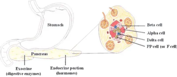
1.1. The pancreas and its endocrine fonctionThe pancreas is a unique structure that perfonns both exocnne and endocrine functions. On one hand, the exocrine cells, present within the pancreas, secrete digestive enzymes including for instance trypsin, carboxypeptidases and lipases that will break down carbohydrates, proteins, and lipids in the chyme. On the other band, the endocrine portion of the pancreas consists of four distinct cell types, found in substructures called the islets of Langerhans. They are mostly composed of P cells (60-80%), which produce insulin and amylin, and a cells (20%), which secrete glucagon. The remaining cells are the somatostatin-secreting 8 cells and the PP cells (or F cells) producing pancreatic polypeptide (Guo and Hebrok 2009) (Fig.l-1). '•,.-:- / / "
,
"(,.
-
---L .
.
1 Panc:n.•as ~· -..-/- + - -- --=---,...
~-
---
1-
---; - 1Exorl'illt' Endonint' fWI'tion (dlf;!t>stin t'nz~·mt>s) (honnont's)
A.It,ha c:t'll Dt>lta c:t'll PP c:t>ll ( 01 F c:t>ll)
Figure 1-1. Cellular structure of the pancreas
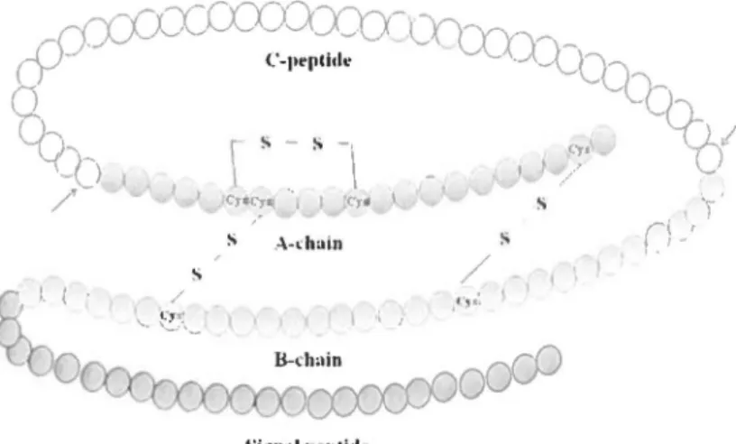
1.1.1. Insu/in hormone
Insulin is a peptide hormone composed of 51 ammo acids. ln the body, insulin is synthesized as a proinsulin precursor. Proinsulin is synthesized in the rough endoplasmic reticulum (RER) from mRNA as preproinsulin. Preproinsulin is a 1 1 0-amino acid structure containing a signal peptide, a B chain, a connecting C peptide and an A chain (Fig. 1-2). Removal of the signal peptide forms proinsulin, which acquires its characteristic 3 dimensional structure in the endoplasmic reticulum. Then, secretory vesicles transfer proinsulin from the RER to the Golgi apparatus, whose aqueous zinc and calcium rich environment favors formation of soluble zinc-containing proinsulin hexamers.
s / '· Q~, __ s A-<h:lin
/
-Signnlpepli<leFigure 1-2. Diagrammatic representation of the amino acid sequence of human preproinsulin
As immature storage vesicles form from the Golgi, enzymes acting outside the Golgi convert proinsulin into insulin. When mature granules are secreted into the circulation by exocytosis, insulin, and an equimolar ratio of C-peptide are released (Fig. 1-3).
,...-("0011 s s :>ill,_ ..\-ch:oin s ' 1 11-•·h:oin
Figure 1-3. Mature human insulin
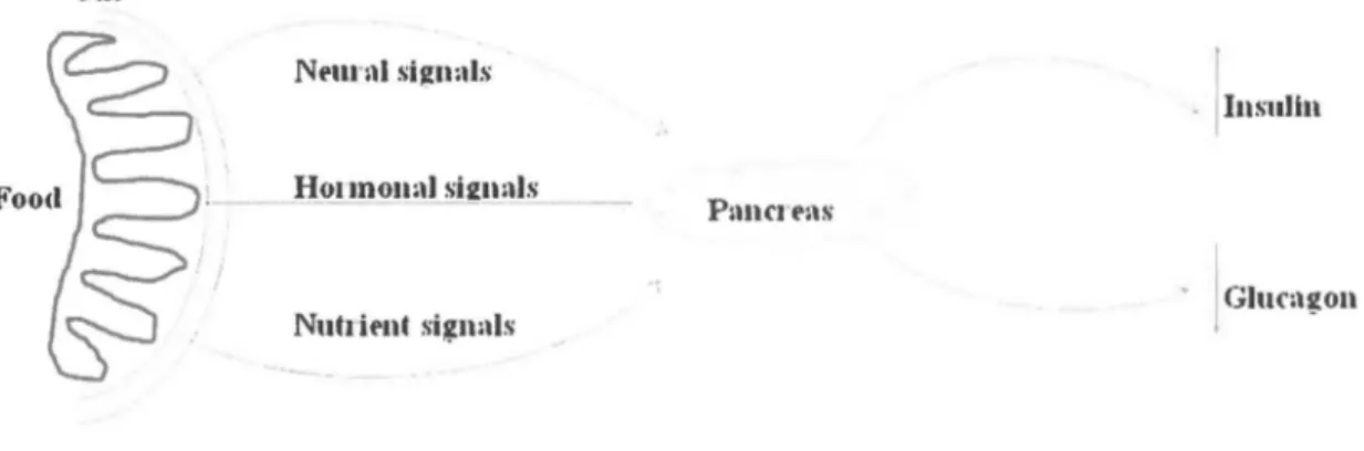
Synthesis and secretion of insulin is regulated by both nutrient and non-nutrient secretagogues, in the context of environmental stimuli and the interplay of other hormones (Fig. 1-4). The exocytosis of insulin is mostly triggered in response to the rising of plasma glucose (nutrient secretagogue). Glucose enters the
p
cells where it is metabolized to generate ATP, increasing ratio of ATP to ADP, therefore activating KATP· Closure of ATP-dependent K+ channels (KArP) results in membrane depolarization and activation of voltage-gated dependent calcium channels (VDCC) leading to an increase in intracellular calcium [Ca2+]i concentration. This increase triggers insulin exocytosis. Non-nutrient secretagogues may act via neural stimuli such as cholinergie and adrenergic pathways, or through peptide hormones and amino acids.Gnl
.j
ln sul in H01 mou:d si2unlsP:mrJt':\s
Nuh it'nt signnls · jGiuragon
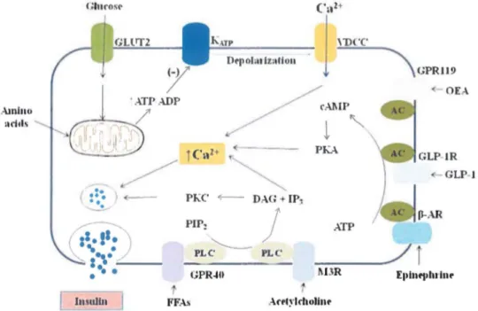
It has been weil recognized that vagus nerve stimulation results in pancreatic insulin secretion. Insulin secretion by thcse mechanisms only occurs in the fccding statc when food is seen, smelled or acutely ingested. Hence, acetylcholine, generated by parasympathetic activity, triggers insulin secretion through the muscarinic acetylcholine receptor M3 (M3R) stimulation, which produces a diacylglycerol (DAO) and protein kinase C (PKC) activation (Nakajima, Jain et al. 2013). The P-adrenoreceptors (P-AR) are also able to transfer signais from neural stimuli to initiale insulin secretion via cyclic adenosine monophosphate (cAMP), but this effect appears to be minor. Also, the incretin hormone glucagon-like peptide (GLP-1), secreted after detection of nutrients in the gut, enhances insulin secretion. lts mechanism of action appears to be mediated by cAMP and activation of a cAMP-responsive protein kinase A (PKA). Infusion of several amino acids such as arginine and leucine leads to significant increases in plasma insulin. The insulinotropic effect of these amino acids is associated to depolarization of the cell membrane and gating of VDCC (Kim and Egan 2008). Sorne fatty acids (FAs) stimulate the G protein-coupled receptor (GPCR) 40 (GPR40) or GPR119 to augment the [Ca2+]i and consequently cause insulin exocytosis (Mancini and Poitout 2013) (Fig. 1-5).
Amlno :uid~ Gluro~l' PIP:
'
-PLC GPR-411 FF.-\.~ <- OE.-\. GLP-1 IP~J
ATP PLC_ _
./
j
t.BRFigure 1-5. Mechanism ofinsulin secretion in
p
cell (Adapted from Kahn, Hull et al. 2006) 9Like other peptide hormones, insulin interacts with a membrane receptor on its target cells. The insulin receptor (IR) exhibits tyrosine kinase activity. The activated IR phosphorylates proteins called the insulin-receptor substrates (IRS). These proteins act through downstream signais to influence transport and cellular metabolism (Grote, Ryals et al. 2013). In the body, insulin is central to regulating carbohydrate and fat metabolism.
Insulin
...
• •[
~ ~
GLUT~RESE
~
.
IR IRS PBK h iglyrt>ridt>s glyro~t'ns\ 1
GhlfO~t'•
•
••
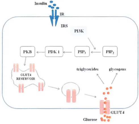
Figure 1-6. Role of insulin in GLUT4 translocation
Insulin causes cells in the liver, skeletal muscles, and fat tissues to absorb glucose from the blood. In the liver and skeletal muscles, glucose is stored as glycogen, and in fat cells (adipocytes), it is stored as triglycerides. lnsulin stimulates glycogen accumulation through a coordinated increase in glucose transport and glycogen synthesis. GJucose transport into fat and muscle cells is largely mediated by a specifie protein known as glucose transporter 4 (GLUT4).
transporter GLUT4 from intracellular sites to the plasma membrane. Insulin activates the IR,
which phosphorylates IRS. Phosphorylated IRS activatc phosphatidylinositol-3-kinase (PI3K) in the cytoplasm, which afterwards converts phosphatidylinositol-4,5-bisphosphate (PIP2) into phosphatidylinositol-3,4,5-trisphosphate (PIP3). PIP3 activates PDPK-1 (3-phosphoinositide-dependent kinase 1), and PDK-1 in tum phosphorylates protein kinase B (PKB) in the cytosol. PKB then stimulates the relocation of the storage vesicles to the plasma membrane (Kahn, Hull et al. 2006) (Fig. 1-6).
The synthesis of glycogen firstly requires glucose to be transported into muscle cells, and then glucose is phosphorylated by hexokinase to form glucose 6-phosphate (G-6-P). G-6-P is converted to uridine diphosphate glucose (UDP-glucose), and glycogen synthase (GS) subsequently catalyzes the synthesis of glycogen by transferring a glucosyl moiety from UDP-glucose to a preexisting glycogen molecule. In skeletal muscle, two isoforms ofhexokinase (type 1 and type Il) are expressed. Between these two isoforms, hexokinase II (HK-Il) predominates, accounting for 90% of the total. Gene transcription of this type of enzyme is regulated acutely by insulin. This hormone also promotes activation ofGS by inactivating glycogen synthase kinase-3 (GSK-3) through phosphorylation (Hoffinan and Elmendorf2011) (Fig. 1-7) .
GSKJ • • 111.\111111
...
IR1
t;hiCOSI' Hrxoklnasl'1
GhiCOSI' 6-P/
l
llllP-Giucosl'Figure 1-7. Role ofinsulin in glycogen synthesis
Insulin also inhibits the production and release of glucose by the liver by blocking gluconcogencsis and glycogcnolysis. This occurs through a direct effect of insulin on the li ver, as weil as by indirect effects of insulin on substrate availability. Insulin can also influence glucose metabolism indirectly by changes in free fatty acids (FFAs) generated from visceral fat.
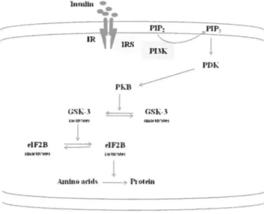
Insulin rapidly activates protein synthesis by activating components of the translational machinery including eiFs (eukaryotic initiation factors) and eEFs (eukaryotic elongation factors). In the long term, insulin also increases the cellular content of ribosomes to augment the capacity for protein synthesis. The rapid activation of protein synthesis by insulin is mediated primarily through PI3K. This involves the activation of PKB. In one case, PKB phosphorylates and inactivates GSK-3, which in tum phosphorylates and inhibits eiF2B. Insulin elicits the dephosphorylation and activation of eiF2B. Because eiF2B is required for recycling of eiF2, a factor required for ali cytoplasmic translation initiation events, this contributes to overall activation of protein synthesis (Fig. 1-8). PKB also phosphorylates the TSCI (tuberous sclerosis complex l)-TSC2 complex to relieve its inhibitory action on the mTOR (mammalian target of rapamycin). The protein mTOR controls translation initiation and elongation. The cap-binding factor eiF4E can be sequestered in inactive complexes by 4E-BPI (e1F4E-binding protein 1).
In~ulln •
===---
-::
IR
;:-·W·fiJ·r
l
-
RS
-
.
_..:.
P
~
l
~
.:...
:
-"-7.a.:,.
P
~
1
l
.
P
~,
-===
PBK PDK GSK-3 I:MU\";lff>ll
.-1F2B .., t"lF2B tU.,lffn'otrPtl
Amlno aritls ~ ProlflnInsulin elicits phosphorylation of 4E-BP1 and its release from eiF4E, allowing eiF4E to form initiation factor complexes. Insulin induccs dephosphorylation and activation of eEF2 to accelerate elongation. Insulin inactivates eEF2 kinase by increasing its phosphorylation at several mTOR-regulated sites. Insulin also stimulates synthesis of ribosomal proteins by promoting recruitment oftheir mRNAs into polyribosomes.
As in the case of carbohydrate metabolism, insulin also promotes the synthesis of lipids, and inhibits their degradation. In adipocytes, glucose is stored primarily as lipids, owing to increased uptake of glucose and activation of lipid synthetic enzymes, including pyruvate dehydrogenase, fatty acid synthase and acetyl-CoA carboxylase. Insulin also profoundly inhibits lipolysis in adipocytes, primarily through inhibition of the hormone sensitive enzyme lipase. This enzyme is acutely regulated by control of its phosphorylation state, which is activated by PKA-dependent phosphorylation, and inhibited as a result of a combination of kinase inhibition and phosphatase activation. lnsulin inhibits the activity of the lipase primarily through reductions in cAMP levels, owing to the activation of a cAMP-specifie phosphodiesterase in fat cells. In
general, insulin exerts multiple effects upon target cells, especially skeletal muscle, liver, and adipose tissue (Wong and Sul2010).
The hormone amylin, in addition to insulin, is secreted from the pancreatic
p
cells. This hormone is co-secreted with insulin from the pancreaticp
cells in the ratio of approximately 1:100. Jt is a 37-residue peptide hormone. Amylin decreases food intake, slows the emptying of the stomach and activates the satiety center in the brain. Amylin reduces the leve) of glucose in the blood by inhibiting the secretion of glucagon.1.1.2. Glucagon hormone
Glucagon is a 29-amino acid peptide that 1s generated from the cleavage of proglucagon secreted by pan crea tic islet a ce Ils.
In intestinal L cells, proglucagon is cleaved to the alternate products glicentin, GLP-1, intervening peptide 2 (IP-2), and glucagon-like peptide 2 (GLP-2) (Fig. 1-9), which promotes intestinal growth. In the body, a cells are equipped with a specifie set of channels that generate action potentials of Na+ and Ca2+ at low Jevels of glucose. Although most of the Ca2+ current
goes through L-typc channcls in a cells, N-type channels mediate the Ca21 required for cxocytosis at low glucose levcls. At low glucose levels, the activity of KATP channels produces a
membrane potential of about -60 mV.
0~ Y NTOl\IOI){lLIN GLUCAGON IP-1
GLICENTIN GRPP GLFC'AGON IP-1
PREPROGLtTC.AGON PS GRPP GLUCAGON IP-1 GLP-1 IP2 GLP-2
PROGLtTC.AGON GRPP GLUCAGON IP-1 GLP-1 IP2 GLP-2
PROGLlT{'AGON 1-61 GRPP GLUCAGON
GLtT{'AGON GLUCAGON
Figure 1-9. Processing of glucagon. PS: Signal peptide; GRPP: Glicentin-related pancreatic
peptide; IP: lntermediate peptide; GLP: Glucagon-like peptide
At activated state, T -type channels open, which depolarizes the membrane potential to
levels where Na+ and P/Q-type Ca2+ channels are activated, leading to regenerative action
potentials. CaH entry through P/Q-type channels induces glucagon secretion (Fig. 1-10).
However, the increase in extracellular glucose levels rises the cytosolic ATP/ADP ratio, which
blocks KATI' channels, thus depolarizing a cells to a membrane potential range where the
channels involved in action potentials become inactivated. As a consequence, electrical activity,
Ca2+ signais and glucagon secretion are inhibited. Moreover, secretion studies prove that glucose
inhibits glucagon release at concentrations below the threshold for
p
cell activation and insulinrelease.
Glucagon secretion is also stimulated by fatty acids. Palmitate and oleate enhance
glucagon secretion and triglyceride accumulation is time- and dose-dependent. Short-term
exposure to these fatty acids stimulates the release of this hormone via enhancing Ca2+ entry
through L-type Ca2+ channels and also by relief of the inhibitory paracrine action of the
somatostatin secreted from ô cells. The principal leve! of control on glycaemia by the islets of
Langerhans depends largely on the coordinated secretion of glucagon and insulin from
a
andp
white hyperglycemie conditions inducc insulin secretion from ~ cclls, u cclls rclease glucagon whcn glucose levels decreasc.
KAt'l' T-t~·pe C'nH
----
x
--=:::;-
]
,....---' "ATP/ADP Glucose SGLT2 Nn' P/Q-type ( •nH1
•• Glucagon•
Figure 1-10. Glucagon synthesis
Insulin and glucagon have opposite effects on glycaemia as weil as on the metabolism of nutrients. Glucagon induces a catabolic effect, mainly by activating liver glycogenolysis and gluconeogenesis, which results in the release of glucose into the bloodstream (Gaisano, Macdonald et al. 2012).
1.1.3. Somatostatin hormone
Somatostatin (SS) is a peptide hormone produced by many tissues in the body,
principally in the nervous system. There are two different forms of somatostatin molecules: one contains 14 amino acids and the other 28. ln the pancreas, ô cells produce SS. This hormone inhibits the secretion of insulin and glucagon. SS is also produced in the gastrointestinal tract where it acts locally to reduce the secretion of gastrointestinal hormones, including gastrin and secretin. Moreover, somatostatin from the hypothalamus inhibits the secretion of growth hormone and thyroid stimulating hormone from the pituitary gland (Weckbecker, Lewis et al. 2003).
1.1.4. Pancreatic polypeptide hormone
Pancreatic polypeptide (PP) is a 36-amino acid peptide hormone secreted by bath PP and acinar cells of the pancreas. Release of PP by protcin-rich meals, fasting, exercisc, and acute hypoglycemia occurs in a biphasic manner. The first rapid release occurs as a result of vagal stimulation; the second, more prolonged rise, occurs predominantly in response to hormonal stimulation. PP inhibits gastric emptying of solid food and dela ys the postprandial ri se in plasma glucose and insu) in. lt also reduces the appetite (Guo and Hebrok 2009).
1.2. Types of diabetes mellitus
The most important disease of the pancreatic endocrine system is diabetes mellitus (DM). DM is characterized by abnormally elevated plasma glucose concentrations {hyperglycemia) resulting from either inadequate insulin secretion or abnormal target cell responsiveness. Chronic hyperglycemia and its associated metabolic abnormalities cause the various complications of DM, including damage to b1ood vessels, eyes, kidneys and the nervous system (Le Roith and Zick 2001; DeFronzo and Tripathy 2009).
Figure 1-11. Types of diabetes
There are three main types of DM (Fig. 1-11 ). Type 1 diabetes mellitus (Tl DM) is a condition of insulin deficiency resulting from
p
ce II destruction. T 1 DM is most commonly an autoimmune disease in which the body fails to recognize thep
cells as "self' and destroys themwith antibodies and white blood cells (Buschard 20 Il). T2DM is known as insulin-resistant diabctcs because in most patients, insulin levels in the blood are normal or even clcvated initially. Later in the disease process, many T2DM become insulin-deficient and require insulin injection. T2DM is actually a whole family of diseases with a variety of causes (Schofield and Sutherland 20 12). The third type of DM is gestational diabetes, a condition in which women without previously diagnosed diabetes exhibit high blood glucose levels during pregnancy.
1.2.1. Type 1 diabetes mellitus
T 1 DM is a complex disorder whose onset in genetically susceptible individuals is sorne times preceded by a viral infection. Many Tl DM diabetics develop the ir disease in childhood. About 10% of ali diabetics have Tl DM. Because individuals with Tl DM are insu lin deficient, the only treatment is insulin injections (Leslie, Williams et al. 2006). Until the arrivai of genetic engineering, most pharmaceutical insulin came from swine, cow and sheep pancreas. However, once the gene for human insulin was cloned, biotechnology companies began to manufacture artificial hurnan insulin for therapeutic use (Werner and Chantelau 2011). In addition, scientists have developed techniques for implanting encapsulated
p
cells in the body, in the hope that individuals with TIDM will no longer need to rely on regular insulin injections (Beek, Angus et al. 2007; Tuch, Keogh et al. 2009; Dufrane and Gianello 2012).1.2.2. Type 2 diabetes mellitus
T2DM accounts for 90% of ali diabetics. The disease is more common in people over the age of 40, but there is growing concem about the increase diagnosis of T2DM in children and adolescents. About 80% of T2DM diabetics are obese (Dixon and O'Brien 2002). A common hallmark of T2DM is insulin resistance, demonstrated by a delayed response to an ingested glucose Joad. Sorne T2DM diabetics have both resistance to insulin action and decreased insulin secretion (Schofield and Sutherland 20 12). In addition, although T2DM diabetics are hyperglycemie, they often have elevated glucagon levels as well (Shah, Vella et al. 2000). The glucagon then contributes to hyperglycemia by promoting hepatic glycogenolysis and gluconeogenesis (Quesada, Tuduri et al. 2008).
1.2.3. Gestational diabetes mellitus
Gestational diabetes mellitus (GDM) is high blood glucose occumng exclusively in pregnant women. About 7% of pregnant women devclop GDM. It usually occurs when the placenta produces large amounts of hormones to help the baby grow. These hormones cause insulin resistance and, unless the woman can produce more insulin to overcome the resistance, the blood glucose will rise. High blood glucose levels may cause the baby to grow large. GDM usually disappears once the baby is born. However, after the birth, 5 to 10 percent of women are found to have DM, usually T2DM (Buchanan, Xiang et al. 20 12).
1. 3. Type 2 diabetes mellitus
1.3.1. Prevalence of type 2 diabetes mellitus
T2DM affects 90% of people with DM around the world, and is largely the result of excess body weight and physical inaetivity. The prevalence ofT2DM is very high and increasing. More than 80% of diabetes deaths occur in low- and middle-income countries. Until 2011, with an estimated 366 million people struggling with the disease, 4.6 million deaths were due to it each year, and annual health-care spending was pegged at $465 billion. Following the prediction of the World Health Organization, by 2030, unless preventive measures are taken, 552 million people worldwide will have diabetes, with the largest increase occurring in developing countries (Grote, Ryals et al. 2013). Over time, diabetes can cause complications such as blindness, heart disease, kidney problems, nerve damage and erectile dysfunction.
1.3.2. Causes of type 2 diabetes mellitus
T2DM is characterized by a slow, progressive loss of
f3
cell function and/or the inability of the body to use insulin (which is called insulin resistance). T2DM typically occurs in individuals over the age of 40 and in individuals who are considered overweight. Normally, insulin activates the glycogen synthesis from glucose by stimulation of the translocation of GLUT4 that transports glucose into the cells and expression of hexokinase, which is essential in the first step of the synthesis. Among the target tissues of insulin, skeletal muscle accounts forthe majority of insulin-stimulated glucose uptake and over 80% of this glucose is stored as
glycogen. Glucose transportation into the eells is largcly mediated by glucose transporter 4
(GLUT4). In obese patients with T2DM, fatty acids {FA) accumulation causes insulin resistance.
Fatty acids compete with glucose for substrate oxidation in muscle. lncreased fatty acid oxidation would cause an increase in the long-chain acyl-CoA (LCCoA) and diacylglycerol (DAG), which triggers a serine/threonine kinase cascade. This ultimately induces serine/threonine phosphorylation of critical IRS-1 sites; thereby inhibiting IRS-1 binding and activation of PI 3-kinase, resulting in reduced insulin-stimulated glucose transport (Fig.1-12).
• • Imulùa
...
IRIR.Sl t)"l'O!>'illt JlbO~'PbOI)'llltton
T
Pl3-kùl :lSt llCii\"llfÏOilIRS 1 .~ti'Ùif' JlhO~JlhOI)'I;lliOII
1'
Srlillt Thnonùat kùanst ncliYil:'· Di:acyl-glycuol
1'
Long danba :uyi-CoA 1' Fl·tt fntty ndds (FFAst ..\kt
T
Cumnillt GLlTT-41
Gbuost GLlTT-4Figure 1-12. Mechanism offatty acid-induced insulin resistance
GLlTT-4
Fatty acid infusion could alter insulin-regulated GLUT4 traffic between intracellular
compartments and the cell membrane. In insulin-resistant skeletal muscle exposed to high fatty
acid levels, glucose oxidation and glycogen synthesis were 50% to 60% lower than control. The
increase of the fatty acid metabolite also induces a rise in intracellular citrate levels, leading to
inhibition of phosphofiuctokinase and phosphate accumulation. Because glucose-6-19
phosphate inhibits hexokinase activity, this would result in intracellular glucose accumulation and dccreased glucose uptake (Savage, Petersen et al. 2005).
T2DM is a progressive disease also characterized by the
p
ccli dysfunction. lt is gencrally accepted thatP
cell failure is caused by the increasing demands associated with insulin resistance; however, recent findings have suggested that impairedp
cell function may be the primary genetic defect in patients with T2DM, with affected individuals being lcss capable of overcoming insulin resistance (Cnop, Welsh et al. 2005; Bonadonna, 2013). Thep
cell function in many patients is already markedly compromised by up to 50% at the time of diagnosis. Furthermore, recent data suggest thatp
cell function might have decreased by up to 80% (Abdul-Ghani, 20 Il). Consequent! y, the pancreatic islet function declines, and this is thought to be due primarily to a significant increase inp
cell apoptosis, ultimately resulting in a deficit inp
cell mass. In patients with T2DM, chronically elevated blood glucose levels have a detrimental effect onp
cells (glucose toxicity). This results in a perpetuai cycle of reduced insulin production, hyperglycemia andP
cell damage.1.3.3. Complications o_ftype 2 diabetes mellitus
Generally, the harmful effects of hyperglycemia are separated into microvascular complications (diabetic retinopathy, nephropathy and neuropathy) and macrovascular complications ( coronary artery disease, peripheral arterial disease, and stroke ). Diabetic retinopathy is the most common microvascular complication of diabetes. The high concentration of glucose leads ta the increase of sorbitol. Osmotic stress from sorbitol accumulation induces the development of this disease. Advanced glycosylated end products (AGEs), promoted by high glucose concentrations, were also associated with the formation of microaneurysms and pericyte loss. Diabetic nephropathy is a progressive kidney disease caused by angiopathy of capillaries in the kidney glomeruli. It is characterized by nephrotic syndrome and diffuse glomerulosclerosis. Although diabetic nephropathy remains the most common cause of renal failure, retinopathy is the leading cause of visual loss in adults and diabetic neuropathy is the leading cause of lower limb amputations. lmprovement in the management of these complications has been substantial. The pathological changes to the kidney include increased glomerular basement membrane thickness, microaneurysm formation, mesangial nodule formation, and ether changes. The
underlying mechanism of injury may also involve sorne or ali of the same mechanisms as diabetic rctinopathy. Diabetic neuropathy is thought to result from diabetic microvascular injury involving small blood vessels that supply nerves. The precise cause of injury to the peripheral nerves from hyperglycemia is related to mechanisms such as polyol accumulation, injury from AGEs, and oxidative stress.
The central pathological mechanism m macrovascular disease is the process of atherosclerosis, which leads to narrowing of arterial walls throughout the body. Atherosclerosis is thought to result from chronic inflammation and injury to the arterial wall in the peripheral or coronary vascular system. Moreover, the combination of increased coagulability and impaired fibrinolysis in T2DM further increases the risk of vascular occlusion and cardiovascular events.
Peripheral vascular disease (PVD), commonly referred to as peripheral artery disease, refers to the obstruction of large arteries not within the coronary, aortic arch vasculature, or brain. PVD can result from atherosclerosis, inflammatory processes leading to stenosis, an embolism, or thrombus formation. It causes either acute or chronic ischemia. T2DM causes endothelial and smooth muscle cell dysfunction in peripheral arteries. The risk of developing lower extremity peripheral arterial disease is proportional to the severity and duration of diabetes. Up to 70% of non-traumatic amputations are performed on diabetics.
1.4. Therapeutic targets used in treatment of type 2 diabetes
Drugs used to treal type 2 diabetes may (1) stimulate
p
cell secretion of insu lin, (2) make target tissues more rcsponsive to insulin, (3) inhibit hepatic glucose output, or (4) slow the digestion or absorption of carbohydrates in the intestine (Bennett, Wilson et al. 20 Il).Table 1-1. Available medications for the treatment ofT2DM
Agents Approval Target Mode of action
Insu lin 1921 Insulin receptor Stimulates glucose uptake
KArP blocker 1955 Sulphonylurea receptor 1 (SURI) Stimulates insulin secretion (Sulphonylurea)
AMP-activated protein 1958 AMP-activated protein kinase Inhibits hepatic gluconeogenesis kinase activator (Metformin) (AMPK)
a-glucosidase inhibitor 1995 a-glucosidase Delays carbohydrate digestion Thiazolidinedione 1997 Peroxisome proliferator-activated lncreases fat cell prolifaration and
receptor y (PPARy) fatty acid uptake
Meglitinide 1998 Sulphonylurea receptor Stimulates insulin secretion Amylin receptor agonist 2005 Brain amylin receptors lnhibits glucagon release and
(Pramlintide) slows gastric emptying
GLP-1 analogs (Exenatide) 2005 GLP-1 receptor Stimulates insulin secretion and inhibits glucagon re1ease DPP-IV inhibitors 2006 Dipeptidyl peptidase IV Prolongs half-life ofGLP-1
Bile-acid sequestrant 2008 Bile acid Increases cholesterol uptake
Sodium/glucose co- 2012 Sodium/glucose co-transporter 2 Inhibits reabsorbtion of glucose in
transporter 2 inhibitor kidney
1.4.1. KAJ'I' blockers
The KATI' channels play an integral role in glucose-dependent insulin secretion (GOIS). Closure of these channels, as a result of increased glucose metabolism, leads to the release of insulin. ln pancreatic
p
cells, the KATI' channel is composed of two subunits: a sulfonylurea receptor (SUR 1) and an in ward rectifying potassium channel (Kir6.2) (Fig. 1-13).SllR 1
Figure 1-13. Structure ofKATPÏD
p
cellSulfonylureas derive from sulfonic acid and urea. Sulfonylureas contain a central S-phenylsulfonylurea structure with a p-substituent (R) on the phenyl ring and various groups terminating the urea N end group (RI). Their differences reside in the types of substitutions at both ends of the molecule.
Introduced in 1955, the sulfonylureas belong to the first group of drugs used for the treatment of type 2 diabetes. The first generation of sulfonylureas includes acetohexamide, chlorpropamide, tolazamide and tolbutamide. These medicines bind to the SURI subunit ofKA'IP and keep the channel closed. This causes an influx of Ca2+ into the cell that results in an
increased release of insulin. Because these drugs of first generation are not potent, a high dose is always suggested. In fact, this first generation of sulfonylureas has been largely replaced in
routine use by agents of second generation, which include glyburide (also known as glibcnclamide), gliclazidc, glipizide, and glimepiride. These drugs of second generation bind more tightly to the sulfonylurea receptor, which makes them more potent, thus requiring a lower dose to produce the secretion of an adequate amount of insulin. Although this new generation of drugs outperforms the first group, side effects such as headache, dizziness, paresthesias, abdominal discomfort, and nausea are observed.
R
Figure 1-14. Structure ofsulfonylureas (adapted from Margolskee, 2012)
Furthermore, ali sulfonylureas stimulate insulin secretion m a glucose-independent manner, and this explains why hypoglycemia is reported frequently. To solve this hypoglycemie activity observed with ali sulfonylureas, meglitinides were developed.
(a)
This is a novel class of non-sulfonylurea insulin secretagogues, including repaglinide and nateglinide (Fig. 1-15). Repaglinidc (Prandin® in USA and GlucoNorm00 in Canada) is a benzoic acid derivative introduced in 1998, and was the first member of the meglitinide class, whereas nateglinide (Starlix® in USA and Canada), a derivative of the amino acid D-phenylalanine, was introduced to the market in 200 l. Meglitinides also bind to the sultonylurea receptor (Fig. 1-16) in
p
ce lis but to a different part of the receptor than sulfonylureas. The interaction of meglitinides with the receptor is not as ''tight" as that of the sultonylureas. Because they are shorter-acting, they do not causef3
cells to push out as much insulin between meals. They are therefore less likely to cause hypoglycemia.Snlfon~·hn·f:t. Jnf211tlnidf
c
-
y
·ATPADP1
Ghi<'O~>f mflabolism Ghi<'OSfj
@
Insu lin \Tl('('Figure 1-16. Mechanism of action of sulfonylureas and meglitinides in
p
cell1.4.2. AMP-activated protein kinase (AMPK) activators
lnsulin resistance is a characteristic feature in T2DM and plays a major role in the pathogenesis of the disease. The application of insulin-sensitizers will help the body to reduce