wcwxwx
Use of RAL Stainer® for the detection of Mycobacterium sp. in
veterinary medicine. Comparison with the reference method.
DUPREZ JN.
1, TCHUENKAM-KAMDEM N.
2, VOLPE R.
2, FETT T.
2, LESENFANTS C.
2, PATERNOSTRE J.
2, MAINIL J.
1, LINDEN A.
21.Bacteriology and bacterial diseases, FARAH, ULg. 2.Health and pathology of wildlife, FARAH, ULg.
Introduction
- Tuberculosis and paratuberculosis of ruminants are two diseases caused by mycobacteria with not only veterinary economic implication (decrease in meat and milk production) but also public health impact (M. bovis and M. paratuberculosis are potentially zoonotic species). Moreover, some wild animal species represent reservoirs of mycobacteria and can be a source of contamination for farm animals in pastures. It is therefore necessary to detect those bacterial species not only in domestic but also in wild ruminants such as roe-deer and red-deer.
- Mycobacteria are acid-fast bacteria and the bacterioscopic reference staining method is the Ziehl-Neelsen after heating (hZN). Its main inconvenience are the production of toxic phenol vapors and the staining timing. The RAL company developed a machine for Gram, Ziehl-Neelsen and Giemsa staining using a cold variant of the hZN method and able to color 20 slides at once. This device is often used and has been validated in human medicine, but not yet in veterinary medicine. In addition, animal samples (faecal materials, mesenteric or mandibular lymph nodes, etc) are more contaminated than human samples (f.i. sputum) and also contain non bacterial components (especially ruminant faeces) that can interfere with the staining procedure. - This study aimed at comparing the RAL Stainer®, the reference hZN staining method on biological samples from domestic and wild ruminants and from a few the wild animal species (wild boars, badgers, porpoise) between each other and with the resuts of growth cultures.
Matérial and Methods
Device and dye: The RAL Stainer® device and the specific RAL STAINER COLD ZN® dye kit were developed by the RAL
company and was loaned by I2A company (Montpellier, France) for this study.
Protocols: The RAL cold ZN staining protocol was the one set by default: fixation for 5 min , Ziehl’s Fuchsine for 10 min,
acid-alcohol discoloration for 3 min and methylene blue 1 min. The reference hZN method was applied as recommended by WHO (1). So far, culture results have been obtained for 81 samples after inoculation in 3 growth media as recommended by OIE (2,3): Loewenstein-Jensen and Colestos (Biorad, Belgium) and HEYM with mycobactin (Becton Dickinson, Belgium).
Controls: The positive controls were a Mycobacterium avium ssp paratuberculosis (Map) from a Cervus elaphus mesenteric
lymph node, a Map of a bovine faecal sample and a culture of Mycobacterium nebraskensi, identified by PCR and sequencing. The negative controls consisted of mixed cultures of non-acid-fast bacteria: Eschericha coli ATCC 25922/Staphylococcus aureus ATCC 25923 and Pseudomonas aeruginosa ATCC 27853/Enterococcus faecalis ATCC 29212.
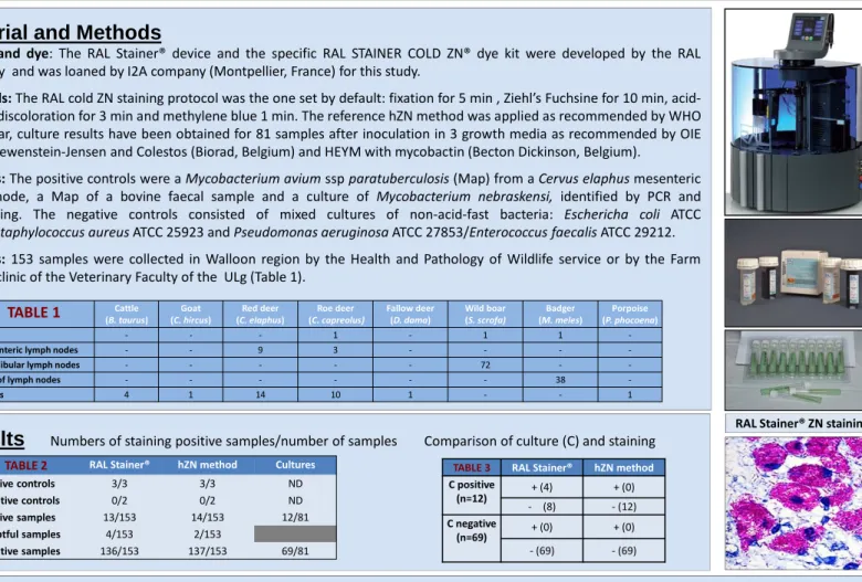
Samples: 153 samples were collected in Walloon region by the Health and Pathology of Wildlife service or by the Farm
Animal clinic of the Veterinary Faculty of the ULg (Table 1).
Results
Numbers of staining positive samples/number of samples Comparison of culture (C) and stainingTABLE 2 RAL Stainer® hZN method Cultures Positive controls 3/3 3/3 ND Negative controls 0/2 0/2 ND Positive samples 13/153 14/153 12/81 Doubtful samples 4/153 2/153
Negative samples 136/153 137/153 69/81
Discussion and conclusion:
- The RAL Stainer® gives the same results as the reference hZN staining method for very positive animal samples highly contaminated with organic materials. In this study, the RAL Stainer® sensitivity is even greater than that of the hZN compared to cultures for less highly contaminated sample. - The RAL Stainer® is a very easy-to-use simple device and the staining obtained presents bright color with high contrast. The closed chamber of the device prevents toxic steam to spread during use. Finally, the existence of two separate racks of 10 allows a rotation increasing yield.
- This study shows that the use of RAL Stainer® in veterinary medicine allows to combine time-saving, security, cleanness, without losing the level of
effectiveness of diagnosis.
References:
1. World Health Organization. Laboratory services in tuberculosis control. Part II: microscopy. WHO/TB/98.258.Geneva, Switzerland: WHO, 1998. 2. Manuel terrestre de l’OIE 2008, 302-318, OIE Paris, France 2008
3. OIE terrestrial manuel 2009, 1-16, OIE Paris, France 2009 TABLE 1 Cattle (B. taurus) Goat (C. hircus) Red deer (C. elaphus) Roe deer (C. capreolus) Fallow deer (D. dama) Wild boar (S. scrofa) Badger (M. meles) Porpoise (P. phocoena) Lungs - - - 1 - 1 1
-Mesenteric lymph nodes - - 9 3 - - -
-Mandibular lymph nodes - - - 72 -
-Pool of lymph nodes - - - 38
-Faeces 4 1 14 10 1 - - 1
RAL Stainer® ZN staining
TABLE 3 RAL Stainer® hZN method C positive (n=12) + (4) + (0) - (8) - (12) C negative (n=69) + (0) + (0) - (69) - (69)
Acknowledgments:
I want to thank the services, "Health and diseases of wildlife“, the ruminant pole of the veterinarian university clinic and the I2a firm without whom this study would not have been possible