Université de Montréal
Levels of immunoglobulin G, white blood cells and
fibrinogen in dairy cows with and without endometritis
during the transitional period
par Ali Bazzazan
Département de sciences cliniques
Faculté de médecine vétérinaire
Mémoire présenté à la Faculté de médecine vétérinaire
en vue de l’obtention du grade Maîtrise ès sciences (M. Sc.)
en sciences vétérinaires option reproduction
Résumé
La performance reproductive des vaches laitières est dépendante de la santé utérine pendant la période postpartum. Dans des circonstances normales, près de 100% des vaches sont
contaminées par l'utérus dans les deux semaines qui suivent le vêlage. Graduellement le système immunitaire inné et reproducteur combine leur effort pour éliminer les agents infectieux, mais une infection utérine persiste chez plus de 40% des vaches en postpartum. Les objectifs de cette étude étaient d'estimer les concentrations d'indicateurs immunitaires systémiques tels que les
globules blancs , les IgG et le fibrinogène pendant la période de transition chez les vaches laitières sans endométrite et de les comparer celles avec une endométrite. Un total de trois troupeaux laitiers commerciaux situés au Québec (Canada) dans la région maskoutaine ont été recrutées ou 61 vaches multipares ont été systématiquement et consécutivement inscrites au moment du tarissement, puis examinées six fois entre 40 jours (J-40) avant et 40 jours (J40) après le vêlage. Les six examens comprenaient une évaluation de la boiterie, la collecte
d'échantillons de lait et de sang, un examen transrectal et vaginal, et une cytobrosse vaginale. Les vaches malades (n = 11) ont été identifiées sur la base de la présence de Troprella pyogenes de grade > 2 dans l'utérus (J21), d'endométrite clinique et subclinique et de cervicite (J35). Les animaux du groupe contrôle (n = 11) ont été caractérisés par l'absence des quatre critères. Pour les 61 vaches sélectionnées au tarissement, les prévalences de l'endométrite clinique, de
l'endométrite subclinique, T. pyogenes et de cervicite étaient respectivement de 23%, 16%, 25% et 31%. Les concentrations d'indicateurs inflammatoires systémiques IgG, les concentrations de globules blancs , et de fibrinogène n'ont pas changé de manière significative durant la période de transition ou varié de manière significative entre les vaches contrôles et malades à tous les temps de prélèvements. En conclusion, les indicateurs systémiques de l'inflammation ne sont pas de
bons marqueurs pour diagnostiquer ou pour faire le suivi de l'endométrite chez les vaches laitières pendant la période de transition.
Mot clés: vaches laitières, Immunoglobulin G, Fibrinogen, globules blancs, neutrophiles,
Abstract
Reproductive performance is particularly dependent on uterine health during the postpartum period. Under normal circumstances, close to 100% of cows have uterine contamination within the first 2 weeks after calving. The innate immune and reproductive system usually eliminate all of the offending microbes, however persistent infection reportedly occurs over 20 % of
postpartum cows. The aims of this study were to estimate concentrations of systemic immune indicators like white blood cells (WBC), IgG, and fibrinogen during the transitional period in dairy cows and to compare these concentrations in dairy cows with and without endometritis. A total of three different commercial dairy herds located in Quebec (Canada) in the Maskoutain region were recruited, and 61 multiparous cows were systematically and consecutively enrolled at drying period and then examined six times between 40 days (J-40) before and after calving (J40). All six exams included an assessment of lameness, the collection of milk and blood samples, a transrectal and vaginal exam, and a vaginal cytobrush. Diseased cows (n = 11) were identified based on the presence of T. pyogenes grade >2 in the uterus, clinical and subclinical endometritis, and cervicitis. The control group animals (n = 11) were characterized by the absence of all four criteria. For the 61 cows, the prevalence for clinical endometritis, subclinical endometritis , T. pyogenes, and cervicitis were 23%, 16%, 25%, and 31% respectively.
Concentrations of the systemic inflammatory indicators (IgG, WBC, and fibrinogen) did not change significantly over time or vary significantly between control and diseased cows at any exam. In conclusion, systemic indicators of inflammation are not good markers for diagnosing or monitoring endometritis in postpartum dairy cows.
Key words: Dairy cows , Immunoglobulin G, Fibrinogen, white blood cells, neutrophils, endometritis, transition period.
TABLE OF CONTENTS
Résumé ……… ΙΙ
Abstract……… ΙV
Table of contents………. VΙ
List of tables……… X
List of figures………. X
List of abbreviation ………. XΙΙ
Acknowledgment ……… XΙV
General introduction………..………. 1
Literature review………. 3
1 Postpartum uterine health in dairy cows……….. 3
1.1 Uterine involution……….. 3
1.2 Contamination of uterus and elimination of
bacteria after calving ………. 4
1.3 Clinical uterine infection……….. 7
2 Definitions of postpartum uterine diseases……… 8
2.1 Puerperal metritis……….. 8
2.2 Clinical endometritis……….. 8
2.3 Subclinical endometritis………. 10
2.4 Pyometra……… 12
3 Diagnostic tools for postpartum endometritis………... 13
3.1 Manual transrectal palpation……….. 13
3.2 Vaginoscopy……….. 13
3.3 Ultrasonography………. 14
3.4 Endometrial cytology………. 15
3.4.1 Uterine biopsy……… 18
3.5 Uterine bacteriology………... 18
4.2 Humoral antibodies……… 21
4.3 Epithelial cells of the endometrium and cytokines…………23
5 General issue……….... 26
6 General hypothesis……… 26
7 Objectives………. 26
ARTICLE
1
8 Introduction………. 28
9 Materials and methods……….. 31
9.1 Animals………... 31
9.2 Experimental design………. 32
9.3 Gynecologic examination………. 33
9.3.1 Vaginal examination……….... 33
9.4 Blood sampling……… 34
9.5 Quantification of serum IgG and
fibrinogen concentration ………..……… 34
9.6 Endometrial cytological sampling………... 35
9.7 Preparation and staining of slides……… 36
9.8 Uterine bacterial culturing and identification………… 37
9.9 Statistical analysis………. 38
10 Results……….. 39
10.1 Descriptive statistics ( study population ) …………. 39
10.2 Descriptive statistics ( study groups ) ……… 41
11 Discussion………. 51
12 Conclusion………. 57
13 Conflict of interest statement……… 57
14 Acknowledgment………. 57
List of tables
Table 1: Summery of studies of the prevalence of endometritis diagnosed by cytology in Postpartum dairy cows (Kasimanickam et al., 2004)………
11
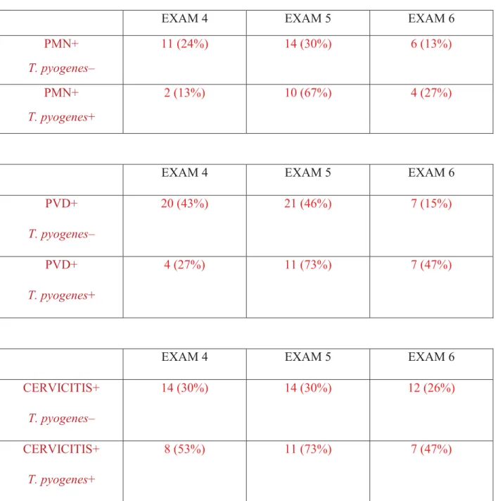
Table 2: Temporal changes in PMN, PVD and cervicitis in cows with and without T. pyogenes. Based on the bacteriological examination at EXAM5, cows with clinical endometritis (PVD ≥ 2), subclinical endometritis (≥ 18% neutrophils) and cervicitis (score = 2) were categorized into two groups: cows with and without T. pyogenes……….
50
List of figures
Figure 1: Classification system for purulent vaginal discharge (Williams et al., 2005) Typical samples of vaginal mucus character: score 0 = clear or translucent mucus; score 1 = mucus containing flecks of white or off-white pus; score 2 = discharge containing ≤50% white or off-white mucopurulent material; and score 3 = discharge containing ≥ 50% purulent material, usually white or yellow, but occasionally sanguineous (score 4). Previously published with (Williams et al., 2005)………...
10
Figure 2: cytobrush and stainless steel instrument.Cytobrush(1), Solid stainless steel rod(2), stainless steel tube(3), sanitary plastic sleeve(4) and
plastic protective sheat (5)……….
16
Figure 3: Image showing how to endometrial cytobrush samples were taken.Cytology device was manipulated through the vagina to the external cervical os (1); the sanitary sleeve was punctured and the instrument was passed through the cervix into the uterine lumen (2); endometrial cytology samples were collected by rotating the cytobrush in a clockwise in contact with the uterine wall (3). ………...…
17
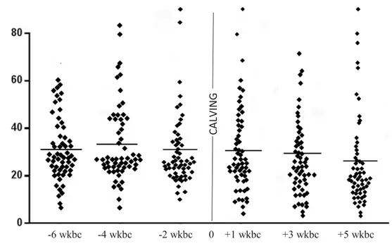
Figure 5: The amount of white blood cells in 61 cows over time of examination………
40
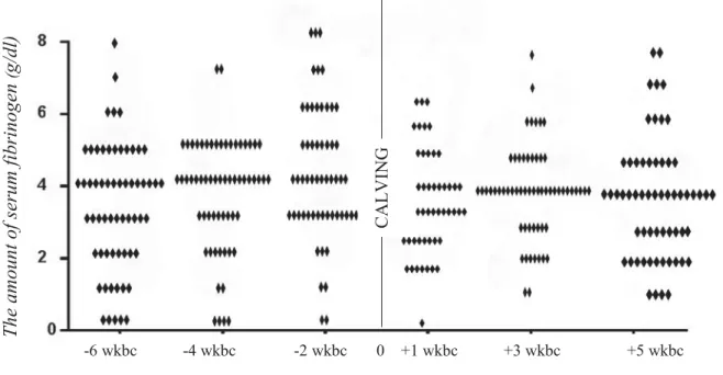
Figure 6: the volume of serum’s fibrinogen in all cows in 6 exams……….
41
Figure 7a: Blood IgG levels in 11 disease cows in 6 exams………. 43
Figure 7b: Blood IgG levels in 11 healthy cows in 6 exams……….
44
Figure 8a: Blood WBC levels in 11 healthy cows in 6 exams………..
45
Figure 8b: Blood WBC levels in 11 disease cows in 6 exams………..
46
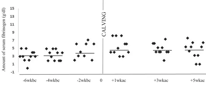
Figure 9a : Blood Fibrinogen levels in 11 healthy cows in 6 exams………... 47
Figure 9b : Blood Fibrinogen levels in 11 disease cows in 6 exams………... 48
Figure 10 :The prevalence of clinical and subclinical endometritis and the percentage of T.pyogenese in all cows at the end of research………..
49
Annexe 1: : Experimental design: Timeline for monitoring of cure and evaluation of genital tract health……….
64
LIST OF ABBREVIATIONS
AMP = antimicrobial peptide APP = acute-phase protein CE = clinical endometritis
DAMP = damage-associated molecular pattern DAP = days after parturition
DBC = days before calving DIM = days in milk
E. coli = Escherichia coli
EnPEC = endometrial pathogenic E. coli ELISA = enzyme-linked immunosorbent assay
F. necrophorum = Fusobacterium necrophorum
FMV = Faculté de médecine vétérinaire, Université de Montréal HP = hepatogloboline
HRP = horseradish peroxidase Ig = immunoglobulin
IL = interleukin
NF-κB = nuclear factor kappa-light-chain-enhancer NK-cells = natural killer cells
NLR = NOD-like receptor
PAMP = pathogen-associated molecular pattern PMN = polymorphonuclear neutrophil
PRR = pattern recognition receptor PVD = purulent vaginal discharge SE = subclinical endometritis SSA = serum amyloid A TLR = Toll-like receptor
T. pyogenes = Trueperella pyogenes
TBS = tris-buffered saline TE = transrectal examination
TMB = 3,3’,5,5’-tetramethylbenzidine TNF-α = tumor necrosis factor alpha VWP = voluntary waiting period WBC = white blood cell
ACKNOWLEDGEMENTS
Completing this master’s has been a great learning experience. A most sincere thank you to my supervisor Dr. Réjean Lefebvre and co-supervisor Dr. Mariela Segura for their guidance during my research at the Université de Montréal. They allowed me to work at my own pace, while remaining accessible and willing to help. Learning under their guidance has been a pleasure, and their work ethic is to be admired.
I would also like to thank Dr. Ndaya Kalidou and Dr. Nicolas Tison for agreeing to be on my advisory committee.
Special thanks to Dr. Nelson Enrique Prieto Cardenas , Daniel Dubuc , Mr. Esmail MokhberiDr. Guy Beauchamp for all their technical and statistical support.
This project would not have been possible without the farmers who so willingly contributed. I would like to thank the Brodeur, Demers and Palardy farms. Also thanks to all my friends for their support and good laughs.
Finally, I would like to thank my wife Samareh for her endless supply of love, optimism, encouragement and patience.
GENERAL INTRODUCTION
About 50% of cows contract a type of uterine infection at the end of the postpartum period, causing the dairy industry significant economic losses due to the loss of uterine function
(fecundation and pregnancy) and ovarian function (folliculogenesis and corpus luteum function). Clinical endometritis, which has a prevalence of about 20-25%, is the most frequent and
economically damaging uterine pathology and the second most prevalent disease after mastitis during the postpartum period (Sheldon et al., 2008). The economic losses are primarily due to treatment costs, decreased of milk production, early culling and long-term infertility.
Conservative estimates suggest that uterine infections in postpartum cows costs Quebec’s 5,743 dairy farms $103 million per year (Canadian Dairy Information Centre). Quebec farms represent 41.3% of all dairy cows in Canada, with a potential revenue of 2.5 billion dollars per year. Clearly, uterine health is an important issue for the Quebec and Canadian dairy industry. The transition period (30 days before to 30 days after calving) is a critical time for dairy cows. It is a period of great stress that requires the cow to make significant metabolic adjustments in response to a negative energy balance, and immunity modulations to allow for a smooth transition
between the end of gestation and the restoration of uterine condition and cyclicity. The latter leads, eventually, to the reestablishment of a new pregnancy (Goff and Horst, 1997). Modulating these demands during the transitional period is essential to maintaining the animal’s general and uterine health.
During gestation, the conceptus is perceived as an allograph (i.e. a foreign body) by the mother’s immune system through the activity of the major histocompatibility complex. Essentially, the
immune system returns to full functionality following calving is thought to influence the risk of uterine infection. The reduced function of granulocytes and monocytes compromises the dairy cow’s capacity to eliminate bacteria, and favors the persistence of uterine infection and
inflammation after calving (Zerbe et al., 2000; Mateus et al., 2002). Peripheral blood leucocytes in cows that develop endometritis show reduced phagocytic capacity compared to those of endometritis-free cows (Kim et al., 2005). The circulating neutrophils of cows that develop metritis show reduced concentrations of glycogen and a decreased tumor necrosis factor alpha (TNF-α) expression (Galvao et al., 2010, 2012). Profiles of immunological mediators
(cytokines/chemokines) in the uterus are also perturbed during uterine infection (Galvao et al., 2011; Ghasemi et al., 2012). The adaptive immune system, through the action of lymphocytes, adds specificity to the actions of the innate immune system and amplifies the response. In addition, immunoglobulins also have a protective role against pathogens in the bovine uterus (Dhaliwal et al., 1996a; Dhaliwal et al., 1996b; Dhaliwal et al., 2001b). However, the specific functions of immunoglobulins, such as immunoglobulins A, G and M (IgA, IgG and IgM) in the genital tract, are still not clear (Dhaliwal et al., 2001a). Globally, these findings show that innate and adaptive immunity, both local and systemic, play an essential role in defending the uterus against postpartum infection.
Microbial challenges to the endometrial inflammatory response in postpartum cows and uterine involution are the two main reasons for an increased local and systemic defense (Aknazarov, 1988; Dhaliwal et al., 2001b). Currently, about 95% of dairy cows exhibit contamination in the uterine cavity during the first 2 weeks following calving (Paisley et al., 1986; Sheldon, 2004). Up to 40% of dairy cows still have contamination in the genital tract 3 weeks after calving (Sheldon et al., 2008). When infection or inflammation persists in the uterus beyond 4 weeks
postpartum, the infected uterus is associated with infertility. Several bacteria, like Escherichia
coli, Trueperella pyogenes, Fusobacterium necrophorum and Prevotella, invade the uterine
cavity after parturition (Sheldon et al., 2008), but endometritis is mainly associated with
T. pyogenes (William et al., 2007). This bacterial species is associated with endometrial lesions and infertility in cows (Bonnett et al., 1998).
LITERATURE REVIEW
1 Postpartum uterine health in dairy cows
1.1 Uterine involution
The female genital tract has several barriers to minimize bacterial entry. These include the vulva, the vestibule-vaginal sphincter, the cervix, cervico-vaginal mucus, and the epithelial barrier of the genital tract. At calving, all of these physical barriers are compromised. Following delivery, the bovine genital tract undergoes an active inflammatory process called uterine involution in order to clear the uterine filling and respond to bacterial contamination. Uterine involution after calving allows the reproductive tract to return to normal function before a new pregnancy is re-established. This phenomenon is very complex and dynamic, involving anatomical,
histologic/bacteriological, biochemical, endocrinal and immunological changes. After calving, the placenta should be expelled between 12 and 24 hours (Sheldon et al., 2011). The genital tract transforms from a large structure that is 1.0 m long and weighs 20 kg to a small tract of 30 cm
period (VWP) has been established at 45 days, after which most cows are able to conceive and maintain a normal pregnancy. At the end of the first week postpartum, necrosis of the caruncles progresses well and the stratum compactum is covered with neutrophils (Weber et al., 2001). In the second week, the necrotic tissues detach and mix with blood, endometrial exudate, and cellular debris in the uterine cavity to make the lochia, which will be gradually be expelled during the first rise of estrogen (Moller, 1970). Discharge of lochia during the first 2 weeks after parturition is normal and must not be mistaken for pyometra or metritis (Dhaliwal et al., 2001a; Sheldon and Dobson, 2004). By day 20 after calving, tissue sloughing and hemorrhaging stop. At this time, uterine size has been reduced by more than 80%, but full restoration of the deeper layer requires more time (about 45 days) (Sheldon et al., 2008). The epithelialization of the
endometrium then progresses to completion (Deng et al., 2014), and by the end of VWP the uterine environment is normally sterile and ready to establish a new pregnancy.
1.2 Contamination of uterus and elimination of bacteria after calving
During pregnancy, the genital tract is assumed to be sterile and non-infected. At calving , the anatomical barriers (cervix, vagina, and vulva) open, resulting in contamination of the uterine cavity (Sheldon, 2004). Bacterial contamination of the uterus from vaginal, fecal and
environmental microflora occurs. Approximately 95% of cows after calving have their uterus contaminated with bacteria, regardless of signs of diseases. (Foldi et al., 2006; Paisley et al., 1986; Sheldon and Dobson, 2004). In addition to the loss of the anatomical barriers, the negative pressure created by the phenomenon of uterine contraction and relaxation enhance the vacuum effect and the entry of bacteria in the uterus. Gram-negative bacteria predominate in the uterus during the first week after calving, but then are gradually replaced by Gram-positive bacteria. By day 15 postpartum, 78% of cows still have bacteria in the uterus; by day 60, only 9% do (Wira et
al., 2005). During normal uterine involution, the uterus clears the bacterial contamination within 5 to 6 weeks postpartum (Sheldon et al., 2002). This whole process is a dynamic one whereby bacteria gain access to the uterine cavity and are then cleared by the uterine defense mechanisms. Pathogens that persist in the uterus at the end of the postpartum period are associated with
postpartum endometritis, and up to 40% of dairy cows are not able to completely eliminate the bacterial infection (Sheldon et al., 2008). Cows with evidence of bacterial pathogens after the fifth week postpartum show a delay in uterine involution (de Vries et al., 2015). There is a correlation between the persistence of certain bacterial species and inflammation of the uterus. Both Gram-positive and Gram-negative bacteria (using the standard culture techniques) play a major role in uterine inflammation and infection.
T. pyogenes, E. coli, F. necrophorum, Prevotella melaninogenicus and Bacterioides spp. are the
most common bacteria associated with postpartum uterine infections (Hazlett and Wu, 2011; Tison et al., 2017) and are recognized as non-specific uterine pathogens. Uterine diseases are dependent on specific bacterial characteristics, like their capacity to adhere to mucosa, colonize and penetrate the epithelium, and release bacterial toxins that lead to the establishment of uterine disease (Sheldon et al., 2009). E. coli is the predominant uterine pathogen during the first week postpartum and is associated with metritis. The endometrial pathogenic E. coli (EnPEC) is able to attach to the endometrium and is associated with uterine infection (Esposito et al., 2014; Sheldon et al., 2010). The bacteria T. pyogenes causes inflammation and lesions in the
endometrium and significantly slows down the process of uterine involution by producing toxins (pyolysin and cytolysin) (Newsholme et al., 1987). It predominates during the second and third week postpartum, becoming the main bacteria associated with endometritis. Isolation of
T. pyogenes in the second week postpartum has been associated with a higher risk of developing
endometritis in the fourth week postpartum (William et al., 2005) and it is associated with
reduced success rate per artificial insemination and increasing the calving interval (Kanneganti et al., 2007; Thompson et al., 2011). One study showed that one third of T. pyogenes isolates have antibiotic resistance genes and one third of E. coli isolates have genes associated with biofilm formation (Kasimanickam et al., 2016). F. necrophorum is capable of inhibiting phagocytosis with a leucocidal toxin (Roberts 1967; Sheldon et al., 2004; Singh et al., 2008). This bacterial species is located primarily deeper in the endometrium, in the lamina propria, whereas
T. pyogenes is found mainly on the endometrial surface. Different interactions and synergistic relationships between bacteria have been reported. For example, T. pyogenes produces catalase and a growth factor that supports the proliferation of F. necrophorum (Singh et al., 2008). Inversely, some reports have indicated that the presence of alpha-hemolytic, coagulase-negative
Staphylococcus is associated with a reduction in endometritis (Balu et al., 2002). The presence of Streptococcus uberis early after calving increases the chance of T. pyogenes growth later and
sustained uterine inflammation (Wagener et al., 2014). Bacterial virulence factors could play an important role in the pathogenesis of inflammation of postpartum uterine conditions in cows. However, the effects of synergetic bacterial interactions are still being debated.
Interestingly, bacteria are found in the uterus of normal and abnormal postpartum cows (Brodzki et al., 2014). The microbiomes of postpartum cows with and without metritis were similar, with the exception of a greater diversity in normal cows (Santos et al., 2012). This suggests that prolonged uterine inflammation and infection in the postpartum period may be attributed to a failure of uterine defense rather than to specific bacterial pathogens (Dadarwal et al., 2017).
1.3 Clinical uterine infection
Reproductive performance is strongly related to uterine health status at the end of VWP
(C. Bartlett et al., 1986). Clinical assessment, the most efficient treatment and the prevention of postpartum uterine pathologies in dairy cows depends on an understanding of the mechanisms behind uterine pathology. Uterine diseases affect about half of dairy cows in the postpartum period (Wira et al., 2011) and cause infertility by disrupting uterine and ovarian function (Wira et al., 2005).
2 Definitions of postpartum uterine diseases
Postpartum uterine diseases in dairy cows vary as a function of days after calving. Puerperal metritis, clinical endometritis, subclinical endometritis and pyometra are the most common reproductive diseases. Genital tract inflammation/infection encountered in the postpartum period are often identified based on the anatomical location involved (vagina, cervix and uterus) during days in milk (DIM).
2.1 Puerperal metritis
Puerperal metritis is an inflammation affecting the endometrium and deeper tissues of the uterine wall. It is associated with increased rectal temperature (> 39.5 ºC), watery and fetid vaginal discharge, flaccid uterine tone on transrectal palpation, decreased milk yield, and dullness within 15 days after calving (Drillich, 2006; Drillich et al., 2001; Sheldon et al., 2006; Sheldon et al., 2008). The predisposing factors are: 1) dystocia, 2) placental retention, 3) poor hygiene in the parturition pen, and 4) metabolic diseases around calving (Chapwanya et al., 2009; Drillich et al., 2001; Foldi et al., 2006; Sheldon et al., 2006).
2.2 Clinical endometritis
Definitions for clinical endometritis that more accurately reflect the clinical situation have been proposed by a number of researchers (C. Bartlett et al., 1986; Sheldon et al., 2008). One
definition is: an inflammation of the endometrium of a normal-sized uterus associated with purulent vaginal discharge (PVD) after 21 DIM (days in milk) in the absence of systemic clinical disease. PVD is generally associated with T. pyogenes infection in the uterus beyond 21 days postpartum (Hazlett and Wu, 2011; Wiesner and Vilcinskas, 2010; Wira et al., 2005). Although
purulent vaginal discharge is an indirect method for diagnosing inflammation and infection of the uterus, the presence of purulent discharge in the anterior vagina or cervix has been
consistently associated with reduced fertility in dairy cows (Fazeli et al., 2005; Hazlett and Wu, 2011; Tison et al., 2017). The endometrium (stratum spongium) is the only inflamed tissue in the uterus (Foldi et al., 2006). The negative impact of clinical endometritis on reproductive
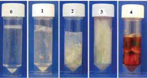
performance is reflected by an increase in the number of services per conception, calving to first service interval, and calving to conception interval (Kanneganti et al., 2007; Thompson et al., 2011), and in reduced risk of pregnancy (Balu et al., 2002) and conception (Salamonsen et al., 2007). The reproductive inefficiency associated with clinical endometritis (Fahey et al., 2005) translates into a significant economic loss for the dairy industry (Tabibzadeh, 1990; Thompson et al., 2011). This financial impact is caused by excessive culling, milk loss, infertility treatment costs and the genetic losses due to a decrease in replacement heifers. Sheldon et al. (2005) devised a 4-point classification system for purulent vaginal discharge where 0 = no discharge, 1 = clear and translucent mucus, 2 = cloudy mucus with or without flecks of pus (<50% pus), 3 = mucopurulent discharge more than 50% pus, and
Figure 1: Classification system for purulent vaginal discharge (Williams et al., 2005)
Typical samples of vaginal mucus character: score 0 = clear or translucent mucus; score 1 = mucus containing flecks of white or off-white pus; score 2 = discharge containing ≤50% white or off-white mucopurulent material; and score 3 = discharge containing ≥ 50% purulent material, usually white or yellow, but occasionally sanguineous (score 4). Previously published
with (Williams et al., 2005)
2.3 Subclinical endometritis
Subclinical endometritis is defined by the presence of neutrophils over a certain threshold and certain DIM in the uterine cavity and on the endometrium in absence of systemic clinical signs and PVD after 21 days postpartum (Kasimanickam et al., 2004). According to previous studies, more than 18% of neutrophils at 20-33 days after parturition or more than 10% at 34-47 days after calving could be used as thresholds to diagnose subclinical endometritis (Kasimanickam et al., 2004). As is the case for clinical endometritis, subclinical endometritis occurs primarily after 3 weeks postpartum (Table 1).
Reference No. cows DIM* Diagnostic method Prevalence Impact** Median DTP*** Unaffected Endometritis (Kasimanickam et al., 2004) 228 in 2 herds 20 to 33 Cytobrush >18% neutrophils 35%( excluding cows with PVD**** the days before 112 141 (Gilbert et al., 2005) 141 in 5 herds 40 to 60 Flush > 5% neutrophils 53% ( no vaginal examination) 118 206 (Barlund et al., 2008) 221 in 8 herds 28 to 41 Cytobrush > 8% Neutrophils 12% ( not excluding cows with PVD) 121 145
(Galvao et al., 2009) 202 in 1 herd 48 to 54 Flush ≥ 5%
neutrophils 29% ( not excluding cows with PVD)
112 126
(Galvao et al., 2010) 406 in 1 herd 32 to 38 Flush ≥ 7% neutrophils 38% ( not excluding cows with PVD) 121 151 (Dubuc et al., 2010) 2072 in 6 herds 32 to 38 Cytobrush ≥ 6% neutrophils 20% (13% cytological only and 7% both cytological and PVD) 132 148 (cytological only) 195 (both cytological and PVD)
Table 1: Summary of studies on the prevalence of endometritis diagnosed by cytology in postpartum dairy cows (Kasimanickam et al., 2004)
DIM*: days in milk
Impact** : All difference in DTP < 0.05 in survival analysis DTP*** : days from calving to pregnancy
2.4 Pyometra
Pyometra is an endometrial inflammatory disease that occurs later in postpartum (> 45 DIM) and is characterized by an accumulation of purulent exudate in the uterine cavity, along with no systemic clinical signs and PVD (closed cervix) in the presence of a persistent corpus luteum (Chapwanya et al., 2009; Foldi et al., 2006;Sheldon and Dobson, 2004; Sheldon, 2004). It is often diagnosed at the first artificial insemination pregnancy check using rectal palpation. The diagnosis is confirmed by ultrasonography.
Collectively, these genital tract conditions indicate a failure of both physical barriers and the innate and acquired immune defense mechanisms that normally limit microbial entry and colonization of the uterine cavity.
3 Diagnostic tools for postpartum endometritis
Historically, all postpartum uterine diseases were diagnosed by manual transrectal palpation. This explains part of the confusion associated with the diagnosis of postpartum uterine diseases (Heuer et al., 1999; Sheldon et al., 2006).
3.1 Manual transrectal palpation
Rectal palpation is a very important part of the general and genital clinical examination of the animal and, therefore, is one of the diagnostic tools used for the female genital tract assessment. However, uterine palpation per rectum to diagnose endometritis is not reliable (Gilbert 1992 ; Foldi et al., 2006 ; Sheldon et al. 2006 ; Palmer et al., 2008). Characterization of the uterus depends on several factors, including breed, and age of the animal, ability of the veterinarian, disposition of the cow, farm facilities, uterine size, etc. This technique is also limited when aiming for a detailed and objective analysis of the pathogenic process (Chapwanya et al. 2009).
3.2 Vaginoscopy, Metricheck and gloved hand techniques
Vaginal examination is the most reliable method for assessing clinical endometritis in postpartum cows. Endometritis can be identified by categorizing the vaginal discharge and inspecting the vaginal and cervical appearance in postpartum to find abnormalities (LeBlanc et al., 2002). The vaginal discharge can be identified using three different methods: 1) gloved hand, 2) Metricheck, and 3) vaginoscopy. The lubricated gloved hand technique allows for an
assessment of the amount of vaginal discharge; however, the use of lubricant makes it impossible to recognize the absence of vaginal discharge. The Metricheck, a long spoon-like instrument, is
However, scraping the vaginal wall makes it harder to assess the appearance of the vaginal discharge. The vaginoscope provides a view of the undisturbed discharge in the vagina, enabling a better assessment of the appearance of the mucosa and the cervix. Drillich et al. (2004)
reported a sensitivity and specificity for the vaginoscopy of 12.3% and 90.2%, respectively, compared to endometrial cytology done by cytobrush. Barlund et al. (2008) found that
vaginoscopy lacks sensitivity when compared to cytobrush cytology for the diagnosis of clinical endometritis and subclinical endometritis. Event with a low sensitivity, vaginoscopy is still the best technique for characterizing vaginal discharge and for identifying clinical endometritis based on reproductive efficiency. It is recommended that the technique be used as a routine diagnostic method by veterinarians in postpartum genital examination (Leblanc et al., 2002).
3.3 Ultrasonography
Ultrasonography is a good practical method, but when used alone it is not reliable enough to identify reproductive diseases (Barlund et al., 2008; LeBlanc et al., 2002). This technique is used extensively as a diagnostic method in veterinary medicine to assess the presence of uterine fluid (Maira et al., 2012) and to measure endometrial thickness (Barlund et al., 2008). Agreement between the ultrasonography and cytobrush methods is not good (Barlund et al., 2008; Kasimanickam et al., 2004). The ultrasonographic measurement of uterine discharge and
endometrial thickness is not a sensitive indicator of pregnancy status at 150 days (Barlund et al., 2008). When ultrasonography and endometrial cytology are combined, both sensitivity (20%) and specificity (80%) are improved (Kasimanickam et al., 2004). Consequently, the use of both methods in clinics is recommended (Barlund et al., 2008). Ultrasonography alone is not a good tool for monitoring uterine health in postpartum cows.
3.4 Endometrial cytology
Endometrial cytology can be performed using two different methods: cytobrush (Figures 2 and 3) and shallow uterine lavage (Barlund et al., 2008). Both techniques are reliable for establishing a diagnosis of endometritis in postpartum cows (Gilbert et al., 2005; Kasimanickam et al., 2005; Kasimanickam et al., 2004; Drillich et al., 2004; Barlund et al., 2008). Gilbert et al. (2005) collected endometrial cells using the uterine lavage method and reported a neutrophil threshold of 5% between 40 and 60 DIM for the diagnosis of endometritis. Uterine lavage has also been described as a method for evaluating the bacterial growth in intrauterine fluid (Barlund et al., 2008; Mateus et al., 2002). The cytobrush method is reported to be a more reliable tool for harvesting inflammatory cells than uterine lavage because it requires less time, is more consistent, and the endometrial cells are better preserved. By contrast, about 17% of uterine lavages fail to collect cells (Barlund et al., 2008; Kasimanickam et al., 2005). Using a neutrophil threshold of 8%, Barlund et al. (2008) showed the sensitivity of endometrial cytobrush cytology to be only 12.9%, although specificity was 89.9%, in cows sampled between 28 and 41 days postpartum based on pregnancy rates at 150 DIM. As a result, the researchers concluded that cytobrush cytology is the most accurate procedure for identifying endometritis.
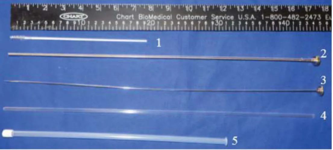
Figure 2: cytobrush and stainless steel instrument.
Cytobrush (1), solid stainless steel rod (2), stainless steel tube (3), sanitary plastic sleeve (4) and plastic protective sheat (5)
Figure 3: The cytobrush system to reduce sample contamination Image showing how to endometrial cytobrush samples were taken
Cytology device was manipulated through the vagina to the external cervical os (1); the the sanitary sleeve was punctured and the instrument was passed through the cervix into
the uterine lumen (2); endometrial cytology samples were collected by rotating the cytobrush in a clockwise in contact with the uterine wall (3).
3.4.1 Uterine biopsy
Endometrial biopsy has been used since the 1960s as a tool for investigating infertility in mare and is also the most reliable method for diagnosing endometritis in mares. However, this is not the case in cows (Barlund et al., 2008; Gilbert et al., 2005; Sheldon et al., 2006). While it is used on cows (Chapwanya et al., 2009), it is very expensive, time consuming and invasive (Barlund et al., 2008). Uterine biopsy enables the assessment of the level of mucosal inflammation (deepness in the tissue) based on inflammatory cells infiltration. However, histologic interpretation is still subjective and imprecise (Bonnet et al., 1991; Meira et al., 2012) and not associated with fertility. Therefore, it is not applicable in the field as a diagnostic tool.
3.5 Uterine bacteriology
Bacterial culturing can be performed on biopsied and cytological tissues, or uterine fluids. After culturing, bacteria can be categorized into three groups: 1) bacteria causing endometrial lesions, 2) bacteria not causing endometrial lesions, and 3) opportunistic bacteria (Williams et al., 2005). Among all of the different bacteria that have been isolated, only T. pyogenes has been associated with endometrial lesions (Williams et al., 2005, Werner et al., 2012, Prunner et al., 2014). However, bacterial culturing is expensive and there is a considerable time lag between sampling and culture results.
4 Uterine defense in postpartum
The immune system plays an important role during the physiological stages of parturition and during the involution of the genital tract after parturition (Chapwanya et al., 2009). After calving, bacteria in the vaginal cavity invade the uterine lumen. The innate immune defense system represents the first line of defense against contamination (Stossel, 1977).
4.1 Postpartum immune response
During pregnancy, the cow is in an immunotolerant state in which the local uterine immune defense is modulated to accept the new fetus and maintain the pregnancy for 9 months. This must then give way to an active uterine immune response after calving in response to a large population of bacteria entering the uterus. Innate immunity includes anatomical and
physiological barriers to prevent bacterial entry, along with local cellular defenses to eliminate contaminating bacteria. Once physical barriers are breached, immune cells and epithelial cells detect and eliminate invading bacteria.
During parturition, a surge in fetal cortisol level around calving triggers peripheral leukocytosis (Preisler et al., 2000(Kim et al., 2005), which is followed by leucopenia during the first week postpartum. This decline in leukocytes is associated with the significant demand for leukocytes by the uterus and mammary glands (Guidry et al., 1976; Singh et al., 2008). Peripheral levels of leukocytes gradually return to normal levels in the following 3 weeks postpartum (Cai et al., 1994; Hussain and Daniel, 1992; Kim et al., 2005; Mateus et al., 2002; Singh et al., 2008). The neutrophils are the most important cells involved in the innate immune response of the uterus
neutrophils from blood vessels and bone marrow by chemotaxis to the uterine cavity. This is associated with increased cytokine secretion until 24 days postpartum (Gabler et al., 2010). Studies show that neutrophils represent 68% of endometrial cells collected for cytology.
The number of neutrophils then declines by about 15% by the fourth week postpartum in normal uterine involution. A neutrophil threshold on endometrial cytology of between 5 and 18%, depending on days after calving, has been used to establish a diagnosis of chronic inflammation of the endometrium in dairy cows in the postpartum period (Kasimanickam et al., 2004).
However, the proper threshold value for neutrophils is still being debated. The healthy uterus should not be completely free of neutrophils, but rather contain a limited number (Kluciński et al., 1990). The effective action of neutrophils not only depends on the number of neutrophils, but also on their phagocytic capacity. Cows with postpartum uterine infections have been shown to have an increased concentration of neutrophils but a lower phagocytic capacity compared to cows with normal uterine involution (Mateus et al.; 2002, Zerbe et al., 2000 and 2003). The bacterial killing mechanism includes chemotaxis, adherence, and the ingestion and digestion of the bacteria (Hussain, 1989). The phagocytic capacity of peripheral blood neutrophils is still being debated because some studies have reported normal neutrophil function throughout the peripartum period (Hoedemaker et al., 1992; Cai et al., 1994; Mateus et al., 2002).
Macrophages are also an important component of innate immune defense. Although they are not present on the endometrium in large numbers (Tison et al., 2017) because they reside in deeper layers of the uterine wall. They are involved in phagocytosis, antigen presentation, regulation of inflammation in the uterine cavity during the postpartum period (Hansen, 2013) and promotion of tissue remodeling. Along with dendritic cells, macrophages are essential for T-lymphocyte activation (Hashimoto et al., 2011) and vary during different stages of gestation. In the first half
of the gestation, their numbers are reduced in order to prevent immunological fetal rejection (Hansen, 1997). In the last half, macrophages are found in the intercaruncular endometrium. The eosinophils and mast cells are rarely found in the uterine cavity; however, they are present beneath the endometrium and possess high affinity receptors for IgE (Bondurant, 1999). Binding to these receptors triggers the degranulation of mast cells, leading to the release of inflammatory mediators (TNF-α, histamine, prostaglandins, interleukins, chemokines, etc.). The release of these mediators increases vascular permeability, which, in addition to induce surface epithelial damage, allows circulating IgG to reach the uterine lumen (Bondurant, 1999).
4.2 Humoral immunity
Since innate immunity plays such a major role in postpartum uterine defense, the role of the local humoral response is limited but important (Mestecky et al., 2005). B-lymphocytes and their role in genital tract immune defense during the postpartum period is not well studied, although their role is demonstrated by the presence of immunoglobulins IgG, IgM, and/or IgA in the mucus of the cervix, vagina and uterus (Dhaliwal et al., 2001b; Herr et al., 2011) (Corbeil et al., 1976; Corbeil et al., 1974; Dhaliwal et al., 2001a; Watson et al., 1990). Antibodies may help in
opsonization and, therefore, phagocytosis by neutrophils and macrophages. Like macrophages, B and T-lymphocytes are present in the subepithelial uterine stroma (Leung et al., 2000).
The production of humoral antibodies in the reproductive tract is stimulated following an exposure to local antigens. A portion of total IgG concentrations in the uterine lumen is synthesized locally in the endometrium; for example, specific antibodies at the bovine uterine mucosal surface against Campylobacter fetus subsp. veneralis have been reported (Butt et al.,
endometrial inflammatory process resulting from microbial challenge, and the prognosis for clinical recovery (Aknazarov, 1988). Local IgG and IgM concentrations decrease after calving in healthy cows (Dhaliwal et al., 2001a). However, both IgG and IgA concentrations in lochia increase drastically in cows with endometritis (Aknazarov, 1988; Dhaliwal et al., 2001a; Herr et al., 2011). The synthesis of immunoglobulins like IgA, IgG and IgM in the genital tract and their significance to local defense mechanisms is still not clearly understood (Dhaliwal et al.,
2001b)Dhaliwal et al., 2001a; Herr et al., 2011).
Systemic biomarkers (either from the innate or the adaptive immune systems) have been studied for their usefulness in diagnosing uterine inflammation and infection. Acute-phase proteins (APPs) like haptoglobin (hp) and serum amyloid A (SAA) has been shown to increase in cows with endometritis compared to normal postpartum cows (Brodzki et al., 2015). Like blood leukocytes and cytokines, they are not specifically associated with inflammation of the reproductive tract alone. Most studies do not control for alternative sources of inflammation. Fibrinogen is used in cattle as an indicator of the presence of inflammation; bacterial infection and surgical trauma (Hirvonen and Pyorala, 1998). It is involved in homeostasis, providing a substrate for fibrin formation and tissue repair, establishing a matrix for the migration of related inflammatory cells (Thomas, 2000; Bakes and Illek, 2006). Fibrinogen binds specifically to CD11/CD18 integrins on the cell surface of migrated phagocytes, triggering a cascade of intracellular signals that lead to degranulation, phagocytosis, cellular cytotoxicity and delayed apoptosis (Ruble et al., 2001). Fibrinogen concentrations in the plasma of cows with subclinical and clinical mastitis have been found to be significantly higher than in the plasma of healthy cows (p < 0.05; Tabrizi et al., 2008). Like other APPs, fibrinogen is not specific to uterine inflammation or infection, and other sources of inflammation and infection need to be ruled out.
4.3 Epithelial cells of the endometrium and cytokines
Following parturition and for a minimum of 2 weeks, uterine caruncles lack epithelial cells. During this period, tissue necrosis increases the risk of uterine infection and inflammation during the postpartum period. Like the immune cells, the epithelial cells lining the uterine cavity can recognize microbes and initiate the innate defense mechanism. Epithelial cells have the capacity to recognize pathogen-associated molecular patterns (PAMPs) and damage-associated molecular patterns (DAMPs) through their various pattern recognition receptors (PRRs). The most
important PRR is the family of Toll-like receptors, which are expressed by epithelial cells (TRL1-7 and 9) and the stroma (TRL1-4, 6, 7, 9, and 10). These receptors on endometrial cells detect PAMPs such as bacterial DNA, lipids and lipopolysaccharides (LPSs) on the surface of bacteria. The initial defense of the endometrium against microbes also includes antimicrobial peptides (AMPs) and APPs (King et al., 2003; Wira et al., 2005). Cows with an inflamed endometrium have been shown to express significantly more TLRs, interleukins, prostaglandin endoproxide synthase-2, lipocalin prostaglandin D synthase, and tumor necrosis factor α (TNF-α) compared to cows with a healthy endometrium (Gabler et al., 2009; Herath et al., 2006b; Kasimanickam et al., 2013).
The binding of PAMPs to PRRs leads to the secretion of inflammatory mediators like cytokines, chemokines and AMPs (Akira et al., 2006; Beutler, 2004). Epithelial cells recognize LPS
through TLR4 by upregulating expression of pro-inflammatory cytokines (Swangchan-Uthai et al., 2012; Fu et al., 2013). Inflammatory mediators include pro-inflammatory cytokines like IL-1α, IL-1β, IL-6 and TNF-α, chemokines like IL-8 and prostaglandin E2, and
anti-et al., 2003; Sheldon, 2004; Turner anti-et al., 2014; Tzianabos, 2000). Cytokines and growth factors released by the maternal endometrium play a key role in the signaling cascade (Fischer et al., 2010; Kauma, 2000; Sharkey, 1998). The role of PRRs in recognizing DAMPs and PAMPs is important for the response of endometrial cells to pathogens and cellular debris during uterine involution. Epithelial cells may also act as immune effectors as IL-1 and IL-17, histones, and complement components can modulate the secretion of AMPs (Kolls et al., 2008; Drab et al., 2014; Kania et al., 2001).
The cytokine IL-8 is a potent neutrophil chemoattractant, directing neutrophils and activating the inflammatory focus (Fischer et al., 2010; Hoch et al., 1996; Struyf et al., 2005). It is also a potent granulocyte-macrophage colony-stimulating factor (GM-CSF) activator and essential for
macrophage maturation (MacKintosh et al., 2013; de Moraes et al., 1999). IL-8 is produced by activated monocytes and macrophages in response to bacterial toxins and inflammatory
cytokines such as IL-1, IL-6 and TNF-α (Baggiolini, 2001; Fischer et al., 2010; Monaco et al., 2004). Ghasemi et al. (2012) reported local elevated concentrations of IL-8 at 30 days
postpartum in cows with endometritis.
IL-6 is released at the apical surface of epithelial cells and contributes to inflammation and neutrophil recruitment. At a later stage of uterine involution it may influence monocyte or macrophage migration (Hurst et al., 2001). Monocytes and macrophages stimulate signaling pathway like NFκB and improve the transcription of the inflammatory cytokines such as IL-6, TNF-α and IL-1 (Ishikawa et al., 2004). IL-6 blood concentration before calving in dairy cows with endometritis is higher than in the healthy cows (Ishikawa et al., 2004) and decreases gradually within 8 days.
TNF-α induces the production of AMPs for the elimination of pathogenic bacteria (Fischer et al., 2010) and may control prostaglandin synthesis in cattle endometrium (Kasimanickam et al., 2013; Okuda et al., 2004). TNF-α is another stimulating factor for IL-8 secretion and increases adhesion molecule expression in vascular endothelial cells (Kasimanickam et al., 2013; Okuda et al., 2004).
IL-10 is the most important anti-inflammatory cytokine (Galvao et al., 2012; Sabat et al., 2010) and is produced by macrophages and/or monocytes, dendritic cells and certain subtypes of T cells (Islam et al., 2013; Sabat et al., 2010); Tizard, 2009). The most important biological
function of IL-10 is to control macrophage and/or monocyte-derived TNF-α, IL-1, IL-6 and IL-8 (Islam et al., 2013; Mosser and Zhang, 2008)(Couper et al., 2008; Herath et al., 2009; Wira and Rossoll, 2003; Xu et al., 2008).
5. General issue
Despite the negative impact of postpartum endometritis on the reproductive efficiency of dairy cows, very little is known about the mechanisms behind the disease and, more particularly, the immune defense mechanisms. Most studies have focused on the local aspect of the uterine defense and rare studies have assessed systemic markers other than cytokines and chemokines. Usual systemic markers of inflammation used in assessing the animal health status (such as neutrophils and fibrinogen) as well as immunoglobulin levels need to be revisited during the whole transition period of dairy cows since these markers are also synthesized elsewhere than the uterus.
6 General hypothesis
Levels of systemic indicators of inflammation (immunoglobulin G, white blood cells , and fibrinogen) during the transition period are associated with postpartum endometritis in normal dairy cows.
7 Objectives
Objective 1: Estimate concentrations of systemic biological indicators of inflammation like white blood cells , fibrinogen and IgG during the transitional period in dairy cows.
Objective 2: Compare systemic biological indicators of inflammation in dairy cows with and without postpartum endometritis.
Article 1
Levels of immunoglobulin G, white blood cells and
fibrinogen in dairy cows with and without endometritis
during the transitional period
Ali Bazzazan
1,
Nelson Enrique Prieto Cardenas
1, Dominic Dolbec
2,
Mariela Segura
2, Réjean C. Lefebvre
11 Department of Clinical Sciences, Faculty of Veterinary Medicine, University of Montreal,
Saint-Hyacinthe, Quebec, Canada
2 GREMIP, Faculty of Veterinary Medicine, University of Montreal, Saint-Hyacinthe, Quebec, Canada
Journal of Theriogenology
Corresponding author: Réjean C. Lefebvre
Faculté de médecine vétérinaire Université de Montréal
C. P. 5000
St-Hyacinthe, QC J2S 7C6 Tel.: 450-773-8521, ext. 8514 Fax: 450-778-8120
Introduction
The reproductive health of cows is the foundation of productivity in the dairy industry. Reproductive performance is particularly dependent on uterine health during the postpartum period, although the whole transition period is a critical time for dairy cows. It is a period of stress management for the cow (Goff and Horst, 1997), requiring metabolic adjustments in the face of a significant negative energy balance, and immunity modulations enabling a smooth transition between the end of gestation and the restoration of uterine condition, cyclicity and the establishment of a new pregnancy (Drillich et al., 2001; Overton and Yasui, 2014; Sheldon et al., 2006).
Under normal circumstances, close to 100% of cows have uterine contamination within the first 2 weeks after calving (LeBlanc, 2008; Sheldon et al., 2008). Although the innate immune system usually eliminates all of the offending microbes, persistent infection or inflammation reportedly occurs in between 15 and 40% of postpartum cows (Sheldon et al., 2008). Postpartum uterine disease is commonly associated with Escherichia coli, Trueperella pyogenes, Fusobacterium
necrophorum and Prevotella species (Ruder et al., 1981; Olson et al., 1984). Numerically, the
most common pathogens are E. coli (37% of pathogenic bacteria isolated) and T. pyogenes (49%) (Williams et al., 2005). When contamination of the uterus persists beyond 4 weeks postpartum, the uterine infection is referred to as endometritis. Clinical and subclinical
endometritis are prevalent conditions in postpartum dairy cows, causing economic losses due to decreases in both milk production and fertility (Sheldon et al, 2004a). Clinical endometritis is defined as inflammation of the endometrium in cows with purulent vaginal discharge (PVD) in the absence of systemic clinical disease 26 days postpartum (Sheldon et al., 2006), while
subclinical endometritis is defined by the presence of > 18% polymorphonuclear neutrophils (PMNs) in uterine cytosmears prepared 21 to 33 days postpartum, or > 10% PMNs in cytosmears prepared 34 to 47 days postpartum (Kasimanickam et al., 2004).
Cellular and humoral mechanisms of nonspecific and specific immunity play an important role in the resolution of postpartum endometritis in cows. PMNs are the first and most significant cell type recruited during uterine inflammation (Subandrio et al., 1997; Herath et al., 2006a; Rogan et al., 2005). PMNs and monocytes are recruited from the circulation into the lumen of the uterus to eliminate bacteria (Tizard, 1996; Wade and Lewis, 1996). Like PMNs and macrophages,
endometrial cells also have Toll-like receptors (TLRs), which recognize pathogens present in the uterus. For example, TLR4 recognizes lipopolysaccharides (LPS) in the cell wall of
Gram-negative bacterial species, resulting in initiation of the innate immune response that is manifested as antibacterial molecules produced by responding cells (Herath et al., 2009). This inflammatory response results in attraction of PMNs among other cells and amplification of the inflammatory loop (Sheldon, Cronin et al., 2009). The endometrial cells also secrete diverse molecules, including cytokines, whose levels in uterine secretion reflect the intensity of the endometrial inflammation (Singh et al., 2008). In addition, immunoglobulins (Ig) also have a protective role against pathogens in the bovine uterus (Curtain et al., 1971; Dhaliwal et al., 1996a; Dhaliwal et al., 1996b; Dhaliwal et al., 2001b; Duncan et al., 1972; Whitmore and Archbald, 1977).
However, the specific functions of the isotopes IgA, IgG and IgM in the genital tract are still not clear. Globally, these findings suggest that innate and adaptive immunity, both local and
systemic, play an essential role in defending the uterus against postpartum infection. As such, the aims of this study were to: 1) estimate concentrations of systemic immune indicators like white
blood cells (WBC), IgG, and fibrinogen during the transitional period in dairy cows; 2) compare these concentrations in dairy cows with and without endometritis.
9 Materials and methods
9.1 Animals
All procedures conformed to the guidelines of the Canadian Council on Animal Care, and the animal care committee of the Université de Montréal approved the experimental protocol. A total of three different commercial dairy herds located in Quebec (Canada) were recruited, and 61 multiparous cows were included in the study between June 2016 and February 2017. Herd size was from 70 to 130 lactating cows. Reproductive and health data were compiled in a databank using health record management software (DSAHR). Rolling herd average milk production was about 9000 kg. Cows from tied stall barns were milked twice daily. Cows were fed a total mixed ration formulated mainly with corn and hay silage to meet the dietary requirements for a
lactating dairy cow (NRC, 2011). Farms were visited weekly by the same veterinarians, and all cows were vaccinated two times against E. coli (J-VAC, Merial Inc., Athens, GA, USA) before calving (2 ml intramuscularly, D–40 and D–26 before parturition) and one time against BVD type 1 and 2, IBR, PI-3, and BRSV (Bovi-Shield GOLD® FPTM 5 L5, Zoetis,Parsippany, New Jersey 07054, USA) (2 ml intramuscularly, D15-40 after calving), and injected with Se (5.0 ml, D–60, MU-SE, Intervet Canada Corp., a subsidiary of Merck and Co. Inc, Kirkland, QC, Canada H9H 4M7).
9.2 Experimental design
This was an observational prospective cohort study in which all cows (n=61) were systematically and consecutively enrolled at drying period and then examined six times between 40 days before and after calving. Exams were carried out on cows at day 40 ± 4 days (D–40), 26 ± 4 days (D– 26) and 12 ± 4 days (D–12) before calving (DBC), and on day 7 ± 4 days (D+7) , 21 ± 4 days (D+21) and 35 ± 4 days (D+35) after parturition (DAP, Annex 1). All six exams included an assessment of lameness, cyclicity, and body condition, the collection of milk and blood samples, a transrectal and vaginal exam (TE), and a vaginal cytobrush (Figures 2 and 3). Before calving, a TE was performed in order to confirm the pregnancy; it involved the assessment of placentomes, fetus viability and the uterine artery (fremitus). After calving, a TE was performed in conjunction with ultrasonography to determine the diameter of the cervix and uterine horns, assess the
presence of fluid in the uterus, and assess the ovarian structures (corpus luteum, dominant follicle and follicular cyst). In addition, a uterine cytobrush and vaginoscopy were performed. Cows were not enrolled in a systematic synchronization protocol, and reproductive data from the cows were collected for at least 300 days in milk (DIM). During the 70-80 days follow-up, cows did not receive any antibiotics except for intramammary mastitis treatment. Diseased cows (n = 11) were identified based on four criteria. In decreasing order of importance these were: 1) cows with the presence of T. pyogenes grade +2 or more in the uterus (EXAM 5), 2) cows with PVD of grade ≥2 (Tison et al., 2017), 3) cows with >18% PMNs on endometrial cytology (number of PMNs/number of total cells (Gilbert et al., 2016)), and 4) cows with cervix of grade 2 (Hartmann et al., 2016). The control group animals (n = 11) were characterized by the absence of all four criteria (T. pyogenes of grade < 2, PVD < 2, PMNs < 5%, cervicitis of grade < 2).
9.3 Gynecologic examination
9.3.1 Vaginal examination
The vaginal examination was performed to assess the cervix based on the classification of Hartmann et al. (2016), and the vaginal discharge based on the classification of Tison et al. (2017) in postpartum cows. Following the transrectal palpation, the vulva was cleaned with a wet paper towel and gauze with alcohol before performing the vaginoscopy. The vaginal speculum (50 cm long) was inserted without lubricant through the vulva and into the vagina up to the outer cervical os. With a light source, the vaginal vault and cervical os were checked visually for the presence of discharge and categorized using a 4-point classification system (Tison et al., 2017). Briefly, the vaginal content was classified as 0 = no discharge, 1 = clear and translucent mucus, 2 = cloudy mucus with or without flecks of pus (< 50% pus), 3 = mucopurulent discharge (> 50% pus), and 4 = purulent discharge with fetid smell (Figure 1). The transrectal examination was performed first for convenience, as the detection of vaginal discharge by vaginoscopy is not enhanced by a preceding transrectal palpation of the uterus (Leutert et al., 2012). The cervix was assessed using the Hartmann et al. (2016) classification. The cervix was classified as C0 = cervix without abnormality, C1 = second cervical fold swollen without redness, and C2 = second
9.4 Blood sampling
Blood samples were taken at all six examinations. The sample was collected from the coccygeal vein of the tail heal and placed in a tube either with an anticoagulant (hematology analysis) or without an anticoagulant (biochemistry analysis). Blood samples were immediately placed on ice and transported to the laboratory within approximately 3 hours. Once clotting was complete, the serum was separated by centrifugation at 3000 RPM for 10 min, and beta-hydroxybutyrate measured with the Freestyle Precision NEO Kit (Abbott Laboratories Ltd, Abbott Park, IL, USA). Finally, 3.0 ml serum aliquots were stored frozen at –80°C for further analysis. After 30 min at room temperature and gentle agitation, the complete hematology analysis was carried out with a VetScan HM5 hematology analyzer (Abaxis Global Diagnostics, Union City, CA, USA).
9.5 Quantification of serum IgG and fibrinogen
concentrations
Bovine IgG levels were measured by ELISA using a commercial Bovine IgG ELISA Quantitation Set (E10-118, Bethyl Laboratories Inc., Montgomery, TX, USA). As per the instructions provided with the kit, 96-well plates were coated with 100 μl of affinity purified sheep anti-bovine IgG antibodies and incubated at room temperature (20-25°) for one hour, after which the excess antibody was removed by washing five times. The wells were blocked using tris-buffered saline (TBS) with 0.05% Tween 20 and incubated at room temperature for 30 min, after which the excess antibody was removed by washing five times. A standard curve was generated by serial dilution of the reference serum provided in the kit. Bovine serum samples were diluted up to 1/640,000. A volume of 100 μl for each sample or standard was transferred
into the wells and incubated for one hour, after which the wells were washed five times. Each well was then incubated for 1 hour with 100 μl of a horseradish peroxidase (HRP) conjugated antibody solution, and then washed again five times. Finally, 100 μl of tetramethylbenzidine (TMB) solution was distributed in each well and incubated as per the instructions provided. The oxidation of TMB by HRP was stopped using 100 μl of a diluted sulfuric acid solution before the absorbance measured 450 nm on an automated well plate reader (μQuant MX200, Biotek, VT, USA). Plasma fibrinogen levels were measured using a heat-precipitation method; protein levels were measured using a refractometer. Briefly, 2 capillary tubes were filled with blood (EDTA) and spun in a high-speed microhematocrit centrifuge at 12 000 x g for 5 min to clear the plasma at room temperature. The total protein concentration in the plasma of one tube was determined using a refractometer. The other tube was immersed in a water bath at 57°C for 3 min to precipitate the fibrinogen. It was centrifuged again at 12 00 x g for 5 min at room temperature, and the total protein concentration of the remaining plasma was determined. The difference between the 2 measurements was the fibrinogen concentration.
9.6 Endometrial cytological sampling
Uterine cytobrush samples (n = 61) were collected for routine endometrial cytology and bacteriology assessment. Briefly, a sterile cytobrush (CytoSoft, Camarillo, CA, USA) was screwed onto a stainless steel rod (65 cm long x 4 mm diameter) within a stainless steel tube (Couto et al., 2012). The apparatus was inserted into a hard protective plastic sheath (IMV Technologies, L’Aigle, France) before insertion into a second protective sheath (30 cm long Sani-Shield Rod Protector, Agtech, Manhattan, NY, USA) in order to reduce the risk of vaginal
instrument into the vagina by parting the vaginal lips and moving it into the cranial vagina. With a sleeved arm in the rectum for manipulating the cervix, the veterinarian pushed the lubricated (J-Lube Powder, Jorgensen Laboratories Inc., CO, USA) instrument through the cervix. The cytobrush was then pushed out of the sheaths and stainless steel tube into the uterus. The sterile cytobrush, now in contact with the mucosa, was gently rotated clockwise (360 degrees) to collect cellular material. The veterinarian then retracted the cytobrush into the stainless steel tube and hard plastic sheath, and withdraw the apparatus from the cervix into the second protective sheaths (double pipette). The slides for cytological examination were prepared at the farm. The bristles of the brush were rolled over a sterile slide (Fischer Scientific, Toronto, ON, Canada), and then the brush tip was cutoff and packaged for bacterial culturing.
9.7 Preparation and staining of slides
The smear on the slide was allowed to dry at room temperature for 10-15 min. Cytology smears were stained with modified Wright Giemsa stain, using an automated slide stainer (Hema-tek Slide Stainer; Miles Scientific, Napierville, IL, USA). All slides were evaluated under the microscope at 250x and 400x magnification to obtain counts of individual inflammatory cell types (PMNs, lymphocytes, and monocytes), and endometrial epithelial cells. For each slide, a total of 300 cells were counted by one observer. Cows were classified as endometritis positive if more than 18% PMNs was present.
9.8 Uterine bacterial culturing and identification
Uterine cytobrush samples were collected only at 21 days after calving (EXAM 5) for routine bacterial culturing (aerobic and anaerobic) using standard methods for bacteriological testing (API system, bioMérieux, Marcy l’Étoile, France). Cytobrush samples were stored in a culture tube (Starplex Scientific Inc., Etobicoke, ON, Canada) and were transported at room temperature to the diagnostic laboratory of the Faculty of Veterinary Medicine within 3 hours. For
microbiological analysis, the brushes were plated onto sheep blood agar with a sterile disposable plastic eye (soy agar with 5% sheep blood, Becton, Dickinson and Co., Sparks, MD, USA). Plates were incubated for 48 h at 35°C under aerobic conditions and then examined. When growth was observed, colony types were identified based on morphology, pigmentation and hemolytic patterns.
Beta hemolytic, catalase-negative minuscule colonies demonstrating coliform Gram-positive rods were identified as T. pyogenes, F. necrophorum and P. melaninogenicus and were
isolated by the standard procedure used at the diagnostic laboratory of the Faculty of Veterinary Medicine (PON-BAC-019). For other bacterial species, cytobrush samples were plated on Brucella agar containing Neomycin (100 g/ml) and incubated anaerobically at 35°C for 5 days. When Gram-negative rods were observed, colonies were examined using the API 20 A gallery system for identification of F. necrophorum and P. melaninogenicus. For isolation of E. coli, cytobrush samples were plated on blood agar and MacConkey agar (Oxoid Inc., Ottawa, ON, Canada) at 37°C. At the OIE Reference Laboratory for Escherichia coli (EcL; Faculty of
Isolates were submitted to three biochemical tests (indole spot, Simmon’s citrate and motility) for confirmation of E. coli. Isolates of E. coli were stored in tryptic soy broth containing 30% glycerol at –80°C (Becton, Dickinson and Co., Sparks, Maryland, USA).
9.9 Statistical analysis
Reproductive data were obtained from the health records databank. We calculated descriptive statistics and then analyzed the data using SAS v. 9.2, with cow as the unit of interest. The model took into account the herd effect as a random effect. Before the study, statistical power and sample size calculations were performed. Based on the results of Tison et al. (2017) and Harr et al. (2011), the following values were deemed indicative of endometritis: 18% PMNs on
endometrial, > grade 2 of PVD, > 2 cervix, and between15-35 mg/ml IgG in serum. Usinga one-sided test with 95% confidence and 80% power, 11 cows per group were required (G*Power, version 3.1.9.2, Germany) (Faul et al., 2007).
The data was not normally distributed and so were transformed using logarithm base 10 to normalize the distribution. A linear mixed model was applied, with farm and cow nested within the farm as random effects, and health status (control and diseased animals) and time (1 to 6) as fixed effects. Means for each health status at each time were compared. The alpha level was adjusted downward using the Benjamin-Hochberg procedure. For ordinal scores, the Cochran-Mantel-Haenszel test was used to examine potential differences in the distribution of score values between healthy and diseased cows. Statistical significance was defined as p < 0.05, while p < 0.1 was deemed as tendency.