P
URIFICATION AND CHARACTERIZATION OF BOVINE DENDRITIC
CELLS
Ximena Renjifo a e, Chris Howard b, Pierre Kerkhofs c, Martine Denis d, Jacques Urbain a, Muriel
Moser a, Paul Pierre Pastoret e
a Laboratoire de Physiologie Anima/e, Universite Libre de Bruxelles. Rue des Chemux 67. B-1640 Rhode
Saint Genese, Belgium
b Institute for Animal Health. Compton Laboratory, Compton. Newbury RG20 7NN, UK c National Veterinary Research Institute, 99 Groeselenberg. Bruxelles I 180, Belgium d SmithKline Beecham Biologicals. Rue de l'Institut 89, Rixensart 1330, Belgium
e Departement of Virology- Immunology, Fae. Vet. Med.. University of Liege. Liege 4000, Belgium
KEYWORDS: Cattle immunology; Bovine blood dendritic cells; Maturation of dendritic cells; Antigen-present- ing-cell-function
ABBREVIATIONS:FCS, Fetal Calf Serum; DC, Dendritic Cells; PBMC, Peripheral Blood Mononuclear Cells; PBS, Phosphate Buffered Saline; HBSS, Hanks' Balanced Salt Solution; AET, 2-Aminoethylisothiouronium bromide
ABSTRACT
Optimal activation of T lymphocytes depends on TCR interaction with peptide/MHC complexes in conjunction with costimulatory signals, which are delivered by specialized cells called antigen-presenting-cells (APC). The population of APC is heterogeneous and includes dendritic cells, B cells and macrophages. The family of dendritic cells (DC) is widely distributed in tissues and plays a major role in the induction of primary T-dependent immune responses. The aim of this paper was to isolate and characterize dendritic cells from cattle. Two methods are described that have been used to isolate dendritic cells from bovine peripheral blood. One method involves sequential depletion of other cells, adherence and isolation of low buoyant density cells on Metrizamide column. The second involves enrichment of cells displaying receptors for plasma fibronectin, followed by adherence and separation on Metrizamide. Both preparations were characterized morphologically by flow cytometry and functionally. Both procedures produced enriched populations that did not express molecules typical of T cells (CD3, CD4, CD8, WC I), B cells (slg, CD21) and monocytes (CD14, Fcg 2R). Procedure 2 yielded cells with a typical veiled DC morphology that were highly effective at stimulating allogeneic T cells. Procedure 1 yielded cells that did not have the veiled morphology and were less effective in the MLR which may represent a more immature stage.
Introduction
Dendritic cells (DC) were first identified in the mouse by Steinman and Cohnin, 1973, and were shown to represent a heterogeneous population distributed in lymphoid and nonlymphoid tissues throughout the body (Ardavin et al., 1993; Austyn et al., 1994; Strober and Kesall, 1996). DC are considered the major presenting cells involved in the activation of antigen-specific NAIVE T cells and therefore play an essential role in the initiation of immune responses (Steinman, 1991).
Extensive work in rodents and humans has demonstrated that the potent accessory properties of DC depend on a process of maturation (O'Doherty et al., 1993). Different maturational stages are evident in vitro (Winzler et al., 1997) and in vivo (Larsen et al., 1990; De Smedt et al., 1996) : immature DC efficiently process native antigens but are relatively poor at activating naive T cells (Romani et al., 1989). In contrast, mature DC have lost the capacity to efficiently process proteins but have upregulated the ability to activate T cells upon the first encounter with the antigen (Koch et al., 1995).
The maturation of DC seems to occur during their migration in vivo (De Smedt et al., 1996). There is evidence that DC circulate through the blood to the spleen and through the lymph to the lymph nodes (Bujdoso et al., 1989; Kudo et al., 1997). The maturation of DC is characterized by profound changes in expression of MHC class II and costimulatory molecules (Inaba et al., 1994) as well as in antigen-processing capacity (Schuler and Steinman, 1985).
Bovine DC isolated from lymph have been characterized phenotypically and functionally. McKeever et al. (1991); McKeever and Morrison (1992) showed that afferent lymph veiled cells rapidly internalize antigens deposited in the periphery, and process them for presentation to naive T cells in the draining lymph node.
Little is known, however, about DC populations in peripheral blood. In this paper, we describe two methods to isolate bovine DC from blood. Our results show that, depending of the isolation protocol, the DC-enriched populations display distinct phenotypes and functional properties and suggest that they may represent DC at different stages of maturation.
Materials and methods
PREPARATION OF CELLS
Protocol 1 : this protocol relies on sequential depletion of blood cells types and was successfully used to isolate human DC by Freudenthal and Steinman (1990). PBMC were isolated by Ficoll-Hypaque (Pharmacia, Uppsala, Sweden) density centrifugation from heparinized whole blood of healthy cattle. T cells were depleted by rosetting with 2-aminoethylisothiouronium bromide (AET, Sigma Chemical. St. Louis, MO) treated sheep red blood cells and the non rosetting population was incubated overnight in RPMI 1640 medium (Gibco BRL, Merelbeke, Belgium) in 5% C02 at 37°C. Non-adherent cells were layered over metrizamide gradient (14, 5% w/v)
(Nycomed Pharma, Oslo. Norway) according to a procedure described by Thomas et al. (1993) and centrifuged at 650g at room temperature for 10 min. The low density cells were collected, incubated with monoclonal antibodies to CD14 (CC-G33), CD3 (MMIA) and IgM 0LA30) (Howard and Naessens, 1993) and stained cells removed by panning on Petri dishes pre-coated with 10 µg/ml rabbit antibodies to mouse immunoglobulins.
a
Mean Cell Number (range) Yields Whole blood
Ficoll-Hypaque column
Blood mononuclear cells PBMC 5.4 108 (1.5 108 - 9.0 108) 100%
sheep erythrocyte rosette
T-cell rosette-depleted 1.7 108 (3.2 108 - 2.0 107) 31%
18 hours culture, adherence over plastic
Monocyte & T-cell depleted population 1.7 107 (2.9 107 - 6.0 105) 5%
Metrizamide column Low Density DC-enriched
Panning (CD3+, CD14+, lgM+)
Final DC enriched population 2.5 106 (9.6 106 - 9.0 104) 0.1%
b
Mean Cell Number (range) Yields Whole blood
Ficoll-Hypaque column
Blood mononuclear cells PBMC 5.0 108 (1.0 109 - 3.0 108) 100%
gelatin-plasma coated surfaces
T-cell rosette-depleted 5.8 107 (7.0 107 - 3.0 107) 11%
18 hours culture, adherence over plastic
Monocyte & T-cell depleted population 2.5 107 (4.8 107 - 1.2 107) 5%
Metrizamide column Low Density DC-enriched
Panning (CD3+, CD14+, lgM+)
Final DC enriched population 3.6 105 (1.5 106 - 2.0 105) 0.7%
Fig. 1. Sequences of steps that reliably enrich the population of bovine blood DC according to protocol I (a) and protocol 2 (b). The mean number of cells obtained after each step, the range (in parenthesis) and the yield are indicated and are representative of four independent experiments.
Protocol 2 : this protocol is a modification of a method used to isolate human monocytes by enrichment of cells expressing receptors for plasma fibronectin (Fig. 1b) (Bevilacqua et al., 1981). Polystyrene 170 cm2.flasks were incubated overnight at room temperature with 2%
gelatin (Sigma lmmunochemicals, St Louis, MO) in bidistilled water. The gelatin solution was removed and the flasks were dried for two hours at room temperature. The flasks were incubated with 10 ml heparinized autologous plasma for one hour at 37°C and washed with phosphate-buffered saline
Table 1. List of antibodies used in this study
Specificity mAb lsotypea Sourceb
CD1W2 CC-20 IgG2a IAH-C CD IW3 CC-43 lgG2b IAH-C CD2 CC42 IgG1 IAH-C CD3 MM1A lgG1 wsu CD4 CC8 lgG2a IAH-C CD5 CC17 lgG1 IAH-C CD6 CC38 lgG2b IAH-C
CD8a CC63 lgG2a IAH-C
CD11a ILA99 lgG2a ILRI
CD11b CC94 lgG1 IAH-C CD11c NAM-4 lgG1 FUNDP CD14 CC-G33 lgG1 IAH-C CD21 CC21 lgG1 IAH-C CD25 ILA111 lgG1 ILRI CD29 FW3-47 lgG1 BII-M CD44 25-32 lgG1 UMELB CD45 CCI lgG1l IAH-C
CD45RO ILA116 lgG3 ILRI
CD45R(B) CC76 IgG1 IAH-C
CD49d 218 IgG1 BIIM
BoWC1 CC15 IgG2a lAH-C
BoWC6 CC98 lgG2a IAH-C
BoWC10 CC28 IgG1 IAH-C
ALVC CC81 IgG1 IAH-C
gran/ monocyte ILA24 IgG1 ILRI
L-selectin CC32 IgG1 IAH-C
lgM ILA30 IgG1 ILRI
FeRll CCG36 IgG1 lAH-C
Fc2R CCG24 lgM IAH-C
MHC II (DQ) CC158 IgG2a IAH-C
MHC II ILA21 IgG2a ILRI
MHC I ILA88 IgG2a IAH-C
CD80/CD86 (B7) hCTLA-4-Ig IgG1 Dr. P. Linsley
a All antibodies are murine antibodies.
b UMELB Centre for Animal Biotechnology, School of Vet Science. University of Melbourne. Parkville 3052. Victoria,
Australia IAH-C AFRC. Institute for Animal Health. Compton Laboratory. Compton, Nr. Newbury. Berkshire RG16 ONN. UK. WSU Dept. of Vet Microbiology and Pathology. WA 99164 7040, USA (ILRJ) International Lifestock Research Institute, P.O. Box 30709, Nairobi, Kenya FUNDP. Facultes Universitaires Notre Dame de la Paix, 61 Rue de Bruxelles, B-5000 Namur. Belgium BII-M. Basel Institute for Immunolgy. Grezacherstrasse 487, Postfach, CH-4005 Basel, Switzerland Dr P. Linsley (Bristol-Myers Squibb Pharmaceu- tical Research Institute, Seattle, WA 98121).
(PBS). 3 X 10⁶ PBMC/ ml were incubated in gelatin /plasma coated flasks for two hours at 37°C.
Non-adherent cells were discarded and adherent cells were collected by incubation in 10 ml EDTA (10 mM in Ca2+, Mg2+-free Hanks' balanced salt solution) for 10 min at room
temperature, washed and cultured in Petri dishes overnight. The non-adherent cells were separated into low and high density fractions on Metrizamide gradient 14,5% (w/v). The low density cells were incubated with mouse antibodies to CD14, CD3 and IgM, as described above for protocol 1, and stained cells were removed by panning on Petri dishes coated with rabbit antibodies specific for mouse immunoglobulins. The DC-enriched, non-adherent fraction was collected and used for phenotypic and functional analysis.
FLOW CYTOMETRY
The antibodies and fusion protein used in immunofluorescence studies are listed in Table 1. Cells were washed twice with ice-cold PBS containing 1% BSA and 0.1% sodium azide. 2x10⁵
were washed with PBS and incubated for 30 min on ice with FITC-(Fab)2 goat anti-mouse IgG1 or PE-(Fab)2 goat anti-mouse IgG2a (Southern Biotechnology Associates, Birmingham, AL), washed again and analyzed on FACScan flow cytometer (Becton Dickinson). Propidium iodide was used to exclude dead cells from analysis.
MIXED LEUKOCYTE REACTION
The assays were carried out in 96 well round-bottomed microwell plates in 0.2 ml RPMI-1640 medium (Gibco BRL) containing 10% PCS and 5 X 10⁻⁵ M 2-mercapto-ethanol and 0.01 M Hepes.
T cells were obtained by rosetting mononuclear cells with AET-treated sheep erythrocytes and consist of at least 90% CD2+ cells. 4 x 10⁵ cells were cultured in the presence of various numbers
of irradiated (2000 rads) allogeneic or autologous DC. Cultures were maintained at 37°C in a humidified incubator (5% C02 ). Cells were pulsed with 0.4 µCi {3H} Thymidine (specific activity,
2 Ci/mmol; Amersham, UK) during the last 10 h of 7 ct-culture and collected onto filter paper using a semiautomated harvester. {3H} Thymidine incorporation was assessed by liquid
scintillation counting. The results are expressed as mean of triplicate cultures.
Results
PURIFICATION OF TWO SUBSETS OF DC FROM BOVINE BLOOD
Two procedures were used to purify DC from bovine peripheral blood. Fig.1 outlines two sequences of steps that reliably enriched the population of bovine blood DC and indicates the mean number of cells obtained after each step in four independent experiments. Both protocols started with about 5 X 10⁸ PBMC from 200 ml of blood and involved T cell depletion, monocyte
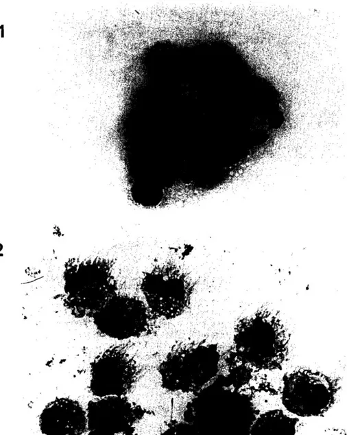
adherence, and separation of low density cells over a Metrizamide column. In the second protocol, monocytes and DC were first enriched by adherence on gelatin/plasma coated flask. The resulting populations contained 75 to 80% of cells which do not express T cell, B cell or monocyte markers. The remaining 20-25% of cells were monocytes, B and T lymphocytes. Under light microscopy, cells isolated by protocol I (DC l ) were round and of medium size (Fig. 2a). In contrast, cells isolated by protocol 2 displayed long cytoplasmic veils and formed aggregates during the culture, a property which has been described as typical of DC (Fig. 2b).
Fig. 2. Morphology of DCI and DC2: cells were prepared by cytospin and were processed for May Grlinwald Giemsa staining. Magnification: 100 x.
PHENOTYPE ANALYSIS
Both DC populations were further characterized using mAbs listed in Table 2. DC purified according to the first protocol expressed high levels of MHC class II molecules but low levels of adhesion and costimulatory molecules (mean fluorescence for MHC class II staining: 101; B7 staining: 16; CD11c staining: 6). DC isolated following the second protocol displayed higher levels of MHC class II, adhesion and costimulatory molecules (mean fluorescence for MHC class II staining: 1251; B7 staining: 67; CD11c staining: 48). Both populations were negative for most lineage markers (Table 2).
Fig.3. Immunostimulatory capacity of DC. Various numbers of irradiated (2000 rads) DC isolated according to protocol 1 (open circle) or protocol 2 (closed circle) were cultured with 4 X I 05 T cells from the same (autologous) or different animal (allogeneic). Proliferation was assessed by thymidine incorporation during the last 10 h of 7-d culture. The data are expressed as cpm, and each point represents the mean ± SD of triplicate cultures. The results are representative of three experiments.
BOTH DC ENRICHED POPULATIONS HAVE THE CAPACITY TO SENSITIZE NAIVE T
CELLS
Dendritic cells are highly effective, as compared to other antigen-presenting-cells. at stimulating T-cells. One method that has been used to compare capacities to stimulate T-cell responses is to determine relative abilities to induce a proliferative response in allogeneic T cells. As shown in Fig. 3, DCl and DC2 induced strong proliferation of allogeneic T-cells, although DC purified according to protocol 2 were 3-to-6 fold more efficient on a per cell number basis.
Discussion
We used two procedures to enrich bovine DC from blood. The isolated cells have distinctive features that include low buoyant density, transient adherence. lack of expression of T, B and monocytes markers (CD3, CD 14, IgM), elevated expression of class II MHC markers and importantly potent immunostimulatory capacity. The unique capacity of both cell populations to sensitize naive T cells as well as the lack of expression of CD14 suggest that these cells belong to the family of DC. It should be noted that purified monocytes from the same animals had a strongly reduced capacity to activate T cells in the same assay, as compared to DC1 and DC2 (data not shown).
Table 2. Comparison of cell surface markers of DC 1 and DC2 populations
Determinant mAb Protocol 1 Protocol 2
MHC molecules MHC II ILA21 + + + + + + CC158 + + + + + + MHC I ILA88 + + + + + + Costimulatory molecules B7 hCTLA41g + + + + Myeloid markers CD14 CC-G33 - - gran/monoeyte ILA24 + + lntegrin / adhesin BoWC6 CC98 CD11a ILA99 + + + CD11b CC94 + + + + CD11c NAM-4 + + + + ALVC CC81 - - L-selectin CC32 + + + + BoWC10 CC28 - B cell markers IgM + ILA30 - - CD21 CC21 - - T cell markers CD2 CC42 - - CD4 CC8 - - CD8 a CC63 - - CD3 MM1 A - - BoWC1 CCIS - CD1 W1 20-27 + - CD1 W2 CC20 + - CD1 W3 CC43 + + CD45 CCI + + + + + + CD45RO ILA116 - - CD5 CCl7 - - CD6 CC38 - CD25 ILA111 + CD49d 218 + - CD44 25-32 + + + CD45 R(B) CC76 + CD29 FW3-47 + + Fe receptors Fe RII CCG36 + + + Fe 2 R CCG24 - -
Cell fluorescence intensity was evaluated by FACS analysis and data are expressed by proportion of cells expressing each marker. + + +, 80-95%: + +, 50-75%: +, 15-45% and -, no detectable staining. Antibodies used for this study are listed in Table I. Similar results were obtained in at least three independent experiments for each antibody.
Both protocols started with 5x10⁸ peripheral blood mononuclear cells and yielded similar
number of cells (0.1% in protocol 1 and 0.7% in protocol 2). The yield of DC obtained in this study is similar to the one (0.4-0.6%) obtained from human peripheral blood (Freudenthal and Steinman, 1990).
Cells of the dendritic family are distinguished from other antigen-presenting-cells by strong MLR stimulatory capacity (Van-Voorhis et al., 1982; Steinman et al., 1983). The unique property to activate naive T lymphocytes in vitro (Young et al., 1992; Streilen and Grammer, 1989) and in vivo (Liu and MacPherson, 1993) correlates with high density of MHC class II, adhesion molecules and costimulatory molecules. However. recent data indicate that the adjuvant capacity of DC is not constitutive but develops during a process of maturation that occurs spontaneously in vitro (Inaba et al., 1994) and can be induced in vivo (De Smedt et al., 1996). The maturation involves strong upregulation of MHC and B7 molecules (Larsen et al., 1994; Symington et al., 1993). It is therefore tempting to speculate that both DC-enriched subsets represent DC at different stages of maturation. DC1 would be immature, as suggested by morphology (absence of cytoplasmic processes), phenotype (relatively low levels of B7 molecules and CD 11c adhesion molecule) (Thomas et al., 1993) and function (lower stimulatory capacity as compared to DC2). In contrast, DC2 exhibit a dendritic morphology, high density of MHC class II, B7 and CD 11c molecules and have a superior capacity to sensitize T cells in vitro (Larsen et al., 1992; Inaba et al., 1994), suggesting that they have undergone a process of maturation in vivo.
Various stages of the DC lineage have indeed been described in blood and include progenitors and migratory DC in the process of maturation (O'Doherty et al., 1994: Romani et al., 1994; Weissman et al., 1995). However, since cells of the dendritic family have been shown to spontaneously mature upon culture, we cannot rule out the possibility that only DC2 matured during the purification steps that involve overnight culture. Indeed, in the second procedure, DC and monocytes were selected on gelatin- plasma coated surface and attachment of human blood monocytes to gelatin-coated surfaces has been shown to promote differentiation and some mononuclear phagocyte function (Bevilacqua et al., 1981). We have indeed observed that monocytes isolated by adherence on gelatin plasma coated dishes expressed higher density of activation markers, as compared to monocytes purified by adherence on plastic (data not shown). Whether the spontaneous in vitro maturation is autonomous or requires factors released by other cell types is presently unknown (O'Doherty et al., 1993; Bender et al., 1996). There is evidence, however, that TNFa and GM-CSF may be involved in this process (Heufler et al., 1988; Koch et al., 1990; Holt et al., 1993; Cumberbatch and Kimber, 1995). Experiments are underway to test whether addition of monocyte conditioned medium would induce maturation of the DC1 'immature subset'.
Veiled cells prepared from bovine afferent lymph have been shown to act as potent stimulators in mixed leukocyte cultures (McKeever et al., 1991), like both populations obtained in this study, but display differential expression of the adhesion molecules CD11a, b and c.
In conclusion, we have purified two DC-enriched populations that display features of immature and mature DC. Additional studies using fresh DC. purified without a step in culture, will be required to determine whether these subsets reflect two different stages of maturation that occurred in vivo or in vitro. The availability of both populations will help define the status and
function of bovine DC in health and disease, and will be a useful tool to study the induction of T cell dependent immune responses against various pathogens in vitro.
Acknowledgements
We thank Drs. S. Barcy, 0. Leo, E. Hanon, G. Urbain and F. Willems for interesting discussions and valuable help ; G. Charlier, G. Dewasme, M. Swaenepoel, F. Tielemans, and L. Karelle for excellent technical assistance. The work was supported by the Biotech Programme of the European Commission (contract No 93.0324). The laboratory of Animal Physiology was supported by grants of Fond National de la Recherche Scientifique (FNRS) and by the Fond de la Recherche Fondamentale Collective, by the Biotech Programme of the European Commission (contract No. BI02-CT93-0489), and by the Belgian Programme on Interuniversity Poles of attraction initiated by the Belgian State, Prime Minister's Office, Science Policy Programming. M. Moser is Research Associate from the Belgian FNRS.
References
Ardavin, C., Wu, L., Li, C.L., Shortman, K., 1993. Thymic dendritic cells and T cells develop simultaneously in the thymus from a common precursor population. Nature 362, 761-763.
Austyn, A.M., Hankins, D.F., Larsen, C.P., Morris, P.J., Roake. J.A., 1994. Isolation and characterization of dendritic cells from mouse heart and kidney. J. Immunol. 152, 2401-2410.
Bender, A., Sapp, M., Schuler, G., Steinman, R.M., Bhardwaj, N., 1996. Improved methods for the generation of dendritic cells from nonproliferating progenitors in human blood. J. Immunol. Meth. 196, 121-135. Bevilacqua, M.P., Amrani, 0., Mosesson, M.W., Bianco, C., 1981. Receptors for cold-insoluble globulin (plasma fibronectin) on human monocytes. J. Exp. Med. 153, 42-60.
Bujdoso, R.J., Hopkins, B.M., Dutia, P., Young, P., McConnell, I., 1989. Characterization of sheep afferent lymph dendritic cells and their role in antigen carriage. J. Exp. Med. 170, 1285-1301.
Cumberbatch, M., Kimber, I., 1995. Tumor necrosis factor-a is required for accumulation of dendritic cells in draining lymph nodes and for optimal contact sensitization. Immunology 84, 31-35.
De Smedt. T., Pajak. B., Muraille, E., Lespagnard, L., Heinen, E., De Baetselier, P., Urbain. J., Leo. 0., Moser, M., 1996. Regulation of dendritic cells numbers and maturation by lipolysaccharide in vivo. J. Exp. Med. 184, 1413-1424.
Freudenthal. P.S., Steinman. R.M., 1990. The distinct surface of human blood dendritic cells. as observed after an improved isolation method. Proc. Natl. Acad. Sci. USA. 87, 7698-7702.
Heufler, C., Koch, F., Schuler, G., 1988. Granulocyte/macrophage colony-stimulating factor and interleukin 1 mediate the maturation of epidermal Langerhans into potent immunostimulatory dendritic cells. J. Exp. Med. 167, 700-705.
Holt. P.G., Oilver, J., Bilyk. N., McMenamin. C., Kraal. G., Thepen. T., 1993. Downregulation of the antigen presenting cell function(s) of pulmonary dendritic cells in vivo by resident alveolar macrophages. J. Exp. Med. 177, 397-407.
Howard. C., Naessens, J., 1993. General summary of workshop findings for cattle: Section 4. Vet. Immunol. lmmunopathol. 39, 25-48.
Inaba. K., Witmer-Pack, M., Inaba, M., Hathcock. K.S., Sukuta, H., Azuma. M., Yagita. H., Okumura. K., Linsley. P.S., Ikehara. S., Muramatsu, S., Hodes. R.J., Steinman. R.M., 1994. The tissue distribution of the B7-2 costimulator in mice: Abundant expression on dendritic cells in situ and during maturation in vitro. J. Exp. Med. 180, 1849-1860.
Koch. F., Heufler. C., Kampgen. E., Schneeweiss. D., Bock. G., Schuler. G., 1990. Tumor necrosis factor a maintains the viability of murine epidermal Langerhans cells in culture, but in contrast to granulocyte/macrophage colony-stimulating factor, without inducing their functional maturation . J. Exp. Med. 171, 159-171.
Koch. F., Trockenbacher. B., Kampgen, E., Grauer. 0., Stossel. H., Livingstone. A.M., Schuler, G., Romani. N., 1995. Antigen processing in populations of mature murine dendritic cells is caused by subsets of incompletely matured cells. J. Immunol. 155, 93-100.
Kudo. S., Matsuno, K., Ezaki. T., Ogawa. M., 1997. A novel migration pathway for rat dendritic cells from the blood: Hepatic sinusoids-lymph translocation. J. Exp. Med. 185, 777-784.
Larsen. C.P., Steinman, R.M., Witmer-Pack, M., Hankins. D.F., Morris. P.J., Austyn. J.M., 1990. Migration and maturation of Langerhans cells in skin transplants and explants. J. Exp. Med. 172, 1 483-1493.
Larsen. C.P., Ritchie, S.C., Pearson, T.C., Linsky, P.S., Lowry. R.P., 1992. Functional expression of the costimulatory molecule. B7/BB1. on murine dendritic cell population. J. Exp. Med. 176, 1 215-1 220. Larsen. C.P., Ritchie, S.C., Hendrix. R., Linsley. P.S., Hathcock. K.S., Hodes, R.J., Lowry, R.P., Pearson. T.C., 1994. Regulation of immunostimulatory function and costimulatory molecule (B7-1 and B7-2) expression on murine dendritic cells. J. Immunol. 152, 5208-5219.
Liu. L.M., MacPherson. G.G., 1993. Antigen acquisition by dendritic cells: Intestinal dendritic cells acquire antigen administered orally and can prime naïve T cells in vivo. J. Exp. Med. 177, 1299-1307.
McKeever. D.J., MacHugh. N.D., Goddeeris, B.M., Awino. E., Morrison. I.W., 1991. Bovine afferent lymph veiled cells differ from blood monocytes in phenotype and accessory function. J. Immunol. 147, 3703-3709.
McKeever, D.J., Morrison, W.I., 1992. Afferent lymph veiled cells prime CD4+ T cell responses in vivo. Eur. J. lmmunol. 22, 3057-3061.
O'Doherty, U., Steinman, R.M., Peng. M., Cameron, P.U., Gezelter, S., Kopeloff, I., Swiggard. W.J., Pope. M., Bhardwaj. N., 1993. Dendritic cells freshly isolated from peripheral blood express CD4 and mature into typical immunostimulatory dendritic cells after culture in monocyte-conditioned medium. J. Exp. Med. 178, 1067-1078.
O'Doherty, U., Peng. M., Gezelter, W .. Swiggard, M., Betjes, N., Bhardwaj. N .. Steinman, R.M .. 1 994. Human blood contains two subsets of dendritic cells, one immunologically mature and the other i mmature. Immunology 82, 487-493.
Romani. N., Lenz, A., Glassel, H., Stossel, H., Stanzl, U., Majoic. 0., Fritsch, P., Schuler. G., 1989. Cultured human Langerhans cells resemble lymphoid dendritic cells in phenotype and function. J. Invest. Dermatol. 93, 600-609.
Romani, N., Gruner, S., Brang. D., Kampgen. E., Lenz, A., Trockenbacher. B., Konwalinka. G., Steinman. R.M., Schluer. G., 1994. Proliferation dendritic cell progenitors in human blood. J. Exp. Med. 180, 83-93.
Schuler, G., Steinman, R.M., 1985. Murine epidermal Langerhans cells mature into potent immunostimulatory dendritic cells in vitro. J. Exp. Med. 161, 526-546.
Steinman, R.M., Cohn, Z.A., 1973. Identification of a novel cell type in peripheral lymphoid organs of mice: I. Morphology, quantification, tissue distribution. J. Exp. Med. 137, 1 142.
Steinman, R.M., Gutchinov, B., Witmer, M.D., Nussenzweig, M.C., 1983. Dendritic cells are the principal stimulators of the primary mixed leukocyte reaction in mice. J. Exp. Med. 157, 613-627.
Steinman, R.M., 1991. The dendritic cell system and its role in immunogenicity. Ann. Rev. Immunol. 9, 271-296.
Streilen, J.W., Grammer, S.F., 1989. In vitro evidence that Langerhans cells can adopt two functionally distinct forms capable of antigen presentation to T lymphocytes. J. Immunol. 143, 3925-3933.
Strober, W., Kelsall, B.L., 1996. Distinct populations of dendritic cells are present in the subepithelial dome and T cell regions of the murine Peyer's Patch. J. Exp. Med. 183, 237-247.
Symington, F., Brady, W., Linsley, P.S., 1993. Expression and function of B7 on human epidermal Langerhans cells. J. Immunol. 150, 1286-1295.
Thomas, R., Davis, L.S., Lipsky, P.E., 1993. Human peripheral blood dendritic cell subsets. Isolation and characterization of human peripheral blood dendritic cells. J. Immunol. 150, 821-834.
Van-Voorhis, W.C., Hair, L.S., Steinman, R.M., Kaplan, G., 1982. Human dendritic cells: enrichment and characterization from peripheral blood. J. Exp. Med. 155, 1172-1187.
Weissman, D., Li, Y., Ananworanich, J., Zhou, L., Adelsberger, J., Tedder, T.F., Baseler, M., Fauci, A., 1995. Three populations of cells with dendritic cells morphology exist in peripheral blood, only one of which is infectable with human immunodeficiency virus type 1. Proc. Natl. Acad. Sci. 92, 826-830.
Winzler, C., Rovere, P., Rescigno, M., Granulocci, F., Penna. G.. Adorini. L.. Zimmermann, V.S., Davoust, J., Ricciardi-Castagnoli. P., 1997. Maturation stages of mouse dendritic cells in growth factor-dependent long-term cultures. J. Exp. Med. 185, 317-328.
Young, J.W., Koulova, L., Soergel, S.A., Clark, E.A., Steinman, R.M., 1992. The B7/BB1 antigen provides none of several costimulatory signals for the activation of CD4+ T lymphocytes by human blood dendritic cells in vitro. J. Clin. Invest. 90, 229-237.