EFFECTS OF REFLUX LARYNGITIS ON LARYNGEAL
CHEMOREFLEXES IN NEWBORN LAMBS
Anne-Marie Carreau†, Hugues Patural¶, Nathalie Samson†, Alexandre A Doueik§, Julie
Hamon†, Pierre-Hugues Fortier*, Jean-Paul Praud†
†Neonatal Respiratory Research Unit, Departments of Pediatrics and Physiology; §Department of Pathology; *ENT Division, Department of Surgery, Université de
Sherbrooke, QC, Canada – J1H 5N4
¶Pediatric Intensive Care Unit, CHU St Étienne, 42055 – St Étienne, Cedex 02, France
Short title: Reflux laryngitis and laryngeal chemoreflexes
Address for correspondence and proofs:
Jean-Paul Praud MD PhD Phone: (819) 346-1110, ext 14851 Departments of Pediatrics and Physiology Fax: (819) 564-5215
Université de Sherbrooke email: [email protected] J1H 5N4, QC Canada
ABSTRACT
It has been suggested that reflux laryngitis (RL) is involved in apneas-bradycardias of the newborn. The aim of the present study was to develop a unique RL model in newborn lambs in order to test the hypothesis that RL enhances the cardiorespiratory components of the laryngeal chemoreflexes (LCR) in the neonatal period. Gastric juice surrogate (2 mL HCl + pepsin, pH 2) (RL group, n = 6) or saline (Control group, n = 6) was repeatedly injected onto the posterior aspect of the larynx, 3 times a day for 6 consecutive days, via a retrograde catheter introduced into the cervical esophagus. Lambs instilled with gastric juice surrogate presented clinical signs of RL, as well as moderate laryngitis on histological observation. Laryngeal chemoreflexes were thereafter induced during sleep by injection of 0.5ml of HCl (pH 2), milk, distilled water or saline into the laryngeal vestibule via a chronic, transcutaneous supra-glottal catheter. Overall, RL led to a significantly greater respiratory inhibition compared to the Control group during LCR, including longer apnea duration (p = 0.01), lower minimal respiratory rate (p = 0.002) and a more prominent decrease in SpO2 (p = 0.03). No
effects were observed on cardiac variables. In conclusion, 1) our unique neonatal ovine model presents clinical and histological characteristics of RL; 2) the presence of RL in newborn lambs increases the respiratory inhibition observed with LCR, at times leading to severe apneas and desaturations.
KEYWORDS: Polysomnography, sleep, apparent life-threatening event, apnea of prematurity, sudden infant death syndrome.
INTRODUCTION
Laryngeal chemoreflexes (LCR) are a group of reflexes triggered in mammals by the contact between a liquid, such as milk or gastric juice, and receptors of the laryngeal mucosa. These reflexes, comprised of laryngeal closure, cough, swallowing (and arousal if necessary) in a mature organism, constitute a major protection mechanism against tracheal aspiration of liquids (20,21). However, in certain conditions, LCR can conversely be responsible for apneas, bradycardias and hypoxemia. The latter, while reminiscent of fetal-type LCR, can be so severe that they are likely responsible for a certain proportion of apneas of prematurity, most life-threatening events of infancy and some cases of sudden infant death syndrome (11,13,20,21,30).
Gastric refluxes, especially laryngopharyngeal refluxes, are a very frequent and normal occurrence in the neonatal period. While most gastric refluxes in infants are weakly acid, mainly due to frequent feeds, it is nevertheless well documented that acid refluxes occur in infants. This includes healthy, preterm neonates (34 weeks postconceptional age), where a mean number of 16 acid laryngopharyngeal refluxes per day has been reported (17). In such cases, the acid-activated pepsin present in the refluxate can induce reflux laryngitis (RL) (1,12).
Reflux laryngitis has been suggested to be one condition responsible for LCR with severe cardiorespiratory events in infants (31). However, evidence for the responsibility of RL in increasing the occurrence and severity of cardiorespiratory events during LCR is very weak. The main objective of our study was thus to compare LCR characteristics in RL lambs and control lambs, testing the hypothesis that increased cardiorespiratory
inhibition is present in RL lambs, as compared to controls. However, given that we were unable to find a neonatal RL model in the literature, a neonatal ovine RL model had to be designed beforehand in order to perform the study.
MATERIAL AND METHODS
AnimalsA total of twelve newborn lambs, distributed equally into two groups (RL group and Control group), were included in this study. The study protocol was approved by the ethics committee for animal care and experimentation of our institution.
Surgical instrumentation and recording equipment Surgical instrumentation
Aseptic surgery was performed under general anesthesia (2% isoflurane, 30% N2O,
68% O2) upon the day of arrival (postnatal day 1). Anesthesia was preceded by an
intramuscular injection of ketamine (10 mg/kg), atropine sulfate (0.1 mg/kg) and morphine (0,016 ml/kg) and an intra-venous bolus (10 ml/kg) of Ringer’s lactate solution. One dose of ketoprofen (3 mg/kg) was also injected intramuscularly for analgesia and repeated if needed 12h later. Antibiotics (5 mg/kg gentamicin and 0.05 ml/kg duplocilline) were administered intramuscularly prior to surgery and daily thereafter. Lambs were mechanically ventilated through an orotracheal tube (4.5 mm) throughout the surgical procedure. Bipolar platinum needle electrodes (made from F-E2 needle-electrodes, Grass Technologies, West Warwick, RI, USA) were inserted into both thyroarytenoid muscles (TA; a glottal adductor) through the lateral aspect of the thyroid cartilage for electrical activity (EMG) recording. Both electrodes were then glued on the external surface of the thyroid cartilage. Two right-angled needle electrodes (E7-12, Grass Technologies) were inserted into the parietal cortex directly through the skull,
at the level of the lambda suture, 1 cm from the midline, for electrocorticogram (ECoG) recording. A third needle electrode (F-E2M, Grass Technologies) was inserted under the scalp as a ground. Two platinum needle electrodes (F-E2, Grass Technologies) were also inserted subcutaneously into the outer lower and the inner upper region of the right eye socket for electrooculogram (EOG) recording. For electrocardiogram (ECG) recordings, two additional needle electrodes (F-E2M, Grass Technologies) were inserted under the periosteum of the 5th rib, on both sides of the thorax, and directly
glued on the rib. A catheter was introduced into the right carotid artery to monitor arterial blood gases and arterial pressure. Leads from all electrodes were subcutaneously tunneled to exit on the back of the lambs. In addition, a supraglottal catheter was inserted transcutaneously to allow injection of liquids into the laryngeal vestibule, as previously described (6). Briefly, a 16 G infusion catheter was securely positioned such that its tip protruded 5-7.5 mm above the anterior part of the glottis. A plastic tubing (internal diameter 1 mm) was subcutaneously tunneled in the neck of the lamb and connected to the external part of the supraglottal catheter, which protruded 15-20 mm externally at the level of the anterosuperior aspect of the thyroid cartilage. Correct catheter positioning above the glottis was monitored throughout the insertion procedure by direct laryngoscopy.
Finally, a retrograde catheter was introduced into the esophagus for reflux laryngitis induction (see below). After identification of the thyroid gland, the esophagus was exposed caudally over 10 cm to visualize the right recurrent laryngeal nerve trajectory. An 8 Fr Levine catheter connected to a trocar from a Hemovac drainage tube was introduced into the esophagus via the mouth of the lamb and exited by an incision in the
cervical esophagus wall, which avoided the recurrent laryngeal nerve. Under direct laryngoscopic visualization, the rostral extremity of the esophageal catheter was positioned 5 mm above the posterior rim of the larynx. The catheter was then secured to the cervical esophageal incision with biological glue, then sutured to the skin. The caudal part of the Levine catheter was finally subcutaneously tunneled to exit on the lamb’s back.
Neonatal ovine model of reflux laryngitis
A unique, chronic neonatal ovine model of reflux laryngitis was designed for the purpose of the present study. Following surgical instrumentation, lambs were randomly assigned to either the RL group or Control group. In the RL group, 2 ml of a solution containing pepsin (300 U/ml, Sigma-Aldrich Canada Ltd, Oakville, ON) and chlorohydric acid (HCl, pH 2.0) was instilled 3 times a day for 6 consecutive days using the esophageal catheter. Application of an acidified pepsin solution on the larynx to mimic an acid gastric reflux has been previously shown to induce laryngitis (1,12). In addition, our acidified pepsin solution was chosen to be representative of the gastric content of a newborn infant, whose pepsin concentration of gastric aspirates varies between 43 and 683 U/ml in the neonatal period (M Armand, personal communication, Marseille, France). Meanwhile, lambs in the Control group were instilled with 2 ml of physiological saline 3 times a day for 6 consecutive days, via their esophageal catheter.
Validation of our neonatal reflux laryngitis model was based on clinical signs and histological evaluation. Daily clinical follow-up included weighing of lambs and monitoring to detect the presence of chronic cough, difficulties to feed and altered
bleating. For the purpose of the study, a semi-quantitative score on a scale of 10 (with 10 = 100% normal bleat, 9 = 90% normal bleat, ...) was used to evaluate altered bleating in each lamb and was assessed daily by the same observer. Correct positioning of the esophageal catheter was confirmed before euthanasia by direct laryngoscopy. Following euthanasia, the larynx was observed macroscopically for the presence of edema, ulceration and erythema. Finally, a pathologist expert in larynx assessment (AD) scored epithelial inflammation based on a scale used in a previous reflux laryngitis model. The glottis, epiglottis and first tracheal ring were scored on a scale of 15 depending on the worse criteria encountered for each following category: epithelial changes (intraepithelial inflammation, squamous metaplasia, erosion, ulceration) and subepithelial changes (stromal inflammation, periglandular inflammation and granulation or fibrosis) (12).
Recording equipment
Lamb instrumentation was completed immediately before recordings. Nasal flow was recorded from both nostrils using a double thermocouple wire (iron/constantan, type J; Omega Engineering, Stamford, CT) secured in an adapted dog muzzle. Rib cage and abdominal respiratory movements were also recorded by respiratory inductance plethysmography (Respitrace, Monitoring systems, Miami Beach, FL). A pulse oximeter sensor (MasimoRadical, Masimo, Irvine, CA) was attached at the base of the tail for continuous monitoring of arterial hemoglobin saturation (SpO2) and pulse wave. Finally,
a custom-designed, radiotelemetry-driven injector, where the liquids to be injected were warmed and maintained at the lamb’s body temperature.
In order to obtain data from prolonged recordings in non-sedated lambs, under the least possible restraining conditions, all leads from the EMG, EOG, ECoG and ECG electrodes, as well as nasal flow, were connected to our custom-designed radio telemetry transmitter (14). The pulse oximeter sensor was also connected to a second custom-designed radiotelemetry transmitter. The raw EMG signals were rectified, integrated and moving time averaged (100 ms). All parameters were continuously recorded using AcqKnowledge software (version 3.7.3, Biopac Systems, Santa Barbara, CA). In addition, an observer was continuously present to note all events occurring during recordings.
Design of the study
All lambs were housed with their mother in our animal quarters until experimental day. Following surgical instrumentation and RL induction, polysomnographic recordings were performed in non-sedated lambs on postnatal day 9 and 10 between 6:00 AM and 12:00 PM. Lambs were placed in a Plexiglas chamber (1.2 x 1.2 x 1 m) and comfortably positioned on a mattress without contention during recordings. Laryngeal chemoreflexes were induced by random injection of 0.5 ml of saline (0.9% NaCl, pH = 5.5, osmolarity = 326 mosM), distilled water (pH = 5.7), hydrochloric acid (HCl) diluted in saline (pH = 2, osmolarity = 295 mosM) and ewe milk (pH = 6.6, osmolarity = 298 mosM) via the supraglottal catheter. All four solutions were injected once in quiet sleep (QS) and once in active sleep (AS) on each experimental day (i.e., a total of 2 injections per solution
and per state of alertness). In addition, the supraglottal catheter was systematically flushed with 1 ml of saline between each injection of test solutions (dead space of the catheter 0.5 ml). Each animal was given at least 15 min of recovery time between two injections. Events such as agitation, cough, arousal and/or full awakening were noted by an observer throughout the recording sessions. Finally, following completion of the recordings, euthanasia was performed by an intravenous injection of 100 mg/kg of pentobarbital. Correct electrode and catheter positioning was systematically verified at autopsy.
Data analysis States of alertness
Standard electrophysiological and behavioral criteria were used to define sleep states (22). Cortical arousal from quiet sleep (QS) was defined by the association of a change in ECoG (decrease in amplitude + increase in frequency) for 3 seconds or more, with at least two of the following modifications: a 10% increase in heart rate (HR) or change in respiratory rate (RR) or movement (9). Arousal from active sleep (AS) was recognized by direct observation of the lamb and disappearance of intense EOG activity. Full awakening was defined when the lamb was still awake after 1 minute (7).
Laryngeal chemoreflexes
The main objective of the study was to assess the effect of RL on LCR. In addition, we queried whether this effect was different in QS vs. AS, and from one experimental solution to another. Analysis of the LCR was performed as described previously within
the first minute following each laryngeal injection (27). First, the cardiorespiratory responses were assessed as follows. The percentage of decrease in heart rate [%dec HR = (HRBL – HRmin)* 100/ HRBL] was calculated, with HRBL representing the baseline
HR value averaged over 30 s before injection, and HRmin representing the minimal HR
value observed after injection. The percentage of increase in mean arterial pressure, decrease in respiratory rate and decrease in transcutaneous oxygen saturation (SpO2) were calculated in the same manner. Any presence of bradycardia (defined by a %dec HR > 30%) was noted, and the number and total summed duration of bradycardias were tabulated. The number of apneas (defined as at least two missed breaths relative to baseline breathing) and total summed duration of apneas were recorded within the first minute after laryngeal stimulation. Respiratory LCR duration was measured as the time duration between the onset of the LCR and resumption of three consecutive breaths similar to baseline RR. In addition to cardiorespiratory responses, the number of swallows (recognized as a brisk, high-amplitude and short-duration TA EMG) was tallied. Total summed duration of TA EMG activity longer than 0.3 sec (considered as indicating laryngeal closure not related to the respiratory pause to accommodate swallowing) was calculated. The number of coughs (including laryngeal expiratory reflexes, which could not be discerned from coughs in our study) was also inferred from visual observation, as well as analysis of TA EMG and respiratory inductance plethysmograph signals. Finally, the presence of arousal or full awakening was noted. Statistical analysis
Quantitative variables were expressed as mean ± SD, whereas qualitative variables (arousal and awakening) were expressed as relative frequency. Statistical analyses
were performed on raw data for all variables. The effect of RL was assessed for baseline physiological values by the Student’s t test. For the laryngeal chemoreflexes, quantitative variables were analyzed through a general linear model 3-way ANOVA for repeated measures using Proc mixed procedures (SAS software version 9.1.3, Cary, NC) with group, solutions and sleep states as the independent variables. Qualitative variables were analyzed with a logistic regression model. Differences were deemed significant if p < 0.05. In addition, given the relatively small number of studied lambs (related both to the complexity of the ovine model and ethical constraints), it was decided to give full consideration to the presence of a significant trend, defined as p < 0.1.
RESULTS
A total of 94 laryngeal stimulations were performed in the Control group (n = 6) [QS: 11 saline, 12 distilled water, 12 HCl, 12 milk. AS: 12 saline, 11 distilled water, 12 HCl, 12 milk] compared to 81 laryngeal stimulations in the RL group (n = 6) [QS: 12 saline, 11 distilled water, 12 HCl, 12 milk. AS: 8 saline, 9 distilled water, 8 HCl, 9 milk].
Reflux laryngitis group
Clinical presentation of RL was variable from one lamb to the other. Hoarsened bleat (score over 7/10) at the end of the 6 days of instillation, up to absent bleating in 2 lambs, was observed in 5/6 lambs from the RL group vs. 1/6 lambs in the Control lamb. Chronic cough was also observed in 2 lambs from the RL group. In addition, one lamb from the RL group, who presented cough during attempts at bottle-feeding and refused to feed, was tube-fed. Another RL lamb presented an apparent life-threatening event following simulated laryngopharyngeal refluxes and required manual stimulation and nasal administration of oxygen for a few minutes. Finally, a third RL lamb developed a recurrent neck extension with writhing movements of the limbs and an arched back following laryngopharyngeal refluxes (Sandifer syndrome). Overall, though variability was present within each group, the RL group presented more swallowing activities, coughing and agitation than the Control group at the time of simulated laryngopharyngeal refluxes. Mean weight gain was 190 ± 18 g per day for the Control group and 99 ± 67 g per day for the RL group (p = 0.05).
Macroscopic assessment
of the control lambs. The presence of erythema on the posterior aspect of the vocal cords and edema was also noted in lambs in the RL group.
Histological scoring of laryngeal inflammation
Histological scoring of laryngeal inflammation was performed on 5 larynges in the RL group and 3 in the Control group (technical problems did not allow assessment of all larynges). Based upon histological criteria, a moderate laryngeal inflammation was induced in the RL group when compared to the Control group (4/15 ± 2/15 vs. 0/15 ± 1/15).
Baseline parameters
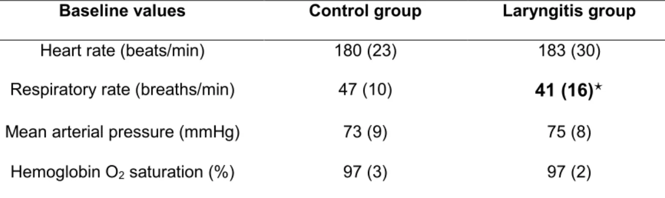
Baseline parameter values obtained in each group are detailed in Table 1. Compared with the Control group, lambs in the RL group presented a lower baseline respiratory rate (41 ± 16 vs. 47 ± 10 min-1, p < 0.0001). No other significant differences were
observed between Control and RL groups for baseline parameters.
Effects of reflux laryngitis on laryngeal chemoreflexes
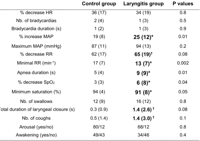
Results obtained during laryngeal chemoreflexes (LCR) are summarized in Table 2 and detailed in Tables 3 and 4, including significant interactions. Overall, LCR elicited a significantly greater respiratory inhibition in the RL group compared to the Control group (all solutions and both sleep states taken together). This respiratory inhibition included a greater % decrease in RR, longer apnea duration, lower minimum respiratory rate and a more prominent decrease in SpO2 following LCR stimulation. In addition, apneas longer
than 10 and 20 sec, as well as decreases in SpO2 greater than 10%, were more
during LCR, with intense swallowing-related hypoventilation leading to a marked decrease in SpO2 down to 64% after milk and 54% after HCl (figure 1). While no overall
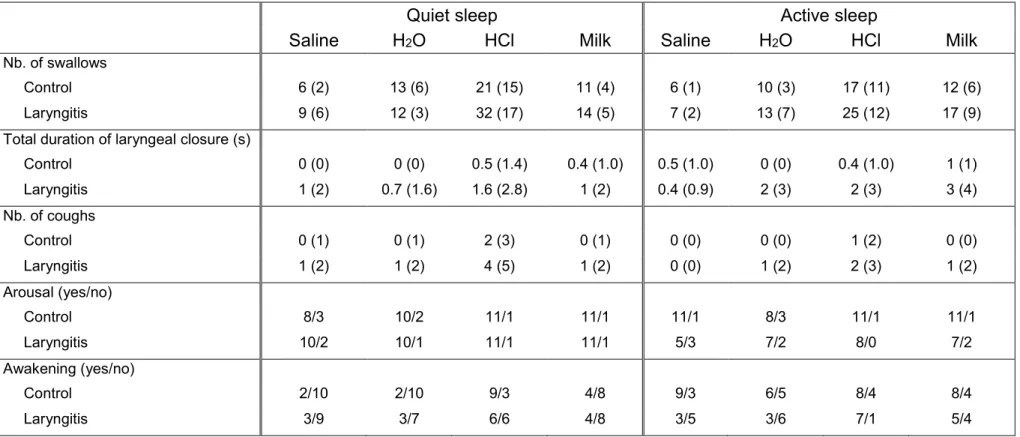
significant differences were observed for cardiac parameters, including % of decrease in HR and bradycardia duration, the % of increase in mean arterial pressure was higher in the RL group than in the Control group. Finally, while no differences between groups were observed for arousal or awakening, a greater number of coughs and total duration of laryngeal closure, denoting enhanced lower airway protective mechanisms, was observed in the RL group.
Regardless of the solution tested, no statistical interaction between group and sleep state was observed, indicating an absence of a sleep state effect on the differences between RL and Control groups.
On the contrary, a significant interaction was observed between the type of solution and group of lambs, such that HCl triggered longer apnea duration in the RL as opposed to the Control group (respectively 13.2 ± 9.7 vs. 5.5 ± 7.8 sec, p = 0.0008). In fact, HCl triggered 7 of the 10 apneas longer than 20 seconds observed in the RL group. No other statistical interactions were observed between group and solution.
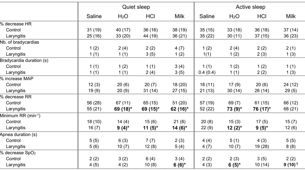
Finally, several significant interactions between group, sleep state and type of solution were observed, including for minimal RR, % decrease in RR and % decrease in SpO2
(see Table 3 for details). Overall, those interactions suggest that the effects of RL can be observed with any solution but saline, and independent of the sleep state.
DISCUSSION
The present study provides novel findings on laryngeal chemoreflexes in a new, non-sedated newborn lamb model of reflux laryngitis, which was developed for the purpose of the study. The relevance of this model is shown by clinical and/or histological signs of reflux laryngitis presented by all lambs. Overall, our findings reveal that the presence of reflux laryngitis in newborn lambs enhances the respiratory inhibition observed during laryngeal chemoreflexes.
Our ovine neonatal model of reflux laryngitis
An extensive review of the literature revealed that there are only a few animal models of RL. And while a total of 6 RL models have been described in adult rabbits, dogs or rats, no neonatal model, to our knowledge, has been previously described. In addition, most of these models were specifically designed to characterize the inflammation involved in subglottic stenosis on a traumatized larynx. (1,4,12,24). Only 2 models were designed to study an untraumatized larynx in mildly sedated adult rabbits or non-sedated adult rats (2,18). In contrast, our model was designed to closely reproduce clinical conditions, by posterior laryngopharyngeal instillations of a gastric juice surrogate (pepsin + HCl) in non-sedated newborn lambs. Parameters of our RL model were particularly chosen in order to reproduce what is observed in the neonatal period. A previous study showed that preterm infants are able to maintain an acidified gastric pH < 2, even at 24 weeks of gestational age (10). In addition, a recent study demonstrated that acidified gastric refluxes occur between feedings and, on average, represent 25% of all gastro-esophageal refluxes observed per day in healthy preterm infants (17). Our acidified
pepsin solution was chosen to be representative of the gastric content of term newborns of 1-2 months (M Armand, personal communication, Marseille, France).
Our neonatal ovine model accurately mimics clinical findings in RL, including cough, weak bleat and reduction in weight gain when compared to the Control group. Such subacute clinical manifestations were not reported in any of the previous studies cited, except for the presence of a biphasic stridor on exertion in rabbits with untraumatized larynx (18). While variable among lambs, simulated laryngopharyngeal refluxes with the gastric juice surrogate induced greater reactions in the RL group than in the Control group, even leading to an ALTE in one lamb. This lamb presented considerable clinical manifestations of RL, including unwillingness to feed. A previous study also showed that the response to simulated reflux was variable among individuals and also led to an ALTE in one adult animal (18). Our observations are consistent with the fact that, while laryngopharyngeal refluxes are present in virtually all infants, chronic consequences are very variable and only 1-2% will ultimately present with an ALTE (5). Further studies are hence needed in order to understand these individual variations and predispositions to an ALTE, which do not appear solely related to the degree of RL.
Effect of reflux laryngitis on neonatal laryngeal chemoreflexes
The present study confirms that RL influences neonatal LCR. More specifically, RL enhanced the respiratory inhibition observed during LCR, including longer apnea duration, lower minimum respiratory rate and a more prominent decrease in SpO2.
Though this respiratory inhibition was most often moderate, severe respiratory events were observed in three RL lambs, which also presented with important clinical
manifestations. On the other hand, RL did not appear to enhance cardiac inhibition following LCR. Interestingly, a previous study in newborn lambs infected with the respiratory syncytal virus (16) showed an enhanced respiratory inhibition following LCR compared to controls, but no changes in cardiac parameters. Moreover, our recent study in full-term lambs exposed to cigarette smoke for the first 14 postnatal days showed a somewhat more significant respiratory than cardiac inhibition during LCR (28). However, other studies in lambs have shown that conditions, such as postnatal nicotine injection (29) and prematurity (27), can enhance both respiratory and cardiac inhibition during LCR. Providing a clear explanation for the different effects on the cardiac and respiratory components of the LCR in various conditions is not straightforward. Various mechanisms can be at play with regards to respiration and cardiac inhibition during LCR in lambs. For instance, while control of cardiac activity during LCR involves some level of sympatho-vagal co-activation (3), respiratory inhibition can in part be driven by the reciprocal inhibition brought about by intense swallowing activity (present results and 27). In fact, cardiac and respiratory activity during LCR likely results from a complex interaction of various mechanisms, which may depend on the offending condition. Conceivably, the latter, e.g. cigarette smoke exposure, RL, nicotine or prematurity, can variably alter the function of one or several components of the LCR loop, giving rise to variable consequences on cardiorespiratory components of the LCR. One hypothesis stems from the association between upper airway inflammation and increased cytokines in the brainstem, which in turn could enhance LCR-linked respiratory inhibition (15,30). Interestingly, increased cytokines in the brainstem have been reported in SIDS victims, compared to controls (8).
Aside from cardiorespiratory responses, our present study in full-term lambs did not reveal any alteration in swallowing activity or arousal in the RL group, whereas enhanced laryngeal closure and coughing were observed, all of which are important responses for liquid clearance from the laryngeal mucosa and prevention of tracheal aspiration. Previous observation of the blunting effect of premature birth on these airway protective mechanisms during LCR (27) suggests that the respiratory consequences of RL are likely worse in preterm neonates, as reported in infants (31).
Influence of sleep states and the type of solution used to induce laryngeal chemoreflexes
In the present study, no systematic effects of sleep states on LCR-related cardio-respiratory events were observed. While such results may seem at variance with one study in lambs infected with respiratory syncytial virus (16), they are in agreement with other results obtained in preterm as well as in full-term lambs under various experimental conditions (19,27,28).
In contrast, a systematic effect of HCl was observed herein, irrespective of the sleep state, such that the longest apnea duration in the RL group was specifically observed with HCl-triggered LCR. The fact that this was not the case in our previous studies (26,27,28) suggests that the apneic effect of HCl is specifically enhanced in the presence of RL. While this intriguing possibility requires further confirmation, it may be hypothesized that repeated exposure to HCl and/or pepsin during the 6 preceding days led to a specific sensitization of the laryngeal chemoreceptors, such as the C fiber endings (25).
Clinical implications
Our results add further support to the suggestion that acid laryngopharyngeal refluxes can trigger clinically significant cardiorespiratory events in infants, up to an ALTE. In addition, enhancement of LCR-linked respiratory inhibition, especially by an acid solution, in the presence of RL, supports the involvement of laryngitis in certain cardiorespiratory events of preterm infants (31), and plausibly in some SIDS cases. Further studies combining the present RL model with our previous models of preterm lambs and/or lambs exposed to cigarette smoke are needed to better understand the importance of RL in the pathophysiology of apneas of prematurity, ALTE and SIDS.
ACKNOWLEDGMENTS
The authors gratefully acknowledge the expert technical assistance of Jean-Philippe Gagné. The study was supported by grants from the Canadian Institutes for Health Research and the Foundation of Stars (Quebec) allocated to J-P Praud. A-M Carreau was a MD-MSc scholar of the Fonds de la recherche en santé du Québec (FRSQ). Jean-Paul Praud is a member of the FRSQ-funded Clinical Research Center Étienne-Le Bel, Sherbrooke University Hospital, and the holder of the Canada Research Chair in Neonatal Respiratory Physiology.
REFERENCES
1. Adhami T, Goldblum JR, Richter JE, Vaezi MF. The role of gastric and duodenal agents in laryngeal injury: an experimental canine model. Am J Gastroenterol 11: 2098-2106, 2004.
2. Asaoka D, Nagahara A, Oguro M, Mori H, Nakae K, Izumi Y, Osada T, Hojo M, Otaka M, Watanabe S. Establishment of a reflux esophago-laryngitis model in rats.
Dig Dis Sci 2010 Oct 9. [Epub ahead of print]
3. Beuchée A, Nsegbe E, St Hilaire M, Carrault G, Branger B, Pladys P, Praud J-P. Prolonged dynamic changes in autonomic heart rate modulation induced by acid laryngeal stimulation in non-sedated lambs. Neonatology 91: 83-91, 2007.
4. Carron JD, Greinwald JH, Oberman JP, Werner AL, Derkay CS. Simulated reflux and laryngotracheal reconstruction: A rabbit model. Arch Otolaryngol Head Neck
Surg 127: 576-580, 2001.
5. Edner A, Wennborg M, Alm B, Lagercrantz H. Why do ALTE infants not die in SIDS? Acta Paediatr 96: 2: 191-194, 2007.
6. Fortier PH, Reix P, Arsenault J, Dorion D, Praud J-P. Active upper airway closure during induced central apneas in lambs is complete at the laryngeal level only. J Appl Physiol 95: 97-103, 2003.
7. Goto K, Mirmiran M, Adams MM, Longford RV, Baldwin RB, Boeddiker MA, Ariagno RL. More awakening and heart rate variability during supine sleep in
preterm infants. Pediatrics 103: 603-609, 1999.
8. Kadhim H, Deltenre P, De Prez C, Sébire G. Interleukin-2 as a neuromodulator possibly implicated in the physiopathology of sudden infant death syndrome.
Neurosci Lett 480:122-126, 2010.
9. Kato I, Franco P, Groswasser J, Scaillet S, Kelmanson I, Togari H, Kahn A. Incomplete arousal processes in infants who were victims of sudden death. Am J
Respir Crit Care Med 168: 1298-1303, 2003.
10. Kelly EJ, Newell SJ, Brownlee KG, Primrose JN, Dear PR. Gastric acid secretion in preterm infants. Early Hum Dev 35: 215-220, 1993.
11. Kinney HC, Thach BT. The sudden infant death syndrome. N Engl J Med 361: 795-805, 2009.
12. Koufman JA. The otolaryngologic manifestation of gastroesophageal reflux disease (GERD): a clinical investigation of 225 patients using ambulatory 24-hours pH monitoring and an experimental investigation of the role of acid and pepsin in the development of laryngeal injury. Laryngoscope 101: 1-78, 1991.
13. Leiter JC, Böhm I. Mechanisms of pathogenesis in the sudden infant death syndrome. Respir Physiol Neurobiol 159: 127-138, 2007.
14. Létourneau P, Dumont S, Kianicka I, Diaz V, Dorion D, Drolet R and Praud J-P. Radiotelemetry system for apnea study in lambs. Respir Physiol 116: 85-93, 1999.
infants with respiratory tract infection. Acta Paediatr 85: 798-803, 1996.
16. Lindgren C, Lin J, Graham BS, Gray ME, Parker RA, Sundell HW. Respiratory syncytial virus infection enhances the response to laryngeal chemostimulation and inhibits arousal from sleep in young lambs. Acta Paediatr 85: 789-797, 1996.
17. Lopez-Alonso M, Moya MJ, Cabo JA, Ribas J, del Carmen Macias M, Silny J, Sifrim D. Twenty-four-hour esophageal impedance-pH monitoring in healthy preterm neonates: Rate and characteristics of acid, weakly acidic, and weakly alkaline gastroesophageal reflux. Pediatrics 118: e299-308, 2006.
18. Ludemann JP, Manoukian J, Shaw K, Bernard C, Davis M, al-Jubab A. Effects of simulated gastroesophageal reflux on the untraumatized rabbit larynx. J
Otolaryngol 27: 127-131, 1998.
19. Marchal F, Corke BC, Sundell H. Reflex apnea from laryngeal chemo-stimulation in the sleeping premature newborn lamb. Pediatr Res 16: 621-627, 1982.
20. Praud J-P. Upper airway reflexes in response to gastric reflux. Paediatr Respir Rev 11: 208-212, 2010.
21. Reix P, St-Hilaire M, Praud J-P. Laryngeal sensitivity in the neonatal period: from bench to bedside. Pediatr Pulmonol 42: 674-682, 2007.
22. Renolleau S, Letourneau P, Niyonsenga T, Praud J-P. Thyroarytenoid muscle electrical activity during spontaneous apneas in preterm lambs. Am J Respir Crit
23. Roh JL, Lee YW, Park HT. Effect of acid, pepsin, and bile acid on the stenotic progression of traumatized subglottis. Am J Gastroenterol 101: 1186-1192, 2006.
24. Roh JL, Yoon YH. Effect of acid and pepsin on glottic wound healing: A simulated reflux model. Arch Otolaryngol Head Neck Surg 132: 995-1000, 2006.
25. Roulier S, Arsenault J, Reix P, Dorion D, Praud J-P. Effects of C fiber blockade on cardiorespiratory responses to laryngeal stimulation in concious lambs. Respir
Physiol Neurobiol 136: 13-23, 2003.
26. St-Hilaire M, Nsegbe E, Gagnon-Gervais K, Samson N, Moreau-Bussière F, Fortier PH, Praud J-P. Laryngeal chemoreflexes induced by acid, water, and saline in nonsedated newborn lambs during quiet sleep. J Appl Physiol 98: 2197-2203, 2005.
27. St-Hilaire M, Samson N, Nsegbe E, Duvareille C, Moreau-Bussiere F, Micheau P, Lebon J, Praud J-P. Postnatal maturation of laryngeal chemoreflexes in the preterm lamb. J Appl Physiol 102: 1429-1438, 2007.
28. St-Hilaire M, Duvareille C, Avoine O, Carreau AM, Samson N, Micheau P, Douiek A, Praud J-P. Effects of postnatal smoke exposure on laryngeal chemoreflexes in newborn lambs. J Appl Physiol 10: 1820-1826, 2010.
29. Sundell HW, Karmo H, Milerad J. Impaired cardiorespiratory recovery after laryngeal stimulation in nicotine-exposed young lambs. Pediatr Res 53: 104-112, 2003.
30. Thach BT. Laryngeal chemoreflexes and development. Paediatr Respir Rev 11: 213-218, 2010.
31. Vermeylen D, Franco P, Hennequin Y, Pardou A, Brugmans M, Simon P, Hassid S. Laryngeal oedema in neonatal apnoea and bradycardia syndrome (a pilot study). Early Hum Dev 81: 361-367, 2005.
FIGURE LEGENDS
Figure 1:Laryngeal chemoreflexes triggered in a lamb with reflux laryngitis on postnatal day 9, following instillation of 0.5 ml of HCl onto laryngeal mucosa during active sleep. EEG, electroencephalogram; EOG, electrooculogram; TA, electrical activity of the thyroarytenoid muscle (a glottal constrictor muscle); nasal flow, nasal airflow; lung volume, sum signal of the respiratory inductance plethysmograph, allowing qualitative measurement of respiration (inspiration upward); ECG, electrocardiogram; HR, heart rate (beats/min); AP, arterial pressure; SpO2, oxygen hemoglobin saturation measured
TABLES
Table 1: Influence of reflux laryngitis on baseline cardiorespiratory values Baseline values Control group Laryngitis group
Heart rate (beats/min) 180 (23) 183 (30)
Respiratory rate (breaths/min) 47 (10) 41 (16)
Mean arterial pressure (mmHg) 73 (9) 75 (8)
Hemoglobin O2 saturation (%) 97 (3) 97 (2)
Values are expressed as mean (standard deviation). : p < 0.05 compared to Control group. All other p values are greater than 0.1
Table 2: Overall effects of reflux laryngitis on laryngeal chemoreflexes Control group Laryngitis group P values
% decrease HR 36 (17) 34 (19) 0.8 Nb. of bradycardias 2 (4) 1 (3) 0.5 Bradycardia duration (s) 1 (2) 1 (3) 0.9 % increase MAP 19 (8) 25 (12)* 0.01 Maximum MAP (mmHg) 87 (11) 94 (13) 0.2 % decrease RR 62 (17) 65 (19)† 0.08 Minimal RR (min-1) 17 (7) 13 (7)* 0.002 Apnea duration (s) 5 (4) 9 (9)* 0.01 % decrease SpO2 3 (3) 6 (8)* 0.04 Minimum saturation (%) 94 (4) 91 (8)* 0.05 Nb. of swallows 12 (9) 16 (12) 0.8
Total duration of laryngeal closure (s) 0.3 (0.9) 1.4 (2.6) † 0.08
Nb. of coughs 0.5 (1.4) 1.4 (3.0) † 0.1
Arousal (yes/no) 80/12 68/12 0.8
Awakening (yes/no) 49/43 34/46 0.4
Quantitative variables are expressed as mean (SD) and qualitative variables (arousal and awakening) are expressed as relative frequency, with both sleep states and solution types taken together, for each group. Abbreviations: HR, heart rate; nb, number; MAP, mean arterial pressure; RR, respiratory rate; SpO2, hemoglobin O2
Table 3: Influence of sleep state and type of solution on the effects of reflux laryngitis on the cardiorespiratory components of laryngeal chemoreflexes
Quiet sleep Active sleep
Saline H2O HCl Milk Saline H2O HCl Milk
% decrease HR Control 31 (19) 40 (17) 36 (16) 38 (19) 35 (15) 33 (18) 36 (18) 37 (14) Laryngitis 25 (16) 33 (20) 44 (19) 36 (21) 35 (22) 30 (11) 37 (15) 36 (23) Nb. of bradycardias Control 1 (2) 2 (4) 2 (2) 4 (7) 1 (2) 2 (4) 2 (2) 2 (1) Laryngitis 1 (1) 1 (1) 3 (5) 1 (2) 1(1) 1 (2) 2 (3) 1 (3) Bradycardia duration (s) Control 1 (1) 1 (2) 1 (1) 3 (4) 1 (1) 1 (2) 1 (2) 1 (1) Laryngitis 1 (1) 1 (1) 2 (4) 3 (5) 0.4 (0.4) 1 (1) 2 (2) 1 (3) % increase MAP Control 12 (3) 20 (6) 20 (7) 18 (20) 18 (11) 17 (9) 20 (6) 24 (12) Laryngitis 19 (9) 20 (9) 31 (14) 27 (15) 21 (13) 30 (14) 26 (14) 29 (5) % decrease RR Control 56 (28) 67 (11) 65 (15) 51 (20) 57 (19) 69 (7) 61 (15) 66 (12) Laryngitis 55 (21) 69 (18)* 69 (15)* 62 (16)* 52 (22) 73 (9)* 76 (17)* 68 (21) Minimum RR (min-1) Control 18 (10) 14 (4) 15 (6) 21 (8) 20 (8) 15 (3) 17 (5) 15 (7) Laryngitis 16 (7) 9 (4)* 11 (5)* 14 (6)* 22 (9) 12 (2)* 9 (5)* 12 (6) Apnea duration (s) Control 5 (5) 6 (3) 7 (7) 2 (3) 4 (4) 5 (1) 4 (3) 5 (5) Laryngitis 5 (6) 10 (7) 12 (8) 5 (4) 4 (7) 10 (7) 19 (28) 8 (8) % decrease SpO2 Control 2 (2) 3 (2) 6 (4) 3 (4) 2 (2) 2 (3) 3 (5) 2 (2) Laryngitis 4 (5) 4 (2) 10 (8) 6 (6)* 4 (3) 6 (5)* 10 (14) 9 (10) †
Table 4: Influence of sleep state and type of solution on the effects of reflux laryngitis on the non-cardiorespiratory components of laryngeal chemoreflexes
Quiet sleep Active sleep
Saline H2O HCl Milk Saline H2O HCl Milk
Nb. of swallows
Control 6 (2) 13 (6) 21 (15) 11 (4) 6 (1) 10 (3) 17 (11) 12 (6)
Laryngitis 9 (6) 12 (3) 32 (17) 14 (5) 7 (2) 13 (7) 25 (12) 17 (9)
Total duration of laryngeal closure (s)
Control 0 (0) 0 (0) 0.5 (1.4) 0.4 (1.0) 0.5 (1.0) 0 (0) 0.4 (1.0) 1 (1) Laryngitis 1 (2) 0.7 (1.6) 1.6 (2.8) 1 (2) 0.4 (0.9) 2 (3) 2 (3) 3 (4) Nb. of coughs Control 0 (1) 0 (1) 2 (3) 0 (1) 0 (0) 0 (0) 1 (2) 0 (0) Laryngitis 1 (2) 1 (2) 4 (5) 1 (2) 0 (0) 1 (2) 2 (3) 1 (2) Arousal (yes/no) Control 8/3 10/2 11/1 11/1 11/1 8/3 11/1 11/1 Laryngitis 10/2 10/1 11/1 11/1 5/3 7/2 8/0 7/2 Awakening (yes/no) Control 2/10 2/10 9/3 4/8 9/3 6/5 8/4 8/4 Laryngitis 3/9 3/7 6/6 4/8 3/5 3/6 7/1 5/4