PEOPLE‘S DEMOCRATIC REPUBLIC OF ALGERIA
ةرازو
ﻢﯿﻠﻌﺘﻟا
ﻲﻟﺎﻌﻟا
و
ﺚﺤﺒﻟا
ﻲﻤﻠﻌﻟا
MINISTERY of HIGHER EDUCATION AND SCIENTIFIC RESEARCH
ﺔﻌﻣﺎﺟ
ﻲﺟﺎﺑ
رﺎﺘﺨﻣ
.
ﺔﺑﺎﻨﻋ
.
BADJI MOKHTAR UNIVERSITY - ANNABA
FACULTY OF SCIENCES
DEPARTMENT Of BIOLOGY
SUBMITTED FOR THE
OBTAINOF DOCTORAL THESIS
IN ANIMAL BIOLOGY
ENTITLED:
Presented by: M
issLOUDJANI Farida
Member of the Jury:
KHELILI Kamel (Pr) Chairman University of Annaba ABDENNOUR Cherif (Pr) Supervisor University of Annaba LALAOUI Koraichi (MC) Examiner University of Constantine NECIB Youcef (MC) Examiner University of Constantine MAALEM Leila (MC) Examiner University of Annaba
The protective role of vitamin C and virgin
olive oil in Wistar rats fed a Pb
I would like to express my deepest gratitude to him for his supervision, help and
guidance to prepare and complete this thesis.
Professor Khelili Kamel
I am gratefully for him to be the chairman of my thesis, sincere thanks and deepest
respect for him.
Doctor Lalaoui Koraichi,
I would thank him for agreeing to participate in my thesis editorial board, all my
gratitude and my profound respect.
Doctor Nacib Youcef,
I would thank him for agreeing to participate in my thesis editorial board, all my
gratitude and my profound respect.
Doctor Maalam Leila,
Thanks are given for her to be an examiner in my thesis, please accept my sincere
appreciation.
Finally, I would like to extend my thanks to everyone who helped me and made this
work possible, who have contributed in one way or another to make this work
fruitful, in particular my sister Amina.
I thanked him before, he gave me. I thank him today to give me more and more
“Allah”
I heartily offer my success to the most two persons I ever loved;
To my dear mother source of love and affection which has always shown me her
blessing and her sacrifices in the most important moments.
T o my dear father source of courage and trust, who always supported and
helped me, he was always by my side since my childhood that I succeed.
May Allah keep them and protect them.
To my precious pearl “TOUFIK”
To my dear brothers: Faycel and Mohamed
To my dearest sisters: Samia, Abla, Amina.
To my nieces: Mouhamed, Nadir, Adlen, Selma, Abedelrahmen, Hadil, Chames
Eddin, Yaakoub.
Abstract:
In this study, an attempt was carried out to detoxify lead poisoning by
antioxidant substances; vitamin C and olive oil. Two separate experimental protocols were carried out:
The first experimental protocol:
In order to investigate the antioxidant role of vitamin C in the Wistar rat
subjected to a Pb contaminated diet, females received either Pb alone or combined with vitamin C in drinking water for the first 4 weeks, where half the animals were sacrificed. In the second period, treatment method has been reversed for the remaining animals and continued for 2 other weeks (6 weeks in total).
Serum albumin, immunoglobulins (Igs), Ca, Fe, leukocytes, and relative
organ weights were evaluated. During the trial of four weeks, the most important results showed a significant decrease of albumin and Neutrophils with a significant increase in Igs of Pb group. However, the group of Pb-Vitamin C resulted in significant inverse results.
Regarding the levels of Ca and Fe, no significant difference was observed
compared to control.
During the second period, a significant decrease (albumin and lymphocytes)
and a significant increase (Igs, Neutrophils and monocytes) were observed in the Pb group.
By comparing the two periods, albumin, Igs, Ca and Fe were returned to
normal in the C-Vitamin Pb, accompanied by a decrease (albumin), an increase (Igs, Ca and Fe) or no change (Neutrophils) in group Pb. The relative organ weights of kidneys and spleen were higher in both groups by both treatments compared to control. Results therefore indicate a significant effect of vitamin C against the toxicity of lead on the concentration of albumin and IGS.
The second experimental protocol:
Vitamin C and olive oil were supplemented separately or together to rats fed
a Pb contaminated diet during a period of four weeks. The following parameters were estimated; total cholesterol, HDL-cholesterol, LDL-cholesterol, triglycerides, calcium, iron, immunoglobulins, leucocytes, red blood cells and haemoglobin. The histological study of liver and kidney was also carried out.
Regarding the biochemical parameters, the Pb-treated group has a decreased
when rats were given a combination of vitamin C and olive oil.
The most important results have revealed increased levels of white blood
cells and Igs in the Pb-group. But, all groups supplemented with Vit C and olive oil showed completely opposite results. In the Pb-group there were a decreased number of red blood cell counts and the heamoglobulin concentration, which have been returned to normal values by the addition of Vit C and olive oil.
Concerning the histological study, necrosis and swelling to hepatocytes were
observed in the Pb-group. Pb has caused also renal degeneration and necrosis, mainly at the proximal tubules. However, the presence of Vit C has reduced necrosis of both organs, but olive oil was more efficient than Vit C.
In conclusion, Pb contaminated food containing a sufficient amount of
vitamin C could reduce the toxicity of metals to some extent if the supplementation was given at the beginning or at the end of intoxication.
In addition, after the removal of vitamin C during the second treatment, the toxicity of Pb was evident in some cases.
This study also suggests that virgin olive oil is also useful in protecting rats
from intoxication of lead by strengthening the immune system.
In this study, an attempt was carried out to detoxify lead poisoning by antioxidant substances; vitamin C and olive oil. Two separate experimental protocols were carried out:
The first experimental protocol:
In order to investigate the antioxidant role of vitamin C in the Wistar rat subjected to a Pb contaminated diet, females received either Pb alone or combined with vitamin C in drinking water for the first 4 weeks, where half the animals were sacrificed. In the second period, treatment method has been reversed for the remaining animals and continued for 2 other weeks (6 weeks in total).
Serum albumin, immunoglobulins (Igs), Ca, Fe, leukocytes, and relative organ weights were evaluated. During the trial of four weeks, the most important results showed a significant decrease of albumin and Neutrophils with a significant increase in Igs of Pb group. However, the group of Pb-Vitamin C resulted in significant inverse results.
Regarding the levels of Ca and Fe, no significant difference was observed between the two treatments groups, which decreased significantly compared to control. The weight of the kidneys and spleen were significantly increased compared to control.
During the second period, a significant decrease (albumin and lymphocytes) and a significant increase (Igs, Neutrophils and monocytes) were observed in the Pb group.
By comparing the two periods, albumin, Igs, Ca and Fe were returned to normal in the C-Vitamin Pb, accompanied by a decrease (albumin), an increase (Igs, Ca and Fe) or no change (Neutrophils) in group Pb. The relative organ weights of kidneys and spleen were higher in both groups by both treatments compared to control. Results therefore indicate a significant effect of vitamin C against the toxicity of lead on the concentration of albumin and IGS.
The second experimental protocol:
Vitamin C and olive oil were supplemented separately or together to rats fed a Pb contaminated diet during a period of four weeks. The following parameters were estimated; total cholesterol, HDL-cholesterol, LDL-cholesterol, triglycerides, calcium, iron, immunoglobulins, leucocytes, red blood cells and haemoglobin. The histological study of liver and kidney was also carried out.
Regarding the biochemical parameters, the Pb-treated group has a decreased concentration of HDL-cholesterol and iron, accompanied by a significant increase in LDL. When vitamin C or olive oil is added, the results were reversed, especially for LDL and HDL. However, these parameters were
almost identical to the control when rats were given a combination of vitamin C and olive oil.
The most important results have revealed increased levels of white blood cells and Igs in the Pb-group. But, all groups supplemented with Vit C and olive oil showed completely opposite results. In the Pb-group there were a decreased number of red blood cell counts and the heamoglobulin concentration, which have been returned to normal values by the addition of Vit C and olive oil.
Concerning the histological study, necrosis and swelling to hepatocytes were observed in the Pb-group. Pb has caused also renal degeneration and necrosis, mainly at the proximal tubules. However, the presence of Vit C has reduced necrosis of both organs, but olive oil was more efficient than Vit C.
In conclusion, Pb contaminated food containing a sufficient amount of vitamin C could reduce the toxicity of metals to some extent if the supplementation was given at the beginning or at the end of intoxication.
In addition, after the removal of vitamin C during the second treatment, the toxicity of Pb was evident in some cases.
This study also suggests that virgin olive oil is also useful in protecting rats from intoxication of lead by strengthening the immune system.
The findings prove the nutritional benefits of virgin olive oil and vitamin C to preserve the health of animals exposed to chronic Pb intoxication.
Résumé
Nous avons tenté d’étudier le rôle détoxifiant du plomb inorganique en utilisant des antioxydants tels que la vitamine C et l’huile d’olive, pour cela nous avons suivi les deus protocoles expérimentaux suivants :
Premier protocole expérimental :
Afin d'étudier l’effet antioxydant de la vitamine C chez le rat Wistar soumis à un régime alimentaire pollué de plomb, en premier temps période dans laquelle, les femelles ont reçu pendant 4 semaines soit le Pb seule ou combinée à la vitamine C dans l'eau potable, après cela la moitié des animaux a été égorgée . Dans la deuxième période, la méthode de traitement a été inversée pour les rats restants et poursuivie pendant 2 autres semaines (6 semaines en
total). L'albumine sérique, des immunoglobulines (Igs), le Ca++, le Fe++, les
leucocytes, et le poids relatif des organes ont été évalués. Au cours du procès de 4 semaines, les résultats les plus importants ont montré une diminution significative de l'albumine et des neutrophiles avec une augmentation significative des Igs du groupe traité par le Pb. Toutefois, le groupe de Pb-Vit C a provoqué d'importants résultats inverses.
En ce qui concerne les niveaux de Ca et de Fe, aucune différence significative n'a été observée entre les deux groupes traités, qui ont diminué significativement par rapport au témoin. Le poids des reins et la rate ont été
augmentés de manière significative par rapport au témoin. Au cours de la deuxième période, une diminution significative (albumine et lymphocytes) et une augmentation significative (les Igs, les neutrophiles et les monocytes) ont été observés dans le groupe Pb. En comparant les deux périodes de traitement, de l'albumine, Igs, Ca et Fe ont été retournés à la normale dans le C Pb-Vit, accompagnée d'une diminution (albumine), une augmentation (Igs, Ca et Fe) ou aucun changement (neutrophiles) chez le groupe Pb.
Le poids relatif des organes des reins et la rate ont été plus élevés dans les deux groupes par les deux traitements par rapport au témoin. Résultats, par conséquent, indiquent une importante action de la vitamine C contre la toxicité du plomb, sur les taux d'albumine et de l'Igs.
Deuxième protocole expérimental :
On ce qui concerne cette période du traitement qui a enregistrer la supplémentassions de la vitamine C et l’huile d’olive ensemble ou chacun séparer chez les rats soumis a un régime contaminés par le plomb pendant une période de 4 semaines concernant le cholestérol total, HDL-cholestérol,
LDL-cholestérol, T triglycérides, Ca++ , Fe++, Immunoglobulines totales, globules
rouges, hémoglobine et les leucocytes ont été comme suite :
Concernant les paramètres biochimiques du groupe traité par le Pb
accompagnée d’une augmentation significative du taux de LDL. Toute fois les groupes traités par le ou et la vitamine C\ extra Huile d’olive les résultats ont de complément inverser surtout au niveau de la concentration du LDL et HDL. Par contre, chez le groupe traité par Pb + vitamine C + l’huile d’olive tous ces paramètres été presque identique au groupe témoin. Les résultats les plus importants ont révélés une augmentation des concentrations des globules blancs et les Igs chez le groupe traité par le plomb. Cependant, tous les groupes supplémenter par les produits curatifs ont révèles des résultats totalement inverse.
L’examen hématologique du groupe Pb a montré une diminution du nombre des globules rouges et de taux l’hémoglobuline. La vitamine C retourné les globules rouge a leurs valeurs normales. Toutefois, l’huile d’olive a été positivement efficace avec ces deux paramètres hématologiques.
On ce qui concerne l’étude histologique du foie et des reins nous avons marque un gonflement avec une more cellulaire accidentelle (nécrose) concernant les hepatocytes pour le groupe traité par le plomb. Ainsi l’intoxication par le plomb à causée une dégénérescence et une nécrose rénale. Plus spécifiquement au niveau des tubes rénal proximal. Donc la présence de la vitamine C a réduit cette nécrose au niveau des deux organes, mais l’huile d’olive été plus efficace que la vitamine C a ce niveau.
Pour conclure, l'alimentation Pb contaminés contenant une quantité suffisante de vitamine C, pourrait contribuer à réduire la toxicité des métaux pour une certaine mesure, si sa supplémentation a été donnée au début de l'intoxication. En outre, lorsque la suppression de la vitamine C dans le second traitement, la toxicité du Pb était évidente dans certains cas.
Cette étude suggère également que l'huile d'olive vierge et également de la vitamine C sont utiles dans la protection des rats d'une intoxication plombique en renforçant le système immunitaire. La conclusion suggère également l'avantage nutritionnel et le rôle antioxydant de l'huile d'olive vierge et de la vitamine C pour préserver la santé des animaux.
ﺺﺨﻠﻤﻟا
ةﺪﺴﻛأ تادﺎﻀﻣ لﺎﻤﻌﺘﺳﺎﺑ يﻮﻀﻋﻼﻟا صﺎﺻﺮﻟﺎﺑ ﻢﻤﺴﺘﻟا ﺔﻟازإ ﺔﺳارد ﺎﻨﻣ ﺔﻟوﺎﺤﻣ
و ج ﻦﯿﻣﺎﺘﯿﻔﻟا ﻞﺜﻣ
نﺎﺠﮭﻨﻤﻟا ناﺬھ ﺎﻨﻌﺒﺗا نﻮﺘﯾﺰﻟا ﺖﯾز
)
نﻻﻮﻛﻮﺗوﺮﺒﻟا
:(
لوﻷا ﻲﺒﯾﺮﺠﺘﻟا لﻮﻛﻮﺗوﺮﺒﻟا
:
ناﺮﺌﻓ ﻰﻠﻋ ج ﻦﯿﻣﺎﺘﯿﻔﻟا ﮫﺒﻌﻠﯾ يﺬﻟا ةﺪﺴﻛﻸﻟ دﺎﻀﻤﻟا روﺪﻟا ﺔﺳارد ﻞﺟأ ﻦﻣ
tar
s
i
W
ﺬﻏ مﺎﻈﻨﻟ ﺖﻌﻀﺧ
ﺋا
ﻲ
ثﻮﻠﻣ
صﺎﺻﺮﻟﺎﺑ
,
وأ هﺪﺣﻮﻟ صﺎﺻﺮﻟا ﺎﻣإ ثﺎﻧﻹا ﮫﻟﻼﺧ ﺖﻟوﺎﻨﺗ
ةﺪﻤﻟ ﻰﻟوﻷا ةﺮﺘﻔﻠﻟ بﺮﺸﻟا هﺎﯿﻣ ﻲﻓ ج ﻦﯿﻣﺎﺘﯿﻔﻟا ﻊﻣ ﺎﻌﻤﺘﺠﻣ
4
ﻊﯿﺑﺎﺳأ
,
ﻒﺼﻧ ﺢﺑذ ﻢﺗ ﻦﯾأ
تﺎﻧاﻮﯿﺤﻟا
.
ﺔﯿﻧﺎﺜﻟا ةﺮﺘﻔﻟا ﻲﻓ
,
ﻦﯿﻋﻮﺒﺳﻷ ﺮﻤﺘﺳا و ﺔﯿﻘﺒﺘﻤﻟا تﺎﻧاﻮﯿﺤﻠﻟ ﺔﻠﻣﺎﻌﻤﻟا بﻮﻠﺳأ ﺲﻜﻋ
ﻦﯾﺮﺧآ
)
6
عﻮﻤﺠﻤﻟا ﻲﻓ ﻊﯿﺑﺎﺳأ
.(
ﻢﯿﯿﻘﺗ ىﺮﺟ و
ﻞﺼﻤﻟا ﻦﯿﻣﻮﺒﻟأ ﻦﻣ ﻞﻛ
,
تﺎﻨﯿﻠﯿﺑﻮﻠﻐﻟا
ﺔﯿﻠﻜﻟا ﺔﯿﻋﺎﻨﻤﻟا
,
مﻮﯿﺴﻟﺎﻜﻟا
,
ﺪﯾﺪﺤﻟا
,
ءﺎﻀﻋﻸﻟ ﻲﺒﺴﻨﻟا نزﻮﻟا و ءﺎﻀﯿﺒﻟا مﺪﻟا تﺎﯾﺮﻛ
.
ﺪﻌﺑ
4
ﻦﯿﻣﻮﺒﻟﻷا ﺰﯿﻛﺮﺗ ﻲﻓ يﻮﻨﻌﻣ ضﺎﻔﺨﻧا ﺞﺋﺎﺘﻨﻟا ﻢھأ تﺮﮭﻇأ ﻊﯿﺑﺎﺳأ
,
ﺎﯾﻼﺨﻟا ﺔﺒﺴﻧ
ﺔﺑﻮﺤﺼﻣ ﺔﻟدﺎﻌﺘﻤﻟا
ﻋﻮﻤﺠﻣ ىﺪﻟ ةدﺎﻀﻤﻟا مﺎﺴﺟﻷا ﺰﯿﻛﺮﺗ ﻲﻓ يﻮﻨﻌﻣ عﺎﻔﺗرﺎﺑ
صﺎﺻﺮﻟا ﺔ
.
يﻮﻨﻌﻣ ﻞﻜﺸﺑ ﺔﺴﻛﺎﻌﻣ ﮫﺠﺋﺎﺘﻧ تءﺎﺟ ﺪﻘﻓ ج ﻦﯿﻣﺎﺘﯿﻔﻟا و صﺎﺻﺮﻟﺎﺑ ﻞﻣﺎﻌﻤﻟا جﻮﻔﻟا ﻦﯿﺣ ﻲﻓ
ﺢﺿاو
.
ﺔﻟﻻد تاذ تﺎﻗرﻮﻓ ﻞﺠﺴﺗ ﻢﺗ ﺎﮭﻧﺈﻓ ﺪﯾﺪﺤﻟا و مﻮﯿﺴﻟﺎﻜﻟا ﻦﻣ ﻞﻛ تﺎﯾﻮﺘﺴﻣ ﻰﻟإ ﺮﻈﻨﻟﺎﺑ
ﺔﻋﻮﻤﺠﻤﺑ ﺔﻧرﺎﻘﻣ ظﻮﺤﻠﻣ ﻞﻜﺸﺑ ﺖﻀﻔﺨﻧا ﻲﺘﻟا و ﻦﯿﺘﺠﻟﺎﻌﻤﻟا ﻦﯿﺘﻋﻮﻤﺠﻤﻟا ﻦﯿﺑ ﺔﯿﺋﺎﺼﺣإ
ﺪھﺎﺸﻟا
.
ﯿﺣ ﻲﻓ
ﺪھﺎﺸﻟا ﻊﻣ ﺔﻧرﺎﻘﻤﻟﺎﺑ لﺎﺤﻄﻟا و ﻰﻠﻜﻟا ﻦﻣ ﻞﻜﻟ ﻲﺒﺴﻨﻟا نزﻮﻟا داز ﻦ
.
ﺔﯿﻧﺎﺜﻟا ةﺮﺘﻔﻟا لﻼﺧ
,
يﻮﻨﻌﻣ ضﺎﻔﺨﻧا
)
ﺔﯾوﺎﻔﻤﻠﻟا ﺎﯾﻼﺨﻟا و ﻦﯿﻣﻮﺒﻟﻷا
(
يﻮﻨﻌﻣ عﺎﻔﺗرا و
)
ﺔﯿﻋﺎﻨﻤﻟا تﺎﻨﯿﻠﯿﺑﻮﻠﻐﻟا
,
ةاﻮﻨﻟا ةﺪﯿﺣو و ﺔﻟدﺎﻌﺘﻤﻟا ﺎﯾﻼﺨﻟا
(
ﺔﻠﻣﺎﻌﻤﻟا ﺔﻋﻮﻤﺠﻤﻟا ىﺪﻟ ﻆﺣﻮﻟ
هﺪﺣﻮﻟ صﺎﺻﺮﻟﺎﺑ
.
ﺔﻧرﺎﻘﻣ ﺪﻨﻋ
جﻼﻌﻟا ﻲﺗﺮﺘﻓ
,
ﻦﯿﻣﻮﺒﻟﻷا ﻦﻣ ﻞﻛ تﺎﯾﻮﺘﺴﻣ
,
ﺔﯿﻠﻜﻟا ﺔﯿﻋﺎﻨﻤﻟا تﺎﻨﯿﻠﯿﺑﻮﻠﻐﻟا
,
صﺎﺻﺮﻟا ﺔﻋﻮﻤﺠﻣ ىﺪﻟ ﻲﻌﯿﺒﻄﻟا لﺪﻌﻤﻟا ﻰﻟإ تدﺎﻋ مﻮﯿﺴﻟﺎﻜﻟا و ﺪﯾﺪﺤﻟا
-ج ﻦﯿﻣﺎﺘﯿﻓ
,
ﺔﻗﻮﻓﺮﻣ
ضﺎﻔﺨﻧﺎﺑ
)
ﻦﯿﻣﻮﺒﻟﻷا
(
,
عﺎﻔﺗرا
)
ﺔﯿﻋﺎﻨﻤﻟا تﺎﻨﯿﻠﯿﺑﻮﻠﻐﻟا
,
ﺪﯾﺪﺤﻟا و مﻮﯿﺴﻟﺎﻜﻟا
(
ﺮﯿﯿﻐﺗ نود وأ
)
ﺔﻟدﺎﻌﺘﻤﻟا ﺎﯾﻼﺨﻟا
(
ﻲﻓ
صﺎﺻﺮﻟا ﺔﻋﻮﻤﺠﻣ
.
جﻼﻌﻟا ﻲﺗﺮﺘﻓ لﻼﺧ ﻦﯿﺘﻋﻮﻤﺠﻤﻟا ﻼﻛ ﻲﻓ ﻰﻠﻋأ لﺎﺤﻄﻟا و ﻰﻠﻜﻠﻟ ﻲﺒﺴﻨﻟا نزﻮﻟا نﺎﻛ
ﺪھﺎﺸﻟا ﺔﻋﻮﻤﺠﻤﺑ ﺔﻧرﺎﻘﻣ
.
ﺮﯿﺸﺗ ﮫﯿﻠﻋ اءﺎﻨﺑ
ﺞﺋﺎﺘﻨﻟا
ﺔﯿﻤﺳ ﺪﺿ ج ﻦﯿﻣﺎﺘﯿﻔﻠﻟ ظﻮﺤﻠﻤﻟا ﺮﯿﺛﺄﺘﻟا ﻰﻟإ
ﺔﯿﻋﺎﻨﻤﻟا تﺎﻨﯿﻠﯿﺑﻮﻠﻐﻟا و ﻦﯿﻣﻮﺒﻟﻷا ﺰﯿﻛﺮﺗ ﻰﻠﻋ صﺎﺻﺮﻟا
.
ﻲﺒﯾﺮﺠﺘﻟا لﻮﻛﻮﺗوﺮﺒﻟا
ﻲﻧﺎﺜﻟا
:
ج ﻦﯿﻣﺎﺘﯿﻔﻟا ﻦﻣ ﻞﻛ ﺔﻓﺎﺿإ ﺖﻨﻤﻀﺗ ﻲﺘﻟا و جﻼﻌﻟا ﻦﻣ ﺔﯿﻧﺎﺜﻟا ﺔﻠﺣﺮﻤﻟا ﺺﺨﯾ ﺎﻤﯿﻓ ﺎﻣأ
ءاﺬﻏ ﻲﻓ ىﺪﺣ ﻰﻠﻋ ﺪﺣاو ﻞﻛ وأ ﺎﻌﻣ نﻮﺘﯾﺰﻟا ﺖﯾز و
ناﺮﺌﻔﻟا
ﺎﺑ ثﻮﻠﻤﻟا
ﻟ
ةﺪﻤﻟ ﻚﻟذ و صﺎﺻﺮ
4
ﺔﯿﻟﺎﺘﺘﻣ ﻊﯿﺑﺎﺳأ
,
ﺔﯿﻟﺎﺘﻟا تاﺮﺷﺆﻤﻟا ةﺮﯾﺎﻌﻣ ﺖﻤﺗ ﺪﻗ و
:
لوﺮﺘﺴﻟﻮﻜﻟا
,
LDH
,
LDL
,
ﺛﻼﺜﻟا تاﺪﯾﺮﯿﺴﯿﻠﻐﻟا
ﺔﯿ
,
مﻮﯿﺴﻟﺎﻜﻟا
,
ﺪﯾﺪﺤﻟا
,
ﺔﯿﻋﺎﻨﻤﻟا تﺎﻨﯿﻠﯿﺑﻮﻠﻐﻟا
,
ءاﺮﻤﺤﻟا مﺪﻟا تﺎﯾﺮﻛ
,
ءﺎﻀﯿﺒﻟا مﺪﻟا تﺎﯾﺮﻛ و ﻦﯿﺑﻮﻠﻏﻮﻤﯿﮭﻟا
.
ﻰﻠﻜﻟا و ﺪﺒﻜﻟا ﻦﻣ ﻞﻜﻟ ﺔﯿﺠﯿﺴﻨﻟا ﺔﺳارﺪﻟﺎﺑ ﺎﻨﻤﻗ ﺎﻤﻛ
.
ﺎﯿﻤﯿﻛﻮﯿﺒﻟا تاﺮﺷﺆﻤﻟﺎﺑ ﻖﻠﻌﺘﯾ ﺎﻤﯿﻓ
ﺋ
ﺔﯿ
,
ﺎﻨﻠﺠﺳ ﺪﻘﻓ
ضﺎﻔﺨﻧا
ﺰﯿﻛﺮﺗ ﻲﻓ
LDH
ﺪﯾﺪﺤﻟا و
ﺎﻘﻓﺮﻣ
ﺰﯿﻛﺮﺗ ﻲﻓ يﻮﻨﻌﻣ عﺎﻔﺗرﺎﺑ
LDL
.
ﺖﯾز وأ ج ﻦﯿﻣﺎﺘﯿﻔﻟا ﻦﻣ ﻞﻛ ﻒﯿﺿأ ﺎﻣﺪﻨﻋ ﻦﻜﻟ
ﻦﻣ ﻞﻛ ﺰﯿﻛﺮﺗ ﺺﺨﯾ ﺎﻤﯿﻓ ﺔﺻﺎﺧ ﺎﻣﺎﻤﺗ ﺔﯿﺴﻜﻋ ﺞﺋﺎﺘﻨﻟا تءﺎﺟ ﺪﻘﻓ ﺮﻜﺒﻟا نﻮﺘﯾﺰﻟا
LDL
و
LDH
.
و ﺪھﺎﺸﻟا ﺔﻋﻮﻤﺠﻣ ﻊﻣ اﺪﺟ ﺔﺑرﺎﻘﺘﻣ ﺎﮭﺒﻠﻏأ ﻲﻓ ﺖﻧﺎﻛ ﺪﻘﻓ تاﺮﺷﺆﻤﻟا هﺬھ ﻞﻛ ﻦﯿﺣ ﻲﻓ
صﺎﺻﺮﻟﺎﺑ ﻞﻣﺎﻌﻤﻟا جﻮﻔﻠﻟ ﺔﺒﺴﻨﻟﺎﺑ ﻚﻠﻃ
-ج ﻦﯿﻣﺎﺘﯿﻔﻟا
-نﻮﺘﯾﺰﻟا ﺖﯾز
ﺮﻜﺒﻟا
.
ﻢھأ تﺮﮭﻇأ ﺎﻤﻛ
ﺞﺋﺎﺘﻨﻟا
تﺎﻨﯿﻠﯿﺑﻮﻠﻐﻟا و ءﺎﻀﯿﺒﻟا مﺪﻟا تﺎﯾﺮﻛ ىﻮﺘﺴﻣ ﻲﻓ عﺎﻔﺗرا
صﺎﺻﺮﻟا ﺔﻋﻮﻤﺠﻣ ىﺪﻟ ﺔﯿﻠﻜﻟا ﺔﯿﻋﺎﻨﻤﻟا
.
داﻮﻤﻟا ﺎﮭﻟ ﺔﻔﯿﺿأ ﻲﺘﻟا تﺎﻋﻮﻤﺠﻤﻟا ﻊﯿﻤﺟ ﻦﻜﻟ
ﺔﯿﺋاوﺪﻟا
ﺖﻄﻋأ ﺪﻘﻓ ةﺪﺴﻛﻸﻟ ةدﺎﻀﻤﻟا
ﺞﺋﺎﺘﻧ
ﺔﺤﺿاو ﺔﯿﺴﻜﻋ
.
ضﺎﻔﺨﻧا صﺎﺻﺮﻟا ﺔﻋﻮﻤﺠﻤﻟ مﺪﻟا ﺺﺤﻓ ﺮﮭﻇأ
ﺰﯿﻛﺮﺗ ﻲﻓ
و ءاﺮﻤﺤﻟا مﺪﻟا تﺎﯾﺮﻛ
ﻦﯿﺑﻮﻠﻏﻮﻤﯿﮭﻟا
,
و ج ﻦﯿﻣﺎﺘﯿﻔﻟا ﻦﻣ ﻞﻛ ﺔﻓﺎﺿإ ﺪﻌﺑ ﻚﻟذ و ﺔﯿﻌﯿﺒﻄﻟا ﺎھﺰﯿﻛﺮﺗ ﻰﻟإ ﺖﻌﺟر ﻲﺘﻟا
/
وأ
ﺮﻜﺒﻟا نﻮﺘﯾﺰﻟا ﺖﯾز
.
يﻮﻠﺧ تﻮﻣ ﻊﻣ ﻢﺨﻀﺗ ﻞﺠﺳ ﺪﻘﻓ ﻰﻠﻜﻟا و ﺪﺒﻜﻟا ﻦﻣ ﻞﻜﻟ ﺔﯿﺠﯿﺴﻨﻟا ﺔﺳارﺪﻟا ﺺﺨﯾ ﺎﻤﯿﻓ
صﺎﺻﺮﻟﺎﺑ ﻞﻣﺎﻌﻤﻟا جﻮﻔﻟا ﻲﻓ ﺔﯾﺪﺒﻜﻟا ﺎﯾﻼﺨﻟا ﺺﺨﯾ ﺎﻤﯿﻓ ﺊﺟﺎﻔﻣ
.
ﻚﻟﺬﻛ صﺎﺻﺮﻟا ﺐﺒﺴﺗ ﺎﻤﻛ
ﻲﻓ
يﻮﻠﺧ تﻮﻣ و ﻒﻠﺗ
يﻮﻠﻜﻟا ﺞﯿﺴﻨﻠﻟ
,
ﻰﻠﻋ ﻢﺘﺗ ﻦﯾأ ﺐﯾﺮﻘﻟا يﻮﺘﻠﻤﻟا ﺔﻘﻄﻨﻣ ﻲﻓ ﺎﺻﻮﺼﺧ
ﺼﺘﻣﻻا ةدﺎﻋإ تﺎﯿﻠﻤﻋ ﺮﺒﻛأ هاﻮﺘﺴﻣ
صﺎ
.
ﺎﻣ ﺪﺣ ﻰﻟإ ﺺﻘﻧ ج ﻦﯿﻣﺎﺘﯿﻔﻟا ﺔﻓﺎﺿإ ﺖﻠﺧدأ ﻦﯿﺣ ﻲﻓ
ﺔﺛدﺎﺤﻟا تﺎﺑاﺮﻄﺿﻻا
ﻦﯾﻮﻀﻌﻟا ﻼﻛ ﻲﻓ
,
ﻲﻓ ﺮﺒﻛأ ﺔﯿﻠﻋﺎﻓ ﮫﻟ ﺖﻧﺎﻛ نﻮﺘﯾﺰﻟا ﺖﯾز دﻮﺟو ﻦﻜﻟ
ءاﻮﺳ ﺪﺣ ﻰﻠﻋ ﻰﻠﻜﻟا و ﺪﺒﻜﻠﻟ ﺔﯿﺠﯿﺴﻨﻟا ﺔﯿﻨﺒﻟا ﻦﯿﺴﺤﺗ
.
مﺎﺘﺨﻟا ﻲﻓ
,
ﺎﻛ ﺔﯿﻤﻛ ﻰﻠﻋ يﻮﺘﺤﻤﻟا و صﺎﺻﺮﻟﺎﺑ ثﻮﻠﻤﻟا ءاﺪﻐﻟا نﺈﻓ
ﻓ
ﻦﯿﻣﺎﺘﯿﻔﻟا ﻦﻣ ﺔﯿ
ج
,
هﺮﺧآ ﻲﻓ وأ ﻢﻤﺴﺘﻟا ﺔﯾاﺪﺑ ﻲﻓ ﻲﻄﻋأ ءاﻮﺳ ﺎﻣ ﺪﺤﻟ ندﺎﻌﻤﻟا ﺔﯿﻤﺳ ﻦﻣ ﺪﺤﻟا ﮫﻨﻜﻤﯾ
.
ةوﻼﻋ
ﻚﻟذ ﻰﻠﻋ
,
ﺔﯿﻧﺎﺜﻟا جﻼﻌﻟا ةﺪﻣ ءﺎﻨﺛأ ج ﻦﯿﻣﺎﺘﯿﻔﻟا ﺔﻟازإ ﺪﻨﻋ ﮫﻧﺈﻓ
,
صﺎﺻﺮﻟا ﺔﯿﻤﺳ تﺮﮭﻇ
تاﺮﺷﺆﻤﻟا ﺾﻌﺑ ﻲﻓ حﻮﺿﻮﺑ
.
ةدﺎﻣ ﺮﻜﺒﻟا نﻮﺘﯾﺰﻟا ﺖﯾز نأ ﻰﻟإ ﺎﻀﯾأ ﺔﺳارﺪﻟا هﺬھ ﺮﯿﺸﺗ ﺎﻤﻛ
اﺮﻔﻟا ﺔﯾﺎﻤﺣ ﻲﻓ ةﺪﯿﻔﻣ
ﻲﻋﺎﻨﻤﻟا ﻢﻈﻨﻟا ﺰﯾﺰﻌﺗ لﻼﺧ ﻦﻣ صﺎﺻﺮﻟﺎﺑ ﻢﻤﺴﺘﻟا ﻦﻣ ن
.
ﺞﺋﺎﺘﻧ
و ﺔﻤﯿﻘﻟا ﻰﻟإ ﺎﻀﯾأ ﺮﯿﺸﺗ ﺚﺤﺒﻟا اﺬھ
دﺎﻀﻤﻟا لﺎﻌﻔﻟا روﺪﻟا و ﺔﯿﺋاﺬﻐﻟا ةﺪﺋﺎﻔﻟا
ﺔﯿﻧاﻮﯿﺤﻟا ﺔﺤﺼﻟا ﻰﻠﻋ ظﺎﻔﺤﻟا ﻲﻓ ﺮﻜﺒﻟا نﻮﺘﯾﺰﻟا ﺖﯾز و ج ﻦﯿﻣﺎﺘﯿﻔﻟا ﻦﻣ ﻞﻜﻟ ةﺪﺴﻛﻸﻟ
.
Fig 1 Sources of lead exposure and its health effects 5
Fig 2 Nutritional factors known to influence susceptibility to lead effects. 6
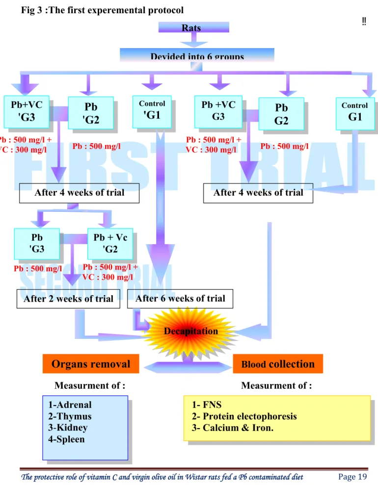
Fig 3 Experimental Protocol 1 19
Fig 4 Experimental protocol 2 20
Fig 5
Comparison of albumin (Alb) concentration (X± SD) in the serum of female Wistar rats in the control (G1), Pb (G2) and Pb +Vitamin C (G3)
after 4 weeks.
38
Fig 6 Comparison of immunoglobulins (Igs) concentration (X± SD) in the serum of female Wistar rats in the control (G1), Pb (G2) and Pb +Vitamin C (G3)
after 4 weeks.
38
Fig 7 Comparison of white blood cells concentration (X± SD) in the blood of female Wistar rats in the control (G1), Pb (G2) and Pb +Vitamin C (G3)
after 4 weeks.
39
Fig 8 Comparison of Neutrophils (Neu) concentration (X± SD) in the blood of female Wistar rats in the control (G1), Pb (G2) and Pb +Vitamin C (G3)
after 4 weeks.
39
Fig 9 Comparison of eosinophils (Eos) concentration (X± SD) in the blood of female Wistar rats in the control (G1), Pb (G2) and Pb +Vitamin C (G3)
after 4 weeks.
40
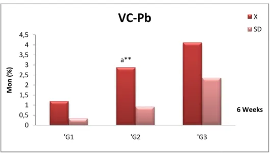
Fig 10 Comparison of monocytes (Mon) concentration (X± SD) in the blood of female Wistar rats in the control (G1), Pb (G2) and Pb +Vitamin C (G3)
after 4 weeks.
40
Fig 11 Comparison of calcium (Ca++) concentration (X± SD) in the serum of female Wistar rats in the control (G1), Pb (G2) and Pb +Vitamin C (G3)
after 4 weeks.
41
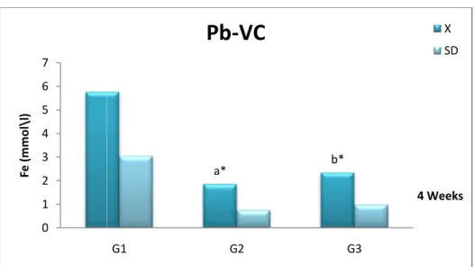
Fig 12 Comparison of iron (Fe++) concentration (X± SD) in the serum of female Wistar rats in the control (G1), Pb (G2) and Pb +Vitamin C (G3) after 4
weeks.
41
Fig 13 Ccomparison the relative organ weigh (%) of adrenal female Wistar rats in the control (G1), Pb (G2) and Pb +Vitamin C (G3) after 4 weeks.
42
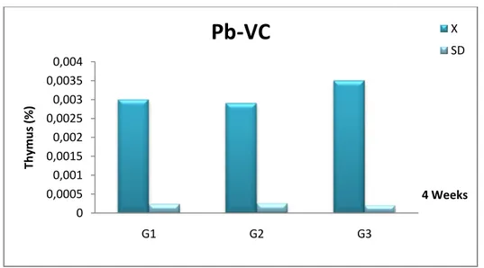
Fig 14 Comparison the relative organ weigh (%) of thymus female Wistar rats in the control (G1), Pb (G2) and Pb +Vitamin C (G3) after 4 weeks.
42
Fig 15 Comparison the relative organ weigh (%) of kidney female Wistar rats in the control (G1), Pb (G2) and Pb +Vitamin C (G3) after 4 weeks.
43
Fig 16 Comparison the relative organ weigh (%) of adrenal female Wistar rats in the control (G1), Pb (G2) and Pb +Vitamin C (G3) after 4 weeks.
43
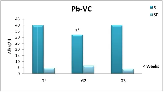
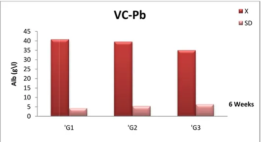
Fig 17 Comparison of albumin (Alb) concentration (X± SD) in the serum of female Wistar rats in the control (‘G1), Pb +Vitamin C (‘G2) and Pb (‘G3)
after 6 weeks.
44
Fig 18 Comparison of immunoglobulins (Igs) concentration (X± SD) in the serum of female Wistar rats in the control (‘G1), Pb +Vitamin C (‘G2) and Pb
(‘G3) after 6 weeks.
44
Fig 20 Comparison of Neutrophils (Neu) concentration (X± SD) in the blood of female Wistar rats in the control (‘G1), Pb +Vitamin C (‘G2) and Pb (‘G3)
after 6 weeks.
45
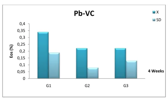
Fig 21 Comparison of eosinophils (Eos) concentration (X± SD) in the blood of female Wistar rats in the control (‘G1), Pb +Vitamin C (‘G2) and Pb (‘G3)
after 6 weeks.
46
Fig 22 Comparison of monocytes (Mon) concentration (X± SD) in the blood of female Wistar rats in the control (‘G1), Pb +Vitamin C (‘G2) and Pb (‘G3)
after 6 weeks.
46
Fig 23 Comparison of calcium (Ca++) concentration (X± SD) in the serum of female Wistar rats in the control (‘G1), Pb +Vitamin C (‘G2) and Pb (‘G3)
after 6 weeks.
47
Fig 24 Comparison of iron (Fe++) concentration (X± SD) in the serum of female Wistar rats in the control (‘G1), Pb +Vitamin C (‘G2) and Pb (‘G3) after 6
weeks.
47
Fig 25 Comparison the relative organ weigh (%) of adrenal female Wistar rats in the control (‘G1), Pb +Vitamin C (‘G2) and Pb (‘G3) after 6 weeks.
48
Fig 26 Comparison the relative organ weigh (%) of thymus female Wistar rats in the control (‘G1), Pb +Vitamin C (‘G2) and Pb (‘G3) after 6 weeks.
48
Fig 27 Comparison the relative organ weigh (%) of kidney female Wistar rats in the control (‘G1), Pb +Vitamin C (‘G2) and Pb (‘G3) after 6 weeks.
49
Fig 28 Comparison the relative organ weigh (%) of spleen female Wistar rats in the control (‘G1), Pb +Vitamin C (‘G2) and Pb (‘G3) after 6 weeks.
49
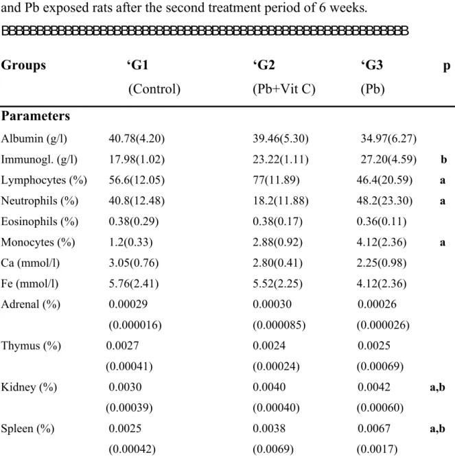
Fig 29 Comparison of Immunoglobulins (Igs) and Albumin (Alb) concentrations in serum of female Wistar rats after the two treatment periods.
50
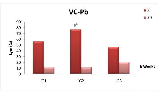
Fig 30 Comparison of Lymphocytes (Lym) and Neutrophils (Neu) in the blood of female Wistar rats after the two treatment periods.
51
Fig 31 Comparison of Eosinophils (Eos) and Monocytes (Mon) in the blood of female Wistar rats after the two treatment periods.
52
Fig 32 Comparison of Calcium (Ca++) and Iron (Fe++) concentrations in serum of female Wistar rats after the two treatment periods.
53
Fig 33 Comparison of relative weight of Adrenal (Adr), Thymus (Thy), Kidney (Kid) and Spleen (Spl) of female Wistar rats after the two treatment
periods.
54
Fig 34 Comparison of white blood cells concentration (X± SD) in the blood of female Wistar rats in the control (G1), Pb (G2) and Pb +Vitamin C (G3)
after 4 weeks.
55
Fig 35 Comparison of white blood cells concentration (X± SD) in the blood of female Wistar rats in the control (G1), Pb (G2) and Pb +Olive Oil (G4)
after 4 weeks
56
Fig 36 Comparison of white blood cells concentration (X± SD) in the blood of female Wistar rats in the control (G1), Pb (G2) and Pb +Vitamin C+ Olive
Oil (G5) after 4 weeks
weeks
Fig 38 Comparison of white blood cells concentration (X± SD) in the blood of female Wistar rats in the control (G1), Pb (G2) and Pb + Olive Oil (G4)
after 4 weeks
58
Fig 39 Comparison of white blood cells concentration (X± SD) in the blood of female Wistar rats in the control (G1), Pb (G2) and Pb +Vitamin C + Olive
Oil (G5) after 4 weeks
58
Fig 40 Comparison of lymphocytes concentration (X± SD) in the blood of female Wistar rats in the control (G1), Pb (G2) and Pb +Vitamin C (G3) after 4
weeks
59
Fig 41 Comparison of lymphocytes concentration (X± SD) in the blood of female Wistar rats in the control (G1), Pb (G2) and Pb + Olive Oil (G4) after 4
weeks
60
Fig 42 Comparison of lymphocytes concentration (X± SD) in the blood of female Wistar rats in the control (G1), Pb (G2) and Pb +Vitamin C + Olive Oil
(G5) after 4 weeks
60
Fig 43 Comparison of monocytes concentration (X± SD) in the blood of female Wistar rats in the control (G1), Pb (G2) and Pb +Vitamin C (G3) after 4
weeks
61
Fig 44 Comparison of monocytes concentration (X± SD) in the blood of female Wistar rats in the control (G1), Pb (G2) and Pb +Olive Oil (G4) after 4
weeks
62
Fig 45 Comparison of monocytes concentration (X± SD) in the blood of female Wistar rats in the control (G1), Pb (G2) and Pb +Vitamin C+ Olive Oil
(G5) after 4 weeks
62
Fig 46 Comparison of immunoglobulins concentration (X± SD) in the serum of female Wistar rats in the control (G1), Pb (G2) and Pb +Vitamin C (G3)
after 4 weeks
63
Fig 47 Comparison of immunoglobulins concentration (X± SD) in the serum of female Wistar rats in the control (G1), Pb (G2) and Pb +Olive Oil (G4)
after 4 weeks
64
Fig 48 Ccomparison of immunoglobulins concentration (X± SD) in the serum of female Wistar rats in the control (G1), Pb (G2) and Pb +Vitamin C+ Olive
Oil (G5) after 4 weeks
64
Fig 49 Comparison of triglycerides concentration (X± SD) in the serum of female Wistar rats in the control (G1), Pb (G2) and Pb +Vitamin C (G3) after 4
weeks
65
Fig 50 Comparison of triglycerides concentration (X± SD) in the blood of female Wistar rats in the control (G1), Pb (G2) and Pb +Olive Oil (G4) after 4
weeks
66
Fig 51 Comparison of triglycerides concentration (X± SD) in the serum of female Wistar rats in the control (G1), Pb (G2) and Pb +Vitamin C+ Olive Oil
(G5) after 4 weeks
66
Fig 53 Comparison of total cholesterol concentration (X± SD) in the serum of female Wistar rats in the control (G1), Pb (G2) and Pb +Olive Oil (G4)
after 4 weeks
68
Fig 54 Comparison of total cholesterol concentration (X± SD) in the serum of female Wistar rats in the control (G1), Pb (G2) and Pb +Vitamin C+ Olive
Oil (G5) after 4 weeks
68
Fig 55 Comparison of HDL concentration (X± SD) in the serum of female Wistar rats in the control (G1), Pb (G2) and Pb +Vitamin C (G3) after 4 weeks
69
Fig 56 Comparison of HDL concentration (X± SD) in the serum of female Wistar rats in the control (G1), Pb (G2) and Pb +Olive Oil (G4) after 4 weeks
70
Fig 57 Comparison of HDL concentration (X± SD) in the serum of female Wistar rats in the control (G1), Pb (G2) and Pb +Vitamin C+ Olive Oil (G5) after
4 weeks
70
Fig 58 Comparison of LDL concentration (X± SD) in the serum of female Wistar rats in the control (G1), Pb (G2) and Pb +Vitamin C (G3) after 4 weeks
71
Fig 59 Comparison of LDL concentration (X± SD) in the blood of female Wistar rats in the control (G1), Pb (G2) and Pb +Olive Oil (G4) after 4 weeks
72
Fig 60 Comparison of LDL concentration (X± SD) in the serum of female Wistar rats in the control (G1), Pb (G2) and Pb +Vitamin C+ Olive Oil (G5) after
4 weeks
72
Fig 61 Comparison of calcium concentration (X± SD) in the serum of female Wistar rats in the control (G1), Pb (G2) and Pb +Vitamin C (G3) after 4
weeks
73
Fig 62 Comparison of calcium concentration (X± SD) in the blood of female Wistar rats in the control (G1), Pb (G2) and Pb +Olive Oil (G4) after 4
weeks
74
Fig 63 Comparison of calcium concentration (X± SD) in the serum of female Wistar rats in the control (G1), Pb (G2) and Pb +Vitamin C+ Olive Oil
(G5) after 4 weeks
74
Fig 64 Comparison of iron concentration (X± SD) in the serum of female Wistar rats in the control (G1), Pb (G2) and Pb +Vitamin C (G3) after 4 weeks
75
Fig 65 Comparison of iron concentration (X± SD) in the serum of female Wistar rats in the control (G1), Pb (G2) and Pb +Olive Oil (G4) after 4 weeks
76
Fig 66 Comparison of iron concentration (X± SD) in the serum of female Wistar rats in the control (G1), Pb (G2) and Pb +Vitamin C+ Olive Oil (G5) after
4 weeks
76
Fig 67 Comparison of red blood cells concentration (X± SD) in the blood of female Wistar rats in the control (G1), Pb (G2) and Pb +Vitamin C (G3)
after 4 weeks
77
Fig 68 Comparison of red blood cells concentration (X± SD) in the blood of female Wistar rats in the control (G1), Pb (G2) and Pb +Olive Oil (G4)
after 4 weeks
78
Fig 69 Comparison of red blood cells concentration (X± SD) in the blood of
Wistar rats in the control (G1), Pb (G2) and Pb +Vitamin C (G3) after 4 weeks
79
Fig 71 Comparison of haemoglobin concentration (X± SD) in the blood of female Wistar rats in the control (G1), Pb (G2) and Pb +Olive Oil (G4) after 4
weeks
80
Fig 72 Comparison of haemoglobin concentration (X± SD) in the blood of female Wistar rats in the control (G1), Pb (G2) and Pb +Vitamin C+ Olive Oil
(G5) after 4 weeks
80
Fig 73 Transversal cross section of rat kidney from the control (X400) 81
Fig 74 Transversal cross section of rat kidney treated by Pb (X400) 82
Fig 75 Transversal cross section of rat kidney from group treated by Pb + Vitamin C (X400)
82
Fig 76 Transversal cross section of rat kidney treated by Pb + Olive Oil (X400) 83
Fig 77 Transversal cross section of rat liver from the control (X400) 84
Fig 78 Transversal cross section of rat liver treated by Pb (X400) 85
Fig 79 Transversal cross section of rat liver treated by Pb + Vitamin C (X400) 85
1. General Introduction
Nutritional factors and lead toxicity Total food intake
Calcium Iron Vitamin C Olive Oil Vitamin E Objective
2. Material & Methods
Biological materials and treatment First experimental protocol
Second experimental protocol
Blood collection and laboratory analysis Haematological analysis Biochemical analysis Protein electrophoresis Histological analysis Statistical analysis 3. Results
Impact of vitamin C during The first experimental treatment The first treatment
Immune system Minerals 1 5 7 7 9 11 12 14 15 17 17 18 21 21 21 28 29 34 35 38 38 41
The second treatment Immune system Minerals
Relative organs weigh
The second experimental treatment Immune system Lipid parameters Minerals Haematological parameters Histological study 4. Discussion 5. Abstract (English) 6. Résumé (French) 7. Abstract (Arabic) 8. References 44 44 47 48 55 55 65 73 78 81 87 104 107 111 115
The pollution is an unfavourable modification of the natural environment, which can affect the man directly or through his agricultural resources in water and the other organic products or by distorting the physical objects which he possesses.
Metals, particularly heavy metals such as lead, mercury, cadmium and arsenic constitute a significant potential threat to human health, both
occupational and environmental (Halliwell, 2000). The environmental
persistence of metal in concert with their intensive use by modern society has, over the years, created a concentration of metal in the biosphere. Thus, there is ample opportunity for exposure to toxic metal both in and outside the workplace.
According to a comparative study (Borella and Giardino, 1990) was built on some of the above-mentioned metals, lead was found to be the most toxic. In view of its wild spread on one hand and in its flexibility and resistance to corrosion and rust on the other hand.
Lead is used during the Bronze Age with the antimony and the arsenic .We finds it mentioned by the Sumerian cuneiforms about 5000 years ago, or still in the Exodus, drafted more than 2500 years ago.
It is interesting to notice that in the middle Ages the alchemists believed that the lead was the oldest metal and associated it with the planet “Saturn”. Doubtless from there that we find nowadays the notion of "Saturnism" which is a disease bound to the Pb intoxication.
Lead does not have a distinct function in human body. In general, the uptake of lead from various sources suggests a three compartmental tool for lead metabolisms; namely blood lead, lead accumulated in soft tissues and lead of the skeleton, where it appears to compete with calcium for binding sites and acts as a
calcium substitute in second messenger metabolism (Goldstein, 1993). In other
experimental animal methods, absorption of lead from the gastrointestinal tract has been shown to be enhanced by dietary calcium depletion or administration of
vitamin D (Mykkanen and Wasserman, 1981, 1982).
Lead competes also with iron for erythrocytes binding sites (Moor, 1988).
However, some increase in lead absorption may be found in iron deficiency
states in humans (Watson and Hume, 1983) and in animals (Flamagan et al.,
1979). In rats, iron deficiency increase the gastrointestinal absorption of lead, possibly by enhancing binding of lead to iron binding proteins in the intestine (Bannon et al., 2003; Barton et al., 1978; Morrison and Quarteman, 1987). Thus, supplementation of diet of lead-poisoned rats with iron and ascorbic acid prevent
the growth depression and anaemia associated with lead (Moor, 1988).
Lead exposure also has been shown to be associated with DNA damage, for example, battery plant workers have significantly elevated levels of DNA breaks
in lymphocytes compared to unexposed subjects (Fracasso et al., 2002). The
DNA damage has been observed also in a mice model of lead inhalation (Valverde et al., 2002). Reports have appeared in recent literature, which link
autoimmune diseases to environmental factors (Hengstler et al., 2003). In many
exposed populations, some individuals are extra sensitive while some extra
In addition to the well-documented and numerous toxic effects of lead on various targets organs, a number of studies have shown that acute and chronic exposure to inorganic lead may result at high level in an immediate suppressive
effect (Koller and Kavavic, 1974, Luster et al., 1978). But at low levels, it has
been reported an increase (Borella and Giardino, 1990), a decrease (Koller,
1973) or a no change (Rott and Charles, 1979) on immune function in
experimental systems. Many factors could account for such effects, including dose, metal chemical forms, animal strain and/or species. Thus, these controversies regarding the influence of Pb on immune system give a very vast possible domain of research.
The mean possible mechanisms of lead toxicity are two firstly, generation of reactive oxygen species (ROC) secondly disruption of tissue oxidant/antioxidant balance. Free radicals or ROC attack lipids, proteins, enzymes and DNA to cause pathological events and cancer.
The current therapeutic approach to lead toxicity is to increase the excretion of lead by chelation. Nutritional factors are often mentioned as important modifier of the metabolism and toxicity of lead. Ascorbic acid is one of the strongest reductants and radical scavengers and reduces stable oxygen, nitrogen, and thyl radical and acts as a primary defense against aqueous radicals in the blood. Animal experiments have demonstrated that essential elements, such as calcium, zinc, iron, selenium, and various vitamins can counteract the toxic
effects of lead (Miller., 1990, Patra et al., 2001, Panda and Flora, 2002). In
humans, particularly children low dietary intakes of iron, zinc, and calcium have
been associated with increased blood lead levels (Osman et al., 1998, Ahamed et
delta-aminolevulinic acid dehydratase (δ-ALAD) (2nd enzyme in heam
biosynthetic pathway) (Singh et al., 1994). Low iron intake enhances the
impairment of iron utilization for heam biosynthesis due to lead (Labbe, 1990).
Rats raised on a low-calcium diet have much higher blood lead concentrations
among the calcium-deficient animals (Miller et al., 1990), although lead
ingestion did not differ. Similar differences have also been observed in tissue lead concentrations.
The extents of understanding how nutritional factors affect susceptibility to lead vary from nutrient to nutrient. Techniques used to investigate nutrient/lead interactions include isolated cells studied in vitro, in situ–in vivo methods, whole animal studies, cross sectional and longitudinal studies with humans, and clinical trial with nutrient therapy. The latter have typically been poorly controlled with respect to other variables. Adequate nutrition may be a key factor in reducing the risk of lead exposure. Nutrients supplementation modifies the process of absorption, transport, storage, and inactivation of lead, thus reducing its toxicity (Dorea and Donangelo, 2006).
To explain the importance of using exogenous nutrients in treating
environmental lead toxicity the following topics are addressed: (Figure 1)
different sources of lead exposure influencing the blood lead levels and (Figure 2)
possible protective effects of nutrients supplementation (some essential metals and vitamins) in lead toxicity.
Figure 1: Sources of Pb 2004).
Nutritional factors and lead toxicity
Nutrients and their effects on lead toxicity can be viewed from a number of perspectives. If one is
exposure, followings are very common nutritional factors that can reduce lead toxicity(Figure2).
Pb exposure and its health effects (from
utritional factors and lead toxicity
utrients and their effects on lead toxicity can be viewed from a number of perspectives. If one is seeking strategies to reduce the toxicity of lead exposure, followings are very common nutritional factors that can reduce lead
exposure and its health effects (from Fewtrell et al.,
utrients and their effects on lead toxicity can be viewed from a number seeking strategies to reduce the toxicity of lead exposure, followings are very common nutritional factors that can reduce lead
Total food intake: Overall patterns of food consumption and frequency of
food intake influence absorption of lead from the gastrointestinal tract (ATSDR,
1999). Although the mechanisms may be complex, Lead that is ingested during
fasting is absorbed to a much greater extent than if ingested with food. For
example, Rabinowitz et al., 1980 reported that among adult male subjects, lead
without food was 35% absorbed; tracer lead ingested with food was absorbed to the extent of 8.2% and lead in food was 10.3% absorbed. Similar results have
been reported by Blake and Mann, 1983 and Flanagan et al., 1982.
Inter-individual variability of oral absorption was shown in the study by
Heusler-Bitschy et al., 1988 in which gastrointestinal absorption ranged from 10% to 80% in eight fasted volunteers receiving a single exposure of 0.007 or 0.002 mg lead/kg/day in drinking water.
The practical role of this information depends on the reason people have limited food intake. Frequency of food intake is controlled by a number of cultural and economic factors. Certainly, shortages of food exist in many parts of the world.
Calcium: The experimental literature on this topic is extensive and rather
consistently supports the observations that ingestion of diets low in calcium
increases lead absorption and toxicity. For example, Mahaffey-Six and Goyer,
1970 observed that lowering dietary calcium from 0.7% to 0.1% significantly
enhanced the body lead burden of rats exposed for 10 weeks to 200 ppm lead in their drinking water. Under these conditions, blood, kidney, and femur levels of lead increased significantly as did urinary excretion of delta amino-levulinic acid (δ-ALA). A significant increase in renal intra-nuclear lead inclusion bodies also was observed in lead exposed animals consuming the low calcium diet. In a
similar study, metabolic and morphologic manifestations of lead exposure were compared in adult rats receiving 2, 13, 48, 96, and 200 ppm lead in their drinking
water and either a 0.1% or 0.7% calcium diet (Mahaffey et al., 1973). Animals
consuming the low calcium diet have increased blood, kidney, and femur lead levels, urinary excretion of δ-ALA, and renal intra-nuclear lead inclusion bodies at lower doses of lead than those on the adequate calcium diet.
However, interpretation of these results is confounded by the fact that the animal receiving the low calcium diet without lead supplementation demonstrated sign of enhanced lead uptake as evidenced by increased levels of lead in femur and kidney, as well as elevated urinary excretion of δ-ALA. This may be result of enhanced uptake of ingested ambient lead that does not bode well for inadequately nourished children. Also, exposure to lead resulted in a decline in rate of weight gain and food consumption in animals receiving the low calcium diet. Decreased lead absorption with increasing dietary intake of calcium
also has been observed in humans (Heard and Chamberlain, 1982). In 8 adult
male subjects fasted overnight, it was observed that absorption of 100 mg lead as
lead chloride containing 203Pb decreased from 60% to approximately 10% when
followed by a dose of 200 mg calcium (as calcium carbonate) and 140 mg of phosphate (as sodium diphosphate).
Calcium alone reduced lead uptake by a factor of 1.3, and phosphate alone by a factor of 1.2 whereas both together reduced uptake by a factor of 6. The calcium and vitamin D status of subjects may have influenced these results;
however, they were not determined. A similar result has been reported by Blake
few clinical studies. An inverse relationship between dietary calcium and lead absorption was found. Although calcium glycerophosphate supplement
prevented lead absorption (Sargent et al., 1999), another study concluded that
calcium supplementation should not be routinely prescribing for mild to
moderately lead-poisoned children with adequate calcium intakes(Markowitz et
al., 2004).
Although bone has predominantly been considered a storage site for sequestering absorbed lead, bone is not simply an inert storage site. Once deposited in bone, lead can be remobilized from bone in response to both physiological (e.g. pregnancy or lactation) and pathological (e.g. osteoporosis)
conditions (Silbergeld, 1991). It has been shown that during the first few years
after onset of menopause, there can be marked mobilization of calcium from bone matrix. Analysis of data from the Second National Health and Nutrition Examination Survey (NHANES II) has demonstrated a highly significant increase in whole blood lead concentration after menopause. Mobilization of long-term stores of lead from the maternal skeleton may be a major determinant in transfer of lead from mother to infant during pregnancy and lactation. Because of concern that maternal blood lead concentrations be maintained as low as possible during pregnancy, this remobilization of lead from bone has substantial public heath interest.
Iron: Iron-deficiency is recognized worldwide as one of the most prevalent
nutritional problems (Grigg, 2004). The public health implications for enhanced
lead toxicity among iron deficient persons are substantial. Both iron and lead affect the heam biosynthetic process. The cellular basis for greater susceptibility of iron deficient animals to lead is that limited iron in the mitochondria
apparently enhances the impairment by lead of iron utilization for heam
synthesis (Labbe, 1990). Lead is known to interfere with mitochondrial energy
metabolism that is necessary to reduce ferric iron to ferrous iron before insertion of iron into porphyrin ring. Where there is insufficient ferrous iron for incorporation by ferrochelatase into heam, protoporphyrin accumulates.
Ferrochelatase activity is sensitive to both lead and iron. Kappor et al., 1984
have reported that the enzyme kinetics of ferrochelatase in isolated human erythrocytes change with both iron and lead concentrations. When iron deficiency is present, ferrochelatase is more sensitive to lead effects. The cellular basis for greater susceptibility of iron deficient animals to lead is that limited iron in the mitochondria apparently enhances the impairment by lead of iron utilization for heam synthesis.
In an earlier study, blood lead levels were negatively correlated with blood
iron level (Ahamed et al., 2007) is in agreement with a study of preschool
children; an increase in blood lead with decreasing dietary iron intake was found (Hammad et al., 1996). Finding of Osman et al., 1998 also supports who has reported that children with low serum iron level have a tendency to higher blood lead levels, indicating increased gastrointestinal absorption of lead at reduced
iron stores. However, Flanagan and Chamberlain, 1982 found no change in lead
absorption with increasing iron absorption due to iron deficiency and inconsistent results regarding lead absorption in adults were observed in a study by Watson and coworkers, 1986. Therefore, there is probably not a single pathway for the absorption of these two elements.
The impairment of cognitive function among iron-deficient children has
accompanying iron-deficiency are similar to those observed during prolonged exposure to low levels of lead. Interference with iron absorption and/or metabolism by lead exposure could result in a synergism of deleterious effects, especially in young children where iron deficiency is one of the most common
nutritional deficiencies (Grigg, 2004).
Vitamin C: Vitamin C (ascorbic acid) is a low molecular mass antioxidant
that scavenges the aqueous free radicals by very rapid electron transfer that
inhibits lipid peroxidation (Halliwell and Gutteridge, 1999). Administration of
vitamin C significantly inhibited the lipid peroxidation levels of liver and brain,
and increased the catalase levels of kidney in lead-exposed rats (Patra et al.,
2001). Lead-induced free radicals indicated by rat sperm chemiluminescence
generation were reduced by 40% with supplementation of 500 mg vitamin C/L
drinking water (Hsu et al., 1998).
Another beneficial role of ascorbic acid supplementation in lead-exposed rats was associated with serum biochemical alterations in the haemopoietic system and drug metabolizing enzymes. Intraperitoneal administration of lead in rats produced a significant inhibition of heam synthesis in blood and liver, and reduced drug metabolism in liver. Vitamin C supplementation in lead-exposed animals significantly reduced blood, liver, and renal lead levels. This result indicated a significant protective action of vitamin C against toxic effects of lead
on heam synthesis and drug metabolism (Vij et al., 1998). A study found the
combination of vitamin C and thiamine was effective in reducing lead levels in blood, liver, and kidney. In addition, both lead-induced inhibition in the activity of blood δ-ALAD and elevation in the level of blood zinc protoporphyrin (ZPP)
There has been considerable debate concerning the relationship between vitamin C nutritional status and heavy metal body burden in lead-induced toxic effects. Early reports found that vitamin C might act as a possible chelator of
lead, with similar potency to that of EDTA (Goyer and Cherion, 1979). Vitamin–
mineral supplementation in African-American women was found to reduce blood lead levels from 5.1 to 1.1 μg/dL, which was negatively correlated with serum
levels of vitamin E and C (West et al., 1994). Dhawan et al., 1988 found that
ascorbic acid might increase urinary elimination of lead and reduced the hepatic and renal lead burden in rats. A cross-sectional study analyzed 4213 young and 15 365 adult Americans with mean blood lead levels of 2.5–3.5 μg/dL, respectively, and showed an inverse relationship between serum vitamin C and
blood lead level (Simon and Hudes, 1999). In another study of 85 volunteers
who consumed a lead-containing drink, vitamin C supplementation produced
small reductions in lead retention (Dawson and Harris, 1997).
On the other hand, some studies suggested that vitamin C supplementation did not significantly lower blood lead levels. In workers occupationally exposed to lead, and with blood lead levels ranged from 28.9 to 76.4 μg/dL, supplementation of vitamin C and zinc did not significantly reduce blood lead
levels (Lauwerys et al., 1983).
Olive oil: In the Mediterranean basin, olive oil, along with fruits,
vegetables, and fish, is an important constituent of the diet, and is considered a major factor in preserving a healthy and relatively disease-free population. Epidemiological data show that the Mediterranean diet has significant protective effects against cancer and coronary heart disease. We present evidence that it is the unique profile of the phenolic fraction, along with high intakes of squalene
and the monounsaturated fatty acid, oleic acid, which confer its health-promoting properties. The major phenolic compounds identified and quantified in olive oil belong to three different classes: simple phenols (hydroxytyrosol, tyrosol); secoiridoids (oleuropein, the aglycone of ligstroside, and their respective
decarboxylated dialdehyde derivatives); and the ligands
[(+)-1-acetoxypinoresinol and pinoresinol]. All three classes have potent antioxidant properties. High consumption of extra-virgin olive oils, which are particularly rich in these phenolic antioxidants (as well as squalene and oleic acid), should afford considerable protection against cancer (colon, breast, skin), coronary heart disease, and ageing by inhibiting oxidative stress.
Recent studies (Marubayashi et al., 1985, Bukowska et al., 2000) showed
that diets rich in monounsaturated fatty acids, especially oleic acid, produce fapoprotiens. Three papers in the June 1988 issue of the American Journal of
chemical Nutrition (Duchnowicz and Koter, 2003, Ozcan and Uner, 2000)
suggest that increased consumption of oleic acid in the form of Olive Oil, at the expense of either saturated fatty acids or carbohydrates, is a proper approach to lowering coronary disease risk.
Wiseman et al., 1996 indicated that dietary Olive Oil specifically causes increased concentrations of liver cholesterol in laboratory animals. Although it is not known whether this effect is harmful and whether the animal data can be extrapolated to man.
A study by Lions et al (1991) has suggested that there may be beneficial
anti-inflammatory effects of Olive Oil consumption on Rheumatoid Arthritis (RA). This study compared the relation risk of development of RA in relation to
lifelong consumption of Olive Oil in a Greek population and it demonstrated that high consumers of Olive Oil (almost every day throughout life) were four times less likely to develop RA than those who consumed Olive Oil less than six times
per month on average throughout their lives (lions et al, 1991).
Vitamin E: Vitamin E is the generic term used to describe at least eight
natural-occurring compounds that possess the biological activity of α-tocopherol. The group is comprised of α-, β-, γ- and tocopherol and α-, β-, γ- and
δ-tocotrienol. The α-tocopherol has the highest biological activity (Weister &
Vecchi, 1983), the other tocopherols and tocotrienols are less biologically active
but they are at least as abundant as α-tocopherol in certain foods (Sheppard et al.,
1993). Vitamin E is nature's major lipid soluble chain-breaking nutrient that is
known to protect biological membranes and lipoproteins from oxidative stress (Packer, 1991). The main biological function of vitamin E is its direct influencing of cellular responses to oxidative stress through modulation of
signal-transduction pathways (Azzi et al., 1992). One of the protective roles of
vitamin E on lead toxicity was preventing lead's effects on productions of free
radicals in liver (Chaurasia and Kar, 1997).
The interaction between vitamin E and other nutrients might have a more efficient protective action against lead toxicity. Vitamin E and C jointly protect
lipid structures against peroxidation (Buettner, 1993). Although vitamin E is
located in membranes and vitamin C in aqueous phases, vitamin C is able to
recycle oxidized vitamin E (Frei, 1991). Vitamin C repairs the tocopherol
radical, thus recovering the chain-breaking antioxidant capacity of vitamin E (Buettner, 1993). Vitamin E alone or in combination with conventional chelator,
CaNa2EDTA, was found to decrease the lead-induced lipid peroxide levels of
liver and brain in rats (Patra et al., 2001).
A study of the influence of vitamin C on the tissue deposition of lead in rats suggested that it might be useful as a prophylactic agent for lead poisoning (Simon, 2003). Later studies in rats demonstrated that vitamin C decreases Pb
intestinal absorption and increases its renal clearance (Fotherby et al., 2000).
Further studies indicated that ascorbic acid may chelate lead and decrease the risk of its toxic effects on immune system; it helps this system to fight off foreign invaders and tumour cells possibly by stimulating the production of white blood cells, primarily neutrophiles, which attack foreign antigens. It also boosts the body’s production of antibodies and interferon, proteins that helps
protect cells from viral invaders (Null, 1994). In addition to its great action
against free radical produced by lead, vitamin C works along with vitamin E, a fat-soluble antioxidant, and the enzyme glutathione peroxidase to stop chain reaction of free radicals.
The objectives:
The aim of the present study, however, is to assess the possible protective roles of natural substances (ascorbic acid and local virgin olive oil) in Pb intoxicated rats. The test includes the immune response (leucocytes and total immunoglobulins Igs), liver and kidney function, relative organ weights of kidney, spleen, thymus and adrenal and the level of some biochemical parameters.
Natural substances supplementation was given at the beginning of the Pb intoxication or after a while. The suppression of vitamin C from Pb intoxicated rats was also tested.
As part of the Mediterranean basin, Algerians consume much of the virgin olive oil of different strains.
A- What is the effect of this eating habit on exposed people to Pb in the environment?
B- Can they withstand such oxidative stress produced by these pollutant? C- Can olive oil and Vitamin C be able to protect and/or boost the immune
2.1. Biological materials and treatment
Wistar rats were brought from Pasteur Institute, Algiers, and then reared in the Biology Department rearing house, University of Annaba under standard conditions of temperature, humidity and light. Animals were five week old and weighting an average of 1003g. They were given food and deionised drinking water ad-libitum.
2.1.1The first experimental protocol:
The first experimental protocol is divided into the first and the second treatment:
2.1.1.1. First treatment:
Rats were randomly divided into three groups of ten individuals and exposed either to 500mg Pb/l as lead acetate in their drinking water (G2) or 500mg Pb/l combined with 300mg vitamin C/l (G3) for a period of 4 weeks. The control group was given deionized water only (G1).
After decapitating half of each group, part of peripheral blood was collected in tubes contained EDTA for haematological study. The other part of blood was collected in dry tubes and used for biochemical measurement after obtaining serum, which then frozen immediately at -20°C. Immunoglobulins were separated by cellulose acetate electrophoresis.
The internal organs; Thymus, Adrenal, Kidney and Spleen were removed and their wet weights were obtained.
2.1.1.2.1. Second treatment:
In the second period, the treatment method was inversed for the remaining animals and continued for other two weeks (6 weeks in total). In this period, vitamin C was suppressed from the combined treatment group, and then added to the group which has already received Pb alone.
After decapitation, the same steps of the first period were repeated (figure 3).
2.1.2. The second experimental protocol:
Fresh virgin olive oil was obtained from Skikda region at an altitude of 500m. Also vitamin C was obtained from the pharmacy. Both lead and vitamin C were dissolved in deionized water.
Lead was given at 500 mg lead acetate/l, vitamin C was supplemented at 300mg/l and virgin olive oil was supplemented at 5% diet.
Animals were randomly divided into two main groups. The first main group was fed on basal diet as a negative control (G1); the second main group received lead contaminated drinking water ad-libitum for a period of four weeks. This group was divided into the following four subgroups; Pb alone (G2), Pb-vitamin C (Pb-VC (G3)), Pb-virgin olive oil (Pb-OO (G4)) and Pb-vitamin C-virgin olive oil (Pb-VC-OO (G5)).
After decapitation, peripheral blood was collected in tubes contained EDTA, and was used for counting leucocytes subpopulations, red blood cells and haemoglobin using Automatic Coulter Counter Machine (T 540). The other part of blood was collected in dry tubes and used for the measurement of serum HDL,
LDL, total cholesterol, triglycerides, Ca++ and Fe++ using an automat machine.
The serum pools sampled for protein measurements were frozen immediately at -20° C until they were separated by cellulose acetate electrophoresis (figure 4).