O
pen
A
rchive
T
oulouse
A
rchive
O
uverte
(OATAO)
OATAO is an open access repository that collects the work of some Toulouse
researchers and makes it freely available over the web where possible.
This is
an author'sversion published in:
https://oatao.univ-toulouse.fr/23085Official URL :
https://doi.org/10.1148/radiol.2018162093
To cite this version :
Any correspondence concerning this service should be sent to the repository administrator:
Cartayrade, Noëlle and Lapègue, Franck and Cambon, Zoé and Sans, Nicolas and Faruch,
Marie Case 264. (2018) Radiology, 289 (3). 873-875. ISSN 0033-8419
OATAO
Case 264
Noëlk Cartayrade, MD
•
Franck Lapègue, MD
•
Zoé Cambon, MD
•
Nicolas Sans, MD, PhD
•
Marie Faruch, MD
From the Depanmem of Radiology, CHU Toulouse-Purpan, Plaœ du docteur Baylac, 31059 Toulouse Cedex 9, Franœ. Address correspondence to N.C. (e-mail: ,we/k.canayratk@gmailcom).
Conflicrs of interest are listed at the end of this article.
Hi
s
tor
y
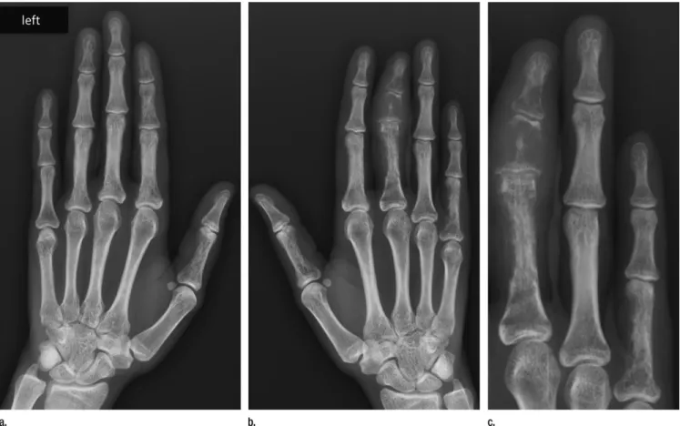
A 28-year-old woman presented to the emergency department with painful swelling of the third finger on her right hand, which devdoped quickly. She had no rdevant medical or surgical history. Her pain was worse at night, with stiffness decreasing during the moming. Clinical examination revealed generalized swelling of the third finger, cyanotic skin, and fingernail splitting on the second finger of the left hand (Fig 1). l.aboratory test results were normal, with no evidence ofinflammatory disease. Radiographs ofboth hands were obtained (Fig 2). CT scanning (Fig 3) and MRI (Fig 4) were also performed.2018 Diagnosis Please Learning
Objectives
In submitting a diagnosis for this case, participants
dem-onstrate the ability to
• Recognize normal and abnormal findings as presented
in the diagnostic images
• Identify pathologie conditions indicated in the
diagnos-tic images
• Use clinical reasoning skills to generate a lise of differ-ential diagnoses
Accreditation and Designation Statement
The RSNA is accredited by the Accreditation Council for
Continuing Medical Education (ACCME) to provide
continuing medical education for physicians. The RSNA designates this journal-based CME activity for a
maxi-mum of 1.0
A.MA PRA Category 1 Credi{.
Physiciansshould daim only the credit commensurate with the ex-tent of their participation in the activity.
Disclosure Statement
The ACCME requires chat the RSNA, as an accredited
provider of CME, obtain signed disclosure statements
from authors, editors, and reviewers for this case. For this
educational activity the authors, editors, and reviewers have indicated chat they have no relevant relationships to disclose.
Submit Diagnosis
Submit the most likely diagnosis to
http://rma.org/dxplease
(use only for submission of diagnosis). Select the case
from the Active Case List for which you are submitting
a diagnosis. Only one case, one name, and one diagnosis per e-mail submission. Multiple diagnoses and multiple
submissions will not be considered. Deadline: Midnight
U.S. Central Time, February 10, 2019. Answer will ap-pear in the April 2019 issue. Authors wishing to submit
cases for Diagnosis Please should first write to the Editor
to obtain approval for the case and further information.
Figure 3: Coronal reconstructed CT image of the right hand ob-tained with the bone window setting.
Figure 2: (a, b) Anteroposterior radiographs of the left (a) and right (b) hands. (c) Close-up view of the last three fingers of the right hand.
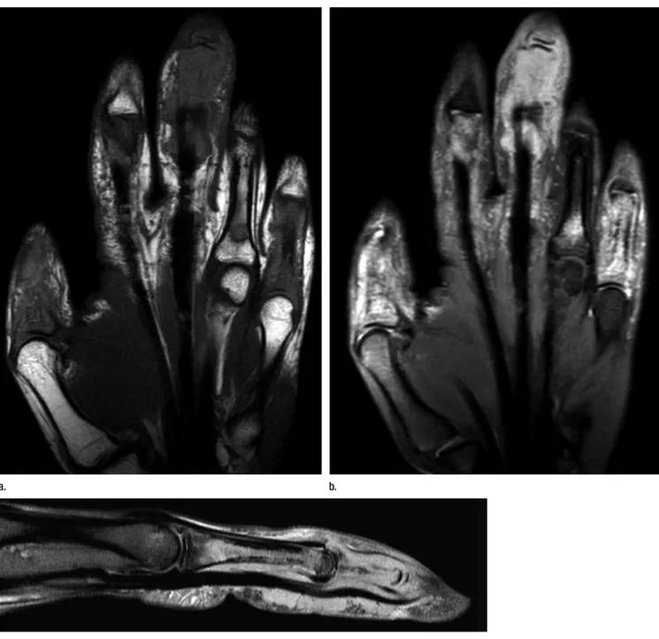
Figure 4: (a) Coronal T1-weighted (repetition time msec/echo time msec, 508/20) MR image of the right hand. (b) Coronal proton-density fat-suppressed (2357/30) MR image of the right hand. (c) Sagittal gadolinium-enhanced fat-fat-suppressed T1-weighted (658/20) MR image of the third finger on the right hand.
Disclosures of Conflicts of Interest: N.C. disclosed no relevant relationships. F.L. disclosed no relevant relationships. Z.C. disclosed no relevant relationships. N.S. disclosed no relevant relationships. M.F. disclosed no relevant relationships.