Cysl T1 R modulation by Th1 Th2 and Th3 type cytokines in BSMC
Par
Karina Espinosa Service d'immunologie Département de pédiatrie
Mémoire présenté à la Faculté de Médecine en vue de l'obtention du grade de maître ès sciences (M.Sc.) en Immunologie
l+I
Acquisitions et Acquisitions and
Bibliographie Services services bibliographiques 395 Wellington Street
Ottawa ON K1A ON4 Canada
395, rue Wellington Ottawa ON K1A ON4 Canada
The author has granted a non-exclusive licence allowing the National Library of Canada to reproduce, loan, distnbute or sell copies of this thesis
in
microform, paper or electronic formats.The author retains ownership of the copyright in this thesis. Neither the thesis nor substantial extracts from it may be printed or otherwise
reproduced without the author' s penmss1on.
Your 6Je VD118 rélérenœ
Our fie Notre rrif8Iwlcs
L'auteur a accordé une licence non exclusive permettant à la
Bibliothèque nationale du Canada de reproduire, prêter, distribuer ou vendre des copies de cette thèse sous la forme de microfiche/film, de reproduction sur papier ou sur format électronique.
L'auteur conserve la propriété du droit d'auteur qui protège cette thèse. Ni
la
thèseni
des extraits substantiels de celle-ci ne doivent être imprimés ou autrement reproduits sans son autorisation.0-612-80583-2
L'inflammation asthmatique chronique est caractérisée par le remodelage des voies aériennes, la dénudation de l'épithélium, l'épaississement des parois bronchiques, la fibrose sous-épithéliale, la métaplasie des glandes à mucus, ainsi que par l'hyperplasie et l'hypertrophie des m.uscles lisses bronchiques. Certaines études suggèrent un rôle possible pour les cystéinyl-leucotriènes (CysLTs) dans le remodelage des voies aériennes. Ce travail vise à caractériser la modulation du récepteur CysL T1 (CysL T1 R) par des cytokines de type Th1, Th2 et Th3 dans un pr~cessus fonctionnel tel que le remodelage des voies aériennes. MÉTHODES: L'expression du CysL T1 R dans des cellules de muscle lisse bronchique humaines (CMLB) provenant de cultures primaires a été évaluée par cytométrie de flux, avec un anticorps polyclonal anti-CysL T1 R, après stimulation avec les cytokines pendant 24h. Une analyse par microscopie de fluorescence a été effectuée pour compléter les résultats obtenus par la cytométrie de flux. L'expression de l'ARNm de CysLT1 Ra été mesurée par RT-PCR à l'aide d' amorces spécifiques pour ce récepteur et pour GAPDH. La prolifération a été évaluée par une analyse colorimétrique dans des cellules traitées pendant 24h avec les cytokines puis pendant 72h avec L T04. RESULTATS: L'expression de CysLT1 R chez les CMLB a été modulée à la hausse par TGF-13, IL-13 et IFN-y. IL-13 et IFN-y, mais pas TGF-13, ont augmenté l'expression de l'ARNm de CysLT1 R. La modulation à la hausse de CysLT1 R
L TD4. L'antagoniste de Cysl T1 R, Montelukast, a inhibé cet effet, ce qui suggère que l'effet de prolifération est sélectif pour Cysl T1 R. Ensemble, nos résultats soutiennent le rôle des Cysl Ts dans le remodelage des voies aériennes observé chez les patients asthmatiques.
Table of Contents ···-~··· i
Diagrams and Tables ... iii
Abbreviation Iist ... iv
Résumé ... . Introduction ... 1
p ART 1 ASTHMA ... 1
1.1. Definition ... 1
1.2. The early and late reaction ... 1
The early phase reaction ... 1
The la te phase reaction ... 2
PART 2. FACTORS IN THE DEVELOPMENT AND REGULATION OF AIRWA Y INFLAMMATION .. 4
2.1. Airway smooth muscle cells in asthma ... 4
The passive role ... 4
The active role ... 8
2.3. Biological actions of IFN-y, IL-13 and TGF-{3 ... 13
IFN-y ... 13
TGF-~ ... 15
IL-13 ... 19
2.4. Cysteinyl Leukotrienes ... 21
Biological actions ... 24
Cysteinyl Leukotriene Receptors ... 28
Objectives and strategies ... 32
Article ... 34
Introduction ... ~ ... 37
Discussion ... 59
Conclusions ... 68
Diagrams and Tables
Diagram 1 ... 22
Table 1 ... 27
Abbreviation list
AHR: Airway hyperresponsiveness ASMC: Airway smooth muscle cells
BAL: Bronchoalveolar lavage
BFGF: Basic fibroblast growth factor BSMC: Branchial smooth muscle cells
cAMP: Cyclic adenosine monophosphate COX: Cyclo-oxygenase
CysLTs: Cysteinyl leukotrienes
CysL T1 R: Cysteinyl leukotriene 1 receptor CysL T2R: Cysteinyl leukotriene 2 receptor
ERK: Extracellular signal regulated kinase Fc-e-Rll: Fragment crystallizable receptor type Il
GM-CSF: Granulocyte monocyte-colony stimulating factor GPCRs: G-protein coupled receptors
HASMC: Human airway smooth muscle cells ICAM: lntercellular adhesion molecule
IFN-y: lnterferon gamma LPS: Lipopolysaccharide L T: Leukotriene
L TAs: Leukotriene antagonists
MCP: Monocyte chernoattractant protein OSM-R: Oncostatin M receptor
PAR2: Proteinase-activated receptor 2 Pl3 K: Phosphatidylinositol 3-kinase PGE2: Prostaglandin E2
RV: Rhinovirus
SES: Socio-econornic status
STAT: Signal transducer and activator of transcription Th: T helper
TNF-a: Turner necrosis Factor alpha TNFR: Turner necrosis Factor receptor VCAM: Vascular cell adhesion rnolecule
Introduction
Part 1 Asthma
1.1. Definition
Asthma is a chronic lung disease characterized by obstruction, inflammation, and hyperresponsiveness of the airway to a variety of nonspecific stimuli. The obstruction is recurrent, partially reversible due to airway remodeling (Fahy et al., 2000) and manifests clinically as dyspnea, wheezing and/or coughing (Monteleone and Sherman, 1997).
ln asthma, there is an early and a late phase reaction;
1.2. The early and late reaction
The ear/y phase reaction
The early asthmatic reaction develops within 20 min after exposure to allergen and is characterized by bronchospasm due to classical immediate-type hypersensitivity, triggered through allergen-induced cross-linking of specific lgE antibody bound to mast cells through high-affinity receptors (Host, 2000).
The activated mast cells release proinflammatory mediators which are divided into three groups: secretory granule preformed mediators (histamine, heparin, tryptase, chymase, cathepsin G and carboxypeptidase), cytokines produced upon activation (IL-3, IL-4,IL-5, IL-6 and TNF-alpha) and lipid mediators produced upon activation (Leukotrienes C4,D4,E4, Prostaglandin D2 and Platelet Activating
Factor). These mediators are capable of inducing constriction of airway smooth muscle, increased secretion of mucus, and vasodilatation, as well as several important biological events, including modulation of the expression of adhesion molecules in endothelial and epithelial cells, and of inflammatory cell recruitment. Clinically, acute airway inflammation is characterized not only by a bronchoconstrictive response but also by a series of airway events that lead to enhanced branchial hyperresponsiveness, mucus secretion and increased asthma severity (Abbas, A, 2000), (Nadel, 1998).
The late phase reaction
Cellular recruitment to the airway is a key factor in this process. There is an initial increase in neutrophils followed by the appearance of eosinophils. There is less evidence for the involvement of neutrophils in asthma than for eosinophils, but neutrophils may have a greater importance in the pathogenesis of severe asthma. Persistent neutrophil inflammation is present in bronchoalveolar lavage (BAL) fluid, biopsy specimens and sputum from patients with symptomatic sever asthma treated with high doses of glucocorticoids (Leckie, 2000).
Eosinophils show several biological effects that can modulate inflammatory cellular reactions and promote further inflammation. The preformed mediators in eosinophils include several proteins, such as Major Basic Protein, which is cytotoxic to epithelial cells, it has been identified in areas of bronchial denudation. Eosinophils also have specific granules that contain eosinophil peroxidase and Eosinophil Cationic Protein. They can synthesize other mediators including leukotrienes C4, 04, E4 and lipoxins as well as proinflammatory cytokines (IL-4 and IL-13) that promote Th2 differentiation. Eosinophils can thus contribute to bronchoconstriction, mucus secretion and changes in vasopermeability (Nadel, 1998), (Abbas, A, 2000).Th2 lymphocytes are essential for the late-phase response, because they produce cytokines that promote allergie responses like IL-4, IL-13 and IL-5. ln contrast, Th1 lymphocytes produce IFN-y, IL-2 and TNF-~.
Part 2. Factors in the deve/opment and regu/ation of
airway inflammation
2.1. Airway smooth muscle cells in asthma
Branchial smooth muscle cells (BSMC) can have a passive and active raie in asthma. For many years, researchers have considered BSMC as a passive tissue subjected to the actions of different cell mediators that could induce airway remodeling and branchial smooth muscle contraction in asthmatic patients. But new findings demonstrate that BSMC could also have an active raie. They can secrete several prainflammatory mediators and express different receptors that contribute to the chronic inflammation seen in the asthmatic airways.
The passive role
Airway remodeling occurs in chranic asthmatic inflammation, features of this response include epithelium denudation, wall thickening, subepithelial fibrasis, mucus metaplasia, myofibroblast hyperplasia, and myocyte hyperplasia and hypertrophy. Vascular abnormalities have also been described (Elias, 2000), (Wiggs et al., 1992), (Aikawa et al., 1992). The consequences of airway remodeling in asthma may include incompletely reversible airway narrawing, branchial hyperresponsivenesss, airway edema, and mucus hypersecretion.
Airway remodeling in asthma thus may predispose persans with asthma ta asthma exacerbations and even death fram airway obstruction caused by smooth muscle contraction, airway edema, and mucus plugging (Fahy et al., 2000)
The number and size of branchial blood vessels is increased in asthma, and these vessels may have an important raie in regulating airway caliber, because an increase in vascular volume may swell the mucosa and narraw the airway lumen. Many inflammatory mediators cause vasodilation, a response that may be accompanied by increased permeability at the postcapillary venule, plasma extravasation, and airway mucosal edema (Laitinen et al., 1987),(McDonald, 1987). There is little evidence for branchial narrawing due ta the mechanical effect of peribranchial edema, or by swelling of the branchial mucosa. However, edema foam may terminally cause grave airway obstruction (Snashall and Chung, 1991).
Compared with nonasthmatic subjects, airway wall thickness is increased from 50 ta 300% in cases offatal asthma and from 10 ta 100% in cases of nonfatal asthma. There is aise an increase of 50-230% and 25-150% in the area of airway smooth muscle (ASM) in fatal and nonfatal asthma, respectively (Elias, 2000). lncreased thickness of the airway may have an important amplifying effect in the contractile response of ASM and may be a major mechanism contributing ta airway responsiveness in asthma (James et al., 1989), (Wiggs et al., 1992).
The degree ta which hyperplasia versus hypertraphy contributes ta this response is contraversial. Ebina and colleagues (Ebina et al., 1993), described two
differing responses; ln Type 1 asthma the increase in muscle mass was associated only with hyperplasia (increase in cell number) and was restricted to large central airways. ln Type Il asthma, comparatively mild hyperplasia was seen in the large airways and hypertrophy (increase in cell size) was detected throughout the branchial tree, particularly in small peripheral airways. IL-1 ~and
IL-6 can induce hyperplasia and hypertrophy of cultured guinea pig airway smooth muscle cells (De et al., 1995).
Several mediators can contribute to the proasthmatic changes in airway remodeling. For example, human mast cell mediators can cause human airway smooth muscle (HASM) proliferation, encourage fibrosis, and thereby contribute to airway remodeling and narrowing; they can cause contraction and increase the responsiveness of HASM to contractile mediators, also contributing to airway narrowing; and last but perhaps most important, these mediators can increase proinflammatory cytokine release and surface protein expression on HASM, thus favoring inflammatory cell infiltration and activation (Page et al., 2001). Studies have reported other differences between the airway smooth muscle from asthmatic and normal subjects. Data suggest that in asthmatic patients, the airway smooth muscle velocity of shortening is increased and the relaxation decreased (Que et al., 2000), (Solway, 2000). There are two important consequences of increased velocity of smooth muscle shortening. First, experiments demonstrate that sensitized tissues with increased velocity of shortening exhibit a greater extent of total shortening in response to contractile stimulation. Second, (which has not yet been proved experimentally) theory
suggests that a faster velocity of shortening should induce contractile hyperresponsiveness, by minimizing the bronchodilating effect of a deep inhalation (Black et al., 2001).
When a bronchospasm occurs, deep inspirations may be one of the most potent bronchodilating strategies, and they comprise the first line of defense. But in the spontaneous asthmatic attack, this potent bronchodilating mechanism fails and deep inspirations only make matters worse. The negative intrathoracic pressure associated with deep inspiration occurring in the context of increased leakiness of the airway vasculature may temporarily increase airway oedema and thus reduce luminal diameter in subjects with asthma (Burns and Gibson, 2002).
ln asthma it exists a perturbed equilibrium of myosin binding (few cross-links attached at any moment, but cycling rapidly), where the fully activated muscle becomes much less stiff and much more viscous, almost as if the muscle had "melted" (Fredberg, 2000).
ln asthma, not only the contractile responses of airway smooth muscle are altered, data show that airway smooth muscle from asthmatic patients have a higher proliferation rate compared with that of non-asthmatic patients (Johnson et al., 2001). There is a strong possibility, that this phenomenon is regulated by the ERK (extracellular signal regulated kinase) and PlK (phosphatidylinositol 3-kinase) pathways, because they appear to be major positive regulators of airway smooth muscle proliferation (Page and Hershenson, 2000).
The active role
As we mentioned before, BSMC not only have a passive raie in asthma. They may also have an active immunomodulatory raie by secreting chemokines, cytokines and expressing cell adhesion molecules or receptors that are important in modulating airway inflammation (Lazaar and Panettieri, 2001 ).
Production and expression of inflammatory mo/ecules
Data suggest that HASM could express and release the C-X-C chemokine IL-8, (a major neutrophil chemoattractant involved in asthma exacerbations), induced by TNF-a and IL-1-f3. This effect was inhibited by Th2 cytokines such as IL-4, IL-10, IL-13 and also by Dexamethasone (John et al., 1998), (Pang and Knox, 2000).
TNF-a induced IL-6 and RANTES secretion from HASM and this effect could be inhibited by [cAMP](i) elevating agents such as isoproterenol, PGE2, the cAMP
ana log dibutyl-cAMP, or by the phospho diesterase inhibitors rolipram and cilomilast (Ammit et al., 2002).
lnterleukin-13 and lnterleukin-4 induced the release of Eotaxin by HASMC. This effect is synergistic with interleukin-1 f3 and is mediated by the lnterleukin-4 receptor alpha-chain (Hirst et al., 2002). Additional chemokines secreted by HASM are MCP-1, MCP-2 and MCP-3 (Pype et al., 1999), (Ghaffar et al., 1999).
We can also find several cytokines and their receptors expressed by BSMC, such as the receptors for TGF-131 (de Boer et al., 1998). RT-PCR studies showed that HASM cells express transcripts for IL-4, IL-13RI, and IL-13Rll, but not for the common IL-2R chain. JAK1, JAK3, and Tyk2 are expressed in cultured HASM cells, whereas JAK2 protein is not. IL-4 and IL-13 stimulation bath lead to STAT-6 and ERK MAP kinase phosphorylation, but the time course of activation of STAT-6 differs for the two cytokines. IL-13 reduces the ability of isoproterenol to decrease HASM cell stiffness and to increase cAMP formation, whereas IL-4 does not (Laporte et al., 2001).
Human ASM endogenously express bath Th2-type (IL-5, GM-CSF) and Th1-type cytokines (IL-2, IL-12, and IFN-y) and their respective receptors. These molecules are sequentially upregulated in the atopic asthmatic sensitized state and they act to upregulate and downregulate proasthmatic perturbations in ASM responsiveness, respectively (Lazzeri et al., 2001), (Chung et al., 1999), (Hakonarson et al., 1999a), (Hakonarson et al., 1999b).
Other groups have reported the presence of bath 50-60 kDa type 1 TNF receptor (TNFR1) and 70-80 kDa type Il TNFR (TNFR2) receptor subtypes in ASM cells and that this receptors selectively activated the stress kinases, c-Jun N-terminal kinase and p38 mitogen-activated protein kinase (p38 MAPK) (McFarlane et al., 2001), (Amrani et al., 2001a).
IL-6R, IL-11 R, and OSM-R (oncostatin M receptor) are expressed in Human airway smooth muscle cells. The activation of these receptors by members of the IL-6 cytokine family induced the phosphorylation of STAT3 and increased COX-2 expression and/or PGE2 release in HASM cells. OSM is produced by
macrophages and neutrophils, hence, it is possible that it might contribute to COX-2 expression and PGE2 release with illnesses characterized by airway
inflammation, such as asthma and other chronic obstructive pulmonary diseases (Lahiri et al., 2001), (Elias et al., 1997).
Interactions between T cells and ASM
Findings suggest that the interaction between activated T cells and HASM could induce proasthmatic-like changes in ASM and may have significant implications for inflammatory diseases.
The adhesion of anti-CD3-activated T cells to the surface of naïve human ASM cells, upregulated the cell surface expression of CD25 in bath cell types, as well as the mRNAs and protein expression of the cell adhesion molecules (CAMs)/costimulatory molecules, CD40, CD40L, CDBO, CD86, ICAM-1 (CD54) in HASM. This upregulation increased contractile responsiveness to acetylcholine and impaired relaxation responsiveness to isoproterenol in isolated rabbit ASM tissues. This effect was abrogated by the pretreatment of ASM cells and tissues with mAbs directed either against CD11a or the combination of CD40 and CD86 (Hakonarson et al., 2001 ).
Similar data found by Lazaar and colleagues (Lazaar et al., 1997), demonstrated that anti-CD3-stimulated peripheral blood T cells also adhere to ASM and markedly upregulate the expression of ICAM-1 and induce the expression of MHC class Il on HASM (although they can not present alloantigen to CD4+ T cells).
Results found by the same group, showed that the expression of VCAM-1 in HASM cells is linked to multiple signaling pathways and may function to augment growth factor-induced responses (Lazaar et al., 2001 ). They have also demonstrated that cultured human ASM express CD40 and that this expression can be upregulated by treatment with TNF-a or IFN-y (Lazaar et al., 1998).
Proteinase-activated receptor 2 (PAR2) expression was detected in primary cultures of human branchial smooth muscle cells. PAR2 activation mobilizes intracellular Ca2+ and induces contraction. PAR2 agonists, including trypsin and tryptase, induce bronchoconstriction of human airway by stimulating smooth muscle contraction (Schmidlin et al., 2001).
Evidence suggests that human ASM tissue expresses Fc-E-Rll. lnterestingly the expression of this receptor is selectively increased in atopic asthmatic ASM. This phenomenon is associated with lgE immune complex/Fc-E-RI 1-mediated elaboration of IL-113 by the ASM itself (Heise et al., 2000).
Under non-inflamed conditions, branchial epithelial and smooth muscle cells of both human and mouse lung expressed C3a and C5a receptor protein (C3aR
and C5aR) and mRNA. C3aR expression increased significantly on both branchial epithelial and smooth muscle cells in mice treated with LPS (Drouin et al., 2001).
Two important receptors expressed in human airways, are Cysl T1 R and Cysl T2R, the receptors for the cysteinyl leukotrienes L TC4, L TD4, and L T~. They are members of the G protein-coupled receptor family and they mediate the contractile and inflammatory actions in human airways (Lynch et al., 1999), (Heise et al., 2000), (Nothacker et al., 2000).
Evidence suggests that Cysl Ts may participate in the remodeling of human airways by potentiating the effects of BSMC growth factors. ln this study we wanted to test the hypothesis that the interaction between cytokines increased in asthmatics patients (such as TGF-p, IL-13 and IFN-y) and Cysl Ts can induce BSMC proliferation when exposed to LTD4.
2.3. Biological actions of IFN-y, IL-13 and
TGF-~.So far, we have discussed the passive and active raie of BSMC in asthma. We have seen that this cell type can secrete several mediators that contribute to inflammation. Now, we will discuss the importance of other mediators released by T cells such as cytokines. There are many cytokines that play a raie in asthma, but we will review the biological functions of the three cytokines used in this study; IFN-y (Th1 type), IL-13 (Th2 type) and TGF-f3 (Th3 type).
IFN-y
Viral infections play an important raie in the exacerbation of asthma. The production of interferons is well known to limit viral spread, but IFN-y can also prime alveolar macrophages to release more inflammatory cytokines, such as tumor necrosis factor-alpha and macrophage inflammatory protein-1a.. Thus,
1 F N-yproduced at times such as du ring viral infections, can prime alveolar
macrophages for enhanced release of inflammatory mediators during allergie reactions (Dery and Bissonnette, 1999).
During rhinovirus (RV) infections, the immune response is considered to contribute to upper respiratory symptoms and may also be an important contributor to lower airway dysfunction in patients with asthma (Parry et al., 2000). Cells producing IFN-y may contribute to RV-induced wheezing, possibly through induction of leukotriene release (van Schaik et al., 2000).
Another mechanism by which IFN-y may promote the development of virus-induced branchial hyperresponsiveness is by enhancing ASM contractile responses to L TD4 (Amrani et al., 2001 b). However, further clinical studies are necessary to determine whether leukotriene antagonists (L TAs) may be useful in treating virus-induced branchial hyperresponsiveness and cough.
IFN-y can also upregulate the expression of several mediators ·and receptors in HASM. For example, Lazaar and colleagues (Lazaar et al., 1998), demonstrated that this cytokine increases the expression of CD40 in human ASM. The CD40-mediated signal-transduction pathway in ASM results in protein tyrosine kinase-dependent calcium mobilization, NFK-8 activation, and IL-6 production. lt is thus possible that cell-cell interactions between T cell and smooth muscle may potentiate airway inflammation.
IFN-y can also potentiate RANTES mRNA expression and protein release by TNF-a in human ASM cells and this effect can be partly inhibited by the Th2-derived cytokines IL-4, IL-10, and IL-13, as well as dexamethasone (John et al., 1997).
Other results suggest an important raie for IFN- y in the chronic inflammation of asthma. As reviewed above, activated mast cells, T cells and their mediators participate in the asthmatic early- and late-phase reaction. The interactions between the two cell types can induce and enhance IFN-y production by T cells up to 60-fold. Thus, mast cells may constitute a negative feedback system locally down-regulating allergen-induced Th2 responses (De Pater-Huijsen et al., 2002).
Although there is no unanimity on the matter, there is evidence showing that the expression levels of IFN-y in asthmatics subjects are increased. Bufe and colleagues (Bufe et al., 2002), found differences between patients with allergie asthma and patients with nonallergic asthma with respect to the capacity to produce IFN-y. Although atopy is thought to be associated with a Th2 cytokine response, IFN-y release from blood cultures was not reduced in allergie children. ln contrast, patients with allergie rhinitis showed a significant increase in IFN-Y release compared to patients with nonallergic asthma. Similar data showed that using a segmental bronchoprovocation model to mimic asthma exacerbations, IFN-y levels in BAL fluid of asthmatics was significantly higher than that in healthy controls (Guo et al., 2000). Data demonstrate that there is also an increased frequency of IFN-y producing CD4+ and CD8+ T cells in asthmatic compared with normal subjects (Cho et al., 2002).
Genetie studies, have demonstrated that several loci in chomosome 6q24-q25 are linked either to asthma or wheezing. lnterestingly, this region contains some candidate genes such as the gene coding for the IFN-y receptor ligand-binding chain (Alcais et al., 2001).
TGF-{3
Th3 cells are CD4+ regulatory cells associated with immune mechanisms involving oral tolerance towards antigens. They form a unique T-cell subset which
primarily secretes transforming growth factor (TGF)-f3, provides help for lgA and has suppressive properties for bath Th1 and Th2 cells (Weiner, 2001)
There is compelling evidence ta suggest that TGF-f3 is over-expressed in several respiratory disorders and that it may have a predominant raie in airway remodeling;
Asthmatic individuals exhibite a greater expression of TGF-f3 mRNA and immunoreactivity in the airway submucosa than normal contrai subjects, and these increases were directly related ta the basement membrane thickness and disease severity (Minshall et al., 1997).
Similar results showed that, in contrast ta normal human lung, TGF-f3 is detected in increased concentrations in asthmatic BAL fluid before and after antigen challenge (Renauld, 2001). lt is aise over expressed in pulmonary fibrosis, cryptogenic fibrosing alveolitis, cystic fibrosis (Corrin et al., 1994). There are aise several non-fibrotic diseases where the expression levels of TGF-f3 are increased, such as interstitial lung disease, extrinsic allergie alveolitis, giant cell interstitial pneumonia, smoking-induced chronic bronchitis, emphysema (Li et al., 2002), chronic sinusitis (Min et al., 1999) and neutrophil-mediated lung injury (Suzuki et al., 1994).
TGF-f3 is a patent profibrotic cytokine, produced in large quantities by eosinophils, fibroblasts and epithelial cells wich may play a raie in structural changes within the airways, including subepithelial fibrosis, as well as chronic
eosinophilic inflammation. Within asthmatic airways, it has been demonstrated that activated eosinophils are a major source of this cytokine. The exact mechanisms responsible for this tissue remodeling have to be established (Minshall et al., 1997).
TGF-~ may contribute to the thickening of the reticular lamina by the deposition of collagen fibers (Renauld, 2001) and induce human ASM cell proliferation by increasing the expression of insulin-like growth factor binding protein-3 (IGFBP-3). The mitogenic action of TGF-~ may result in the hyperplastic nature of ASM cells in chronic asthma and bronchopulmonary dysplasia (Cohen et al., 2000).
The biologically active TGF-~ released by plasmin, a serine protease, induces
ASM cells to synthesize collagen 1 in an autocrine manner. The autocrine
induction of collagen expression by ASMCs may contribute to the increase in airway smooth muscle cell mass and connective tissue proteins in the smooth muscle layer of airways, as seen in the airways of some asthmatics (Coutts et al., 2001).
TGF-~ may play an important role in the pathophysiology of asthma, by inducing
the secretion of IL-8 and the expression of COX-2 and PGE2 in human ASM
(Fong et al., 2000).
Contraction of type 1 collagen gels is an in vitro model of tissue remodeling. ln
epithelial cells might contract extracellular matrices and have the potential to directly participate in the remodeling of the lung after alveolar injury. TGF-f3 is able to augment the contraction by human branchial epithelial cells (HBEC) and A549 cells, plated on top of type 1 collagenous gels in a concentration-dependent manner (Umino et al., 2000). Sorne Th2 cytokines overexpressed in asthmatic patients, such as IL-4 and IL-13, can stimulate the mRNA expression levels and production of other TGF-f3 isoform (TGF-f32) by human branchial epithelial cells in a time- and concentration-dependent manner. IFN-y, in contrast, can inhibit TGF-f32 release bath under basal conditions and following IL-4 or IL-13 stimulation. The ability of these cytokines to modulate TGF-f3 release may contribute to bath normal airway repair and to the development of subepithelial fibrosis in asthma (Wen et al., 2002).
IL-13
The third cytokine used in this work was IL-13, a Th2 type cytokine. Cytokines produced by Th2 lymphocytes have been implicated in the asthmatic airway inflammation and airway hyperresponsiveness and their expression is increased in airway tissues of asthmatics and animal models of asthma (Wills-Karp et al., 1998). There is strong support for the concept that IL-13 can produce airway hyperresponsiveness (AHR) indirectly by promoting the recruitment of inflammatory cells or by direct effects on resident airway cells (Venkayya et al., 2002). Evidence shows that asthmatic individuals exhibit a greater expression of IL-13 than normal contrai subjects (Wills-Karp et al., 1998).
Lee and colleagues (Lee et al., 2001 b) demonstrated that the global effects of IL-13 on gene expression in airway cells could contribute ta the phenotypic features of asthma. By using Genechips that contained probes for approximately 6,500 human genes, the authors observed that, despite activating a common signaling pathway, IL-13 induced dramatically different patterns of gene expression in primary cultures of airway epithelial cells, airway smooth muscle cells, and lung fibroblasts, with little overlap among cell types. The most prominent effects of
IL-13 were on branchial smooth muscle, where it induced the expression of several signaling effectors (e.g.,components of MAP kinase signaling pathways, phospholipase A2 and diacylglycerol kinase), signaling receptors (e.g.,signaling molecules of the Src family, CXCR2 and the signaling IL-13R1 subunit), contractile proteins (e.g.,sarcolipin, dystroglycan-associated protein, smooth
muscle myosin heavy chain, and cardiac myosin heavy chain), and ion channels (e.g., KCNQ2, KVLQT1 and CLCL3). IL-13 also induced expression of secreted factors in airway smooth muscle cells, such as the basic fibroblast growth factor (bFGF) that induces BSMC proliferation and the IL-6 family cytokine leukemia inhibitory factor, which could contribute to the asthma phenotype through autocrine or paracrine effects on other airway cells.
Overexpression of IL-13 in the lungs causes a mononuclear and eosinophilic inflammatory response, mucus hypersecretion, deposition of Charcot-Leyden-like crystals, eotaxin production, airways obstruction, nonspecific AHR and airway fibrosis (Zhu et al., 1999). These results suggest that IL-13 could also have an effect in airway remodeling. lnterestingly, data suggest that both IL-4 and IL-13 cause inflammation but only IL-13 causes subepithelial fibrosis which is mediated, to a great extent, by the production and activation of TGF-~1 in lung
macrophages (Lee et al., 2001 a). Similar data demonstrate that epithelial activation by 1 L-13 plays a critical role in initiating remodeling through release of
TGF-~2. The latter can then activate the underlying myofibroblasts to secrete
matrix proteins as well as smooth muscle and vascular mitogens to propagate remodeling changes into the submucosa (Richter et al., 2001).
Data support the role of IL-13 in airway contraction and narrowing. Liu and Colleagues (Liu et al., 2002) observed that IL-13 augmented the contraction of human airway smooth muscle cells embedded inside native type 1 collagen gels,
reduction in HASMC stiffness induced by the beta-agonist isoproterenol. This effect may contribute at least in part to the airway narrowing observed in patients with asthma (Laporte et al., 2001).
ln conclusion, IL-13 may modulate airway smooth muscle activities leading to inflammation, remodeling and contraction. Therefore, it could play a raie in the altered airway tissue which characterizes asthma.
2.4. Cysteinyl Leukotrienes
Cysteinyl leukotrienes (Cysl Ts) research began in 1940 when Kellaway and Trethewie demonstrated in a bioassay that the effluent from antigen-stimulated guinea pig lung tissue contracted gastrointestinal smooth muscle. This material was designated "slow reacting substance" . Later, in 1960, it was renamed "slow reacting substance of anaphylaxis" by Brocklehurst (Brocklehurst, 1981 ).
The active substances were discovered in 1979 and named leukotrienes because the parent molecule was originally isolated from leukocytes, and its carbon backbone contained four double bonds (hence the 4 subscript), 3 of which are in a conjugated triene structure (Samuelsson et al., 1979).
Synthesis
Cysteinyl leukotrienes are powerful proinflammatory mediators generated from arachidonic acid, an essential fatty acid component of phospholipids in membrane of all cells.
The enzyme 5-lipoxygenase metabolizes arachidonic acid to 5-hydroperoxyeicosatetraenoic acid (5-HPETE), which is spontaneously hydrolyzed to 5-hydroxyeicisatetraenoic acids (5-HETE) or further converted by 5-lipoxygenase into an unstable epoxide, the leukotriene ~ . The latter can be transformed into L T84 by a L TA4 hydrolase or conjugated with the tripeptide glutathione (Cys-Glu-Gly) by L TC4 synthase, leading to the formation of L TC4, the first cysteinyl leukotriene. L TC4 is then transported out of the cell where it is converted to L T04 and then to L TE4. During this process, peptidases act on the glutathione side chain of L TC4 to eliminate glutamic acid and then glycine. L TC4 is converted into L T04 by a gamma-glutamyl-transpeptidase and L TD4 into L T~ by a dipeptidase (Devillier et al., 1999). (Diagram 1)
LTB4 Diagram 1. ~OOH Arachidonic acid 5-lipoxygeiwse
l
+ FLAP Cysteinyl lcukO(ricncs ... - ... .. CyrloarvKe1u1Jè\ - ... Prostano1ds OH ~COOl-1 LTC..i rnu 7 -G/111wnyl!
lran,,peptidase~OOH
u·o,1
DipeptiJase OH ~COOH LTE4The formation of leukotrienes and prostanoids (thromboxane A2 and prostaglandins) via arachidonic acid breakdown, with associated enzymes. FLAP=5-lipoxygenase activating protein. (Devillier et al., 1999).
Biological actions
The biological activities of Cysl Ts (Table 1 ), suggest a prominent raie in the pathology of asthma, as well as in aspirin and exercise-induced asthma (Szczeklik et al., 2001),(Sun et al., 2002).
BSMC are one of the principal targets for Cysl Ts. They can induce smooth muscle contraction resulting in long-lasting bronchoconstriction with a potency at least 1000-fold greater than histamine (Schmidt and Rabe, 2000).
The bronchoconstriction that occurs during the asthmatic early and late response, is in large part caused by allergen-induced synthesis and release of these proinflammatory mediators. The Cysl Ts are also the most important mediators causing exercise-induced bronchoconstriction in individuals w i th asthma. Studies have demonstrated marked attenuation of exercise-induced bronchoconstriction after pretreatment with a variety of different Cys-L T1 R antagonists, thereby blocking the action of the Cysl Ts on their receptors in human airways (O'Byrne, 2000).
The L T04-induced contractile responses in human airways elicit a small, transient change in intracellular calcium ion concentration, suggesting that the transduction mechanism linked to BSMC contraction is partly independent of Ca2+ and may involve the activation of PKC-a (Accomazzo et al., 2001).
Cysl Ts are important mediators in vascular permeability, vasoconstriction, increased mucus production (Buccellati et al., 2002) and branchial
hyperresponsiveness (Leff, 2000) due ta an enhanced responsiveness of ASM, particularly ta L TC4 (Schmidt and Rabe, 2000).
Growing data suggest that CysL Ts may also induce ASM remodeling; Panettieri and colleagues (Panettieri et al., 1998) demonstrated that LT04, could potentiate the induction of DNA synthesis and proliferation induced by the mitogen, Epidermal Growth Factor on HASM. Similar results indicate that CysL Ts may participate in the pathogenesis of smooth muscle hyperplasia and subepithelial fibrosis in a mouse model (Henderson et al., 2002).
ln Brown Norway rats, the increase in branchial responsiveness induced by inhaled antigen is directly related ta an increase in the mass of BSMC, and bath these increases are blocked by the LTAs MK-571, suggesting a mitogenic effect of CysLTs on smooth muscle (Salmon et al., 1999).
Epithelial denudation, in chronic inflammation of the asthmatic airway, may contribute ta branchial hyperresponsiveness by increasing access of stimuli ta the branchial mucosa and afferent nerve fibers, and by reducing the availability of bronchodilator and antiinflammatory mediators such as PGE2. CysL Ts may exacerbate the sensitizing effects of epithelial denudation by directly stimulating local afferent nerves ta release tachykinins, leading ta bronchoconstriction and plasma exudation (Holgate and Sampson, 2000).
Minoguchi and colleagues (Minoguchi et al., 2002) demonstrated that CysLTs have a raie in airway eosinophilic inflammation in patients with asthma. These
mediators can significantly up-regulate Mac-1 expression and induce eosinophil chemotaxis in a dose-dependent manner (Fregonese et al., 2002) .
The interactions between eosinophils from healthy volunteers and activated epithelial cells are associated with increased biosynthesis of Cysl Ts, suggesting a possitive feedback in chronic eosinophil airway inflammation in asthma (Jawien et al., 2002).
ln a complex reaction, such as the chronic inflammation observed in asthma, a number of mediators and modulators with different targets are likely to be present. The biologie response to an externat or internai stimulus is not exerted by Cysl Ts atone, but rather represents the composite result of interactions in order to maintain homeostatic contrai in the intact organism. Vasodilating prostaglandins, lipoxins, and histamine are examples of endogenous substances that may interact with Cysl Tsin the inflammatory process (Hedqvist et al., 2000).
The proinflammatory effects of the Cysl Ts also play an important rote in other disorders such as allergie rhinitis, chronic hyperplastic sinusitis with nasal polyposis (Borish, 2002) and atopic dermatitis (Chari et al., 2001 ). Recent evidence also suggest a rote in interstitial cystitis (Bouchelouche et al., 2001) and cardiovascular disease (Folco et al., 2000).
ln vitro and in vivo data have demonstrated that l Ts may play a key role in atopic dermatitis (Chari et al., 2001), allergie rhinitis and chronic hyperplastic sinusitis
with nasal polyposis (CHS/NP)(Borish, 2002). There is a enhanced biosynthesis of Cys-L Ts during atopic eczema/dermatitis syndrome exacerbations (AEDS) and preliminary clinical observations show the efficacy of leukotriene antagonists in alleviating the symptoms of this syndrome (Adamek-Guzik et al., 2002). Clinical evidence of the use of antileukotrienes in atopic dermatitis is limited , but initial results have been promising and these agents may one day serve as corticosteroid-sparing treatments for atopic dermatitis (Chari et al. , 2001 ), and they might be an alternative to repeated surgical therapy in CHS/NP (Modrzynski et al. , 2002),(Borish, 2002).
SUMMARY OF Bl.OLOGICAt ACTI ONS OF LEUKOTRIENES
Agonist Receptor BL T rnc.eptor Cysl T 1 rœep to r Cysl T z rœop to r rncct• L•ukocyu ch•motaxis L•ukooyte Hcretion C)'/Olcirtt? 5t?CŒtion lgE sy11ltte5ÎS
Nuclear lrart>Cripticxr (FPA Rex)
Bronchospasm Plasma oxudation Eosinophil re1cruitment Micous secretiorr Vasoconslric:I ion Vasodilation DJrdiodepm5Sio11
Smoot.'r muscle proliferntion
Vascular re~pon,os
SmooU1 mi15cle comraction
• rnects dcmotoo in t::oldf ace h avo t::ee.n characleriz.ed wi th respoct to rei...eptor in -vdvemenl. rnects in italics remain to bl? furt11er characterizl?d.
Table 1.
Cysteinyl Leukotriene Receptors
The receptors for Cysl Ts (Cysl TR), are heterogeneous; at least two different classes have so far been recognized (Table 2), named Cysl T1 R which is blocked by the so-called classical antagonists, such as zafirlukast (Accolate, ICI 204,219), montelukast (Singulair, MK-476) and pranlukast (Onan, ON0-1078) and Cysl T2R which is insensitive to the classical antagonists, but sensitive to BAY u9773 (Nicosia et al., 2000).
Two groups (Lynch et al., 1999), (Sarau et al., 1999) reported the molecular and pharmacological characterization of the cloned human Cysl T1 R. The full-length cDNA for this receptor has a 1014-bp sequence and encodes a protein of 337 ami no acid residues. The Cysl T1 R gene was mapped to the long arm of the human X chromosome at bands 13-21(Xq13-Xq21). Analysis of the DNA sequence indicated homology of this polypeptide sequence to the seven-transmembrane-spanning G-protein coupled receptors (GPCRs). ln addition, hydrophobicity plot analysis showed the existence of seven hydrophobie reg ions, each containing approximately 20 to 30 amino acids, which are likely ta represent the membrane-spanning demains found among the GPCRs. The rank order of affinities of Cysl T1 R for the leukotrienes was L TD4 >L TE4
=
L TC4 >L TB4. Cysl T1 R possesses 28% identity with the cloned L TB4 receptor. The sequencing of a genomic clone confirmed the DNA sequence and also revealed that the coding region of Cysl T1 R was intron-less.Cysl T1 R mRNA of approximately 3.0 kb was revealed by Northern blots in spleen, peripheral blood leukocytes and lung. ln normal human lung, expression of the Cysl T1 R mRNA was confined to smooth muscle cells and tissue macrophages (Lynch et al., 1999). ln situ hybridization of the Cysl T1 R mRNA in human lung showed very confined localization to bronchial smooth-muscle and macrophages. There is also expression of Cysl T1 R mRNA and protein in most peripheral blood eosinophils and pregranulocytic CD34+ cells, and in subsets of monocytes and B lymphocytes (Figueroa et al., 2001).
Maekawa and colleagues (Maekawa et al., 2001) isolated the mouse Cysl T1 R from a mouse lung cDNA library and found two isoforms. A short cDNA isoform containing two exons encoded a polypeptide of 339 amino acids with 87.3% amino acid identity to the human Cysl T1 R. A long isoform has two additional exons and an in-frame upstream start codon resulting in a 13-amino acid extension at the N-terminus. The dominant mouse isoform with the N-terminal amino acid extension encoded by an additional exon, has the same ligand response profile as the spliced form and the human receptor.
The same group generated Cysl T1 R-deficient mice by targeted gene disruption (Maekawa et al., 2002). Their in vivo findings revealed a major role for the Cysl Ts-Cysl T1 R signal in innate and adaptive immune responses with enhanced vascular permeability during Zymosan-induced peritoneal inflammation and lgE-mediated, mast cell-dependent local anaphylaxis. They also
demonstrated that Cysl T1 R is the major functional Cysl T receptor on peritoneal macrophages in terms of intracellular calcium mobilization.
Three groups (Heise et al., 2000) (Nothacker et al., 2000) (Takasaki et al., 2000) reported the cloning and characterization of a second Cysl TR, Cysl T2, a 346-amino acid protein with 38% amine acid identity to the Cysl T1 R. lt is expressed in lung macrophages and airway smooth muscle, cardiac Purkinje cells, adrenal medulla cells, placenta, spleen, peripheral blood leukocytes, and brain. The receptor gene was mapped to chromosome 13q14, a region aise linked to atopic asthma.
Evidence suggests that Cysl T2R has biological significance in the cardiovascular system. High levels of Cysl T2R mRNA were detected in the human atrium and ventricle and intermediate levels in the coronary artery (Kamohara et al., 2001). Furthermore, in situ hybridization revealed that Cysl T2R mRNA was expressed in myocytes, fibroblasts, and vascular smooth muscle cells, but not in endothelial cells. Cysl T2R may aise be implicated in myocardial ischemia (Szczeklik et al., 2002).
Ago nists
LTC,i
Table 2.
IU PHAR CLASSIFICATION OF LT RECEPTORS Recoptor Cl ns~ BLT C~- LT Subclnss An t.'.lg o nist LY-223982 LY-255283 ON0- 4057 SC-41930 RG-14 893 RP-69598 SB-201993 ICl-198,615 MK -57 1 ICl-204, 21 ~J (zn rirlukast ) ON0-1078 (pran luknst) CGP-45 715A (irn luknst) ~lK - 47 6 (montelukast ) SKF- 104353 {pobilukast) BAY u9773
BAY u9773
IUPHAR Classification of the different leukotrienes receptors. (Nicosia et al., 2000).
Objectives and strategies
In the present work, we aimed at studying the modulation of Cysl T1 R expression in human BSMC by Th1, Th2 and Th3 type cytokines with potential relevance to airway remodeling.
First, we studied the modulation of expression of Cysl T1 R in BSMC by TGF-~.
IL-13 and IFN-y using flow cytometry and immunofluorescence microscopy. Our results indicated an up regulation of Cysl T1 R expression in BSMC induced by the three cytokines.
Moreover, we wanted to evaluate whether the increased pratein expression was associated with augmented levels of Cysl T1 R mRNA. For this purpose, we used a RT PCR assay to compare the Cysl T1 R/GAPDH ratio of cytokine treated and contrai cells.
Finally, we wanted to know the functional relevance of the increased receptor expression in BSMC induced by TGF-~, IL-13 and IFN-y. Using a colorimetric assay, we measured branchial smooth muscle cell proliferation because it has been demonstrated that in asthmatic patients, chranic inflammation induces
airway remodeling. Evidence suggests that CysL Ts may participate in this remodeling of human airways by potentiating the effects of BSMC growth factors.
However, no studies had demonstrated previously, the possible interaction between cytokines increased in asthmatics patients (such as TGF-~, IL-13 and
IFN-y) and CysL Ts in the induction of BSMC proliferation. ln our study, we demonstrated that TGF-~- and IL-13-pretreated BSMC showed increased proliferation when subsequently exposed to L TD4. ln order to demonstrate that this proliferation was dependent on CysL T1 R, we used Montelukast, a selective CysT1 R antagonist, and showed that proliferation was prevented.
Arlicle
Cysl T1 Receptor Upregulation by
TGF-~and IL-13 is associated
with Bronchial Smooth Muscle Cell Proliferation ln Response To
LTD4
Karina Espinosa BSc, Jana Stankova PhD and Marek Rola-Pleszczynski MD lmmunology Division, Department of Pediatrics,
Faculty of Medicine, Université de Sherbrooke, Sherbrooke, QC Canada J 1 H 5N4 Correspondence: Marek Rola-Pleszczynski, M.D.
Ward count 2737.
lmmunology Division, Department of Pediatrics, Faculty of Medicine, Université de Sherbrooke, 3001 N. 12th Avenue
Sherbrooke, QC Canada J1H 5N4 Tel: 819-346-1110, ext 14851 Fax: 819-564-5215
Airway remodeling is a feature of chronic asthma and involves a number of structural changes, including branchial smooth muscle cell (BSMC) hyperplasia and hypertrophy. Cysteinyl leukotrienes (cysl Ts) have been suggested to play a role in airway remodeling in addition to their numerous other physiopathological effects. The present work is aimed at characterizing the potential modulation of Cysl T1 receptor expression by cytokines and the eventual functional relevance of this modulation. When human BSMC were exposed to TGF~. IL-13 or IFNy, their expression of Cysl T1 receptor was significantly augmented in a time- and concentration-dependent manner. lnterestingly, IL-4 had no significant effect on Cysl T1 receptor expression in BSMC. Moreover, IL-13 and IFNy, but not TGF~.
were able to increase Cysl T1 mRNA levels. Finally, when BSMC were pretreated with TGF~ or IL-13, but not IFNy, their responsiveness to LTD4 was markedly enhanced in terms of BSMC proliferation. Whereas TGF~, IL-13 or L TD4 alone had little effect on BSMC proliferation, preexposure of the cells to
TGF~ or IL-13 for 24 h resulted in a greater than 3-fold increase in proliferation in
response to L TD4 . The proliferation was totally prevented by pretreating the
cytokine-primed BSMC with the selective Cysl T1 receptor antagonist, montelukast. Taken together, our findings indicate a synergy between certain cytokines and cysl Ts, mediated by the augmented expression of the Cysl T1 receptor and subsequent L TD4-triggered BSMC proliferation. These findings
support a role for cysl Ts in the airway remodeling observed in asthmatic patients and provide a rationale for preventive and therapeutic intervention.
Keywords: bronchial smooth muscle, cysteinyl leukotrienes, leukotriene
Introduction
Airway remodeling occurs in chronic asthmatic inflammation and features of this response include myocyte hyperplasia and hypertrophy. An increase of 50-230% and 25-150% is observed in the area of airway smooth muscle in fatal and nonfatal asthma, respectively 1. lncreased thickness of the airways may have an important amplifying effect in the contractile response of branchial smooth muscle cells (BSMC) and may be a major mechanism contributing to airway hyperresponsiveness in asthma2 3. A growing body of evidence suggests that
cysteinyl-leukotrienes (cysl Ts) may induce airway smooth muscle remodeling in animal models4 5 and potentiate the proliferation induced by growth factors6.
Cytokines are also thought to contribute to chronic inflammation and airway remodeling observed in asthmatic airways, and asthmatic individuals produce more TGF-~1 and IL-13 than normal subjects7 8. TGF-~1 is a patent profibrotic Th3-type cytokine, which may induce structural changes within the airways, such as subepithelial fibrosis 7, thickening of the reticular lamina9 and human airway smooth muscle proliferation by increasing the expression of insulin-like growth factor binding protein-3 (IGFBP-3) 10. IL-13, a Th2-type cytokine, may have important effects on gene expression in airway cells that contribute to the phenotypic features of asthma. IL-13 can induce the expression of secreted factors in airway smooth muscle cells, such as basic fibroblast growth
factor (bFGF) that induces BSMC proliferation which could contribute to the asthma phenotype through autocrine or paracrine effects on other airway cells 11.
Data suggest that IL-13 causes subepithelial fibrosis by the production and activation of TGFj31 in lung macrophages 12. Although there is no unanimity on
the matter, there is evidence showing that the expression levels of IFN-y, a Th1-type cytokine, are increased in asthmatic subjects 13 14. There is an increased frequency of IFN-y producing CD4+ and CD8+ T cells in asthmatics compared with normal subjects 15. Genetie studies have demonstrated that several loci in
chromosome 6q24-q25 are linked either to asthma or wheezing. lnterestingly, this region contains the gene coding for the IFN-y receptor ligand-binding chain 16.
ln human BASM, IFN-y can enhance Cysl T1 R expression and increase the contractile responses to L TD4 and thus possibly contribute to the airway
hyperreactivity (AHR) observed in asthma 17.
Previous studies from our laboratory have demonstrated that Cysl T1 R mRNA and protein expression can be up-regulated by Th2-type cytokines (IL-13, IL-4 and ILS) in monocytes, macrophages and eosinophilic HL-60 cells 18' 19. To
our knowledge, however, there are no studies on interactions of cytokines (such as TGF-13 AL-13) and cysl Ts, in terms of human BSMC proliferation. ln the present work, we studied the modulation of Cysl T1 R expression in human BSMC by TGF-j3, IL-13 and IFN-y and the functional relevance of this modulation in terms of BSMC proliferation in repense to L TD4.
Materials and Methods
Reagents
Human rll-13, rlFN-y and TGF-~1 were obtained from PeproTech Canada,lnc. (Ottawa, ON, Canada); all cytokine preparations contained <0.1 ng endotoxin per micragram (1 EU/µg); rabbit polyclonal anti-human C-terminal Cysl T1 R Ab was developed and characterized in collaboration with Cayman Chemical (Ann Arbour,MI); rabbit lgG isotype contrai was obtained from Southern Biotechnology Associates (Birmingham, AL); FITC-conjugated goat anti-rabbit lgG and Rhodamine (TRITC)-conjugated goat anti-rabbit lgG were obtained fram Jackson lmmuno Research Laboratories lnc. (West Grave, PA). Primers for Cysl T1 R and GAPDH were obtained tram lnvitragen, (Burlington, ON, Canada). L TD4 was obtained from Cayman Chemical; Montelukast was obtained from Merck Research Laboratories (West Point, PA).
Cells
Primary cultures of normal human branchial smooth muscle cells were purchased from the Clonetics Corporation (San Diego, CA). The cells were cultured in 25cm2 flasks, in medium for HBSMCs supplemented with 5% Fetal Bovine Serum (FBS), human recombinant Epidermal Grawth Factor (0.5 µg/ml), lnsulin (5 mg/ml), human recombinant Fibroblast Grawth Factor (1 µg/ml),
Gentamicin (50 µg/ml), and Amphotericin B (0.05 µg/ml) (SmGM-2 BulletKit; Clonetics) in an atmosphere of 5% C02 and 95% air at 37°C. When the cells became confluent, they were passaged using 0.025% trypsin in 0.01 % ethylenediaminetetraacetic acid (EDTA). Cells at passages 2 to 6 were used for experiments.
Flow cytometry
The expression of Cysl T1 R in BSMC was assessed using a polyclonal anti-Cysl T1 R Ab directed against the carboxyl-terminal portion of the receptor18. The
Ab was raised against a peptide corresponding to amino acids 318-337 of the C terminus of human Cysl T1 R. For flow cytometry studies, BSMC cells were washed with PBS and fixed with 2% paraformaldehyde for 15 min at room temperature followed by permeabilization with 0.1 % saponine for an additional 15 min at room temperature. Cells were resuspended with PBS/2% BSA and labeled for 30 min at 4°C with anti-Cysl T1 R Ab (or with contrai, nonpertinent Ab). Cells were then washed with cold PBS and incubated for 30 min at 4°C with FITC-conjugated goat anti-rabbit lgG. Finally, cells were washed again and resuspended in PBS before single-color immunofluorescence analysis of 5000 cells was performed on a FACScan flow cytometer (Becton Dickinson, San Jose, CA). A 1/250 v/v dilution of the anti-Cysl T1 R antiserum and nonpertinent Ab was used in all cytometry studies.
RT-PCR
Total RNA was extracted from BSMC using Tri-Pure® Isolation Reagent (Roche). Two µg of RNA were converted to cDNA by the reverse transcriptase enzyme reaction (AMV transcriptase-reverse; Promega, Madison, WI) in a total volume of 20 µI. PCR was performed in a final volume of 50 µI containing 5 µI of RT reaction product. Samples were placed in a Biometra T1 thermo cycler; (Montréal,Biotech lnc.) for 34 cycles consisting of 2 min denaturation at 94°C, 30s annealing at 62°C, and 90s extension at 72°C, followed by a final 5-min extension at 72°C. Cysl T1 R was amplified with the primers derived from the published cDNA sequence for Cysl T1 R 20, 5'-CGGGATCCGATG AAACAGGAAATC-3' as sense and 5'-CCGGAATTCAATG GGTTTAAACTATAC-3' as antisense. Samples were subjected to parallel amplification of the constitutively expressed, housekeeping gene, GAPDH using the following primers: 5'-GCTAGAGTAAGTAGTT-3' as sense and 5'-AACGACGAGCGTGAC-3' as antisense. A 10-µI aliquot from each PCR was allowed to migrate by electrophoresis in a 1 % agarose gel. The Cysl T1 R-amplified fragment contained 1014 bp. The gel was then colored with ethidium bromide and photographed under UV transillumination. No PCR products were obtained when reverse transcriptase was omitted, indicating that there was no DNA contamination.
Fluorescent microscopy.
BSMC were grown to confluence in six-well plates containing sterile coverslips. Supernatants were eliminated and cells were fixed with paraformaldehyde 2% and permeabilized with saponin 0.1 %. Cells were then incubated overnight with antibody against the C-terminus of Cysl T1 R18 (1/250 v/v), washed and incubated for 2h with rhodamine-labelled goat anti-rabbit antibody (1/1000). Indirect immunofluorescence was examined without counterstain using an Axioscop 2 fluorescent microscope (Carl Zeiss, lnc., Thornwood, NY) equipped with a band pass filter for fluorescence of FITC (Ex. 0480/30 : Em. 0535/40) or tetramethylrhodamine isothiocyanate (TRITC) (Ex. 0560/40 : Em. 0630/60) (both from Chroma Technology Corp.). Photomicrographs of 1315 x 1033 pixels were captured using 40x oil immersion objective and a cooled SPOT color digital camera (Diagnostic Instruments lnc, St. Sterling Heights, Ml). The images were processed using SPOT software.
Proliferation assay
BSMC were grown in 96-well plates (3000cells/well), starved 24h (medium 1 % FBS) and then incubated for 24h with cytokines or medium. Supernatants were removed and washed cells were incubated for 72h with LTD4 10-7 M. To assess
the specificity of the effect for Cysl T1 R, separate cell cultures were pre-incubated for 30 min with Montelukast 10-eM before incubation for 72h with L TD4
10-7M. The cells were fixed in 70% ethanol at -20° C, dryed and stained with crystal violet (1 % in water). After washing, stained cells were solubilized in 33% acetic acid and the absorbance was determined in a Thermomax microplate reader (Molecular Devices) at 570 nm. The analysis was performed at least in quintuplicate in four independent experiments.
Results
Cytokine induced Cysl T1 R protein expression.
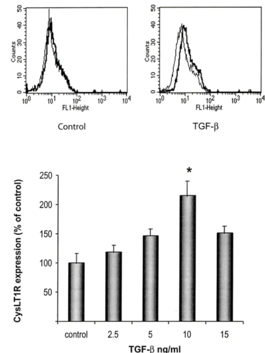
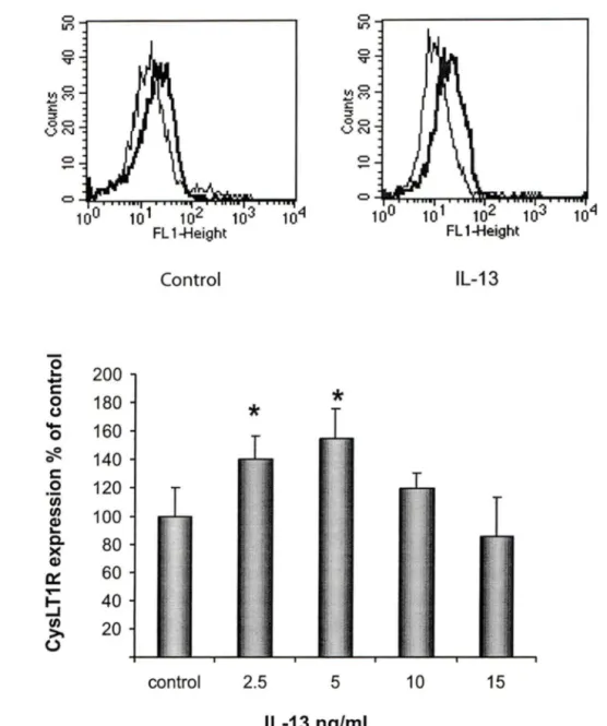
The expression of Cysl T1 R protein in cultured human BSMC was investigated by flow cytometry using a specific anti-Cysl T1 R Ab. BSMC were treated for 24h with graded concentrations of TGF-f3 and IL-13 ranging from 2.5 to 15 ng/ml, and concentrations of IFN-y ranging from 50 to 200 U/ml. Whereas the basal expression of Cysl T1 R in cultured BSMC was low, exposure of the cells to TGF-f3, IL-13 and IFN-y, markedly increased their expression of the receptor (Fig 1-3).
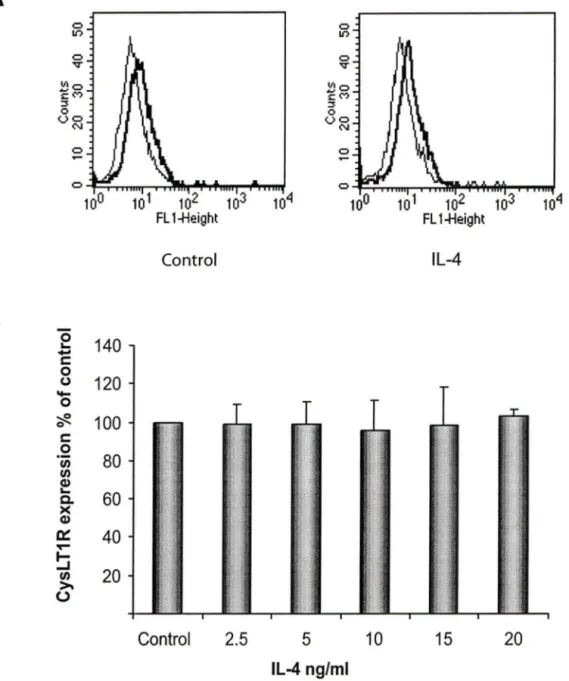
Basal expression levels of Cysl T1 R protein were adjusted to 100% and Cysl T1 R expression is illustrated as % of the contrai cells. TGF-f3 (Fig 1) and IFN-y (Fig 3) induced a 200% increase in the expression of Cysl T1 R as compared to non-stimulated cells, whereas, IL-13 (Fig 2) induced a 150% increase. The effects of TGF-f3, IL-13 and IFN-y were maximal at 10 ng/ml, 5ng/ml and 1 OO U/ml, respectively. Given that IL-4, a prototypic Th2-type cytokine, could also contribute to chronic inflammation in asthma, we tested graded concentrations of this cytokine but failed to observe any significant effect on Cysl T1 R expression in human BSMC (Fig 4).
ln kinetic studies, the effects of the three active cytokines on CysL T1 R expression were found to be maximal after 24h of stimulation (data not shown).
ln order to complement the results obtained by flow cytometry and to evaluate the distribution of CysL T1 R within BSMC, we assessed CysL T1 R protein expression by immunofluorescence (Figure 5). Control cells expressed low levels of CysL T1 R. ln contrast, TGF-~, IL-13 or IFN-y-treated cells showed an increased expression of the protein. The distribution of CysL T1 R was observed throughout the cell. Although CysL T1 R expression was increased by the three cytokines, some BSMC in each treatment group remained CysL T1 R-negative. The reason for this phenomenon is unknown, but unpublished results from our laboratory show that other cell types such as monocytes and macrophages also show a partial and random distribution of CysL T1 R.
Although BSMC also expressed basal levels of CysL T2R, no modulation of expression was observed with either of the cytokines (data not shown).
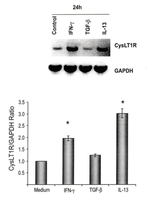
Cytokine-induced Cysl T1 R mRNA expression.
ln order to assess whether increased CysL T1 R protein expression was associated with augmented CysL T1 R mRNA levels, BSMC were stimulated with
TGF-~. IL-13 or IFN-y, respectively, and their steady-state levels of CysL T1 R
mRNA were analyzed (Fig 6). BSMC constitutively expressed low levels of CysLT1 R mRNA. IL-13 and IFN-y, but not TGF-~. induced an augmentation of
transcript levels following 24h of stimulation. Exposure of BSMC to cytokines for a shorter time period (8h) failed to show any significant modulation of CysL T1 R mRNA (data not shown). Scanning densitometry analysis of data from all experiments, showed that IL-13 and IFN-y augmented CysL T1 R mRNA levels 3-fold and 2-fold, respectively, over baseline levels (Fig 68). TGF-B did not significantly modulate CysL T1 R mRNA expression.
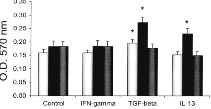
L TD4-induced proliferation of cytokine-primed BSMC.
The functional relevance of increased CysLT1 R expression in TGF-B, IL-13 or IFN-y-stimulated BSMC was investigated in terms of proliferation in response to L TD4. ln control cells, L TD4 alone failed to induce any significant
proliferation (Fig 7). However, when BSMC had been exposed for 24 h to TGF-~ or IL-13, a significant proliferation was induced by subsequent treatment with L TD4 (Fig 7 and 8). lnterestingly, IFN-y stimulated BSMC failed to respond to
L TD4 in terms of proliferation (Fig 8). Finally, in order to evaluate whether the
L TD4-induced proliferation observed in TGF-~ or IL-13-stimulated cells was
mediated through the enhanced expression of CysL T1 R, BSMC were pre-incubated for 30 min with the CysL T1 R antagonist, Montelukast (1
o-
6M), before the addition of L TD4 10-7M. As shown in figure 8, Montelukast was capable oftotally preventing the proliferation induced by L TD4 in both TGF-B- and IL-13-pretreated cells.
0 0 V V 1110 ~M ~M 1110 c: c: :3 :3 Oo VN VN Oo 0 0 ~ 0 100 102 104 102 FL 1-}leight FL 1-}leight Control TGF-~
B
-
250
*
0....
-
c: 0200
().._
0 'èft.150
-
c: 0 If) If)100
Q)....
c.. >< Q) ~50
'l'"" ~ If) >. (.) contrai2.5
5
10
15
TGF-~ ng/mlFig 1.
Flow cytometric analysis of CysLT1 R expression in cells stimulated with TGF-~.BSMC were incubated with different concentrations of TGF-~ for 24h. Cells were subsequently labeled with anti-Cysl T1 R or isotype-matched contrai Abs, followed by incubation with FITC-conjugated goat anti-rabbit lgG. (A) Histograms of a single experiment, representative of at least three. Solid thin lines represent labeling with isotype contrai Ab. Solid thick lines represent labeling of medium- and 10 ng/ml TGF-~-treated cells, respectively, with anti-Cysl T1 R Ab. (B) The graphie illustrates Cysl T1 R expression following graded concentrations of TGF-~. with the contrai normalized to 100%. (n=3} (*;p<0.006).
B
~---, C) V C) VI Mc
::i: 0 C) U N 0 ...-
c: 0 (J...
0 ~ 0 c: 0 Ill Ill Q) ... c. >< Q) c::: ""'"' ~ Ill >. (.) Control 200*
180*
160 140 120 100 80 60 40 20 contrai 2.5 5 IL-13 ng/ml IL-13 10 15Fig 2.
Flow cytometric analysis of CysL T1 R expression in cells stimulated with IL-13.BSMC were incubated with different concentrations of IL-13 for 24h. Cells were subsequently labeled with anti-Cysl T1 R or isotype-matched contrai Abs, followed by incubation with FITC-conjugated goat anti-rabbit lgG. (A) Histograms of a single experiment, representative of at least three. Solid thin lines represent labeling with isotype contrai Ab. Solid thick lines represent labeling of medium- and 5 ng/ml IL-13-treated cells, respectively, with anti-CysLT1R Ab. (B) The graphie illustrates Cysl T1 R expression following graded concentrations of IL-13, with the contrai normalized to 100%. (n=3) (*;p<0.05).
B
0'--
c: 0 (,)-
0 ~ 0 c: 0"' "'
Cl) '-c. >< Cl) a::: """" ~"'
>. (.) 0 V 0 102 FL 1-Height Co nt roi 250 200 150 100 r 50!l
0 ' control,,
' il' ,!) 0 V 0-*
. [r: . tL· ::':i '{:' \; 50 100 IFN""f U/ml 102 FL 1-Height IFN-'Y ~:t
200Fig 3. Flow cytometric analysis of CysLT1R expression in cells stimu/ated with IFN-y.
BSMC were incubated with different concentrations of IFN-y for 24h. Cells were subsequently labeled with anti-Cysl T1 R or isotype-matched contrai Abs, followed by incubation with FITC-conjugated goat anti-rabbit lgG. (A) Histograms of a single experiment, representative of at least three. Solid thin lines represent labeling with isotype contrai Ab. Solid thick lines represent labeling of medium- and 1 OO U/ml IFN-y-treated cells, respectively, with anti-Cysl T1 R Ab. (B) The graphie illustrates CysLT1 R expression following graded concentrations of IFN-y, with the contrai normalized to 100%. (n=3) (*;p<0.003).