OATAO is an open access repository that collects the work of Toulouse
researchers and makes it freely available over the web where possible
Any correspondence concerning this service should be sent
to the repository administrator:
[email protected]
This is an author’s version published in:
http://oatao.univ-toulouse.fr/25606
To cite this version:
Conzatti, Guillaume
and Cavalie, Sandrine
and Gayet, Florence
and Torrisani,
Jérôme and Carrere, Nicolas and Tourrette, Audrey
Elaboration of a thermosensitive
smart biomaterial: From synthesis to the ex vivo bioadhesion evaluation. (2019)
Colloids and Surfaces B Biointerfaces, 175. 91-97. ISSN 0927-7765
Elaboration of a thermosensitive smart biomaterial: From synthesis to the
ex
vivo
bioadhesion evaluation
G. Conzatti
\
S. Cavalie
a, F. Gayet
\
J.
Torrisani
C,
N. Carrère
c,
d, A. Tourrette
a,
*
• ClRIMA T, Un1"'1'Slti de Toulouse 3 • Paul Sabalier, CNRS, Toulouse, Pranœb LCC, Univenlli de Toulouse 3 • Paul Sabatier, CNRS, Toulouse, Franœ
• msERM, Universlti Toulouse 3 • Paul Sabatier, CentTe de Recherches en Canc.!rologie de Toulouse, Toulouse, Franœ
d G<lstrolnttstlnal Swge,y dtpartmmt, Plllpon HospltJJl • CHU de Toulouse, Toulouse, France
ART ICLE I NFO ABSTR ACT
Keywords: Polysaccltarldes PEC PNIPAM Smart polymer Controlled bioadheslcn
Alginate and chitosan are polysaccharides that are widely used in the biomedical field, especially as wound dressings. Controlled bioadhesion is an advanced functionality that offers the potential to reduœ injuries due to the stripping-off of the biomaterial. Herein, we report the efficient grafting of poly•N(isopropylacryamide) (PNIPAM), a thermosensitive polymer that exhibits a lower critical solution temperature (LCST) at 32 •c on the alginate/chitosan polyelectrolyte complex (PEC) surface. bt vitro studies did not exhibit a cytotoxic effect, and cells adhered preferentially on the LCST on PNIPAM grafted surfaces, as reported in the literature. Ex vivo investigations revealed that the adhesive behavior of the biomaterials was not the same on the liver and pan· creas. The effect of the temperature on the bioadhesion to organs was unexpected, as PNIPAM surfaces exhibited higher adhesion at low temperature. The PNIPAM was therefore able to confer PEC matrix thermosensitivity, but due to the application force, interactions between PNIPAM chains and their substrate could influence bioad· hesion on tissues.
1. Introduction
Bioadhesion during wound dressing is an important part of bioma terial science. Indeed, bioadhesion, defined as the ability of material to adhere to a biological entity, is required to achieve an effective healing. However, a highly bioadhesive material could cause injuries and be counterproductive. Thus, the manipulation of such materials during surgery is a critical consideration, as it is sometimes neœssary to adjust the material position precisely when applied to soft organs.
In this context, a controlled bioadhesion would be a great way to obtain a balance between position holding and easy manipulation. Bioadhesion is a complex phenomenon that involves proteins such as fibronectin that act as an anchor for the cells. (1) The ability of a sur face to support cell adhesion is depaident on its ability to support protein adsorption. Many factors have to be considered, such as the biomaterial stiffness, surface topography and hydrophilicity. To control bioadhesion, it is necessary to control the interactions between cell adhesion proteins and the biomaterial surface, i.e. the biomaterial surface state.
Numerous studies have been focused on smart biomaterials ob tained by grafting stimuli responsive polymers, such as pH or thermo
• Corresponding author.
sensitive polymers. [2,3) Poly N (isopropylacrylamide) (PNIPAM) is a widely studied thermosensitive polymer that changes its hydrophilicity around 32
•c,
called its lower critical solution temperature (LCST) [4,5). Therefore, this is an interesting material, as the human body temperature is close to 37 •c. Once grafted, its ability to change from a passive state at room temperature to an active state at body tempera ture is depaident on chain length and graft density. Nonetheless, the difficulty in efficiently characterizing the surface state of polymer scaffolds has oriented the use of PNIPAM for bioadhesion studies to ward the surface modification of well controlled surfaces, such as glass [6) or gold [7). These surfaces are amenable to theoretical models [8).However, the development of biodegradable biomaterials that would allow controlled bioadhesion is of interest when considering implanted devices. Polysaccharides are a class of such polymers that are generally considered biocompatible. Among others, alginate and chit osan are relatively inexpensive biopolymers that are well tolerated by the immune system. [9,10) Their combination, in the form of poly electrolyte complexes (PECs), allows the production of biodegradable absorbent and resistant matrixes that are able to 1imit fluids, such as bleedings. They can also be used to disseminate or to release drugs [11,12). Previously, we reported that calcium reticulated PECs showed
E-mail address: Audrey.tourrette@univ•t1se3.fr (A. Tourrette).
12,000 g/mol and dried under vacuum.
The theoretical molecular weight, Mnth, of the polymer was calcu
lated with Eq.1:
= × × Mn mono CTA M M [ ] [ ] th 0 mono CTA 0 (1)
where Mmonorepresents the molecular weight of the monomer, [mono]0
its initial concentration, %convis the percentage of conversion of the
monomer (considered as 100%) and [CTA]0the initial concentration of
the CTA.
2.4. Characterization of the PNIPAM COOH
The molar mass of the PNIPAM was measured by size exclusion chromatography (SEC) using DMF/LiBr (10 mM, refractive index of 1.431) as solvent. Before analysis, the samples werefiltered through a 10μm nylon filter. Detection was performed by the measurement of the refractive index variation (Optilab rEX detector) combined to a light scattering analyzer (miniDawn, 690 nm). Mass determination was per formed using the ASTRA software with a dn/dc of PNIPAM in DMF equal to 0.0731 mL/g. [18]
The functionality of PNIPAM was determined by COOH pH metric titration of the polymer (0.5 g in 20 mL H2O) with NaOH (0.02 M). The
pH was measured using a 766 Calimatic pH meter (KCl electrode). 2.5. Surface functionalization
The grafts on the surface of the PECs were achieved through an NHS/EDC coupling. To increase the reactivity of the PECs (PEC and PEC EDA), an acidic treatment was performed by immersing the PECs in water (HCl, pH 2.5) for 1 h prior to any reaction.
2.5.1. Grafting of EDA on alginate/chitosan PECs
To increase the grafting density on thefilms, EDA was previously grafted on the desired samples (PEC EDA). Briefly, PECs (30 g) were immersed in DMSO (0.6 mL) containing NHS (100 mmol/L) and EDC (200 mmol/L) for 3 h before EDA addition (0.6 mL,final concentration of 7.2 mol/L). The mixture was left to react for 12 h. The mixture was slightly agitated on a gyratory rocker. Films were thoroughly rinsed with EtOH and DMSO to remove any unreacted species.
2.5.2. Grafting of PNIPAM COOH on alginate/chitosan PECs
The grafting of the PNIPAM COOH was performed both on EDA grafted PECs (PEC EDA) and on native PECs (PEC). The PNIPAM COOH (2 mmol/L) was activated for 3 h with NHS (25 mmol/L) and EDC (50 mmol/L) in DMSO (1 mL). PEC of PEC EDA (30 g) were added to the reaction mixture. The mixture was left to react for 12 h. The mixture was slightly agitated on a gyratory rocker. Films were thor oughly rinsed with EtOH and DMSO to remove any unreacted species. 2.5.3. Surface characterization
The surface characterization before and after modification was performed via X ray photoelectron spectroscopy (XPS), on an ES CALAB 250, Thermo Electron. The excitation was monochromatic, Al Kα ray at 1486.6 eV. The diameter of analysis was 400μm and a charge com pensation was done through an electron beam at−2 eV. The signal of aliphatic carbons was used as reference and set to 285.0 eV.
Before analyses, samples were stored in glass containers, under vacuum to avoid contamination.
2.6. In vitro evaluation
Cytotoxicity studies were performed with hPNE (human Pancreatic Nestin positive Epithelial) cells expressing green fluorescent protein (GFP) to facilitate their visualization. The samples (5 x 5 mm) were immersed in the culture medium for 20 min before the cells were an interesting resistance toward enzymes, along with a good balance
between degradation and resistance in saline environment [12]. How
ever, only a few studies report the development of biodegradable wound dressings with thermo controlled bioadhesion by the grafting of PNIPAM on polysaccharide substrates [13]. To date, no studies are
devoted to the exploration of a thermosensitive PEC.
Herein, the grafting of a functional PNIPAM COOH polymer in a brush structure on alginate/chitosan PEC film surfaces through NHS/ EDC coupling was studied. To control the chain length of the grafted PNIPAM, it was synthesized by reversible addition fragmentation chain transfer (RAFT) polymerization, a controlled radical polymerization, [4] and compared to a commercial polymer. The grafting strategy was
also set to modulate the graft density of the PNIPAM brush. The re sulting films were characterized by X ray photoelectron spectroscopy (XPS) prior to characterization of cytotoxicity and thermo controlled bioadhesion with human cells (in vitro) and organs (ex vivo).
2. Materials and methods 2.1. Materials
Sodium alginate, chitosan, calcium chloride, acetic acid, N Isopropylacrylamide (NIPAM), poly (N Isopropylacrylamide) (PNIPAM, Mw = 5000 g.mol−1), N Hydroxysuccinimide (NHS), N (3
Dimethylaminopropyl) N’ ethylcarbodiimide hydrochloride (EDC), ethylene diamine (EDA), Tetrahydrofuran (THF), dimethylsulfoxide (DMSO), Methyl 2 (dodecylthiocarbonothioylthio) 2 methylpropio nate (CTA) and 4.4′ Azobis(4 cyanovalericacid) (radical initiator, ACVA), as well as all solvents, were provided by Sigma Aldrich.
The alginate and chitosan were characterized as previously re ported. [12] The G/M unit ratio of alginate was estimated using a water
suppressive NMR method to M/G = 2.7 [14]. Viscosimetry determined
its molecular weight to be 340 kDa, and α and κ parameters to be 1.13 and 6.9 × 10 − 4 mL.g 1, respectively. [15] Using the Shigemasa
method, the degree of acetylation (DA) of the chitosan was estimated to be 23% [16] with Mw = 1 300 kDa (α = 0.93, κ = 1.81 × 10 − 1 ml.
g 1). [17] NIPAM was purified by hexane recrystallization prior to use.
Other products were used in the as received state. 2.2. Alginate/chitosan PECs elaboration
The procedure for the PEC elaboration was based on our previous studies with minor modifications. [11,12] The sodium alginate was
dissolved in water at a concentration of 1.5% w/v and mechanically stirred overnight (500 rpm). In parallel, the chitosan was dissolved in acidic water (acetic acid, 0.34% v/v) at a concentration of 1.5% w/v and mechanically stirred overnight (500 rpm). After dissolution, 50 g of the alginate solution and 30 g of the chitosan solution were mixed to obtain a mass of 63/37 alginate/chitosan. The mixture was homo genized with an Ultra Turrax® (11 000 rpm) for 10 min to form the PEC.
The mixture was poured in a 24 well culture plate (0.2 mL/well) and dried at 50 °C for 2 h. The films were reticulated with a CaCl2 solution
(0.5 mL, 1% m/V) for 30 min, washed with distilled water and dried for another 2 h to obtain thin semitransparent films.
2.3. Synthesis of PNIPAM COOH
Functional PNIPAM COOH was synthesized through a RAFT poly merization in THF. The monomer (NIPAM) was added to a balloon containing the CTA dissolved in THF. The reactor was closed with septa and argon was bubbled in the reaction mixture for 30 min to remove dissolved oxygen. The initiator was added into the mixture and the reaction was maintained for 12 h. After the polymerization, the polymer was precipitated in ether, filtered and washed with ether. This step was repeated three times to remove the ether soluble monomer. The precipitate was dialyzed in water for 3 days with a cut off of
= − ×
T Q Q
Q
(%) (37) (27)
(27) 100 (2)
where Q(27) is the number of adherent cells at 27 °C and Q(37) is the number of adherent cells at 37 °C.
2.7. Ex vivo adhesion evaluation
Porcine organs (liver and pancreas) were furnished from a local slaughterhouse, where they were excised directly after slaughtering, kept in physiological solution at 4 °C and used within the day. Before the measurement, samples of fresh organ external part were excised and immersed in a physiological solution at 25 °C or 40 °C. The samples werefixed on a 10 mm diameter probe and contact was made with the organ surface for 5 min with an application force of 500 g. The bio material was separated from the surface of the organ (0.5 mm/s). The maximal force required for this separation was measured with a TA.XTplus texturometer equipped with a 5 kg captor. The associated constraint corresponded to the adhesive force. The experiment was conducted on three samples of each type.
3. Results and discussion
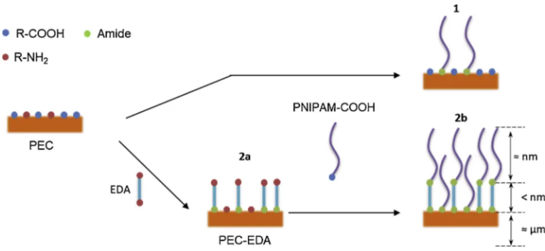
Alginate and chitosan display reactive sites with R COOH carboxylic acids and R NH2amines, respectively. A PNIPAM end functionalized
with a carboxylic acid can be grafted on the amino functions of the chitosan via an amide formation (Fig. 1, step 1). The "grafting to" strategy proposed here consisted of an increase in the number of re active sites at the PECs surface through the grafting of a diamine on the carboxylic acids of the alginate (Fig. 1, step 2a). This resulted in an amination of the alginate and the covalent binding of the PNIPAM
COOH to both diamine and amines of the chitosan (Fig. 1, step 2b). As a result, a higher grafting density of the PNIPAM was expected. In ad dition, the control of the molecular weight of the PNIPAM could modulate the chain length of the grafted brush.
3.1. PNIPAM COOH synthesis
To graft PNIPAM onto alginate/chitosan PECfilms, the synthesis of a functionalized polymer was needed. The RAFT polymerization allows the production a functional polymer with well controlled chain lengths. [19 21]. The COOH functionality mainly resulted from theR group re initiation of the reaction (see supporting information, S1).
Table 1reports the experimental conditions. As observed for PNI N, the polymerization of 2 g of NIPAM without CTA generated a high molecular weight polymer (Mn= 100 000 g/mol) and polydispersity
index Ð equal to 2.17, which is demonstrative of a non controlled polymerization. As the control agent was added (Mn,thof 25 000 g/
mol), a control over the polymerization was observed with a sig nificative diminution of the Mn(PNI 1, 26 000 g/mol) and Ð (1.19). The
obtained molecular weight was close to the theoretical mass calculated with Equation1, which is consistent with a RAFT mechanism. However, this polymerization was performed at a low content of NIPAM (2 g) and to use the synthesized polymer for surface grafting, it was necessary to master the polymerization of a higher content of polymer. Therefore, 15 g of NIPAM was RAFT polymerized using the same conditions (PNI 2). The obtained polymer showed a well defined size with a Mnof 24
000 g/mol and Ð equal to 1.09. The scaling up of this polymerization did not affect the efficiency of the RAFT mechanism.
For the following surface modification, it was necessary to control the functionality of the used polymer. Titrations were performed on PNI 2. The COOH/PNI ratio of 1.0 obtained for PNI 2 is consistent with its low Ð, confirming the absence of significant recombination of the RAFT polymerization. To compare to a smaller PNIPAM, a commercial PNIPAM COOH was studied and showed a Mnof 6 000 g/mol, Ð of 1.56
and a functionality COOH/PNI of 1.4. Both polymers were suitable for PEC surface grafting through their carboxylic functionality.
3.2. Surface functionalization
To provide thermosensitivity to the PEC surfaces, the PNIPAM COOH, commercially obtained or obtained from RAFT synthesis, was grafted onto alginate/chitosan matrix through NHS/EDC process. To this purpose, after their elaboration, PECs were acidified to improve the reactivity of functional groups. Two strategies were tested to modulate the graft density (Fig. 2).
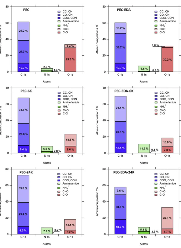
All the reactions were conducted in DMSO, a non swelling solvent for PECs, to limit internal grafting and alginate/chitosan cross reaction. Surface characterization was performed using XPS (Fig. 3). First, the grafting of EDA (PEC EDA) was revealed by the increase of the overall nitrogen content, which increased from 4.4% to 7.4% compared to non treated PECs (PEC). This nitrogen was non protonated, which was consistent with the formation of amide linkage between the amines of EDA and the carboxylic acids of PEC surfaces. At the same time, the CO, CN increased from 27.7% for PEC to 36.7% for PEC EDA, which could confirm the presence of EDA on the surface. The XPS signal of PNIPAM was characterized by a strong contribution of aliphatic carbons (CC, CH, 73.5%) as approximately 10% of nitrogen, which was exclusively in the form of amides hence without protonation (see supporting in formation, S2). Without EDA, PEC 6 K exhibited a slight increase of aliphatic carbons to 31.8% as compared to 23.2% for PEC. The nitrogen content increased to 8.6% and was mainly non protonated (amine/ amide), which is consistent with the presence of amides. The decrease of oxygen content, combined with the increase of their proportion in C = O form, was also attributed to the presence of PNIPAM. All of these results confirmed the presence of PNIPAM over the PEC surfaces. The same measurements were acquired for PEC 24 K: an increase of CC, CH seeded to release from the materials any potential reagent remnants.
After 20 min, the medium was aspirated and the cells were poured in the culture wells at a density of 103 cells per well (V = 1 mL). Control
experiments, without material, were performed in parallel. Cultures were incubated at 37 °C for 4 days. The materials were gently removed, and the cells were removed from their culture plate by the action of 1 mL of trypsin EDTA. A sample of 100 μL was withdrawn and put in an isotonic solution to perform counting with a Beckman Coulter Z1 par ticle counter, which was set to count cells with a diameter greater than 10 μm. The control experiment was considered to be 100% and the percentage of proliferation of the wells containing samples were cal culated as the ratio of the number of cells in the sample, Nsample/Ncontrol
x 100. The experiment was separately repeated three times for each type of sample with freshly prepared biomaterials. Two parallel samples were evaluated for each experiment. The mean was calculated with the higher value of each separate experiment.
Bioadhesion studies were performed with hPNE cells. The samples were elaborated directly on glass slats to ensure the complete recovery of culture wells. Once at the bottom of the wells, the samples were covered with culture medium (1 mL) for 20 min to allow swelling and to evacuate any toxic compounds adsorbed on the surface. The medium was aspirated and the hPNE cells were gently seeded on the PECs (60,000 cells/well, 1 mL). Cultures were incubated at 37 °C or 27 °C. After 8 h, the medium was aspirated, the PECs were transferred to clean culture wells and rinsed twice with PBS to eliminate non adherent cells. In the case of cultures carried out at 37 °C, the flushing PBS was heated at 37 °C before being used. Sample surfaces was observed with a Zeiss Axio Vert A 1 inverted fluorescence microscope with a wavelength of excitation of 493 nm (emission at 517 nm). The number of cells on the surface was counted by image processing using ImageJ. The experiment was separately repeated three times for each type of sample with freshly prepared biomaterials. Two parallel samples were evaluated for each experiment. The mean was calculated with the higher value of each separate experiment. The thermosensitivity, T, was calculated using Eq.
(33.8%) and overall nitrogen (8.1%), the majority of which was in a neutral form and a diminution of oxygen rates (18.6%), mainly in C = O form. It appeared that the increase of the chain length of the PNIPAM COOH was not detrimental to its graft efficiency on the algi nate/chitosan PEC surfaces.
Concerning the grafting of PNIPAM COOH on PEC EDA, the grafting of a small acrylamide of 6000 g/mol was efficient, with the same evo lution of the characteristic signals of PNIPAM compared to PEC EDA 6 K. However, in the case of the grafting of higher molecular PNIPAM COOH (PEC EDA 24 K), it was not possible to confirm the presence of PNIPAM over the surface, as aliphatic carbons diminished and the protonated amines (NH3+) increased. A possible surface passivation
due to an EDA diamine bridging over the surface could obstruct the
surface, and the higher molecular weight PNIPAM could react less ef fectively with the hindered reactive COOH, explaining this grafting limitation. Also to mention, the possible alginate/chitosan cross linking, while not evaluated, could limit the grafting efficiency on all the biomaterial surfaces.
In brief, the grafting of PNIPAM COOH was efficiently performed, leading to various surfaces by modulating the graft density and the chain length of the PNIPAM. As no PNIPAM was observed for PEC EDA 24 K, this sample was not further studied.
Fig. 1. Grafting strategies to control the chain length and graft density of PNIPAM on PEC substrates.
Table 1
Experimental conditions for the polymerization of PNIPAM and the resulting polymer characteristics.
Sample Volume mM0a) [M
0]/[I0]/[CTA0] T Mn,thb Mnc Ð pKa COOH/PNI
(mL) (g) (mol/L) (°C) (g/mol) PNI-N 5 2 1400/1/- 70 – 100 000 2,17 nd nd PNI-1 5 2 1400/1/7 70 25 000 26 000 1,19 nd nd PNI-2 38 15 1400/1/7 70 25 000 24 000 1,09 3.9 1.0 Commercial – – – – – 6 000 1.56 4.0 1.3 a monomer mass. b
theoretical molar mass of PNIPAM calculated through Eq.1.
c determined with SEC.
3.3. Toward surface adhesion 3.3.1. Cytotoxicity
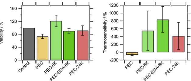
Cytotoxicity testing was performed by comparing the average number of hPNE cells after four days in a culture medium containing biomaterial samples to a control experiment. The results were expressed as percentage compared to control (100%), as shown inFig. 4, Right panel. It appeared that all cell viability and cell proliferation were greater than 70%, which is the considered limit by the ISO 10993 5 to suspect a cytotoxic effect. Therefore, no sample showed cytotoxicity
whether EDA was used, or not, and whether the PNIPAM was synthe sized or from a commercial source.
3.3.2. In vitro bioadhesion
Bioadhesion was evaluated on all the samples to determine whether a thermosensitive effect was caused by the PNIPAM. As shown in the literature, bioadhesion on the PNIPAM brush structure is dependent on the chain length and the graft density on its substrate, as these para meters play a role in the adsorption of the proteins involved in cell bioadhesion. [4,22] Bioadhesion is a process that occurs in thefirst
hours after cell seeding [23]. For this reason, and to avoid proliferation influence, the cell cultures were maintained only for 8 h. After this time, non adherent cells were removed. Adherent cells were quantified by fluorescence image treatment. On PEC, cells tended to aggregate in clusters. The thermosensitivity was calculated as the difference between adherent cells at 27 °C and at 37 °C using Equation2(Fig. 4, Left panel) (don’t we read from left to right? why did you put the Right panel before the Left panel in the description of your results?). The negative thermosensitivity for non modified PEC ( 64%) was indicative of the number of adherent cells at 37 °C compared to 27 °C, which decreased by approximately two thirds.
It is risky to explain this phenomenon, but it could be that a lower temperature diminished the cluster formation kinetics (see supporting information, S3), while these clusters were more prone to detach off the surface during the washing steps. For the PNIPAM grafted biomaterials (PEC 6 K, PEC EDA 6 K and PEC 24 K), the thermosensitivity was re versed and became positive, meaning that a better bioadhesion was achieved by the hPNE cells at 37 C rather than at 27 C due to the presence of PNIPAM. The gain of bio adhesiveness seemed to be higher when the graft density was increased by the use of EDA, with the number of cells multiplied by up to 9.5 (PEC EDA 5 K, 840%) above the LCST of PNIPAM. However, no statistical difference was observed. The size of the grafted PNIPAM (6 000 g/mol or 24 000 g/mol) did not show to affect the bioadhesion thermosensitivity, which would result of the suppression of primary adsorption bioadhesion proteins. [22] These results confirmed the potential of obtaining smart responsive biopoly mers.
3.3.3. Ex vivo evaluation
Adherence between these surfaces and porcine organs was eval uated. Ex vivo experiments allowed afirst approximation of the beha vior of a developed biomaterial before further in vivo assays. Modified and unmodified PEC bioadhesion was tested using pancreas and liver freshly extracted from pigs (Fig. 5). The non thermosensitive PEC samples adhered two times harder on the pancreas than on the liver, which emphasized the importance of the considered organ itself on the bioadhesion. Specifically, the nature of the tissue (cells, extra cellular matrix), its roughness or softness are parameters that can affect bioadhesive properties.
Data obtained from the pancreas were ambiguous. Whereas no statistical difference was observed between the two temperatures, for all the other samples, only PEC 6 K was affected by the passage of the LCST. It had a strong decrease in the adhesive force, from 16 kPa at 25 °C to 6 kPa at 40 °C. Therefore, long PNIPAM chains or high graft densities tended to limit the control over bioadhesion.
When in contact with the porcine liver, non modified biomaterials (PEC) did not show any variation between the measured adhesive force at 25 °C and 40 °C (4 kPa). The PEC 6 K, PEC EDA 6 K and PEC 24 K, however, exhibited a significant difference between the temperatures below and above the LCST of PNIPAM. Indeed, at 40 °C, the bioadhe sion between these PNIPAM grafted samples was lower, reflecting poorer interactions between PNIPAM chains and the liver surface above the LCST.
The PNIPAM surfaces on retinal implants were reported to exhibit similar magnitude of adhesive force (25 kPa). [24] Nonetheless, it was difficult to determine whether the observed phenomenon was due to physical or biological interactions, so as to extract some trends con cerning the impact of the length of the grafted PNIPAM or its graft density, as it was also dependent of the studied organ. Nonetheless, although the thermosensitivity of the surfaces tended to favor interac tions at low temperatures, the observed adherence at warm tempera tures is in the range of those considered as safe [25]. The PNIPAM in teractions were the result of a balance between intra/interchain and water molecules. The influence of a highly polar substrate, composed of alginate and chitosan, could, however, play a critical role on this bal ance.
4. Conclusions
Functional PNIPAM was efficiently synthesized via RAFT poly merization for comparison with a shorter commercial PNIPAM to study the influence of the chain length on bioadhesion. Two different stra tegies were used to graft these polymers on alginate/chitosan PECs through NHS/EDC coupling. The PNIPAM was directly grafted or used in combination with EDA to increase the graft density. Studies of the chemical of the surface through XPS revealed the presence of PNIPAM for PEC 6 K, PEC EDA 6 K and PEC 24 K, without being able to confirm whether the EDA allowed a higher graft density or not. However, the combination of EDA with a high molecular weight PNIPAM did not allow its efficient grafting.
No cytotoxicity was found for all samples, and it was shown that the PNIPAM grafted surfaces exhibited a higher bioadhesion toward cells above the LCST of PNIPAM, whatever the chain length or graft density. Thermosensitivity was also observed using porcine organs. A higher chain length or graft density did not allow a controlled bioadhesion with pancreas above the LCST, whereas it did not strongly affect the thermosensitivity with the liver.
Further work has to be considered to better understand the inter actions between the grafted samples and the surface of organs, taking in consideration possible interactions between the PNIPAM chains and
Fig. 4. (Right panel) Number of cells after 4 days in presence of a biomaterial versus a control without any biomaterial. (Left panel) Thermosensitivity, calculated with Eq.2, of bioadhesions as measured after 8 h of culture at 27 C and 37 C.
their substrate. Acknowledgment
Authors would like to thank the French National Research Agency for itsfinancial support (ANR 14 CE17 0002 01, FP BioPrev project). Appendix A. Supplementary data
Supplementary material related to this article can be found, in the online version, at doi:https://doi.org/10.1016/j.colsurfb.2018.11.084. References
[1] M. Gao, M. Sotomayor, E. Villa, E.H. Lee, K. Schulten, Molecular mechanisms of cellular mechanics, Phys. Chem. Chem. Phys. 8 (2006) 3692–3706,https://doi.org/ 10.1039/b606019f.
[2] L. Moroni, M. Klein Gunnewiek, E.M. Benetti, Polymer brush coatings regulating cell behavior: passive interfaces turn into active, Acta Biomater. 10 (2014) 2367–2378,https://doi.org/10.1016/j.actbio.2014.02.048.
[3] Y. Stetsyshyn, J. Raczkowska, O. Lishchynskyi, A. Bernasik, A. Kostruba, K. Harhay, H. Ohar, M.M. Marzec, A. Budkowski, Temperature-controlled three-stage switching of wetting, morphology, and protein adsorption, ACS Appl. Mater. Interfaces 9 (2017) 12035–12045,https://doi.org/10.1021/acsami.7b00136. [4] G. Conzatti, S. Cavalie, C. Combes, J. Torrisani, N. Carrere, A. Tourrette, PNIPAM
grafted surfaces through ATRP and RAFT polymerization: chemistry and bioadhe-sion, Colloids Surf. B Biointerfaces 151 (2017) 143–155,https://doi.org/10.1016/j. colsurfb.2016.12.007.
[5] R.M.P. da Silva, J.F. Mano, R.L. Reis, Smart thermoresponsive coatings and surfaces for tissue engineering: switching cell-material boundaries, Trends Biotechnol. 25 (2007) 577–583,https://doi.org/10.1016/j.tibtech.2007.08.014.
[6] H.Y. Tsai, K. Vats, M.Z. Yates, D.S.W. Benoit, Two-dimensional patterns of poly(N -isopropylacrylamide) microgels to spatially controlfibroblast adhesion and tem-perature-responsive detachment, Langmuir 29 (2013) 12183–12193,https://doi. org/10.1021/la400971g.
[7] K.N. Plunkett, X. Zhu, J.S. Moore, D.E. Leckband, PNIPAM chain collapse depends on the molecular weight and grafting density, Langmuir 22 (2006) 4259–4266,
https://doi.org/10.1021/la0531502.
[8] A. Halperin, M. Kröger, Theoretical considerations on mechanisms of harvesting cells cultured on thermoresponsive polymer brushes, Biomaterials 33 (2012) 4975–4987,https://doi.org/10.1016/j.biomaterials.2012.03.060.
[9] K.Y. Lee, D.J. Mooney, Alginate: properties and biomedical applications, Prog. Polym. Sci. 37 (2012) 106–126,https://doi.org/10.1016/j.progpolymsci.2011.06. 003.
[10] D. Sahoo, P.L. Nayak, Chitosan: the most valuable derivative of chitin, Biopolym. Biomed. Environ. Appl. (2011) 129–166,https://doi.org/10.1002/
9781118164792.ch6.
[11] M. Castel-Molieres, G. Conzatti, J. Torrisani, A. Rouilly, S. Cavalie, N. Carrere, A. Tourrette, Influence of homogenization technique and blend ratio on chitosan/
alginate polyelectrolyte complex properties, J. Med. Biol. Eng. 38 (2018) 10–21,
https://doi.org/10.1007/s40846-017-0304-7.
[12] G. Conzatti, D. Faucon, M. Castel, F. Ayadi, S. Cavalie, A. Tourrette, Alginate/ chitosan polyelectrolyte complexes: a comparative study of the influence of the drying step on physicochemical properties, Carbohydr. Polym. 172 (2017) 142–151,https://doi.org/10.1016/j.carbpol.2017.05.023.
[13] J.-P. Chen, C.-Y. Kuo, W.-L. Lee, Thermo-responsive wound dressings by grafting chitosan and poly(N-isopropylacrylamide) to plasma-induced graft polymerization modified non-woven fabrics, Appl. Surf. Sci. 262 (2012) 95–101,https://doi.org/ 10.1016/j.apsusc.2012.02.106.
[14] E.M. Vilén, M. Klinger, C. Sandström, Application of diffusion-edited NMR spec-troscopy for selective suppression of water signal in the determination of monomer composition in alginates, Magn. Reson. Chem. 49 (2011) 584–591,https://doi.org/ 10.1002/mrc.2789.
[15] A. Martinsen, G. Skjåk-Bræk, O. Smidsrød, F. Zanetti, S. Paoletti, Comparison of different methods for determination of molecular weight and molecular weight distribution of alginates, Carbohydr. Polym. 15 (1991) 171–193,https://doi.org/ 10.1016/0144-8617(91)90031-7.
[16] Y. Shigemasa, H. Matsuura, H. Sashiwa, H. Saimoto, Evaluation of different ab-sorbance ratios from infrared spectroscopy for analyzing the degree of deacetylation in chitin, Int. J. Biol. Macromol. 18 (1996) 237–242,https://doi.org/10.1016/ 0141-8130(95)01079-3.
[17] M.R. Kasaai, Calculation of Mark-Houwink-Sakurada (MHS) equation viscometric constants for chitosan in any solvent-temperature system using experimental re-ported viscometric constants data, Carbohydr. Polym. 68 (2007) 477–488,https:// doi.org/10.1016/j.carbpol.2006.11.006.
[18] F. Audouin, A. Heise, Surface-initiated RAFT polymerization of NIPAM from monolithic macroporous polyHIPE, Eur. Polym. J. 49 (2013) 1073–1079,https:// doi.org/10.1016/j.eurpolymj.2013.01.013.
[19] Y. Yang, X. Song, L. Yuan, M. Li, J. Liu, R. Ji, H. Zhao, Synthesis of PNIPAM polymer brushes on reduced graphene oxide based on click chemistry and RAFT poly-merization, J. Polym. Sci. Part A: Polym. Chem. 50 (2012) 329–337,https://doi. org/10.1002/pola.25036.
[20] C. Barner-Kowollik, T.P. Davis, J.P. a Heuts, M.H. Stenzel, P. Vana, M. Whittaker, RAFTing down under: tales of missing radicals, fancy architectures, and mysterious holes, J. Polym. Sci. Part A: Polym. Chem. 41 (2003) 365–375,https://doi.org/10. 1002/pola.10567.
[21] A. Veloso, W. Garcia, A. Agirre, N. Ballard, F. Ruiperez, J.C. de la Cal, J.M. Asua, Determining the effect of side reactions on product distributions in RAFT poly-merization by MALDI-TOF MS, Polym. Chem. 6 (2015) 5437–5450,https://doi. org/10.1039/C5PY00838G.
[22] C. Xue, N. Yonet-Tanyeri, N. Brouette, M. Sferrazza, P.V. Braun, D.E. Leckband, Protein adsorption on poly(N-isopropylacrylamide) brushes: dependence on grafting density and chain collapse, Langmuir 27 (2011) 8810–8818,https://doi. org/10.1021/la2001909.
[23] K. Anselme, L. Ploux, a Ponche, Cell/Material interfaces: influence of surface chemistry and surface topography on cell adhesion, J. Adhes. Sci. Technol. 24 (2010) 831–852,https://doi.org/10.1163/016942409X12598231568186. [24] M. Tunc, X. Cheng, B.D. Ratner, E. Meng, M. Humayun, Reversible thermosensitive
glue for retinal implants, Retina 27 (2007) 938–942,https://doi.org/10.1097/IAE. 0b013e318042ae81.
[25] M. Waring, S. Bielfeldt, M. Brandt, Skin adhesion properties of three dressings used for acute wounds, Wounds UK 5 (2009) 22–31.