doi: 10.17265/2159-5828/2016.05.002
Effect of PLA/ZnO Packaging and Gamma Radiation on
the Content of Listeria innocua, Escherichia coli and
Salmonella enterica on Ham during Storage at 4 °C
Antonella Marra1, Afia Boumail2, Sossio Cimmino1, Paula Criado2, Clara Silvestre1 and Monique Lacroix21. Institute of Polymers, Composites and Biomaterials (IPCB), Consiglio Nazionale delle Ricerche (CNR), Via Campi Flegrei 34, Comprensorio Olivetti, Pozzuoli (NA) 80078, Italy
2. INRS-Institut Armand-Frappier, Research Laboratories in Sciences Applied to Food, 531 des prairies blvd. Laval, Québec H7V 1B7, Canada
Abstract: The effect of ZnO and low γ-radiation on PLA based-films was investigated to be used for food packaging application.
Ham slices were inoculated with E. coli, L. innocua and S. enterica and then covered with PLA and PLA/5% ZnO films. The samples were irradiated with a γ-radiation dose of 0.3 kGy at dose rate of 13.5 kGy/h. Microbiological analysis was performed at 0, 1 and 5 days on samples stored at 4 °C. Results showed that no consistent reduction of bacteria was obtained, even at the fifth day of storage, when the ham was covered with PLA/5% ZnO film and no γ-radiation was performed. The use of γ-radiation results necessary to reduce the bacteria growth. In fact E. coli and S. enterica were not detected after 5 days of storage; whereas in the case of test with L. innocua a reduction of 1.3 log CFU/g was observed after 5 days of storage. The antibacterial results indicate that the presence of ZnO in PLA film is effective only for E. coli. The differences of the results obtained here with those reported in literature (where ZnO particles are reported to be very effective as antimicrobial material) are accounted for the different methodologies used. In conclusion considering the positive results, even if small, obtained here at least only for the E. Coli and considering that PLA/5% ZnO film shows, compared to plain PLA film, good tensile properties (especially Young’s modulus and stress at yielding) and good permeability (to O2 and CO2) induce to consider the PLA/5% ZnO composite film usable for food packaging when long shelf life and
food safety are required, considering also that it is biodegradable and compostable.
Key words: Polylactic acid, zinc oxide, biocomposite, food packaging, γ-radiation, antimicrobial property.
1. Introduction
The use of plastics has permitted the development of a wide range of food and beverage products, their transportation over long distances and safely storage, without compromising the quality and reducing product loss. Plastics enable the production of both rigid and flexible packaging, and maintain their characteristics even for very long periods and they possess, generally, satisfactory barrier properties, which reduce the penetration of gas and help prevent the loss of the organoleptic characteristics of food [1].
Corresponding authors: Monique Lacroix, full professor,
research fields: food science and irradiation; Clara Silvestre, senior researcher, research field: polymers for food packaging.
The great advantage of the plastics is the possibility to use a wide range of materials of different compositions and capable of providing the most convenient design according to the specific needs required by specific food product. Petroleum-based polymers have salient features such as strength, flexibility, stiffness, barrier to oxygen and moisture, and resistance to food component attack. However, their resistance to degradation and their recycling and disposal difficulties are very challenging in today’s heightened strong global interest in environmentally sustainable materials. The development of biopolymers from renewable resources represents an important need [2]. Biopolymers are particularly attractive because they are biodegradable due to their
D
susceptibility to microbial enzyme degradation [3]. Biopolymers are being introduced into the market to serve as food packaging materials to primarily address concerns about plastic waste accumulation [4-6] and their market is expected to grow between 20% and 25% until 2020 [7]. The research is oriented to design new composite materials to help avoid in food development of rancidity, colour loss or change, loss of nutrients, dehydration, microbial growth, gas production, development of odours and senescence.
Nanotechnology has potential applications in all aspects of food chain including food processing, food quality monitoring, food packaging and storage [8, 9]. Major areas of food industry which could benefit from nanotechnology [8] are development of new functional materials for food packaging [4], microscale and nanoscale processing [10], product development [11] and methods and instrumentation design for improved food safety and biosecurity [4]. Nanotechnology is applicable in food packaging to improve packaging performances such as gas, moisture, UV and volatile barriers, mechanical strength, heat resistance and flame retardancy and weight [10, 11]. The use of biodegradable nanocomposites will help to reduce packaging waste while extending shelf life of processed foods. Numerous studies concern the development of PLA matrix composites with antibacterial properties to assure food safety and to prevent food borne disease [12-16]. In Europe, approximately 1,000 of food borne diseases cases per year have been attributed to the presence of Listeria monocytogenes, which around 20% of them led to death [17]. Contaminations by L.
monocytogenes mainly occur on ready-to-eat food
products such as dairy, vegetables and meat [18]. However, Escherichia coli and Salmonella typhimurium can also be found in food products and
cause diseases. It has been estimated that, in Canada, 4 millions of people will develop food borne diseases each year [19]. However, the use of radiation as a cold treatment on packaged foods has some benefits on
food decontamination and can assure food safety. The mechanism of irradiation on foods is basically based on the inactivation or on the reduction of parasites and bacteria [20]. Because of its penetrating properties, γ-rays directly affect DNA resulting in cells’ death, but can also produce oxidation of unsaturated lipids and lead to off-flavors. The maximum dose that can be applied on meat should not exceed 3 kGy [21]. However, low γ-radiation doses may not be fully efficient to eliminate pathogenic bacteria such as
Listeria monocytogenes, Escherichia coli or
Salmonella typhimurium, which are frequent sources
of food contaminations.
It has been shown that the use of active agents incorporated in the polymer material for packaging enables significantly an increase of the shelf-life of the packaged product, going to act on those mechanisms of degradation which cause the deterioration by reducing the growth rate of microorganisms [22, 23].
The antibacterial activity of a polymer is usually obtained by adding organic compounds or metal particles [24-31]. Sub-micrometric and nanometric particles of metal or metal oxides and carbon nanotubes are the most used particles to develop antimicrobial biopolymers. In literature it is reported that silver (Ag) exerts antibacterial and fungicidal against about 150 different types of bacteria when the metal particles have nanoscale size, and Ag particles are already found in many commercial products. Metal oxides, such as zinc oxide (ZnO), titanium dioxide (TiO2), magnesium oxide (MgO) and silicon dioxide (SiO2) are also known to act as antibacterial agents in addition to their ability to block UV radiation and to act as disinfectant agents [26]. Compared to Ag, ZnO and MgO particles are presented as the best and safest solution for food packaging.
Particularly investigated it is the effect of ZnO on the properties of polymer films to be used for food packaging, because it has antimicrobial property [29, 30, 32] and it is defined as “Generally Recognized as
Safe” (GRAS).
In a previous paper [33], the effect of ZnO on the mechanical (tensile), permeability and antimicrobial properties of PLA based films was investigated. The study was performed on three films, with 1, 3 and 5 wt% of ZnO, and the antimicrobial test was done only with E.coli. of particular interest it was found that the film with 5 wt% of ZnO which showed 99.99% reduction for E. coli after 24 hours demonstrating a very efficient antibacterial activity of ZnO against E.
coli. Moreover, ZnO had also some positive effects on
the mechanical properties of the composite film compared to the plain PLA film, such as: increase of Young’s modulus and stress at yielding and decrease of permeability to O2 and CO2 of about 17% and 14%, respectively. Regarding the water vapour permeability it was instead found a slight increase of permeability (from 0.21 for the plain PLA to 0.24 [(cm3/(24 h m2)] (cm/bar) for the composite with 5 wt% of ZnO).
The aim of this study is to investigate further the property of PLA film with 5 wt% of ZnO in order to determinate the synergetic effect of the ZnO particles and the γ-radiation at low irradiation dose, against the contamination of 3 non-pathogens, Salmonella
enterica, Escherichia coli and Listeria innocua,
inoculated on ham slices.
2. Material and Methods
2.1 Materials and Sample Preparation
The materials used in this research are: (1) Polylactic acid (PLA) in pellets, code PLA 4032D, with molecular weight Mw = 2.1 105, acquired from Nature Works (USA); (2) ZnO powder, supplied by Pylote (France), obtained by spray pirolysis with particles size of 100-500 nm.
Before mixing, the PLA pellets and ZnO powders were dried in an oven for 24 h at 65 °C under vacuum. To improve the dispersion of ZnO in PLA a masterbatch with composition PLA 80% and ZnO 20% was prepared by mixing of the components by
using a twin-screw extruder, Collin ZK 25 (D = 25 mm and L/D = 56) Ebersberg (Germany). Then, the masterbatch was diluted in PLA in such quantities as to obtain the desired composition (5% by weight of ZnO). For comparison also the PLA without ZnO was extruded. The extrusion was conducted by using the following temperatures, from the hopper to the die: 150/170/170/170/160 °C. The screw speed of the dispenser was 20 rpm, while the speed of the extruder screws was 25 rpm. The films were prepared by using a single screw extruder and before the extrusion the pellets (PLA/5% ZnO and PLA) were again dried in an oven for 24 h at 65 °C under vacuum. The single screw extruder has, as terminal, a calender characterized by two counter rotating cylinders that allow the film passing through them still in the plastic state and the third useful to direct the output material until the collecting cylinder. The extruder and the chill roll unit are Collin E 20T and Collin CR 72T, Ebersberg (Germany), respectively. The thickness of the film obtained depends on the light between the two counter-rotating cylinders and the speed of rotation of the collecting cylinder; films with thickness of about 60 μm were obtained for both the compositions. The extrusion was conducted using the following temperatures, from the hopper to the die: 160/170/180/170/180 °C. The screw speed of the extruder was 40 rpm.
2.2 Gel Permeation Chromatography (GPC)
The Gel permeation chromatography was performed on GPC-150C Waters Chromatography instrument (Milford, Massachusetts-USA) equipped with two linear columns Polymer Laboratories and elution with tetrahydrofuran (THF). The flow rate was 1 mL/min. The eluent was monitored by PL-ELS 2100 detector. Molecular weights and molecular weight distributions were calculated by reference to a PS standard calibration curve, with the Kuhn–Mark–Houwink equation for poly (l-lactide) in THF: Mn (PLA) = 0.4055 × Mn (PS)1.0486 [34].
2.3 Scanning Electron Microscopy (SEM)
The morphology of the fractured surfaces of the PLA and PLA/5% ZnO films were determined by the scanning electron microscope (SEM), the model used is a Fei Quanta 200 SEM Feg, Hillsboro, Oregon (USA).
2.4 Thermal Analysis (DSC)
The analysis was performed using a calorimeter Mettler DSC-822e, Schwerzenbach, Switzerland.
The thermoanalytical technique measures the difference of the heat required to increase the temperature of a sample pan compared to a reference pan, as a function of temperature, when the sample and the reference pans are submitted to the same heating or cooling rate. The sample pan and reference pan are maintained as possible at the same temperature throughout the experiment.
Each sample (approximately 4 mg) was subjected to the following temperature program:
from room temperature to -30 °C, then heated to 200 °C at heating rate of 10 °C/min.
2.5 Thermogravimetric Analysis (TGA)
The thermal stability of the samples was studied by TGA. The instrument used was a Perkin Elmer Diamond, Albany St. Boston, Massachusetts (USA). The measurements were conducted from 30 °C to 800 °C at a heating rate of 20 °C/min, using about 2.0 mg of the sample in alumina pan. The experiments were done under air atmosphere at flow rate of 200 mL/min. From thermograms it is determined the temperature at the weight loss is 5% (Tonset), at the
degradation rate maximum (Tmax) and in
correspondence of the final plateau (Tendset).
2.6 UV-Visible Spectrophotometry
UV-visible spectra were monitored, from 200 to 850 nm, with Jasco V-570 spectrophotometer, Columbia, Maryland (USA). The spectra were recorded by using films in transmission mode.
2.7 Bacterial Suspension
Escherichia coli ATCC 8739, Listeria innocua
ATCC 51742 and Salmonella enterica subsp. enterica
serovar Typhimurium ATCC 53648 were stored at
-80 °C in TSB in presence of glycerol (150 g/L). Before each experiment, bacteria were propagated through 2 consecutive cycles of 24 h in TSB at 37 °C to obtain cultures containing approximately 109 CFU (colony forming unit)/mL. Cultures were then diluted in order to obtain bacterial suspensions with a concentration of approximately 105 CFU/mL.
2.8 Inoculation and Treatment of Ham
Ready-to-eat ham was purchased in a local supermarket (Metro, Laval, QC, Canada) and cut into pieces under sterile conditions. Ham samples were inoculated by adding 250 µL of bacterial suspensions on the surface of 10 g of ham. Final concentrations were 3.3 log CFU/g for L. innocua and S. enterica, 3.2 log CFU/g for E. coli. The films of PLA and PLA/5% ZnO were then applied on the ham and the samples were stored at 4 °C for 5 days. Irradiation treatments were done at the Canadian Irradiation Center, using a UC-15 A (SS canister) underwater calibrator (Nordion Inc., Kanata, ON, Canada) equipped with a 60Co source. A γ-radiation dose of 0.3 kGy was delivered at a dose rate of 13.5 kGy/h. Samples were stored at 4°C and microbiological analysis was performed at 0, 1, and 5 days. Ham samples (10 g) were immersed in 30 g of peptone water (1 g/L) and homogenized using a Lab-blender 400 stomacher (Laboratory Equipment, London, UK). Appropriate 10-fold dilutions were made and 1 mL of each dilution was spread on Petri dishes before medium was poured. Palcam agar (Alpha Biosciences, Baltimore, MD, USA) supplemented with antiobiotics acriflavin (5 mg/L) and polymyxin B (10 mg/L), MacConkey agar supplemented with sorbitol (Alpha Biosciences) and DCLS agar (Alpha Biosciences) were used for L.
innocua, E. coli and S. enterica respectively. Petri
bacterial enumeration. Detection level was calculated as 0.6 log (4 CFU/g).
2.9 Experimental Design and Statistical Analysis
PLA and PLA/5% ZnO films were cut in of the same shape and dimension larger than the piece of ham in order to be sure that the whole ham was in contact with the film. Samples were separated into 3 groups: (1) control; (2) PLA film; (3) PLA/5% ZnO film. Microbial analyses during storage were done using a 3 3 2 3 3 factorial design: 3 replicates, 3 treatments, 2 gamma radiation doses, 3 bacteria, 3 days of storage. Analysis of variance and Duncan’s multiple-range test were performed by using SPSS 16.0 software (IBM Corp., Somers, NY, USA). Differences between means were considered to be significant at a 5% level.
2.10 Atomic Absorption Spectrometry
The amount of Zn released in the solution was determinated by Atomic Absorption Spectrometry (AAS) using 1 g of film immersed in 10 mL of RS (composition for one liter: 0.150 g of potassium chloride, 2.25 g of sodium chloride, 0.05 g of sodium bicarbonate, 0.12 g of calcium chloride hexahydrate and pH 7.0) and kept on a wrist-action shaker for 0, 6, 24 and 48 h. The AAS measurements were performed using a Perkin Elmer Analyst 800 instrument (Norwalk, CT, USA) and AS 90 plus flame auto sampler. All the data were recorded and processed by WinLab 32 software. A stock zinc solution in 2.5% HNO3 was used for standard calibration curve.
3. Results and Discussions
3.1 GPC
Table 1 shows the molecular parameters of PLA pellets as received (PLApellet) after mixing (PLAmix) and after calender (PLAfilm), and the same last two samples with ZnO. The mixing of PLA pellets does not affect significantly the molecular weights of neat PLA; in fact PLApellet and PLAmix have similar molecular parameters; the second process step (in the calender), from PLAmix to PLAfilm, causes a decrease in Mn of about 25%. It is known that ZnO particles in PLA induce hydrolysis at high temperature [35] and so affect the molecular weight. In fact the reduction of molecular weight comparing PLAmix and the PLA/5% ZnOmix is about 38%, and the same percentage reduction is found comparing the molecular weight of PLAfilm and the PLA/5% ZnOfilm.
3.2 Morphological Analysis
Fig. 1 shows the fractured surface of PLA; the surface results to be homogeneous as expected for a plain polymer.
Fig. 2, micrograph for the fractured surface of the PLA/5% ZnO film, shows agglomeration of ZnO particles fairly distributed in the PLA matrix with an average size of about 1.2 μm. It has to be noted that the ZnO particles, as received from the producer, have sub micron dimensions and they agglomerate in bigger particles during the mixing with PLA because they are incompatible with the organic phase and because of their high surface energy.
Table 1 Molecular parameters for the PLA and composite after mixing (mix) and after calender (film).
Mn × 10-4 Mw × 10-4 Mw/Mn
PLA pelleta 13.0 21.0 1.61
PLA mixb 13.3 20.8 1.56
PLA filmb 10.0 15.5 1.55
PLA/5% ZnOmixb 8.3 13.9 1.66
PLA/5% ZnOfilmb 6.2 10.0 1.60
a Data obtained from the manufacturer.
b Calculated by reference to a PS standard calibration curve with the Kuhn–Mark–Houwink equation for PLA in THF: M
n(PLA) = 0.4055 Mn(PS)1.0486 [34].
Fig. 1 SEM image of PLA film fractured in liquid nitrogen (5,000 ×).
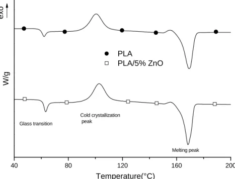
Fig. 2 SEM image of PLA/5% ZnO film fractured in liquid nitrogen (6,000 ×). 40 80 120 160 200 exo PLA PLA/5% ZnO W/g Temperature(°C) Cold crystallization peak Melting peak Glass transition
Fig. 3 DSC heating scans of PLA and PLA/5% ZnO films.
3.3 Thermal Analysis (DSC)
Fig. 3 shows the thermograms of PLA and PLA/5% ZnO in the temperature range from 40 to 200 °C. The curves are shifted along the y axis to illustrate the trends. The heating rate used is 10 °C/min under an inert atmosphere (N2). PLA presents the glass transition temperature at about 61 °C; then an
exothermic peak (cold crystallization peak) extending from about 85 °C to 115 °C with a maximum at about 100 °C; finally the melting peak extending from about 155 °C to 175 °C with a maximum, taken as the melting temperature (Tm), at 169 °C.
The thermoanalytical curve of PLA/ZnO composite does not show significant difference compared to pure PLA. The extension of the endothermic and
exothermic peaks, respectively crystallization and melting peaks, remain fairly unchanged as well as the corresponding maxima, the glass transition temperature and Tm. The main result from the DSC is that both the films, as obtained from the calender, are amorphous, that is the PLA does not crystallize, neither when there are the ZnO particles. This is in agreement with the spectra obtained by the X-ray which show clearly no presence of any crystallization peak [33].
3.4 Thermogravimetric Analysis (TGA)
Fig. 4 shows TGA curves of PLA and PLA/5% ZnO composite in the range from 230 °C to 400 °C. Table 2 reports the values of thermogravimetric analysis when the weight loss is 5% (Tonset), the degradation rate is maximum (Tmax) and in correspondence of the final plateau (Tendset). The analysis of Fig. 4 and the parameters in Table 2 clearly indicate that ZnO has a great influence on the
degradation process of PLA macromolecules, which is shifted to lower temperatures of about 30 degrees. This result is in agreement with that reported in literature and it is explained considering that probably ZnO nanoparticles catalyze the depolymerization of PLA molecules [35].
3.5 UV-Visible Spectrophotometry
Fig. 5 shows the transmittance (T%) for PLA and the composite with 5% of ZnO from 200 nm to 850 nm. For PLA the transmittance is 0% from 200 nm to 227 nm, then there is a sudden increase up to 80% and after that the value still increases at 86%, at wavelength 300 nm, and to 90% at wavelength 850 nm. The composite film with 5 wt% of ZnO has also 0% transmittance in the range 200-227 nm and very low transmittance value up to 850 nm. The low transmittance shown by the composite film is due to the adsorption and reflection of the ZnO particles [36].
240 260 280 300 320 340 360 380 400 0 20 40 60 80 100 PLA PLA/5% ZnO W e ight (% ) Temperature(°C)
Fig. 4 Weight (%) of PLA and PLA/5% ZnO films.
Table 2 Thermal degradation temperature when the weight loss is 5% (Tonset), the degradation rate is maximum (Tmax) and in correspondence of the final plateau (Tendset).
Sample Tonset (°C) Tmax (°C) Tendset (°C)
PLA 307 ± 1 332 ± 1 355 ± 1
200 400 600 800 0 20 40 60 80 100 PLA PLA/5% ZnO T ransm it tanc e (%) Wavelength (nm)
Fig. 5 UV-visible spectrum of PLA and PLA/5% ZnO films.
In the region around 385 nm the transmittance of the composite film is zero and this is due to the intrinsic capacity of the ZnO particles to absorb the UV light [37-39]. The protection mechanism of conventional UV absorbers used as photostabilizers in polymers is based on the absorption of harmful UV radiation and its dissipation as heat [40].
3.6 Antimicrobial Tests
Antimicrobial effects of PLA and PLA/5% ZnO films, and γ-radiation treatment were tested on ham inoculated with three different bacteria, L. innocua, E.
coli and S. enterica, and then stored at 4 °C; the tests
were performed at day 0, 1 and 5.
Table 3 reports the results of the test performed for the detection of L. innocua bacteria.
In the “Control” there are reported the bacterial counts performed on the ham without covering of film and without γ-radiation treatment; the log CFU/g count is practically unchanged during the 5 days. Covering the ham with plain PLA film or with PLA/5% ZnO film, no significant reduction of bacterial count is detected up to the fifth day. It is found that the presence of ZnO particles did not have
significant effect on the decrease of the bacterial growth, up to the fifth day of experiment. Only when the samples are irradiated at 0.3 kGy, it is found at day 5 a significant reduction of L. innocua (about 1 log CFU/g). However, no synergy was observed between the presence of ZnO particles and the use of γ-radiation treatment.
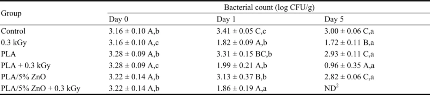
The results for E. coli are presented in Table 4. The “Control” shows no reduction of bacteria at day 5, as expected. The irradiation at 0.3 kGy on the ham induces a significant reduction of E. coli from 3.16 (day 0) to 1.72 log CFU/g (day 5) showing a 1.44 log CFU/gr reduction. Covering the ham with plain PLA film or with PLA/5% ZnO film, no significant decrease of the log CFU/g concentration is detected from day 0 to day 5 (around 0.3 log CFU/g reduction). In order to have a consistent decrease of E. coli bacteria a treatment with γ-radiation was necessary. In fact, the application of 0.3 kGy has permitted a reduction of 1.34 log CFU/gr at day one and this level of bacteria was stable at day 5 of storage. When the ham was packed in PLA based film and then irradiated at 0.3 kGy a 1 log and 2.2 log CFU/gr reduction was observed respectively at day 1 and 5 of
Table 3 Effect of the combined treatment on L. innocua during storage1.
Group Bacterial count (log CFU/g)
Day 0 Day 1 Day 5
Control 3.28 ± 0.04 A,b 3.12 ± 0.05 C,a 3.13 ± 0.08 E,a
0.3 kGy 3.28 ± 0.04 A,c 2.48 ± 0.07 B,b 2.29 ± 0.14 C,a
PLA 3.25 ± 0.07 A,b 3.07 ± 0.07 C,a 3.09 ± 0.07 E,a
PLA + 0.3 kGy 3.25 ± 0.07 A,c 2.19 ± 0.11 A,b 2.06 ± 0.07 B,a PLA/5% ZnO 3.22 ± 0.06 A,c 3.03 ± 0.07 C,b 2.83 ± 0.09 D,a PLA/5% ZnO + 0.3 kGy 3.22 ± 0.06 A,c 2.18 ± 0.11 A,b 1.92 ± 0.08 A,a
1 Means followed by the same uppercase letter in each column are not significantly different at the 5% level. Means followed by the
same lowercase letter in each row for each property are not significantly different at the 5% level.
Table 4 Effect of the combined treatment on E. coli during storage1.
Group Bacterial count (log CFU/g)
Day 0 Day 1 Day 5
Control 3.16 ± 0.10 A,b 3.41 ± 0.05 C,c 3.00 ± 0.06 C,a
0.3 kGy 3.16 ± 0.10 A,c 1.82 ± 0.09 A,b 1.72 ± 0.11 B,a
PLA 3.28 ± 0.09 A,b 3.31 ± 0.15 BC,b 2.93 ± 0.11 C,a
PLA + 0.3 kGy 3.28 ± 0.09 A,c 1.99 ± 0.21 A,b 0.96 ± 0.35 A,a PLA/5% ZnO 3.22 ± 0.14 A,b 3.13 ± 0.37 B,b 2.82 ± 0.06 C,a PLA/5% ZnO + 0.3 kGy 3.22 ± 0.14 A,b 1.86 ± 0.19 A,a ND2
1 Means followed by the same uppercase letter in each column are not significantly different at the 5% level. Means followed by the
same lowercase letter in each row for each property are not significantly different at the 5% level.
2 ND = not detected (< 0.60 log CFU/g).
storage (P < 0.05) showing that PLA act in synergy with irradiation on E. coli inhibition. Moreover, when the ham was packed in PLA/5% ZnO and irradiated at 0.3 kGy, a 1.3 log CFU/gr reduction was observed at day 1, showing similar results that PLA+0.3 kGy. However, the use of PLA/5% ZnO based films was able to inhibit completely the presence of E. coli at day 5 of storage when applied in combination with γ-radiation treatment and showed a 3.16 log CFU/gr reduction. These results indicate that, at least for E.
coli test, the presence of ZnO contributes to the
elimination of the E. coli bacteria during storage and act in synergy with γ-radiation. These results indicate that the PLA and PLA/5% ZnO based films have some antimicrobial activity and can act in synergy with γ-radiation treatment. Also, the presence of ZnO particles in films was essential to assure a complete inhibition of E. coli in ham.
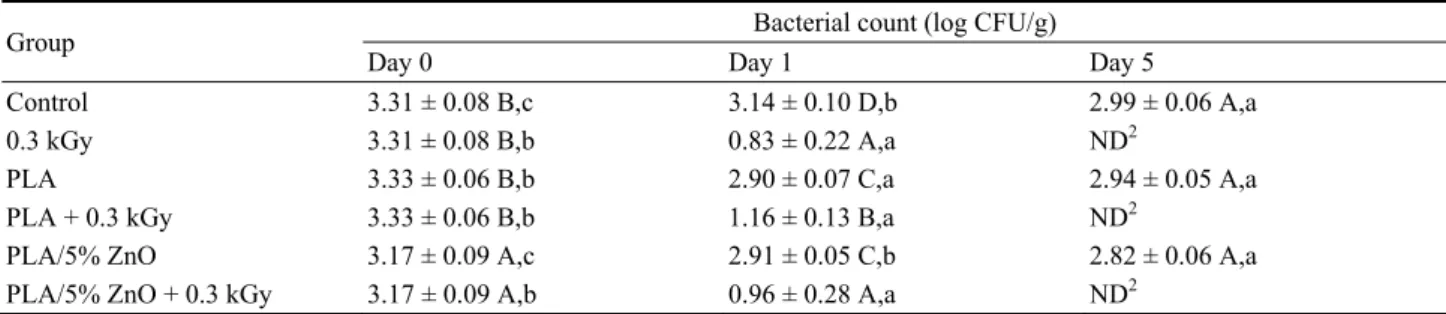
The results of S. enterica during storage are presented in Table 5. As expected, no reduction of bacterial count is observed for the “Control” group
during storage. The treatment with 0.3 kGy radiation induces a 2.43 and 3.31 log CFU/gr reduction at day 1 and 5 respectively and similar results were obtained when the hams were packed with PLA or with PLA/5% ZnO and treated at 0.3 kGy. These results suggested that only γ-radiation had an effect on the S.
enterica elimination (Table 5).
The results in this work seem to be not completely in agreement with those reported in literature. In fact, Espitia et al. [32] have listed numerous studies reporting the antimicrobial activity of ZnO particles against different bacteria. Gordon et al. [41] reported that ZnO particles were effective in inhibiting the growth of E. coli and Staphylococcus aureus. Also, according to Seil and Webster [42], the use of ZnO particles allows reducing the viability of such bacteria. The mechanism of action of ZnO on bacteria has not been completely understood [43]. Gordon et al., Zhang et al., Sawai et al., Yamamoto, Padmavathy and Vijayaraghavan and Jalal et al. [41, 43-47] have proposed that the antimicrobial properties of ZnO could
Table 5 Effect of the combined treatment on S. enterica during storage1.
Group Bacterial count (log CFU/g)
Day 0 Day 1 Day 5
Control 3.31 ± 0.08 B,c 3.14 ± 0.10 D,b 2.99 ± 0.06 A,a
0.3 kGy 3.31 ± 0.08 B,b 0.83 ± 0.22 A,a ND2
PLA 3.33 ± 0.06 B,b 2.90 ± 0.07 C,a 2.94 ± 0.05 A,a
PLA + 0.3 kGy 3.33 ± 0.06 B,b 1.16 ± 0.13 B,a ND2
PLA/5% ZnO 3.17 ± 0.09 A,c 2.91 ± 0.05 C,b 2.82 ± 0.06 A,a PLA/5% ZnO + 0.3 kGy 3.17 ± 0.09 A,b 0.96 ± 0.28 A,a ND2
1 Means followed by the same uppercase letter in each column are not significantly different at the 5% level. Means followed by the
same lowercase letter in each row for each property are not significantly different at the 5% level.
2 ND = not detected (< 0.60 log CFU/g).
be due to its interactions with water present in the food. This would result in the production of radicals before generating H2O2, which, according to Tam et al. [48], could be harmful to cells of living organisms. A second mechanism was explained by the positive charges of ZnO that could interact with the negative charges of bacterial cells [49], leading to membrane damages [43].
Moreover, it has to be also noted that the results obtained here for PLA/5% ZnO film tested for the E.
coli are in disagreement with the result in Marra et al.
[33], where for E. coli, it was reported that already after 1 day, the percent reduction was 99.99 (that is a value of CFU/mL close to 0), whereas, in this work a bacterial count of 2.82 log CFU/g at day 5 is obtained with the ham covered with the PLA/5% ZnO. The different results are very likely derived by to the diverse methodologies used; the methodology used in Marra et al. [33] and those in the above reported literature consisted generally in the immersion of the plain ZnO or films with ZnO immersed in the bacterial solutions; this methodology can be defined as “experiment in solution”; whereas in this work the films were lying on the ham slice, which could be defined as “dry experiment”. The two proposed mechanisms of efficiency of ZnO against bacteria require the presence of water (where H2O2 can be formed or where the bacteria can have enough mobility to in contact with ZnO particles and so to be damaged). In the case of the in situ experiment, that
performed in this work, only very small areas of the ZnO particles (of few µm), as shown on the SEM micrograph of Fig. 2, could be in contact with the bacteria inoculated of the ham slice and not all of them are certainly in contact because it would have required that the film would have been thoroughly in contact with on the ham, which is difficult to obtain during the storage.
The results reported in Tables 3, 4 and 5 indicate clearly that the only presence of 5 wt% of ZnO PLA film, used to cover ham slices stored at 4 °C, is not sufficient to decrease the bacterial counts to a value in order to define the composite film as “antibacterial”, but it is necessary to use the treatment of the γ-radiation, although γ-radiation has different efficiency on the three bacteria used here. In fact, the results showed that the dose of γ-radiation needed to eliminate E. coli is significantly lower than the dose needed to eliminate S. enterica, or L. innocua as previously showed [50, 51]. Chiasson et al. [52] and Huq et al. [53] demonstrated that the D10 who is the radiation dose required reducing by 1 log CFU/gr (90% of the viable bacteria) was 0.126, 0.526 and 0.540 respectively for E. coli, S. Typhimurium and L.
monocytogenes in meat. γ-radiation induces DNA
double-strand breaks that can lead to cell death if damages are not repaired. However, it is suggested in literature that only one double-strand break is sufficient to induce E. coli cell death [54-58]. This would explain why those bacteria are more sensitive
to γ-radiation.
In addition, some authors have studied the antimicrobial effects of ZnO on different bacteria. Reddy et al. [59] reported a toxicity of ZnO nanoparticles on Gram-negative bacteria, such as E.
coli, and Gram-positive bacteria, such as Staphylococcus aureus, with a total inhibition at NP
concentrations of 3.4 mM and 1 mM respectively. Díez-Pascualand and Díez-Vicente [60] have also showed an antimicrobial effect of ZnO on E. coli and
S. aureus.
However, antimicrobial compounds could be more effective in culture media than in meat that can contain more than 20% of lipids. According to Gill [61], the microorganisms, which were growing in nutrient-rich environment such as meat increased resistance to different stress by reaching their maximum replication rate and still can repair the cellular components [61]. Canillac et al. [62], also found that introduction of fat into the test medium decreased the bactericidal effect of Piceaexcelsa essential oil against L. monocytognes. Oussalah et al. [63] also found that L. monocytogenes was highly
resistant in ham even in presence of essential oils from spices.
The growth rate of L. monocytogenes is also dependant mainly on the water activity, the storage temperature, and to a smaller extent on the amount of nitrite in the product [64]. However, according to Mbandi and Shelef [65], nitrite, nitrate and sodium chloride do not inhibit the growth of L.
monocytogenes during storage under refrigerated
temperatures. Indeed, ham contains also other several additives such as sodium chloride, sodium erythrobate and sodium phosphate that may affect the efficiency of antimicrobial compounds, which can explain the results obtained in this study on ham.
3.7 Atomic Absorption Spectrometry Analysis
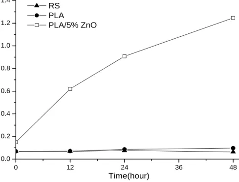
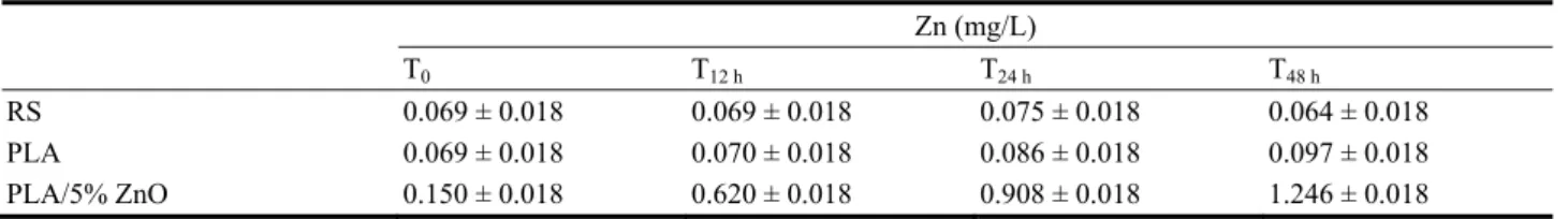
In Fig. 6 it is shown the graph of the zinc concentration (mg/L) in RS after the contact of PLA and PLA/5% ZnO films as a function of the contact time (0, 12, 24 and 48 h). For comparison the RS has been also investigated. The amount found in the RS is due to the content of zinc present in nature. In the case of PLA film in the RS, the value increases slightly
0 12 24 36 48 0.0 0.2 0.4 0.6 0.8 1.0 1.2 1.4 RS PLA PLA/5% ZnO Time(hour)
Table 6 Zinc concentration at different time of contact. Zn (mg/L) T0 T12 h T24 h T48 h RS 0.069 ± 0.018 0.069 ± 0.018 0.075 ± 0.018 0.064 ± 0.018 PLA 0.069 ± 0.018 0.070 ± 0.018 0.086 ± 0.018 0.097 ± 0.018 PLA/5% ZnO 0.150 ± 0.018 0.620 ± 0.018 0.908 ± 0.018 1.246 ± 0.018 because zinc present as contaminant in the PLA adds
to that present in the water, also as contaminant. In the case of composite in RS, the concentration of zinc increases significantly due to the ZnO that migrates from the composite film. The amount increases with the time of contact between the film PLA/5% ZnO and the RS water. Table 6 reports the zinc concentration (mg/L) in RS at different contact time. The results indicate that the Zn content in RS after 48 h is about 1.3 mg/L, which is a low concentration, certainly not harmful for animals or humans considering that the National Institute of Health [66] recommends that adult males should be getting 11 milligrams of zinc each day, and adult females need 8 milligrams and that the tolerable upper intake levels are 40 mg/day for both male and female.
4. Conclusion
It has been investigated the effect of ZnO in PLA film and of γ-radiation treatment, singularly and combined, on the shelf life of packed ham inoculated with bacteria suspension of L. innocua, S. enterica, and E. coli. It was found that the only presence of ZnO 5 wt% in PLA film, covering the ham, was not sufficient to reduce significantly the bacteria, test performed up to 5 days on samples stored at 4 °C. It is necessary the γ-radiation treatment to strongly reduce the bacterium and a complete inhibition is found at day 5 of storage and only for S. enterica and E. coli. A contribution of ZnO particles to the reduction of the bacteria is found only in the case E. coli. The disagreement between the results obtained in this work with those presented in literature, is probably due to the experiment who was done in situ where the presence of nutrients and additives can reduce the antimicrobial efficiency of the ZnO particles.
Acknowledgements
The Natural Science Engineering Research Council (NSERC, discovery program) is acknowledged for their financial support. Nordion is acknowledged for the irradiation operations. It is also acknowledged the Italian Ministry of Foreign Affair (Bilateral Project Italy/Quebec 2014-2016 “Sviluppo di nanomateriali ecosostenibili per l'imballaggio alimentare adatti alla sterilizzazione per radiazione”). In addition, the authors are thankful for the financial support to the research contract No. 17675 and 17704, as well as, the International Atomic Energy Agency through the RCM of the coordinated research project entitled: Application of irradiation technology in the development of advanced packaging materials for food products. Finally, the authors would like to thank Dr. Barbara Immirzi of IPCB-CNR for the GPC experiments, Dr. Viviana De Luca and Dr. Clemente Capasso of the Istituto di Biochimica delle Proteine-CNR (Napoli, Italy), respectively, for the kind help to perform the Atomic Absorption Spectrometry analysis and for the permission to use the instrument.
References
[1] Coles, R., McDowell, D., and Kirwan, M. J. 2003. Food Packaging Technology. Edited by Coles, R., McDowell, D., and Kirwan, M. J. Boca Raton: Blackwell Publishing, CRC Press.
[2] Salvatore, M., Marra, A., Duraccio, D., Pillai, S. D., Cimmino, S., and Silvestre, C. 2015. “Effect of Electron-Beam Irradiation on the Properties of Nanocomposites Based on Polylactic Acid/Montmorillonite for Food Packaging Applications.” J. Appl. Polym. Sci. 133 (2): 42219.
[3] Cai, H., Dave, V., Gross, R. A., and McCarthy, S. P. 1996. “Effects of Physical Aging, Crystallinity, and Orientation on the Enzymatic Degradation of Poly (lactic
acid).” Polym. Phys. 34 (16): 2701-8.
[4] Jamshidian, M., Tehrany, E. A., Imran, M., Jacquot, M., and Desobry. S. 2010. “Poly-Lactic Acid: Production, Applications, Nanocomposites, and Release Studies.” Compr. Rev. Food Sci. Food Saf. 9 (5): 552-71.
[5] Ahmed, J., and Varshney, S. K. 2011. “Polylactides—Chemistry, Properties and Green Packaging Technology: A Review.” Int. J. Food Prop. 14 (1): 37-58.
[6] Sin, L. T., Rahmat, A. R., and Rahman, W. A. 2012. Polylactic Acid: PLA Biopolymer Technology and Applications. Edited by Rahman, W. A., Sin, L. T., and Rahmat, A. R. Oxford: William Andrew-Elsevier Publishing.
[7] Sikkema, R., Steiner, M., Junginger, M., Hiegl, W., Hansen, M. T., and Faaij, A. 2011. “The European Wood Pellet Markets: Current Status and Prospects for 2020.” Biofpr. 5 (3): 250-78.
[8] Weiss, J., Takhistov, P., and McClements, J. 2006. “Functional Materials in Food Nanotechnology.” J. Food Sci. 71 (9): 107-16.
[9] Neethirajan, S., and Jayas, D. S. 2011. “Nanotechnology for the Food and Bioprocessing Industries.” Food Bioprocess Technol. 4 (1): 39-47.
[10] Garcia, M., Forbe, T., and Gonzalez, E. 2010. “Potential Applications of Nanotechnology in the Agro-Food Sector. Cienc.” Technol. Aliment. 30 (3): 573-81.
[11] Ayhan, Z. 2010. “Potential Applications of Nanotechnology in Food Packaging.” In 1st International Congress on Food Technology. November 3-6, Antalya, Turkey.
[12] Rossignol, C., Verelst, M., Dexpert-Ghys, J., and Rul, S. 2006. “Synthesis of Undoped ZnO Nanoparticles by Spray Pyrolysis.” Adv. Sci. Thecnol. 45: 237-41.
[13] Bacsa, R., Kihn, Y., Verelst, M., Dexpert-Ghys, J., Bacsa, W., and Serp, P. 2007. “Large Scale Synthesis of Zinc Oxide Nanorods by Homogeneous Chemical Vapour Deposition and Their Characterization.” Surf. Coat. Tech. 201 (22-23): 9200-4.
[14] Zhou, N., Liu, Y., Li, L., Meng, N., Huang, Y., Zhang, J., Wei, S., and Shen, J. 2007. “A New Nanocomposite Biomedical Material of Polymer/Clay-CTS-Ag Nanocomposites.” Curr. Appl. Phys. 7 (S1): 58-62.
[15] Reuge, N., Caussat, B., Joffin, N., Dexpert-Ghys, J., Verelst, M., and Dxpert, H. 2008. “Modelling of Spray Pyrolysis-Why the Synthesized Y2O3 Microparticles
Hollow?” AlChE J. 54 (2): 394-405.
[16] Caiut, J. M. A., Dexpert-Ghys, J., Kihn, Y., Veelst, M., Dexpert, H., Ribeiro, S. J. L., and Messaddeq, Y. 2009. “Elaboration of Boehmite Nano-powders by Spray-Pyrolisis.” Power Thecnol. 190 (1-2): 95-8.
[17] Duret, S., Gwanpua, S. G., Hoang, H. M., Guillier, L.,
Flick, D., Laguerre, O., Jabri, M. E., Thuault, D., Hezard, B., Lintz, A., Stahl, V., and Geeraerd, A. 2015. “Identification of the Significant Factors in Food Quality Using Global Sensitivity Analysis and the Accept-And-Reject Algorithm. Part II: Application to the Cold Chain of Cooked ham.” J. Food Eng. 148: 58-65. [18] Batz, M. B., Hoffmann, S., Morris, J., and Glenn, J. 2012.
“Ranking the Disease Burden of 14 Pathogens in Food Sources in the United States Using Attribution Data from Outbreak Investigations and Expert Elicitation.” J. Food Prot. 75: 1278-91.
[19] Public Health Agency of Canada. 2015.
[20] Molins, R. A. 2001. Food Irradiation: Principles and Applications. Edited by Molins R. A. New York: John Wiley & Sons.
[21] Komolprasert, V. 2007. “Packaging for Foods Treated by Ionizing Radiation.” Packaging for Nonthermal Processing of Food. Edited by Han, J. H. Iowa: IFT Press, Blackwell Publishing, 87-116.
[22] Kumar, R., and Münstedt, H. 2005. “Silver Ion Release from Antimicrobial Polyamide/Silver Composites.” Biomaterials 26 (14): 2081-8.
[23] Seyfriedsberger, G., Rametsteine, K., and Kern, W. 2006. “Polyethylene Compounds with Antimicrobial Surface Properties.” Eur. Polym. J. 42 (12): 3383-9.
[24] Liau, S. Y., Read, D. C., Pugh, W. J., Furr, J. R., and Russell, A. D. 1997. “Interaction of Silver Nitrate with Readily Identifiable Groups: Relationship to the Antibacterial Action of Silver Ions.” Lett. Appl. Microbiol. 25 (4): 279-83.
[25] Fujishima, A., Rao, T. N., and Tryk, D. A. 2000. “Titanium Dioxide Photocatalysis.” J. Photochem. Photobiol. C: Photochem. Rev. 1 (1): 1-21.
[26] Jones, N., Ray, B., Ranjit, K. T., and Manna, A. C. 2008. “Antibacterial Activity of ZnO Nanoparticle Suspensions on a Broad Spectrum of Microorganisms.” FEMS Microbiol. Lett. 279: 71-6.
[27] Lia, J. H., Honga, R. Y., Lic, M. Y., Li, H. Z., Zhengd, Y., and Dinge, J. 2009. “Effects of ZnO Nanoparticles on the Mechanical and Antibacterial Properties of Polyurethane Coatings.” Prog. Org. Coat. 64 (4): 504-9.
[28] Silvestre, C., Duraccio, D., and Cimmino, S. 2011. “Food Packaging Based on Polymer Nanomaterials.” Prog. Polym. Sci. 36 (12): 1766-82.
[29] Silvestre, C., Cimmino, S., Pezzuto, M., Marra, A., Ambrogi, V., Dexpert-Ghys, J., Verelst, M., Augier, S., Romano, I., and Duraccio, D. 2013. “Preparation and Characterization of Isotactic Polypropylene/Zinc Oxide Microcomposites with Antibacterial Activity.” Polym. J. 45: 938-45.
[30] Cimmino, S., Duraccio, D., Marra, A., Pezzuto, M., Romano, I., and Silvestre, C. 2015. “Effect of
Compatibilizers on Mechanical, Barrier and
Antimicrobial Properties If Ipp/Znonano/Microcomposites for Food Packaging
Application.” J. Appl. Packag. Res. 7 (2): 108-27. [31] Silvestre, C., Duraccio, D., Marra, A., Strongone, V., and
Cimmino, S. 2016. “Development of Antibacterial Composite Films Based on Isotactic Polypropylene and Coated ZnO Particles for Active Food Packaging.” Coatings 6 (1): 4-17.
[32] Espitia, P. J. P., Soares, N. de F. F., Coimbra, J. S. dos R., Andrade, N. J. de, Cruz, R. S., and Medeiros, E. A. A. 2012. “Zinc Oxide Nanoparticles: Synthesis, Antimicrobial Activity and Food Packaging Applications. Food Bioprocess.” Technol. 5 (5): 1447-64.
[33] Marra, A., Silvestre, C., Duraccio, D., and Cimmino., S. 2016. “Polylactic Acid/Zinc Oxide Biocomposite Films for Food Packaging Application.” Int. J. Biol. Macromol. 88: 254-62.
[34] Degree, P., Dubois, P., and Jérôme, R. 1997. “Bulk Polymerization of Lactides Initiated by Aluminum Isopropoxide.” I. Mechanism and Kinetics. Macromol. Symp. 123 (1): 67-84.
[35] Murariu, M., Doumbia, A., Bonnaud, L., Dechief, A. L., Paint, Y., Ferreira, M., Campagne, C., Devaux, E., and Dubois, P. 2011. “High-Performance Polylactide/ZnO Nanocomposites Designed for Films and Fibers with Special End-Use Properties.” Biomacromolecules 12 (5): 1762-71.
[36] Berber, M., Bulto, V., Kliβ, R., and Hahn, H. 2005. “Transparent NanocrystallineZnO Films Prepared by Spin Coating.” Scripta Mater. 53 (5): 547-51.
[37] Brown, H. E. 1976. Zinc Oxide: Properties and Application. International Lead and Zinc Organization, New York.
[38] Ammala, A., Hill, A. J., Meakin, P., Pas, S. J., and Turney, T. W. 2002. “Degradation Studies of Polyolefins Incorporating Transparent Nanoparticulate Zinc Oxide UV Stabilizers.” J. Nanopart. Res. 4 (1-2): 167-74. [39] Lin, O. H., Akil, H. M., and Mahmud, S. 2009. “Effect of
Particle Morphology on the Properties of Polypropylene/Nanometric Zinc Oxide (PP/nanoZnO).” Adv. Compos. Lett. 18 (3): 77-83.
[40] Chandramouleeswaran, S., Mhaske, S. T., Kathe, A. A., Varadarajan, P. V., Prasad, V., and Vigneshwaran, N. 2007. “Functional Behaviour of Polypropylene/ZnO-soluble Starch Nanocomposites.” IOPSci. 18 (38): 385702.
[41] Gordon, T., Perlstein, B., Houbara, O., Felner, I., Banin, E., and Margel, S. 2011. “Synthesis and Characterization of Zinc/Iron Oxide Composite Nanoparticles and Their Antibacterial Properties.” Colloids Surf. Physicochem. Eng. Asp. 374 (1-3): 1-8.
[42] Seil, J. T., and Webster, T. J. 2012. “Antimicrobial Applications of Nanotechnology: Methods and Literature.” Int. J. Nanomedicine 7: 2767-81.
[43] Zhang, L., Ding, Y., and Povey, M. 2008. “ZnOnanofluids—A Potential Antibacterial Agent.” Prog. Nat. Sci. 18 (8): 939-44.
[44] Sawai, J., Shoji, S., Igarashi, H., Hashimoto, A., Kokugan, T., Shimizu, M., and Kojima, H. 1998. “Hydrogen Peroxide as an Antibacterial Factor in Zinc Oxide Powder Slurry.” J. Ferment. Bioeng. 86 (5): 521-2.
[45] Yamamoto, O. 2001. “Influence of Particle Size on the Antibacterial Activity of Zinc Oxide.” Int. J. Inorg. Mater. 3 (7): 643-6.
[46] Padmavathy, N., and Vijayaraghavan, R. 2008. “Enhanced Bioactivity of ZnO Nanoparticles—An Antimicrobial Study.” Sci. Technol. Adv. Mater. 9 (3): 035004.
[47] Jalal, R., Goharshadi, E. K., Abareshi, M., Moosavi, M., Yousefi, A., and Nancarrow, P. 2010. “ZnOnanofluids: Green Synthesis, Characterization, and Antibacterial Activity.” Mat. Chem. Phys. 121 (1-2): 198-201.
[48] Tam, K. H., Djurišic, A. B., Chan, C. M. N., Xi, Y. Y., Tse, C. W., Leung, Y. H., Chan, W. K., Leung, F. C. C., and Au, D. W. T. 2008. “Antibacterial Activity of Znonanorods Prepared by a Hydrothermal Method.” Thin Solid Films 516 (18): 6167-74.
[49] Stoimenov, P. K., Klinger, R. L., Marchin, G. L., and Klabunde, K. J. 2002. “Metal Oxide Nanoparticles as Bactericidal Agents.” Langmuir 18 (17): 6679-86. [50] Lacroix, M., Turgis, M., Borsa, J., Millette, M., Salmieri,
S., Caillet, S., and Han, J. 2009. “Applications of Radiation Processing in Combination with Conventional Treatments to Assure Food Safety: New Development. Radiat.” Phys. Chem. 78 (11): 1015-7.
[51] Beauchamp, S., and Lacroix, M. 2012. “Resistance of the Genome of Escherichia Coli and Listeria Monocytogenes to Irradiation Evaluated by the Induction of Cyclobutane Pyrimidine Dimers and 6-4 Photoproducts Using Gamma and UV-C Radiations.” Radiat. Phys. Chem. 81 (8): 1193-7.
[52] Chiasson, F., Borsa, J., Ouattara, B., and Lacroix, M. 2004. “Radiosensitization of Escherichia Coli and Salmonella TYPHI in Ground Beef.” J. Food Prot. 67 (6): 1157-62.
[53] Huq, T., Vu, K. D., Riedl, B., Bouchard, J., and Lacroix, M. 2015. “Synergistic Effect of Gamma Irradiation and Microencapsulated Antimicrobials against Listeria Monocytogenes on Ready-to-Eat Meat.” Food Microbiology 46: 507-514.
[54] Kaplan, H. S. 1966. “DNA-Strand Scission and Loss of Viability after X Irradiation of Normal and Sensitized Bacterial Cells.” Proc. Natl. Acad. Sci. U. S. A. 55:
1442-6.
[55] Schans, G. P. van der, Bleichrodt, J. F., and Blok, J. 1973. “Contribution of Various Types of Damage to Inactivation of a Biologically-Active Double-Stranded Circular DNA by Gamma-Radiation.” Int. J. Radiat. Biol. Relat. Stud. Phys. Chem. Med. 23 (2): 133-50.
[56] Boye, E., and Krisch, R. E. 1980. “Induction and Repair of Double and Single-Strand DNA Breaks in Bacteriophage Lambda Superinfecting Escherichia Coli.” Int. J. Radiat. Biol. Relat. Stud. Phys. Chem. Med. 37 (2): 119-33. [57] Frankenberg, D., Frankenberg-Schwager, M., Blöcher, D.,
and Harbich, R. 1981. “Evidence for DNA Double-Strand Breaks as the Critical Lesions in Yeast Cells Irradiated with Sparsely or Densely Ionizing Radiation under Oxic or Anoxic Conditions.” Radiat. Res. 88 (3): 524-32.
[58] Schulte-Frohlinde, D. 1987. “Biological Consequences of Strand Breaks in Plasmid and Viral DNA.” Br. J. Cancer. Suppl. 8 (55): 129-34.
[59] Reddy, K. M., Feris, K., Bell, J., Wingett, D. G., Hanley, C., and Punnoose, A. 2007. “Selective Toxicity of Zinc Oxide Nanoparticles to Prokaryotic and Eukaryotic Systems.” Appl. Phys. Lett. 90, 213902-1-213902-3. doi:10.1063/1.2742324.
[60] Díez-Pascual, A. M., and Díez-Vicente, A. L. 2014. “Poly (3-hydroxybutyrate)/Zn Obionanocomposites with
Improved Mechanical, Barrier and Antibacterial Properties.” Int. J. Mol. Sci. 15 (6): 10950-73.
[61] Gill, A. O., Delaquis, P., Russo, P., and Holley, R. A. 2002. “Evaluation of Antilisterial Action of Cilantro Oil on Vaccum Packed Ham.” Int. J. Food Microbiol. 73: 83-92.
[62] Canillac, N., and Mourey, A. 2004. “Effects of Several Environmental Factors on the Anti-Listeria Monocytogenes Activity of an Essential Oil of Piceaexcelsa.” Int. J. Food Microbiol. 92: 95-103.
[63] Oussalah, M., Caillet, S., Salmieri, S., Saucier, L., and Lacroix, M. 2007. “Antimicrobial Effects of Alginate Based Films Containing Essential Oils for the Preservation of Bologna and Ham.” J. Food Prot. 70 (4): 901-8.
[64] Stekelenburg, F. K., and Kant-Muermans, M. L. T. 2001. “Effects of Sodium Lactate and Other Additives in a Cooked Ham Product on Sensory Quality and Development of a Strain of Lactobacillus Curvatus and Listeria Monocytogenes.” Int. J. Food Microbiol. 66: 197-203.
[65] Mbandi, E., and Shelef, L. A. 2002. “Enhanced Antimicrobial Effects of Combination of Lactate and Diacetate on Listeria Monocytogenes and Salmonella sp. in Beef Bologna.” Int. J. Food Microbiol. 76: 191-8. [66] https://ods.od.nih.gov/factsheets/Zinc-HealthProfessional/.