UNIVERSITÉ DE MONTRÉAL
Modulation of
1HERG11Kby Cellular Metabolites:
Implication in the Arrliytlimogenesis
During Myocardial Iscliemia
par
Jing
Xiong WANG
Programme en Sciences biomédicales
Département de Médicine
Faculté de Médicine
f
Thèse présentée à la faculté des
études
supérieures
en
vue
de l’obtention du grade de
Doctorat philosophie (Ph. D.)
en Sciences biomédicales
Août 2005
W
H
V
Université
(ll’h
de Montréal
Direction des bibliothèques
AVIS
L’auteur a autorisé l’Université de Montréal à reproduite et diffuser, en totalité ou en partie, par quelque moyen que ce soit et sur quelque support que ce soit, et exclusivement à des fins non lucratives d’enseignement et de
recherche, des copies de ce mémoire ou de cette thèse.
L’auteur et les coauteurs le cas échéant conservent la propriété du droit
d’auteur et des droits moraux qui protègent ce document. Ni la thèse ou le mémoire, ni des extraits substantiels de ce document, ne doivent être
imprimés ou autrement reproduits sans l’autorisation de l’auteur.
Afin de se conformer à la Loi canadienne sur la protection des renseignements personnels, quelques formulaires secondaires, coordonnées
ou signatures intégrées au texte ont pu être enlevés de ce document. Bien
que cela ait pu affecter la pagination, il n’y a aucun contenu manquant.
NOTICE
The author of this thesis or dissertation has granted a nonexclusive license allowing Université de Montréal ta reproduce and publish the document, in
part or in whole, and in any format, solely for noncommercial educationat and research purposes.
The author and co-authors if applicable retain copyright ownership and moral
rights in this document. Neither the whole thesis or dissertation, nor
substantial extracts from it, may be printed or otherwise reproduced without the author’s permission.
In compliance with the Canadian Privacy Act some supporting forms, contact
information or signatures may have been removed from the document. While this may affect the document page count, it does flot represent any loss of
Université de Montréal
faculté des études supérieures
Cette thèse intitulée:
Modulation of
1HERG/1K
by Cellular Metabolites:
Implication in the Arrliytlimogenesis
During Myocardial Iscliemia
présentée par:
Jing Xiong Wang
a été évaluée par un jury composé des personnes suivantes:
Dr Terence E. Hébert
Président-rapporteur Représentant du doyen de la FES
Dr Zhi Guo Wang
Directeur de recherche
Dr Stanley Natte!
Co-directeur de rechercheDr
AlvinShrier
Examinateur externeDr Lucie Parent
ExaminateurSOMMAIRE
L’ischémie du myocarde induit des altérations de l’électrophysiologie cardiaque selon un mode biphasique. Les anomalies les plus profondes sont associées a la phase précoce, l’ischémie aigue du myocarde (AMI). Elles sont caractérisées par une
,. + + 4’
accumulation extracellulaire d ions K ([K 10—i), un raccourcissement de la duree du potentiel d’action (PA) et de l’intervalle QT (APD—L ou QT—1-), ainsi qu’une accumulation de métabolites de phospholipides, les lysophosphatidylcholines (LPCs). Lors de la deuxième phase, on observe un rallongement de l’APD et de l’intervalle QT ainsi qu’une synthèse “de novo”, une accumulation de métabolites de sphingolipides, les céramides, et de TNf-Œ. Ces modifications sont associées à l’ischémie chronique du myocarde (CMI).
Ces changements opposés de I’APD/QT sont associés à différents types d’arythmies. Il a été montré que la relation entre les perturbations électriques et métaboliques joue un rôle clef dans la genèse des arythmies ischémiques et la mort subite. Cependant les mécanismes ioniques et métaboliques impliqués dans ces modifications séquentielles de l’APD/QT restent mal compris.
Notre hypothèse est que les désordres électriques ischémiques sont la conséquence de la conjugaison de stress métabolique et de déséquilibres électrolytiques. Plus spécifiquement, le courant IKr/’HERG est augmenté durant la phase précoce, l’AMI. Cette augmentation est principalement causée par une surproduction et une accumulation de LPCs. La potentialisation de IIJJHERG par les LPCs contribue de façon critique à la perte de K intracellulaire, a la diminution de l’APD, et par la même, auxarythmies associées a l’ischémie. En revanche, lors de la CMI et l’insuffisance cardiaque, la diminution de l’expression de HERG induite par les céramides et le TNF-Œ contribue a l’augmentation tardive de I’APD/QT et par conséquent entraîne l’apparition de troubles durythme.
Afin de vérifier cette hypothèse, nous avons réalisé des études à différents niveaux. En passant par l’organe, la cellule, pour terminer à l’échelon moléculaire. Ces expériences ont été réalisées en utilisant différentes approches combinées, l’électrophysiologie, la pharmacologie, la biochimie, et la biologie moléculaire.
Tout d’abord, nous avons montré que les LPCs augmentent le courant IHERG
exprimé dans des cellules HEK-293, et que cette augmentation du courant ‘HERGentraînait
un raccourcissement de l’APD ainsi qu’un potentiel de membrane plus négative; LPC-16 augmentait‘HERGde façon plus prononcée pour des potentiels plus négatifs, ceci associé à
un raccourcissement plus important de I’APD. De plus, seulement la forme amphiphile avec 16 hydrocarbonés comme le I -Palmytoyl-lysophosphatidylcholine (LPC- 16) et le 1-palmitoyl-lysophosphatidylglycerol (LPG-16) entraînait une augmentation significative du courant ‘HERG, ainsi qu’un déplacement de son activation vers des potentiels plus
négatifs.
En utilisant le modèle d’ischémie sévère du myocarde chez le lapin, nous avons montré que le LPC-16 exogène mimait l’effet de l’ischémie de faible perfusion correspondant à une augmentation de [K]0 et de l’intervalle QT ces effets étaient prévenus par des bloqueurs de Ij tel que le doflétilide, les autres bloqueurs des canaux
+ ‘ ‘
K n ayant pas d effet. De façon consistante, le dofletilide suppnme efficacement, les tachyarythmies induites par l’ischémie ou le LPC-16. De plus, le LPC-16 raccourcis de façon remarquable l’APD dans les myocytes ventriculaires gauche de lapin, augmente, de façon réversible et dépendante au potentiel, l’amplitude du courant I)Cj dans les myocytes ventriculaires de cochon d’inde. Le doflétilide supprime cet effet sur le courant ‘“HERG,
et limite ainsi le raccourcissement de l’APD.
Nous avons montré que lors de la mise en place de la CMI, le courant Ii< transporté par les canaux HERG était une cible majeure pour l’action des céramides et du TNf-Œ. L’incubation continue de cellules exprimant les canaux HERG, ou de myocytes, avec des céramides ou du TNF-cL diminue de façon significative le courant Iy ou la fonction des canaux HERG. Ces effets sont médiés par la stimulation de ROS (réactive oxygène species) intracellulaires. Puisque les antioxydants tels que la vitamine E ou le MnTBAP suppriment l’effet dépressif des céramides ou du TNF-Œ surJjJHERG. De plus, le céramide comme le TNF-Œ augmentent de façon importante le niveau intracellulaire de ROS, cet effet est prévenu par la vitamine E ou le MnTBAP.
En conclusion, nos résultats suggèrent que l’accumulation de LPC-16 conjuguée a l’augmentation du courant ‘HERG démasquent un réel couplage entre un catalyseur
courant potassium sortant et une réduction de l’intervalle QT lors d’un épisode ischémique. L’ensemble de ce travail constitue la première évidence de l’implication du courant ‘KJJHERG dans l’augmentation post-ischémique du courant potassium sortant et
par ce biais dans la diminution de l’intervalle QT. L’inhibition de la production de LPC 16 ainsi que la réduction de son accumulation pourrait constituer une stratégie thérapeutique prometteuse en vue d’atténuer les arythmies létales associées aux maladies ischémiques du coeur. D’un autre côté, la réduction du courant IHERGparles céramides ou le TNF-Œ principalement en augmentant la production de ROS semble contribuer a la prolongation de l’intervalle QT lors de l’ischémie prolongée du myocarde, ainsi que dans l’insuffisance cardiaque.
Mots clefs: ischémie du myocarde, arythmies, canaux potassium, lysophosphatidylcholine, céramides, TNF-cx,ROS.
ABSTRACT
Ischemic myocardium demonstrates characteristic bi-phasic aiterations in cardiac electrophysiology. The most profound abnormalities associated with the eariy phase of acute myocardial ischemia (AMI) are extracellular K accumulation ([Kj0—t) and shortening of action potential duration or QT interval (APD—I- or QT4) as well as accumulation of phospholipid metabolites iysophosphatidylcholines (LPCs). These are followed by the subsequent lengthening ofAPD or QT inteiwai (APD—t/QT—t) and de nova synthesis and accumulation of sphingolipid metabolite ceramide and tumor necrosis factor—a, as the most prominent electrical and metabolic disturbances in chronic myocardial ischemia (CMI). The opposite changes ofAPD/QT are often accompanied by different types of arrhythrnias. It has been shown thatthe interplay between the electrical and metabolic perturbations is pivotai in the genesis of ischemic arrhytbmias and sudden cardiac death. However, the ionic and metaboiic mechanisms underlying the sequential changes of APD/QT remained poorly understood.
We proposed that ischemic electricai disorders are a consequence of inteneiated metaboiic stress and electrolyte disturbance. More specificaily, I}cj/IHERG is enhanced
during the eariy stage of AMI and the enhancement is mainly caused by overproduction and accumulation of LPCs. The enhancement of‘Kr/’HERGby LPCs is a criticai contributor
to intracelluiar K ioss/[Kj0—t and APD—J,- thereby the associated ischemic arrhythmias. Whereas in CMI and failing heart, where HERG impairment by ceramide and TNF-a contributes to the late APD—t/QT—t, leading to cardiac arrhytbmias. To examine our hypothesis the studies at different ievels including organ, ceilular, and molecuiar ieveis were carried out with combined approaches of electrophysioiogy, pharmacoiogy, biochemistry and molecular bioiogy.
We first revealed that LPCs enhance ‘HERG expressed in HEK293 ceil and the
enhancement of‘HERGmanifested with shorterAPD and at more negative potentiai; LPCs
increased IHERG to a greater extent at more negative potentiais and with shorter APD.
furthermore, oniy the amphiphile with 16 hydrocarbons such as 1-palmitoyl-iysophosphatidyicholine (LPC- 16) and 1 -palmitoyl-lysophosphatidyiglyceroi (LPG- 16)
were found to produce significant enhancement of‘HERG and negative shifts of HERG
activation.
By using a rabbit model of acute global myocardial ischemia, we found that exogenous LPC-16 mimicked the low-perfusion ischemia to produce significant [K]0—t and QT4, which were prevented by Iy blocker dofetilide but not by blockers for other K channels. Consistently, dofetilide efficiently abolished the ventricular tachycardia arrhythmias induced by LPC- 16 or ischemia. Moreover, LPC- 16 remarkably shortened APD in rabbit lefi ventricular myocytes, reversibly and voltage-dependently increased the amplitude of Iyin guinea pig ventricular myocytes. Dofetilide abolished the ‘KrI’HERG
enhancing andAPD shortening caused by LPC-16.
In the setting of CMI, we identified IgjHERG K channel as a new target for the action of ceramide and TNF-a. Chronic exposure of HERG expressing ceils or cardiac myocytes to ceramide or TNf-Œ significantly impaired HERG K channel orIy- function. The impairment of channel function is mediated by stimulating intracellular reactive oxygen species (ROS) because antioxidants vitamin E or MnTBAP abolished the depressing effects of ceramide or TNF-Œ on IcjHERG. Moreover, either ceramide or TNF-Œremarkably elevated the intracellular ROS levels, which was prevented by vitamin E or MnTBAP.
In conclusion, our resuits suggest that LPC-16 accumulation and HERG enhancement form the coupling between metabolic trigger and ionic pathway that may account for ischemic [Kj0—t and QT—1-. This represents the first documentation of I/HERG as the ionic mechanism for ischemic [Kj0—t and QT—..L. Inhibition of LPC-16 production and accumulation andlor ofI/HERG may be a promising therapeutic strategy to attenuate the incidence of lethal arrhytbmias associated with ischemic heart disease. On the other hand, HERG/IK impairment by ceramide or TNF-Œ mainly via ROS overproduction may contribute to QT prolongation in prolonged ischemia of myocardium and heart failure. The inhibition of ceramide and TNF-&TNfR1 signaling may be an effective strategy to prevent not only the arrhythmogenesis but also the cardiac sudden death in chronic myocardial ischemia and CHF.
Key words: Myocardial ischemia, Arrhythmias, Potassium channels, Lysophosphatidylcholine, Ceramide, TNf-alpha, Reactive oxygen species.
ACKNOWLEDGEMENTS
This thesis couid neyer have been possible without the help ofmany great peopie.
First of ail, I offer my most sincere appreciation to my mentor and friend Zhiguo. I am heartiiy indebted to him. Were it flot for his extraordinary supervisor, sustained encouragement, enthusiastic inspiration, pertinent guidance, constructive criticism, meticulous proofreading and editing of au the manuscripts and this thesis, my PhD study and this thesis wouid neyer have been achieved. I am aiso greatly indebted to my co supervisor, Dr. Staniey Nattel, for bis outstanding scientific counsel, bis critical heip in manuscript preparation and for his powerful recommendation in my applications for a feilowship and post-doctorai positions as well.
I have owned a speciai debt of gratitude to my previous mentor, Dr. Jackie R. Vendenheede of Kathoiieke Universiteit Leuven, Beigium, whose infinite encouragement in my research career and no-reservation recommendations for my applications of studentship, feiiowship and post-doctoral positions engraved in my heart.
My sincere thanks naturaiiy go to the Program of Biomedical Sciences, Department of Medicine of Université de Montréal for giving me the opportunity to pursue my doctoral studies in this wonderfui university; to the Montreal Heart Institute for her fabulous milieu and fine peopie.
Dr. Zhiguo Wang’s lab and Dr. Nattei’s lab are wonderflil place in which to work. I realiy want to express my gratitude to all lab members with whom I have interacted for keeping an open and coliaborative spirit. I am grateful to Dr. Wei Han for her introducing me to Dr. Zhiguo Wang; to Mr. Yiqiang Zhang, my doctoral mate and friend, for bis ingenious assistance in many aspects of my study and work; to Dr. Hong Shi for ber hands-on training in patch—ciamp approaches; to Drs. Huizhen Wang and Hong Han for their collaboration in molecular biology studies, to Ange Maguy, for his kindness to help me working out the French version of the abstract of the thesis. Especially, I want to thank Xiaofan Yang, Denis Chartier, Marc-Antoine Giilis, and Louis R. Villeneuve, for their excellent technical assistance. A sincere thanlc-you also goes to many other coileagues, especially, Marc Pourrier, Stephen Zicha, Daniel Herrera, Huixian Lin, Ling Xiao, Liming Zhang, Evenlyn Landry, and Xiaobin Luo, for their just-in-time help and friendship. Speciai recognition is due as weli to the administrative office and audio-vision section staff at Research Center, Montreal Heart Institute.
Finaily, I would like to acknowledge the contributions of my PhD study qualification committee and the thesis evaluation committee: Drs. Céline Fiset, Lucie Parent, Daya Varma, Alvin Shrier, Normand Leblanc, Terence E. Hébert, Stanley Nattel and Zhiguo Wang to this work. I thank them for their suggestions, comments, presence, support and their patience. My special gratitude also goes to the Fonds de Recherche en Santé de Québec for providing me the Doctoral Studentship from 2003 to 2006.
STATEMENT 0F AUTHOR$HIP
The following is a statement regarding the contributions of co-authors and myseif to the fivepapers already published or submitted for publication, included in this thesis.
1. Wang J., Wang H., Han H., Zhang Y., Yang B., Nattel S., and Wang Z. Phospholipid Metabolite 1 -Palmitoyl-lysophosphatidylcholine Enhances Human Ether-a go-go-related Gene (HERG) K Channel Function. Circulation. 2001; 104(22): 2645-8.
In this paper, my supervisors Drs Wang and Nattel offered me close instruction in whoie process, generating the initiai idea, clarifying the notion, and working out the final version of the paper. I designed and performed the experiments, analyzed the data, and wrote the manuscript.
2. Wang J., Zhang Y., Wang H., Han H., Nattel S., Yang B., and Wang Z. Potential Mechanisms for the Enhancement of HERG K Channel function by Phospholipid Metabolites. Br JPharrnacol. 2004; 14 1(4): 586-99.
The initial idea was derived from paper 1. I designed and performed the experiments, analyzed the data, and wrote the manuscript. Yiqiang Zhang participated in analyzing and discussing the data. Dr Wang sewed in overali supervision, clarified the idea, and producing the final version of the paper.
3. Wang J., Gillis M., Zhang Y., Lin H., Xu C., Yang B., Wang Z. Enhancement of HERG Function by Lysophosphatidylcholine Contributes to Extracellular K Accumulation and “Short QT Syndrome” in the Heart with Acute Global Ischemia. Circulation. (Submitted in July 2005).
The initial idea was prompted by literature review and our previous work. I had been responsible for planning and executing experiments, analyzing data, and writing the manuscript. Marc-Antoine Gillis helped me perfonning Langendorff perfusion and ECG recording. Yiqiang Zhang took part in discussing the data. Dr Wang provided overail instruction in ail aspects, clarified the thoughts, and produced the final version of manuscript.
4. Wang J., Zhang Y., Wang H., Lin H., Yang B., Wang Z. Sphingolipid Metabolite Ceramide Causes Metabolic Perturbation Leading to HERG K Channel Dysfunction and Abnormal Siowing of Cardiac Repolarization. J Bio! Chem, (submitted in July 2005).
I designed the experiments and completed majority of the experimental work, analyzed the data, and wrote the manuscript. Yiqiang Zhang participated in data analysis and Huizhen Wang joined in part of experiments. Dr Wang gave his supervision in whole process, helped me in making my thoughts clear, reorganizing the data, and editing the final version of the manuscript.
5. Wang J•*, Wang H.*, Zhang Y., Gao H., Nattel S., Wang Z. Impairment of HERG K Channel Function by Tumor Necrosis Factor-Œ: Role of Reactive Oxygen Species as a Mediator. J Bio! Chem, 2004; 279(14): 13289 — 13292. (* indicating both
authors contributed equally to this study).
I was responsible for planning and conducting most of the experiments, analyzing the data, and wrote the manuscript. Huizheng Wang participated in doing Western blot experiments. Dr Nattel served as co-supervisor for scientific counsel and help in manuscript preparation. Dr Wang paid overail close supervision, including generating the initial ideas, reorgamzing the data, and creating the fmal version ofthe article.
ADDITIONAL PUBLICATIONS
(Durïng my PhD Training Perïod: 05/2000—07/2005)
Peer-reviewed Articles:
1*. Wang J, Han H, Zhang Y, Long H, Wang H, Xu D, and Wang Z. HERG K channel conductance promotes H202—induced apoptosis in HEK293 ceils: Cellular mechanisms. CellPhysioÏBiochem. 2004; 14: 121 — 134.
2. Han H, Long H, Wang H, Wang J, Zhang Y, Wang Z. Progressive apoptotic celi death triggered by transient oxidative insuit in H9c2 rat ventricular ceils: A novel pattem of apoptosis and the mechanisms. Am J Physiol Heart Circ Physiot. 2004; 286(6): H2169 —82.
3. Zhang Y, Han H, Wang J, Wang H, Yang B, and Wang Z. Impairment ofhuman ether-a-go-go-related gene (HERG) K channel fimction by hypoglycemia and hyperglycemia: similar phenotypes but different mechanisms. .L Bio!. Chem. 2003; 278:10417— 10426
4. Zhang Y, Wang H, Wang J, Han H, Nattel S, and Wang Z. Normal function of HERG K channels expressed in HEK293 ceils requires basal protein kinase B activity. FEBSLett. 2003; 534:125 - 132.
5. Wang H, Zhang Y, Han H, Cao L, Wang J, Long H, Nattel S, Wang Z. HERG K Channel: A regulator of tumor celi apoptosis and proliferation. Cancer Research 2002; 62:4843 - 4848.
6*. Rahmutula D, Nakayama T, Izumi y, Ozawa Y, Shimabukuro H Kawamura H, Wang S, Wang J, Aisa M, Yang C, Mahmut M, Mahsut R, and Cheng Z. Angiotensin-converting enzyme gene and longevity in the Xinjiang Uighur Autonomous region of China: An association study. Journal of Gerontotogy: MEDICAL SCIENCES 2002; 57(1): M57 -M60.
7. Wang H, Yang B, Zhang Y, Han H, Wang J, $hi H, and Wang Z. Different subtypes of aipha-adrenoceptor modulate different K cunent via different signaling pathways in canine ventricular myocytes. J. Bio!. Chem. 2001; 276(44): 40811 —40816.
8
Erxidin, Wang J, Yang W, et al. Exploration on the longevity factors of the Uighur nationality in Hotan, Xinjiang, China. Journal of Xinjiang Medicat University 2001; 24:62—63.
Published Abstracts:
1*. Wang J, Zhang Y, Wang Z. Different reactive oxygen species differentially modulate HERG K channel function. Biophysicat Journal2005; 88(1): 608a
2*. Wang J, Wang H, Zhang Y, Nattel S, Wang Z. Sphingolipid ceramide impairs HERG K channels via overproduction of reactive oxygen species and impairment ofHERG trafficking. Biophysicat Journal2005; 88(1): 425a
3*
Zhang Y, Wang J, Wang Z. Ionic mechanisms underlying the QT prolongation in type I diabetic rabbit hearts. Biophysical Journal 2005; 88(1): 472a
4*
Zhang Y, Wang J, Wang Z. IKr/HERG K channel, a potential molecular contributor to the QT prolongation in Type I Diabetic Hearts. 2004 Circulation 110 (17): III—193
5*
Wang J, Gillis M-A, Zhang Y, Nattel S, and Wang Z. The mechanism underlying arrhythmogenesis of lysophosphatidylcholine during early phase of acute global ischemia in isolated perfused rabbit hearts. 2004 International Journal of Cardiotogy 97 Suppi. 2: 544
6*. Wang J, Zhang Y, Yu f, and Wang Z. $phingolipid ceramide impairs HERG K channel function: Involvement of multiple kinases and role of reactive oxygen species as a mediator. 2004International Journal of Cardiology97 Suppl. 2: 557 7*
Wang J, Zhang Y, Wang H, Han H, Naffel $ and Wang Z. Mechanisms for the enhancement of HERG K channel function by phospholipid metabolites. 2003 CirculationMay 20, Page 35
8. Zhang Y, Han H, Wang J, Wang H, Yang B, and Wang Z. Impairment ofHERG (Human Ether-a-go-go Related Gene) K channel function by hypoglycemia and hyperglycemia: similar phenotypes but different mechanisms. 2003 Circulation May 20, Page 35
9. Wang H, Wang H, Wang J, Zhang Y, Han H, Nattel S and Wang Z. TNF-Πdepresses HERG K channel function: a potential molecular contributor to sudden cardiac death inheart failure? 2003 CirculationMay 20, Page 36
10*. Wang J, Wang H, Han H, Zhang Y, Yang B, Nattel S and Wang Z. The phospholipid metabolite lysophosphatidylcholine enhances HERG K channel function. 2002Biophysicat Journal 82(1): 583a
11. Zhang Y, Wang J, Wang H, Han H, Nattel S and Wang Z. Protein kinase B enhances HERG K channel function. 2002Biophysical Journal 82(1): 583a
12*. Zhang Y,Wang J, Han H, Wang H, Long H and Wang Z. Glucose produces dual effects on HERG K channel firnction. 2002 Biophysical Journal 82(1): 583a
13*. Wang H, Wang J,Han H, and Wang Z. Over-expression of HERG K channel in HEK293 cells facilitates apoptotic process induced by ceramide. 2002 Biophysical Journal 82(1): 578-579a
14*. Han H, Wang J, Long H, Wang H, Wang Z and Nattel S. Overexpression of HERG K channels in HEK-293 cells induces DNA fragmentation via activation of MAP kinases. 2001 llth International Conference on Second Messengers. Page 7-6
15*. Wang J, Wang H, Wang H, Han H, Wang Z and Nattel S. Ceramide modulates HERG K channels stably expressed in HEK-293 cells possibly via regulating the synthesis and trafficking of HERG protein. 2002 llth International Conference on Second Messengers. Page 7-8
Note:
(1) for publications in which I am the first author, I generated the idea, designed and performed the experiments, analyzed the data and wrote the manuscripts;
(2) for publications in winch I am a2nd 3rdco-author, I participated in about 30—40% work of the projects, including idea generation, experiments design and cany-out, data analysis, manuscript correction and discussion, etc.;
(3) *
ijfltIesis zddcatec[to:
fMy parents, ZIirongZfiu e1 ZIongfie Wang vIy [augfitei Pnfti
SMy Jing
fMysisters,Yingming an1ingyue
!7vty brotfiers, lingjïe anJYingfong
TABLE 0F CONTENTS
SOMMAIRE. . .in
ABSTRACT. . .vi
ACKNOWLEDGEMENTS.
. .ix
STATEMENTS 0F AUTHORSfflP.
. .x
DEDICATION.
. .xv
TABLE 0f CONTENTS
. . .xvi
LIST 0F SIGNS AND ABBREVIATIONS.
. .xxiii
LIST 0F FIGURES AND TABLES.
. .xxvii
PART I: INTRODUCTION AND
THE REVIEW 0f THE
LITERATURE
...Chapter I: Electrical and Metabolic Disturbances Underlying Ischemic
Arrliythmias in Hearts.
. .2I-1 Overview: Iscliemic Heart Disease—A Global Healtli Problem. . . 3
I-2 General Features of Ischemic Arrhythmias. . .4
I-3 Our Current Understanding of the Potential Ionic Mechanisms for Ischemic Arrhythmias. . . 5
I-3-1 Properties ofionic Channels Underlying Cardiac Action Potentials. . .7 I-3-1-1 Sodium(Na) Channels. . .
9
I-3-1-2 Calcium (Ca2) Channels. . . 10
I-3-1-3 Pacemaker Channels (f-Channels). . . 12
+
I-3-1-4 Potassium (K
)
Channels. . . 13I-3-1-4-2 Ultrarapidly-rectifier Current(‘Kur) . 15
+
I-3-1-4-3 Rapid Delayed Rectifier K Current(ly.). . . 17
I-3-1-4-4 Slow Delayed Rectifier K Current(Iy). . . 21
+
I-3-1-4-5 Inward Rectifier K Current (IKI). . .24
I-3-l-4-6 Acetylcholine-induced K Cunent (I1<ch). . .25 +
I-3-1-4-7 ATP-sensitive K Current(ITP). . . 26
I-3-2 Synopsis oflonic Channels Underlying Cardiac Action Potentials. . . 29 l-4 Disturbancesof Ion Concentrations during Myocardial Ischemia. . .30
I-4-1 Extracellular Potassium Accumulation ([K]0-t) . . .31
I-4-l-1 The Temporal Course of [Kj0—t during Myocardial Ischemia 32
I-4-1-2 Effects of [Kj0-t on Channels and Carriers. . . 35
I-4-1-3 Effects of {Kj0-t on Electrophysiological Properties. . .36 I-4-2 Increase in intracellular calcium concentration ([Ca2]1-t) . . . 36
2+
I-4-2-1 Ca Distnbution.. . 36
I-4-2-2 Mechanisms underlying [Ca2j-t. . . 37
1-4-2-3 Effects of [Ca2411-t on Cunents and Carriers. . . 3$
I-4-2-4 Electrophysiological Effects of [Ca2]-t . . . 3$
I-5 Metabolic Disturbances during Myocardial Ischemia. .39 I-5-1 Phospholipid Metabolites and Ischemic Arrhythmias . . . 39
I-5-l-1 Accumulation of Lysophosphatidylcholine during Myocardial Ischemia. . . 39
I-5-1-2 Effects ofLPCs on Currents and Carriers and the Mechanisms .41
l-5-1-3 Electrophysiological Effects ofLPCs . . . 42 I-5-2 Sphingolipid Metabolites and Myocardial Ischemia . . .43
I-5-2-1 Accumulation of Ceramide during Myocardial Ischemia . . . 44
I-5-2-2 Effects of Sphingolipid Metabolites on Currents and Carriers.. . 45 I-5-2-3 Cardiac Effects of Sphingolipid Metabolites. . . 46
I-5-3 Cytokines and Myocardial lschemiaJHeart Failure. . . 49
49
I-5-3-2 Effects ofTNf-Πon Cardiac Channels and Carriers. . . 50 I-5-3-3 Deleterious Effects ofTNF-Πon Heart. . . 51
I-5-4 ROS and Myocardial IschemialHeart failure.. . 53
I-5-4-1 Generation and Counterbalancing ofROS in Heart. . . 53
I-5-4-2 Effects ofROS on Channels andTransporters. . . 55
I-5-4-3 Electrophysiological Changes Caused by ROS. . . 57
I-6 Electrophysiological Mechanisms Underlying the Ischemic Arrhythmias 58
I-6-1 Electrophysiological Mechanisms for Ischemic [Kj0-t andAPD--I-. . . 59 I-6-2 Electrophysiological Mechanisms for the Arrhytbmias during Chronic
Myocardial Ischemia. . . 62
I-7 Questions Rose from Above Review. . . 65
I-8 Working Hypothesis. . .65
I-9 Specific Objectives of the Project . . .
66
I-10 References . . . 67PART II: ORIGINAL CONTRIBUTIONS
.. .119Chapter II: Modulation
0f ‘HERG”Ktby Phospholipid Metabolites
Contributes to the Arrhythmogenesis during Acute
Myocardial Iscliemia.
. . 120II-1 Phospholipid Metabolite 1-Palmitoyl-Lysophosphatidylcholine Enhances ÷
HumanEther-a-Go-Go-Related Gene (HERG)K Channel Function. . . 122
II-1-l Abstract. . . 124
II-1-2 Introduction. . . 124
II-1-3 Methods . . . 125
II-1-3-1 Celi Culture. . . 125
II-1-3-2 Whole-Cell Patch-Clamp Recording.. . 126
II-1-4 Resuits . . . 126
II-1-5 Discussion. . . 129
II-1-6 Acknowledgments. . . 131 II-1-7 References . . . 131
II-2 Potential Mechanisms for the Enhancement of HERG K Channel Function by Phospholipid Metabolites. . . 134
II-2-1 Abstract. . . 136
II-2-2 Introduction. . . 137 II-2-3 Methods . . . 139
II-2-3-1 Ceil Culture. . . 139
II-2-3-2 Whole-Cell Patch-Clamp Recording. . . 139
II-2-3-4 Data Analysis. . . 141
II-2-4 Resuits . . . 141
II-2-4-1 Enhancement ofJHERG —an Effect Specific to the
Lysophospholipids with 16 Aliphatic Hydrocarbon Chains 141
II-2-4-2 Lack of Influence ofPKC on Lysophospholipid-induced‘HERG
Enhancement. . . 145
II-2-4-3 Lack of Influence of PJP2 on Lysophospholipid-induced‘HERG
Enhancement. . . 146
II-2-4-4 Influence of Antioxidant VitE on Lysophospholipid-induced4IERG
Enhancement. . . 148
II-2-4-5 Comparison ofEffects ofLysophospholipids with Vaiying
Lengths of Aliphatic Hydrocarbon Chain and Differently Charged Groups on‘HERG. .. 151
II-2-5 Discussion. . . 158
II-2-6 Acknowledgments. . . 165 II-2-7 References . . . 166
II-3 Enhancement of HERG Function by Lysophosphatidylcholine Contributes to Extracellular K Accumulation and “Short QT Syndrome” in the Heart with Acute Global Iscliemia. . . 171
II-3-1 Abstract. . . 173
II-3-2 Introduction.
. .
174 II-3-3 Methods . . 176II-3-3-1 Global Myocardial Ischemia Model.
. .
176 II-3-3-2 Experimental Protocol..
177II-3-3-3 Measurement ofQT Interval. . . 178
+
II-3-3-4 Measurement of[K
].
..
178II-3-3-5 Myocyte Isolation.
. .
179 II-3-3-6 Celi Culture . . . 179II-3-3-7 Patch-clamp Techniques.
.
179 II-3-3-8 Data Analysis. . .
180II-3-4 Results
.
. . 181II-3-4-1 Role ofIgJHERG in [K]0-t Induced by Low-perfiision Ischemia OrbyExogenousLPC-16. . .181
II-3 -4-2 Role of I1jHERG in QTc-’.I’ Induced by Low-perfusion Ischemia Or by Exogenous LPC-16. . . 184
II-3-4-3 IJHERG Blocker $uppresses Arrhythmias Induced by Low Perfusion Ischemia or by Exogenous LPC-16.
. .
186II-3-4-4 EnhancementofIxjHERG by LPC-16 Accounts for APD-1
• . • 188
II-3-5 Discussion. • • 193
II-3-5-1 Major Findings ofthe Study.
.
. 193II-3-5-2 Current Knowledge ofMechanisms for Ischemic [Kj0-t and QTc-’.L. . 194
II-3-5-3 I1cjHERG as an Ionic Detenninant and LPC-16 as a Metabolic Trigger Together Contribute to Ischemic [Kj0-t and “Short QT Syndrome”. . . 197
II-3-5-4 Potential Implications and Possible Limitations of Our f indings
• . . 199
II-3-6 Acknowledgments. . . 200
Chapter III: Sphingolipid Metabolite Ceramide Causes Metabolic
Perturbation Leading to HERG K Cliannel Dysfuncfion
and Abnormal Siowing of Cardiac Repolarization
. . . 207 III-1 Summary. . . 209III-2 Introduction. . . 210
III-3 Experimental Procedures. . . 211 III-3-1 Celi Culture. . . 211
III-3-2 Whole-Cell Patch-Clamp Recording. . . 211 III-3-3 Drugs and Treatment. . . 211
III-3-4 Western Blot . . . 212
III-3-5 Immunocytochemistry. . . 213
III-3-6 Intracellular Reactive Oxygen Species (ROS) Measurement. . . 213 III-3-7 Data Analysis. . . 213
III-4 Results . . . 213
III-4-1 Effects of Membrane Permeable Ceramide on‘HERG Expressed in HEK293
.213
III-4-2 Effects of Endogenous Ceramide Generated by Sphingomyelinase on
IHERG. . .215
III-4-3 Effects of Inhibitors to PTK, PKA or PKC on‘HERGModulation by
Ceramide. . . 217
III-4-4 Lack of Effects of Ceramide on HERG Protein Expression Level. . . 221 III-4-5 Role ofReactive Oxygen Species (ROS) in‘HERG Modulation by
Ceramide. . . 221 III-5 Discussion. . . 226
III-6 Acknowledgments. . . 230
III-7 References . . . 230
Chapter IV: Impairment of HERG K Channel Funcfion by Tumor
Necrosis Factor-Œ: Role ofReactive Oxygen Species as a
Mediator..
. 233IV-1 Abstract. . . 235
IV-2 Introduction. . . 235
IV-3 Experimental Procedures. . . 236
IV-3-1 Ceil Disposition. . . 236
IV-3-2 Whole-Cell Patch-Clamp Recording. . . 236
IV-3-3 Western Blot. . . 237
IV-3-4 Intracellular Reactive Oxygen Species (ROS) Measurement. . . 237 IV-3-5 Data Analysis. . . 237
IV-4 Resuits. . . 237 IV-5 Discussion. . . 242
IV-6 Acknowledgments. . . 244 IV-7 References. . . 244
Chapter V: Overall Discussion and Conclusions.
. . 246V-1 Novel findings, Significances and Implications.. . 247
V-1-1 LPCs Enhance I1/HERG: An Additional Mechanism Underlying [Kj0-t And APD—L During the Early Phase of Myocardial Ischemia. . . 247 V-1-2 Impairment ofI/HERGby Ceramide or TNF-Œ in the Prolonged
Ischemia and in Congestive Heart Failure Shares the Common Mechanism: Overproduction ofROS. . . 250
V-2 Limitations ofthe Study. . . 255
V-2-1 No Direct Evidence provided on the DirectLPC—IHERGInteractions
• . . 255
V-2-2 The Lack of Mechanistic Link between Ceramide/TNF—Œ and Intracellular ROS. . . 257
V-3 Future Research Directions . . . 257
V-4 Conclusions . . . 258 V-5 References . . . 259
LIST 0F SIGNS AND ABBREVIATIONS
102: Singlet oxygen0H: Hydroxyl radical
[K]0-t: Extracellular potassium accumulation [Ca2j: Intracellular calcium concentration [Na]: Intracellular sodium concentration [Mg2]: Intracellular magnesium concentration y: Single-channel conductance
AA: Amino acid or arachidonic acid Ach: Acetylcholine
ACS: Acute coronary syndromes AMI: Acute myocardial ischemia AT-Il: Angiotensin II
ATP: Adenosine triphosphate AP(s): Action potential(s) APD: Action potential duration AVN: Atrioventricular node Bis: Bisindolylmaleimide CAD: Coronary artery disease
cAMP: Cyclic adenosine 3’, 5’-monophosphate CF{F: congestive heart failure
CR0: Chinese hamster ovary CICR: Ca2 induced Ca2-release
CM-H2DfDA: 5-(and-6)-chloromethyl-2’, 7 ‘-dichlorodihydrofluorescein diacetate
CMI: Chronic myocardial iscliemia DAD: Delayed early afterdepolarizations E-C: Excitation-contraction
ECG: Electrocardiogram
Em: Membrane potential Erev: Reversai potential
ERP: Effective refractory period GABA: Gamma-amino butyric acid H202: Hydrogen peroxide
HEK: Human embryonic kidney
HERG: Human ether-a-go-go-related gene I/R: Ischemialreperflision
ICU: Intensive care unit Ift Pacemaker funnycurrent IHD: Ischemic heart disease
IIIERG2 HERG K currents
L: Injury current
2+
IL-Ca L-type Ca current IPC: Ischemic preconditioning
2+
IT-Ca T-type Ca current IK: Delayed rectifier K current ‘KI: inward rectifier K current
Iyp: fatty acid and amphiphile activated K cunent IKACh: Acetylcholine-sensitive K current
+
IKATP: ATP-sensitive K current
+ +
Ijc’ Na -activated K current
I: Rapidly-activated delayed rectifier K current
IKs: Slowly-activated delayed rectifier K current
Ito: Transient outward K current
IKur: Ultrarapidly-activated delayed rectifier K cunent
IVf: Idiopathic ventricular fibrillation I-V: Cunent-Voltage
JNK: Jun N-terminal kinase
KChIP: Potassium channel interacting proteins KCOs: K channel openers
IÇ1: Diffusion constant Kv: Voltage-gated potassium LPC: Lysophosphatidylcholine
LPG- 16: 1 -palmitoyl-lysophosphatidylglycerol M celi: Midmyocardial celis
MI: Myocardial ischemia
minK: Minimal potassium channel MiRP 1: minK related peptide 1
mitoKATp: Mitochondrial KATp
MnTBAP: Mn (III) tetrakis(4-benzoic acid) porphyrin chloride; NBF: Nucleotide binding fold
NCX: Sodiumlcalcium exchanger NO: Nitric oxide
NSTEMI: Non—ST-segment elevation myocardial infarction O2: Superoxide radical
PBS: Phosphate-buffered saline PCr: Phosphocreatine p1-1: Intracellular pH pHo: Extracellular pH PLA2: Phospholipase A2 P0: open probability
PTKs: Protein tyrosine kinases PTX: Pertussis toxin
Q1O: Ubiquinone
QT-’I-: Shortening of QT interval RNAi: RNA interference ROS: Reactive oxygen species RP: Resting potential
Si P: Sphingosine- 1-phosphate sarcKATp: Sarcolemmal KATp SCD: Sudden cardiac death
SfLL: SFLLRNPNDKYEPf (Ser-Phe-Leu-Leu-Arg-Asn-Pro-Asn-Lys-Tyr Glu-Pro-Phe), thrombin receptor activator
SM: Sphingomyelin SMase: Sphingomyelinase $OD: Superoxide dismutase SPC: Sphingosylphosphocholine SQTS Short QT syndrome SR: Sarcoplasmic reticulum TdP: Torsade de pointes
TMD : Transmembrane domain TNF-Œ: Tumor necrosis factor-Œ TR: Thioredoxin reductase TIX: Tetrodotoxin
UA: Unstable angina Vf: Ventricular fibrillation
VGCCs: Voltage-gated calcium channels VT: Ventricular tachycardia
LIST 0F FIGURES AND TABLES
CHAPTER I
F igure 1 The temporal relationships between the ventricular AP and the 6 corresponding surface ECG.
Figure 2 Action potential waveforms are variable in different regions of the 8 heart.
Figure 3 Time-course of [Kj0, pH, [Ca2], and plasma LPC during ischemia. 34 Figure 4 Schematic of agonist induced generation of sphingosine afier 44
activation of sphingomyelinase to cause sphingomyelin hydrolysis and ceramide production.
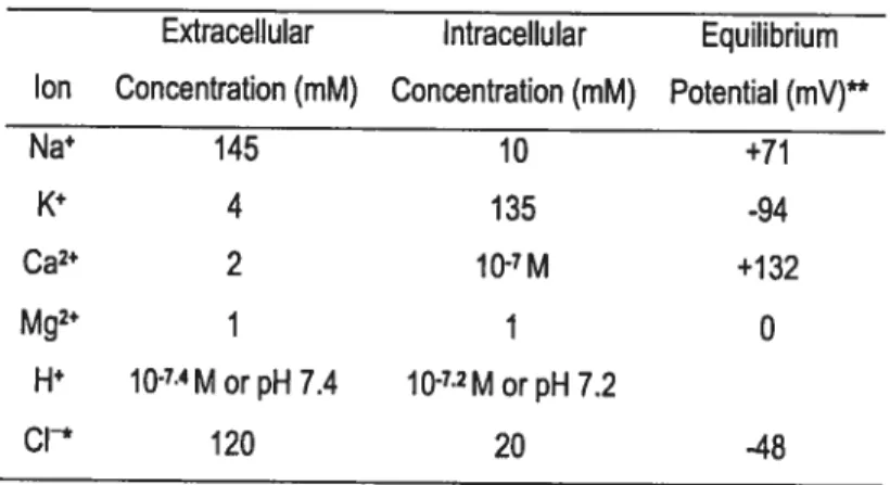
Table 1 Intracellular and Extracellular Ion Concentrations in Cardiocytes 31
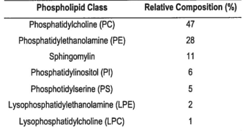
Table 2 Phospholipid Composition of Myocardial Sarcolemma 40
CHAPTER
II
II-1
Figure 1 Enhancement of HERG currents by 1-Palmitoyl- 128
Lysophosphatidylcholine: activation features.
Figure 2 Inactivation property of 1 -Palmitoyl-Lysophosphatidylcholine on 129 HERG currents.
II-2
Figure 1 Structures ofLPCs, LPG-16 and PIP2. 142
Figure 2 Cunent density-voltage relationships ofIHERG showing the effects of 144
various lysophospholipids on‘HERGat various potentials tested.
Figure 3 Time course of IHERG before and afier application of LPC-16 or 145
LPG-16 and after washout ofthe dnigs.
Figure 4 Effects ofPKC on LPC-16- or LPG-16-induced‘HERGenhancement. 147
Figure 5 Effects ofPIP2 on LPC-16- or LPG-16-induced‘HERG enhancement. 149
Figure 6 Effects of antioxidant VitE on LPC-16- or LPG-16-induced ‘FIERa 150 enhancement.
Figure 7 Normalized I—V relationships showing the negative shifi of I—V 152 curve by LPG-16 but not by other lysophospholipids.
Figure $ Percent changes of ‘HERG produced by various lysophospholipids 153
over control as a fimction of depolarizing voltages.
Figure 9 Effects of various lysophospholipids on the steady-state voltage- 154 dependent activation curves.
Figure 10 Effects of various lys ophospholipids on the steady-state voltage- 156 dependent inactivation curves.
II-3
Figure 1 Increases in extracellular K concentration during global ischemia 182 and potential role of 11Cr.
figure 2 [K]0—î induced by lysophosphatidylcholine (LPC-16). 183
figure 3 Shortening of QT interval induced by global ischemia or by LPC- 16. 184 figure 4 Shortening of heart rate-corrected QT interval during global 185
iscliemia and potential roleofIy.
Figure 5 Shortening of Qic interval induced by LPC- 16 and potential role of 187
‘1Cr.
Figure 6 Arrhythmias induced by ischemia or LPC-16 and the effects of 188 IICr/HERG blocker dofetilide.
Figure 7 Shortening of action potential duration induced by LPC-16 and 189 potential role ofIy.
Figure 8 Enhancement of HERG current by LPC- 16 in HERG-expressing 191 HEK293 ceils.
f igure 9 Enhancement of native Iy in left ventricular endocardial myocytes 192 isolated from guinea pigs, and cloned ‘HERG in HEK293 cells, by
LPC-16.
CHÀPTER III
Figure 1 Analog data showing ifie effects of membrane permeable ceramide on 214 HERG current expressed in HEK293 celis.
f igure 2 Characterization of IHERG with prolonged exposure to ceramide. 216 Figure 3 Characterization of ‘HERO depression caused by sphingomyelinase 217
(SMase).
f igure 4 Effects of inhibitors of tyrosine protein kinases (TPKs) and atypical 219 protein kinase C (PKC) on IHERG modulation by ceramide (C2).
Figure 5 Effects of inhibitors of protein kinase A (PKA) or protein kinase C 220 (PKC) on‘HERGmodulation by ceramide (C2).
f igure 6 Expression level of HERG protein determined by immunoblotting 222 with membrane protein preparations extracted from HERG expressing HEK293 cells.
f igure 7 Role of reactive oxygen species (ROS) on IHERG modulation by 223 ceramide (C2).
Figure 8 Effects of vitamin E (VitE) or MnTBAP (an SOD mimic) on 225 intracellular levels of ROS measured by CM-H2DCFDA fluorescence dye.
CHAPTER IV
Figure 1 Impairment ofHERG functions by TNf-Œ. 238
PART I
INTRODUCTION
AND
THE REVIEW
CHAPTER I
Electrical and Metabolic Disturbances Underlying
Iscliemic Arrliytlimias in Hearts
The overall purpose of this chapter is to review the updated experimental findings and notions that have shed light on the mechanisms of electrophysiological and metabolic dismrbances during myocardial ischemia and iscliemic arrhythmias. The specific goals are (1) to set a theoretical foundation for forming my project hypothesis; (2) to raise the questions, which are related to the research project; and (3) to propose the hypothesis and the objectives of the project.
I-1 Perspective: Ischemic Heart Disease—A Global Health Problem
Although a substantial reduction in death rate from cardiovascular causes dunng the past 50 years, ischemic heart disease (IHD) remains the leading cause of morbidity and mortality in the industrialized world. In the United States, IHD afflicts in excess of 6 million Americans annually (American Heart Association, 2004), it causes about 152,000 deaths per year in UK, and world-wide, one in eight deaths is attributed to 1H]) (Ghuran & Camm, 2001). Retrospectively, data from clinical electrophysiological studies and randomized trials have revealed that the majority of IHD deaths are caused by ischemic anhythmias, which appear as the most common pathophysiological cascade involved in lethal arrhythmias degenerating first to ventricular tachycardia (VI), and then to ventricular fibrillation (Vf) and later to asystole and sudden cardiac death (SCD) (Mehta
et al., 1997; Myerburg & Castellanos, 1997; Zipes & Wellens, 1998).
Furthermore, SCD rates also parallel the rates of IHD as a whole in less-developed countries (Huikuri et al., 2001; Zipes & Wellens, 1998). It has been predicted that between 1990 and 2020, mortality from IHD in developing countries is expected to increase by 120% for women and 137% for men. Predictions for the next 2 decades include a near tripling of IHD mortality in Latin America, the Middle East, and sub Saharan Africa (Leeder, 2004). Not only is acute myocardial ischemia (AMI) the most common factor triggering VI, thereafier SCD (Davies, 1992), but the risk of SCD in the population who have had chronic myocardial ischemia (CMI) is four-folds higher than the normal population as well (Abildstrom et al., 2002). Therefore, great efforts have been devoting to understand the mechanisms underlying ischemic anhythmias, and to set the ways for effective treatment, and ultimate prevention of IHD.
Myocardial ischemia (MI) is a condition in which normal myocardial perfusion is arrested, leading to cascades of metabo lic and electrophysiological alterations, which are interrelated and are caused by metabolic stress. The resuits are failure of contraction, deterioration of electrical behavior, and eventual death of the myocytes. At the organism level, the end point may be lethal anhythmias or mechanical pump failure. Despite a remarkable progress of understanding of the pathophysiological and biochemical aspects of MI, our knowledge of which patients with MI will develop sustained VI remains unclear, and the mechanisms underlying the fatal cardiac arrhythmias triggered by MI are far from elucidated. Apparently, more studies, especially the integrated investigations combining in vitro electrophysiological and molecular biological studies and in vivo electrophysiological and biochemical investigations are needed to c1arif,’ the mechanisms so as to interfere with the pathological process of MI and to open the novel avenue in the treatment and prevention ofIHD.
I-2 General Features of Ischemic Arrhythmias
Temporally, ischemic arrhythmias can be categorized into three phases based on the time when they appear: phase I arrhythmias occurring during the first 30 min of ischemia; phase II seen between 5 to 72 hours and phase III arrhythmias of the chronic stage after an infarct. Phase I can be further subdivided in la (between 2 and 10 min of ischemia, while the first burst of VT normally occurs) and lb types (between 15 and 30 mm), with the lb type especially dangerous because they frequently evolve in Vf and SCD in human (Carmeliet, 1999; Pogwizd & Corr, 1987).
VT is the most pronounced arrhythmia in MI. It is defined as three or more consecutive beats arising below the atrioventricular node (AVN) with an RR interval of less than 500 ms (>120 beats/min), being frequently diagnosed with the advert of intensive care unit (ICU) monitoring and long-term ambulatoiy electrocardiogram (ECG) recordings (Akai et aÏ., 2000). VI may be classified by morphological criteria (monomorphic, polymorphic, bundle branch block pattem and axis), by duration (sustained or nonsustained) or by the underlying mechanisms (enhanced automaticity, triggered activity or re-entiy). Clinically, MI has been occasionally found to precede the
onset of a monomorphic VI. More frequently, however, AMI triggers the occurrence of polymorphic VT that may predispose to VF rather than a sustained monomorphic VT.
VI followed by Vf is the lethal arrhythmia that disposes patients who suffer from MI to sudden death. Ihe earlier and more frequently obseiwed abnormalities in ECG recording are the alterations in QT interval, and the changes of SI segment as well. Very intriguingly, ischemic myocardium demonstrates characteristic sequential alterations in electrophysiology with early QT inten’al shortening (QT-1’) in acute ischemia and subsequent QI interval lengthening (QT-t) after a prolonged ischemic period, which are associated with different types of arrhythmias (Boyden & Jeck, 1995; Carmeliet, 1999; Singer et aï., 1981). For instance, in phase I, especially phase la of AMI, QI-J- lias been frequently observed in a wide variety of animal modals and in human (Friedman et al., 1973; Lazzara et aï., 1974; Lazzara et aL, 1978). In coincidence with the QI-.L, the most profound electrophysiological disorder inthisphase of MI may be extracellular potassium accumulation ([K110-t). In contrast to the QT-L’ in early phase of AMI, QI-t lias been found to be a universal change in the CMI (Eick et al., 1976; Lazzara et al., 1975; Wit & Friedman, 1975). Either AMI or CMI, at the cellular and multicellular levels, the electrophysiological disturbances are characterized as depolarization, prolongation of the effective refractoiy period (ERP), decrease in conduction velocity, and change in excitability. Ihe basis of the electrophysiological disturbance is a group of ions and exchangers, which interplay to set the mechanisms of the electrophysiological disturbances in ischemic myocardium.
I-3 Our Current Understanding of the Potential Ionic Mechanisms for Ischemic Arrhythmias
Cardiac myocytes make electrical continuity as a tissue through low-resistance intercellular gap junction. Each individual cardiocyte is like a small battery with the voltage of -50 to -90 mV across its sarcolemmal membrane (measured from the intracellular space with respect to the bathing solution ground). Depolarization is defined
as a change in membrane potential (Em) to more positive voltage, which is mediated by
+ 2+
channels or transporters that enable positively charged ions such as Na or Ca to enter the cell (inward currents) (van Ginneken & Giles, 1991); whereas repolarization refers
to any change to more negative voltage, which is rnainly rnediated via a variety of K channels allowing K movement from the intracellular to extracellular side (outvard ct,rrents) (Marban, 2002; Nattet, 2002). The heart beat is initiated by slow diastolic depolarization of the sinoatrial node (SAN), which drives the membrane potential after action potential (AP) toward the threshold for firing the next AP. Multiple ionic currents with complex interactions are invoÏved in spontaneous diastolic depolarization (Schram et
ai, 2002; Boyett et aï., 2000).
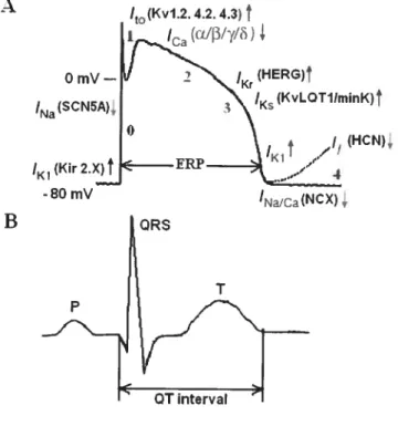
Rapid voltage changes of the mass of the heart are detected as miÏtivolt-sized changes in the surface ECG. Figure I depicts the temporal relationships between the electrical activity ofa typical ventricular rnyocytes, as measured using cellular recordings ofthe transmembrane AP (paner A), and the corresponding ECG (panel B).
À 4.2. 4.3)
t
(i’AS) O mV—J
Kr fHERG)t ‘Na(SCN5A),_____ _••______ ‘NaJCatNCX), BFigure 1. The temporal relationships between the ventricular AP and the corresponding surface ECG. A, a typical ventricular AP with inward current (downward arrows) and outward currents (upward arrows). The molecular basis of each current is indicated in parentheses. Numbers indicate the
phases of the AP. B, a schematic ECG. QRS complex, P wave, T wave and QI interval are indicated.
I< (Kir 2.X)f -80 mV
ORS
P
The upstroke of the AP at the onset of depolarization produces the spiky ‘QRS complex’; repolarization is manifested as the gently rolling T wave. As a first approximation, the time between the beginning of the QRS complex and the end of the T wave the ‘QT inten’al’ — can be used to deduce the overail timing and duration of
ventricular depolarization and repolarization. The frequency of QRS complexes and their sequence relative to the smaller P waves produced by atrial activity allow the clinical detection of normal rhythmor arrhythmias.
I-3-1 Properties of Ionic Channels Underlying Cardiac Action Potentials
Over the last 50 years patch-clamp techniques combined with molecular biology have revolutionized the study of ion chaimels underlying cardiac AP. The AP is virtually an electrical signal integrating with a variety of inward and outward cttrrents, which result from the orchestral opening or closing of number of ion chaimels and transporters in the sarcolemma membrane. TransmembraneAPs form the cellular basis for pacemaker activity, impulse spread, and control of cardiac excitation-contraction coupling (Splawski et aÏ., 2002). APs that originate in the SAN (Lacinova, 2004) and transmit sequentially throughout the atria, atnoventricular node (AVN), His-Purkinje system, and the ventricles, are a summation of precisely orchestrated openings and closings of distinct populations of ion channels. Perturbations in ion channel function andlor structural malformations in heart underlie various cardiac arrhythmias. Moreover, aberrant ion channel function underlies a wide range of diseases in both man and other species (Ashcroft, 2000). Strategic molecular targeting of these proteins will therefore yield novel forms of antianhythmic therapy (Ackerman & Clapham, 1997).
Traditionally, the AP can be divided intofive phases. In the resting ventricular rnyocyte (membrane potential around -90 mV), voltage-gated sodium channels are closed. A propagating impulse causes the membrane potential to become less negative, therefore the sodium channels are opened, and the resulting large rapid inward Na flux (‘Na)
produces phase O depolarization. The initial phase 1 repolanzation results from the opening of the transient outward potassium channels (Ito). During this phase, calcium channels also open, and the longphase 2 plateau of the AP corresponding to ventricular systole (Sorrentino & Reggiani, 1999) reflects a balance between inward calcium current
(JCa) and outward potassium curreni through delayed rectifier channels (IK-, 1, and 1r in
atrial myocytes). Inactivation of calcium channels in phase 3, together with ongoing outward K flux through delayed rectifier and inward rectifier channels (IKI) completes repolarization to resting phase 4. Phase 4 corresponds to ventricutar fihling during diastote (relaxation) and is rnaintained by the open ‘K. In pacemaker myocytes including
SAN, AV node celis, and Purkinje fibers (Knopf et aÏ., 1990), the ceils begin a spontaneously slow depolarization forming pacemaker current (Ii) rather than rernain at the resting potential. This is also referred as phase 4 (Coraboeuf & Nargeot, 1993; ten Eick et aÏ., 1992).
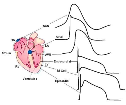
AP configuration and duration vary in specific regions (e.g., atrium versus
ventricle) as well as in specific areas within those regions (figure 2).
Atrium RA
Ventdces
Figure 2. Action potential waveforms are variable in different regions of the heart. Schematic representation of the heart; action potential waveforms recorded in different regions of the heart are illustrated. Action potentials are displaced in time to reflect the temporal sequence of propagation through the heart. [Modified from (Nattel & Li, 2000; Pinto & Boyden, 1999)].
Such physiologic heterogeneities likely reflect variations in expression or function of the repertoire of ion channels and other proteins that constitute cardiac ion currents (Schram et al., 2002). Exaggeration of these heterogeneities, by changes in rate, ion channel mutations, andlor drug exposures, promotes reentrant excitation, a common mechanism for many cardiac arrhythmias. The acute electrophysiological response of a myocyte to exogenous stressors such as myocardial ischemia, autonomic activation, or drugs likeiy reflects changes in fiinction and gating ldnetics of individual ion channels, including channels activated by specific stimuli such as adenosine triphosphate (ATP) depletion, muscarinic stimulation, or stretch.
I-3-1-1 Sodium (Na) Channels
In the heart, voltage-gated Na channels determine the amplitude and siope of the AP upstroke, which are especialiy important in the control of impulse conduction velocity, and in the maintenance of appropriate waves of excitation through the working myocardium. Dysfunction of these channeis can iead to at least 6 types of cardiac rhythm disorders: long QT syndrome (LQTS) (Wang et al., 1995b; Wang et al., 1995a; Wang et al., 1996; Wang et al., 1998a), Brugada syndrome (Brugada & Brugada, 1992; Antzelevitch et al., 2002), idiopathic ventricular fibrillation (Bezzina et al., 2001; Akai et aÏ., 2000), nonfamilial arrhythmia susceptibiiity (Splawski et al., 2002), conduction system disease (Schott et al., 1999), and sick sinus syndrome (Benson et ai., 2003). These conditions are characterized by ventricular tachyanhythmia, heart block, or atrial bradyarrhythmia. Very recently, Na channels have been identified to be associated with atrial fibrillation and heart failure (Oison et aÏ., 2005; Chen et al., 2003b). Ah those disorders can be traced to the abnormal molecular structure and expression of Na channels.
Na channels are believed to consist of a principal pore-forming Œ-subunit (260 kDa) and two auxiliary J3-subunits (36 and 33 kDa each) (Fozzard & Hanck, 1996). In mammals, the genes encoding Na1.1, 1.2, 1.3, and 1.7 (human genes $CN1A, SCN2A, SCN3A and SCN9A) are clustered on human chromosome 2q and appear to have arisen as duplications of a founder gene (Lopreato et al., 2001). There is a second cluster of three genes on human chromosome 3p, ah of which encode tetrodotoxin (Satin et al.,
1992)-resistant charmels. One of these, SCN5A (also designated as hHl) encoding NavI .5, is expressed in heart and brain (Head & Gardiner, 2003).
The Œ-subunit bas four structurally similar domains (DI-DIV); each domain has six helical transmembrane segments (S1-S6). The S4 segments of each domain contain a large number of positive charged residues serving as voltage sensor for channel activation and coupled-inactivation (Kontis et al., 1997). The short intracellular segment between DIII and DIV has been identified as the fast inactivation gate. The charme] pore is formed by four P-loop regions (P region) linldng $5 and $6 segment in each domain. The P region is deduced to be composed of twenty-residues forming the lining of the outer vestibule responsible for ion selectivity and toxin affinity. The more detailed investigations reveal that the primary structure in each P-loop of Na channels is unique, which differs from Ca2 channels in which each P-loop is the same, therefore rendering each of them to different function. for instance, a lysine in the P region of DII critically selects for Na over Ca2 (Heinemann et al., 1992); a cysteine in the P region of DI renders the cardiac channel insensitive to blockade by tetrodotoxin (TTX) or saxitoxin (STX), but sensitive to Cd2 or Zn2 (Satin et al., 1992).
The (3-subunits consist of a single transmembrane domain, a small intracellular C-terminal region and a large extraceflular N-C-terminal domain with an immunoglobulin-like fold (Stevens et al., 2001). Three different isoforms of auxiliary (3-subunits are expressed in cardiac tissue from a range of species including rodents, sheep and human (Dhar et al., 2001; Fahmi et al., 2001; Qu et al., 1995) and are known to modulate the kinetics ofthe cardiac sodium channel Œ-subunit (Kupershmidt et al., 1998), as well as to regulate the expression of sodium channel a-subunits in the heart (Dhar et al., 2001), and therefore attribute to the electrical properties ofwhole channels.
I-3-1-2 Calcium (Ca2) Channels
Voltage-dependent calcium channels (VGCCs) in cardiac myocytes are essential for regulating the influx of Ca2 ions across the sarcolemmal membranes, in response for the upstroke of the AP in the SAN and AVN and play an important role in determining the plateau and eventual spike-dome appearance of the AP in other cardiac cells. Moreover, the Ca2 influx behalves as the electrical signaling player involving in
initiating intracellular events such as Ca2 induced Ca2trelease (CICR) from sarcoplasmic reticulum (SR) (Singer et al., 1981), excitation-contraction (E-C) coupling, and modulation of gene expression. Intracellular concentration of Ca2 ([Ca2j) also modulates the conductance of Ca2tactivated K and CF currents (Wehrens & Marks, 2004). The blockade of Ca2 entry through the cardiac VGCC represents one of the principal approaches for the treatment of IHD.
On the basis of electrophysiological and pharmacological characteristics, VGCCs have been classified into T-, L-, N-, P-,
Q-,
and R-types (Yunker, 2003). In the sarcolemmal membrane of heart celis two types of Ca2-permeable channels have been described and differentiated: the L-type (“long lasting” and “large”) and T-type (“transient” and “tiny”) channels and a background channel (McDonald et al., 1994). The heterogeneity of the T- and L-type channel density in different region of the heart has been long recognized and described (Bourinet et al., 2004; Yunker & McEnery, 2003).VGCCs are heteromultimeric protein complexes. The three-dimensional structure of the bovine cardiac L-type calcium channel has recently been resolved (van der Heyden et al., 2005;Wang et al., 2004c). The largest subunit (-490—240 kDa) is the pore-forming
ai subunit, which is associated with an intracellularly located
f3
subunit (-55 kDa) and amostly extracellularly located disulfide-linked Œ26 subunit (-170 kDa). Several Œi
subunits have been identified to date (alA-alI, aiS, now termed Cavl.l- Cav3.3), and the aic isoform is the one that is expressed at high levels in cardiac muscle, but also in smooth muscle and in the brain (Catterall, 2000; Keef et al., 2001; Jiang et al., 2000a; Striessnig, 1999).
The ŒIC subunit interacting with accessoiy subunits and especially the
f3
subunit isrequired to form fully functional Ca2 channels andJor to alter certain channel properties. Accessoiy subunits determine the activation and inactivation kinetics of the channels. The
f3
subunit also controls targeting of the ŒiC subunit to the membrane. A highly conserved1 8-amino acid sequence in the cytoplasmic loop connecting DI and DII has been identified as the interaction domain oftheaic subunit for the
f3
subunit. The a26 complex,which is less tightly associated with the Œ1C subunit, consists of an extracellularly located
a2 subunit linked to a hydrophobic membrane-spanning 6 subunit. The a2 subunit and 6 subunit are encoded by a single gene. The mature forms ofthese subunits are derived by
post-translational proteolytic processing, but they remain associated through a disulfide bond. The extracellular Œ2 subunit interacts with the $5—S6 linker in DIII of the cxiC
subunit (Norman & Leach, 1994; Striessnig, 1999).
I-3-1-3 Pacemaker Channels (f-Channels)
In 1979, a current named as fuimy current (Ii-, f for “funny” because of its peculiar features) was recorded from small voltage-clamped SAN preparations (Brown et al., 1979). I was inwardly activated on hyperpolarization in the appropriate (diastolic) range ofpotentials, and increased by adrenaline. Following the original report, the I current was described in several cardiac and noncardiac ceils, but the most detailed analysis was conducted in SAN celis. I plays an important roles both in initiating normal rhythmic activity and in mediating the responses to adrenergic and muscarinic neurotransmitters (Baruscotti & DiFrancesco, 2004; DiFrancesco, 1995; Difrancesco & Tromba, 198$).
Unlike the majority of other voltage-gated channels in which the permeability is normally selective for a single ion species, f-channels have a mixed permeability to both Na and K cations (Difrancesco, 1981). The voltage range of current activation is highly variable and depends on structural and modulatory factors. In SAN myocytes, the I is time- and voltage dependent inward cunent upon hyperpolarization, activation threshold can be as positive as -40/-45 mV and being fully activated at -100 mV, and the V112for activation is --80mV, which ensures the current contribution within the whole of the pacemaker phase (Cerbai et al., 1999; Difrancesco & Mangoni, 1994; van Ginneken & Giles, 1991).
Despite the importance of f-channels to functional properties of cardiac myocytes, its cloning was only achieved nearly three decades afier its original description and was accomplished by chance (Santoro et al., 1997). The newly identified sequence bore the hallmarks of a voltage-dependent Ktpermeable ion channel: six putative transmembrane domains and an S4-charged Œ-helix, plus the expected cyclic nucleotide binding domains at the intracellular C-terminus. Following this first sequence, other isoforms were identified (Gauss etal., 1998; Ishii etal., 1999; Ludwig et aï., 199$; Seifert et al., 1999), this group of channels, known as the hyperpolarization-activated cyclic nucleotide-gated family (Pachucki et al., 1999), comprises four members (HCNY to HCN4) and belongs to
the superfamily of voltage-gated potassium (Kv) channels, with which they share many similarities. The four isoforms share a homology of -60% in amino acid (Wang et al., 1996) sequence identity and express in heart except for HCN3 that only in brain (Kaupp & Seifert, 2001). The firnctional expression ofthese channelsresulted in currents with the hallmarks of the cardiac I or of its neuronal equivalent (termed ‘h). Although the properties of different HCN isoforms differ quantitatively, ail isoforms yield cunents that
+ + +.
are activated by hyperpolarization, carry K and Na , are blocked by Cs in a voltage
dependent manner, and are modulated by a direct action ofcAMP on the cytoplasmic side ofthe channel (Santoro et aÏ., 1997).
There is surprisingiy littie direct evidence implicating I in either acquired or inherited disease. Perhaps the best-studied case is in cardiac ventricular muscle, where increases in I magnitude or shifis in voltage dependence have been associated with certain cardiovascular diseases such as spontaneous hypertension in rats (Cerbai et aï., 1994), human failing hearts (Cerbai et al., 1997; Cerbai et al., 2001). It was also reported that HCN2 message level varies with thyroid hormone andlor thyroid hormone receptor level, perhaps contributing to the rapid heart rate associated with hyperthyroidism (Gloss et al., 2001; Pachucki et al., 1999). In addition to these acquired diseases, a recent report describes the first known human mutation in an HCN4 gene. The mutation, identified by genotyping a patient with sinus bradycardia and atrial ventricular fibrillation, results in truncation of the terminal portion of the HCN4 C-terminal, including the CNBD, which leads to a pronounced reduction in the magnitude of I ($chulze-Bahr et al., 2003).
I-3-1-4 Potassium (K) Channels
Cardiac K currents (IK) are a large family of a variety of K channels with different characteristics. ‘K can be distinguished on the basis of differences in their functional and pharmacological properties. In mammalian cardiac cells, K channels can be categorized as voltage-gated (Kv) and ligand-gated channels. The first category includes the rap idly activating and inactivating transient outward current (Ito) (Wang et al.,
1999), the ultrarapid-activated delayed rectifier (IKur) (Wang et al., 1993), rapid-activated delayed rectifier (Iy) and slow-activated delayed rectifier (Iy) (Volders et aï., 1999; Wang et al., 1994) and the inward rectifier (‘ici) (Wang et al., 1998b), whereas the ligand
gated channels include those activated by a decrease in the intraceliular concentration of ATP (KATp) (Wilde et al., 1989), activated by acetylcholine (KAh) (Koumi & Wasserstrom, 1994; Yang et aÏ., 1996), Na-activated K cunents (JKNa) (Kameyama et al., 1984; Luk & Carmeliet, 1990; Noda et al., 1984), arachidonic acid-activated K currents (Iyj,jj (Kim & Duff, 1990), and Ca2- Activated K Channels (Ka) (Cao et al., 2005; Xu et al., 2002).
Cardiac K channeis play a pivotai role in defining resting potential, ceil excitability and membrane repolarization and thereby the iikeiihood of arrhythmias. The configuration and duration of the cardiac APs vary considerabiy among species and different cardiac regions (atria vs. ventricle) and specific areas within those regions (epicardium vs. endocardium) (Figure 2). This heterogeneity mainiy reflects differences in the type andlor expression pattems of the K channeis that participate in the genesis of the cardiac AP. Changes in the expression of K channeis explain the regional variations in the morphology and duration of the cardiac AP among different cardiac regions and are influenced by heart rate, intracellular signaiing pathways, drugs and cardiovascuiar disorders (Coetzee et al., 1999; Nerbonne, 2000; Snyders, 1999). Moreover, the expression and properties of K channels are flot static but are influenced by heart rate, neurohumoral state, phanriacologicai agents, cardiovascular diseases (cardiac hypertrophy and failure, myocardial ischemia and infarction) and arrhythmias.
I-3-1-4-1 Transient Outward K Current (Ito)
Cardiac is responsibie for early rapid repoiarization (Smith et aÏ., 1995) and determines the height of the eariy plateau, thus influencing activation of other cunents that control repolarization, mainiy ‘L-Ca and the delayed rectifier K current (IK).
Furthermore, variations in cardiac repolarization associated with‘to differences strongiy
influence intracellular Ca2 transient by modulating Ca2 entry via ‘L-Ca and
sodiumlcalcium exchange (NCX), potentialiy exacerbating impaired Ca2 cyciing in heart disease (Sah et aÏ., 2003).
It density is 4- to 6-fold higher in atrial tissue, Purkinje fibers, epicardiai and midmyocardiai (M) ceils than in the endocardial ceiis ÇNabauer et al., 1996; Yan & Antzelevitch, 1996). The prominent epicardial ‘to contributes to the selective electncai
depression of the epicardium during ischemia and to the development of a marked dispersion of repolarization between normal and ischemic epicardium and between epicardium and endocardium, thereby providing the substrate for reentrant anhytbmias (Lukas & Antzelevitch, 1993; Yan et aÏ., 2004). CMI and infarction are associated with prolongation of the APD, an effect that lias been attributed, in part, to a downregulation of Ii., (Oudit et aÏ., 2001; Pinto & Boyden, 1999; Tomaselli & Marban, 1999). However, there is also an evidence of the up-regulation of I in cardiac myocytes afier induced myocardial infarction (Pinto & Boyden, 1999; Yao et aÏ., 1999). Paradoxically, using the dynamic clamp technique whicli allows quantitative ‘insertion’ of simulated conductance in real, biological celis, bridging pure computer modeling and experimental electrophysiology, it lias been verified that ‘to does not significantly affect the APD of canine ventricular myocytes, and that the ‘to gradient is not a significant contributor to the transmural APDdispersion in the canine ventricle (Sun & Wang, 2005).
Kv4.3 channels are the leading candidate for encodirig ‘to in human and dog (Kaab et aÏ., 1998; Wang et aÏ., 1999), although a number of Kv Œ-subunits, such as Kvl.4, K3.4, K4.2, and K4.3 known for generating I0-like current when heterologously expressed, have become primaiy candidates for cardiacIt. Potassium channel interacting proteins (KCMP) are Ca2tbinding proteins that act as f3-subunits interacting with the cytoplasmic amino termini ofKv4 Œ-subunits and modify ‘to features (Sanguinetti, 2002). For instance, KChIP2, when coexpressed with hKv4.3, increases surface channel density and cunent amplitude, slows the inactivation, accelerates the recovery from inactivation and shifts the half-maximal inactivation to more positive potentials (An et aÏ., 2000; Pounier et al., 2003a). Thus, feamres of K4.3/KCMP2 currents closely resemble those ofI. In human ventricle KCMP2 mRNA is 25-fold more abundant in the epicardium than in the endocardium, and this gradient parallels the gradient in expression, while K4.3 mRNA is expressed at equal levels across the ventricular wall. Thus, transcriptional regulation of the KCMP2 gene is the primary determinant ofI expression across the ventricular wall (Rosati etaL, 2001).
![Figure 3. Time-courses of [K]0, pH, [Ca2], and plasma LPC during ischemia. A: increase cf [K]0 during ischemia in Langendorff-perfused quiescent and stimulated (4 Hz) rat hearts, measured with K sensitive electrodes inserted in mid left myocardium](https://thumb-eu.123doks.com/thumbv2/123doknet/2054969.5666/63.918.196.757.163.657/ischemia-langendorff-quiescent-stimulated-sensitive-electrodes-inserted-myocardium.webp)