Anti-angiogenic therapy of exudative age-related macular degeneration:
current progress and emerging concepts
Agnès Noël1, Maud Jost1, Vincent Lambert2, Julie Lecomte1 and Jean-Marie Rakic2
1 Laboratory of Tumor and Development Biology, Center for Experimental Cancer Research, Center for Biomedical Integrative
Genoproteomics, University of Liège, B-4000 Liège, Belgium
2 Department of Ophthalmology, CHU, B-4000 Liège, Belgium
Abstract: Age-related macular degeneration (AMD) is the leading cause of blindness in elderly patients. The more aggressive exudative form is characterized by abnormal bloodvessel development that occurs beneath the retina as a result of choroidal neovascularization (CNV). Vascular endothelial growth factor (VEGF) has emerged as the key mediator of CNV formation; this has led to intensive research on VEGF and the recent approval of anti-VEGF compounds by the US Food and Drug Administration. Despite this successful introduction of anti-angiogenic therapies into the clinical setting, there is still a lack of treatments that
definitively reverse damaged vision. Here, we consider the importance of putative molecular targets other than VEGF that might have been underestimated. Emerging cellular mechanisms offer additional opportunities for innovative therapeutic approaches.
Glossary
Bruch's membrane: specialized basement membrane that separates the choroid from retinal pigmented epithelial cells.
Choroid: a thin vascular layer between the sclera and the retina that supplies oxygen and nutrients to the retinal pigment epithelium and the photoreceptors.
Endothelial cells: cells that line the inner lumen of blood vessels.
Extracellular matrix: a three-dimensional network of macromolecules that form a structural scaffold and a signaling environment for cells.
Matrix metalloproteinases (MMPs): a large family of proteolytic enzymes that degrade extracellular-matrix components and control the activity of important mediators of cell functions.
Retinal pigment epithelium (RPE): a monolayer of cells that are specialized to serve the adjacent photoreceptors.
Vascular endothelial growth factors (VEGFs): a family of powerful angiogenic proteins that control growth survival and the migration of endothelial cells.
Age-related macular degeneration
Age-related macular degeneration (AMD) is a leading cause of vision loss in the western world among people aged 50 or older [1]. Ninety per-cent of all vision loss due to AMD results from the exudative form, which is characterized by choroidal neovascularization (CNV), defined as newly formed blood vessels arising from choriocapillaries. The choroid is the vascular layer lying between the retina and the sclera. Exudation of fluid and hemorrhage into the sub-retinal space from hyperpermeable capillaries lead to retinal local detachment and retinal photoreceptor damage, which, in turn, lead to the formation of a disciform scar. The earliest clinically detectable abnormality in AMD, however, is not the consequence of pathological angiogenesis but is the accumulation of abnormal lipopro-teinaceous deposits ('drusen') in the extracellular matrix between the retinal pigment epithelium (RPE) and Bruch's membrane [2].
Etiological research suggests that AMD is a complex disease caused by interactions of multiple gene products and environmental factors. Familial aggregation studies, twin studies and segregation analyses have provided strong evidence for AMD heritability [2]. Among several disease-causing genes, complement factor H (CFH) and HtrA serine peptidase 1 (HTRA1) have recently been identified as major AMD-susceptibility genes [3-5]. Age-related changes that induce pathologic neovascularization are incompletely understood. However, vascular endothelial growth factor (VEGF)-A, a diffusible growth factor that promotes angiogenesis (the sprouting of endothelial cells from preexisting vasculature) and vascular permeability [6], has emerged as a potent angiogenic factor involved in CNV formation. VEGF has, therefore, been the focus of significant research attention, which has led to the development of different strategies aimed at blocking VEGF or VEGF receptors [6] (Table 1). The first anti-VEGF compound, pegaptanib (Macugen®, Eyetech), an aptamer that specifically blocks the 165 amino
acid isoform of VEGF, was approved by the US Food and Drug Administration (FDA) for exudative AMD treatment in 2004 [7]. This achievement validated angiogenesis as an important target for AMD [6].
Administration of bevacizumab (Avas-tin®, Genentech Inc.), an anti-VEGF antibody, appeared to be safe and effective in the short term [8,9]. In 2006, the FDA approved ranibizumab (Lucentis®, Genentech Inc.), an affinity-matured Fab variant derived from bevacizumab (which was already approved for colic cancer treatment) that binds and inhibits all biologically active forms of VEGF-A [9]. Monthly intravitreal administration of ranibizumab arrests the growth of neovascular membrane, prevents severe vision loss in 90% of patients and improves visual acuity in 30% to 40% of patients; it therefore represents a substantial advance against AMD [10] in comparison to previous treatments. Initial pharmacologic CNV treatment consisted of verteporfin
(Visudyne®) photodynamic therapy (PDT), which can slow the progression of vision loss but is usually not associated with a gain in visual acuity [11,12] (Table 1).
Table 1: Current treatments for exudative AMD and examples of trials targeting exudative macular degeneration (also see [69])
Treatment strategies or
drugs
Principle or compound Company Current progress and
limitations
Refs
Argon laser
photocoagulation This earliest and simplest technique relies on the ability to heat and kill target cells locally by focused laser light.
FDA (US Food and Drug Administration) approved. Destroys both CNV and overlying neural cells. Limited, therefore, to extrafoveal neovascularization.
[74]
Photodynamic therapy (PDT)
Photosensitizing agents are delivered intravenously before laser treatment. The resulting local production of free radicals results in localized vascular occlusion.
QLT Inc. and Novartis
FDA and EMEA (European Medicines Agency) approved. Slows the progression of vision loss but does not improve visual acuity.
[75]
Anti-VEGF165
protein strategy Polyethylene glycol (PEG) anti-VEGF aptamer (pegaptanib, marketed as Macugen®).
Eyetech Pharmaceuticals and Pfizer
FDA and EMEA approved. Safe and effective in the short term. No data available regarding long-term effect.
[7]
Anti-VEGF protein
strategy Humanized anti-VEGF monoclonal antibody (bevacizumab, marketed as Avastin®).
Genentech Inc.
and Roche Broad use out of label. NIH-sponsored randomized trial underway.
[76] Anti-VEGF protein
strategy
High affinity anti-VEGF Fab
(ranibizumab, marketed as Lucentis®).
Genentech Inc. and Novartis
FDA and EMEA approved in 2006. Safe and effective in the short term. Improvement of visual acuity. No data available
regarding long-term effect.
[9,10]
AntiVEGF and -PlGF proteins strategy
VEGF-TRAP, a recombinant protein containing the binding domains of VEGF-R1 and -R2.
Regeneron Pharmaceuticals Inc.
Under clinical trial. [77]
Anti-VEGF gene expression strategy VEGF-targeting siRNA (Cand5-bevasiranib). Opko (Acuity Pharmaceuticals Inc.)
Under clinical trial. [37]
Anti-VEGF receptor expression strategy
VEGF-R1-targeting siRNA
(siRNA-027). Merck and Co. Inc. Under clinical trial. [78]
Anti-angiogenic
gene therapy Injection of adenovirus to express high levels of PEDF. GenVec Inc. Under clinical trial. [79] Anti-angiogenic
strategy
Intravenous injection of squalamine (Evizon®).
Genaera Corp. Under clinical trial. [80]
Angiostatic
This successful application of anti-VEGF approaches in the clinic is obviously a turning point in AMD treatment. Nevertheless, despite such important advances, critical issues remain to be addressed. For instance, how do we select patients that are most likely to respond to treatment? Is it possible to decrease the frequency of intravitreal injections by combining anti-angiogenic agents with PDT, conventional laser treatment or another therapy? In addition, because the necessary duration of treatment with anti-VEGF inhibitors is presently unknown, putative long-term local or systemic side effects or progressive appearance of resistance to anti-angiogenic therapy cannot be excluded [6,13]. Furthermore, the current VEGF-centered research might have overlooked other valuable molecular targets. The present review will only focus on choroidal (sub-retinal) angiogenesis, the main feature of exudative AMD. Our main purposes are to summarize recent studies aimed at identifying key molecules involved in CNV and to evaluate issues that need to be addressed before anti-angiogenic approaches to AMD can make further substantial advances.
CNV studies: the need for adequate experimental models
Initial studies on CNV in AMD consisted mainly of mRNA and protein-level analyses of surgically excised human choroidal neovascular membranes in comparison to intact choroids. Animal models represent important tools for elucidating cellular and molecular mechanisms involved in CNV pathogenesis and for the screening of new drugs. At present, the laser-induced Bruch's membrane photocoagulation model is the most widely accepted and most frequently utilized experimental murine CNV model. It relies on laser injury to fracture Bruch's membrane, thus leading to blood-vessel recruitment from the choroid, and it captures many important features of human AMD [2,14]. This murine CNV model offers the possibility of unraveling the molecular mechanisms of CNV by using a set of recently generated transgenic mice. Two models that exhibit the spontaneous
development of drusen, diffuse sub-RPE deposits and CNV have recently been described in senescent animals: knock-in mice with human apoliprotein E-allele maintained under a high-fat diet [15] and superoxide dismutase 1 (SOD1-/-) knockout mice [16]. Although these spontaneous models unravel important physiologic pathways
involved in neural retina degeneration, the present review will mainly consider the earliest AMD models produced by laser injury of Bruch's membrane; a model that is focused on CNV induction and that has proven valuable for testing anti-angiogenic treatments [2].
Inflammation, angiogenesis and vasculogenesis in CNV
CNV is a complex tissue composed of vascular components (endothelial cells, circulating endothelial cells and pericytes) and extravascular cells (inflammatory cells, myofibroblasts, glial cells and RPE) [17]. Inflammatory processes, including immune-complex deposition, complement activation and inflammatory cell infiltration, have been proposed as important mediators of CNV pathogenesis. Inflammatory cells are potent initiators of angiogenesis, in part through their ability to release a set of pro-angiogenic factors. Neutrophils, the most dominant infiltrating leukocyte population in early stages of inflammation, can initiate a complex cascade that leads to a complete inflammatory response [18]. Neutrophil depletion induced by an anti-murine neutrophil-antibody injection reduced CNV formation [19]. Similarly, pharmacological-macrophage depletion reduced the size and leakage of laser-induced CNV and was associated with decreased VEGF production, suggesting a role for these cells as producers or regulators of angiogenic factors [20]. The important contribution of macrophages in CNV formation is further supported by the phenocopy of AMD observed in senescent mice deficient for monocyte chemoattractant protein-1 (Ccl-2, also known as MCP-1) or its chemokine receptor (Ccr-2), which both induce defects in macrophage mobilization [21]. Bone marrow (BM)-transplantation studies revealed that recruitment of blood-derived macrophages, more than resident microglia, seems to be associated with CNV [22]. A fascinating area of research in the field of angiogenesis emerged from the discovery of endothelial progenitor cells (EPCs) in adult BM. It is now more and more accepted that new blood-vessel formation in post-natal events not only relies on angiogenesis, but also on vasculogenesis, the recruitment and differentiation of BM-derived angioblasts into mature endothelial cells. EPCs and hematopoietic progenitor cells (HPCs) are both recruited from BM and contribute to CNV [23,24]. When injected systemically into adult mice, BM-derived cells incorporated into experimentally induced CNV [24]. Engraftment of irradiated C57Bl/6 mice with BM issued from green fluorescent protein (GFP)-transgenic mice led to detection of GFP-positive cells (macrophages, endothelial cells and vascular smooth-muscle cells) in CNV [23]. More recently, hematopoietic stem cells (HSCs) were also shown to differentiate into cells expressing markers specific for astrocytes and RPE [25,26]. Key molecular pathways in CNV
Abnormal vascularization results from shifts in the finely balanced equilibrium between the pro-angiogenic and anti-angiogenic factors that normally regulate angiogenesis. Therefore, initial studies on angiogenesis in CNV
were mainly focused on growth factors such as angiogenic factors and endogenous inhibitors. In addition, genes involved in matrix remodeling and endothelial cell migration, including matrix metalloproteinases (MMPs) and the plasmin system, have been reported as crucial in angiogenesis [27].
VEGF pathway
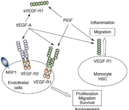
The VEGF family consists of six growth factors (VEGF-A, -B, -C, -D, and -E and placental growth factor, or PlGF) that bind three distinct tyrosine kinase receptors: VEGF-R1 (Flt-1), VEGF-R2 (Flk-1, or KDR) and VEGF-R3 [28]. VEGF-A is a primary stimulator of angiogenesis and is produced by a wide variety of tissues. Alternative exon splicing leads to the production of six VEGF-A isoforms that are characterized by their respective number of amino acids: VEGF121, VEGF145, VEGF165, VEGF165b, VEGF183, VEGF189 and VEGF205 [29]. All VEGF-A splice forms bind to VEGF-R1 and VEGF-R2, whereas PlGF and VEGF-B bind only to VEGF-R1. Most signal transduction through VEGF-A is mediated by VEGF-R2. However, VEGF-R1 displays weak tyrosine kinase activity and is a potent regulator of VEGF-R2 activity [30,31]. VEGF-R3 binds VEGF-C and VEGF-D, which are both regulators of lymphangiogenesis [32]. VEGF-R2 is expressed at the surface of EPCs and is involved in their mobilization during vasculogenesis [33]. PlGF promotes the recruitment and mobilization of HSCs that express VEGF-R1 from marrow to blood circulation [34,35] (Figure 1).
VEGF-A has received considerable attention [29]. Its expression is increased in laser-induced CNV, and blockage of either VEGF-receptor phosphorylation or VEGF expression by small interfering RNA (siRNA) inhibits CNV formation [36,37]. Transcriptional regulation of VEGF-A is mediated by hypoxia-inducible transcription factor 1 (HIF-1) through interaction with hypoxia response element (HRE) in the VEGF promoter. Mice in which HRE is deleted from the VEGF promoter (VEGFδ/δ mice) showed a dramatic reduction of
laser-induced CNV [38].
Figure 1: VEGF pathway. VEGF-R1 and VEGF-R2 are expressed on the cell surface of blood endothelial cells, whereas VEGF-R1 is expressed by monocytes and their progenitors (hematopoietic stem cells indicated by HSC). VEGF-R2 is the major mediator of endothelial-cell proliferation, survival, migration and vascular permeability. VEGF-R1 does not mediate an effective mitogenic signal in endothelial cells and might play a 'decoy' role by sequestering VEGF and preventing its interaction with VEGF-R2. An alternatively spliced soluble form of VEGF-R1 (sVEGF-R1) also displays such a 'decoy' role. VEGF-R1 activation by VEGF-A or PIGF results in monocyte recruitment in injured tissue. Monocytes might, in turn, produce angiogenic factors, and thereby amplify the angiogenic cascade.
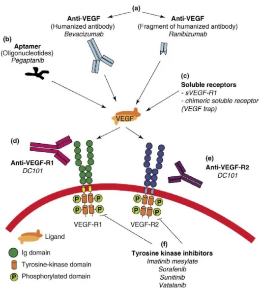
Figure 2: Anti-VEGF strategies currently used. Inhibition of VEGF signaling can be achieved with extracellular or intracellular inhibitors: (a) Monoclonal antibodies that target VEGF-A; (b) Oligonucleotides (aptamers) that bind the heparin-binding domain of VEGF165; (c) Chimeric soluble receptors (VEGF trap, a domain of VEGF-R1 and a domain of VEGF-R2 fused to the Fc fragment of an antibody) or the alternative spliced soluble form of VEGF-R1 (sVEGF-R1, or sflt-1); (d) Monoclonal antibodies that target VEGF-R1; (e) Monoclonal antibodies that target VEGF-R2; (f) Small-molecule VEGF-receptor tyrosine kinase inhibitors that block ligand-dependent receptor autophosphorylation. In addition, antisens and siRNA strategies that target VEGF or VEGF receptors are under development (not shown).
VEGF bioavailability is at least partially regulated by its sequestration in the extracellular matrix and/or interaction with membrane-associated VEGF receptors or truncated soluble VEGF receptors (sVEGFR). VEGF-R1 might serve as a 'decoy' receptor that regulates the amount of free VEGF in extracellular space [39]. In addition, soluble VEGF-R1 (sVEGFR-1, or sflt-1), an alternative spliced, secreted form of VEGF-R1, has the same ligand-binding affinity as the full-length membrane-associated receptor and can function as a trap for secreted VEGF-A. Interestingly, cornea, an avascular tissue, expresses VEGF-A but the presence of this pro-angiogenic molecule is counterbalanced by significant sflt-1 expression, whereas absence of sflt-1 induces corneal neovascularization [40].
Despite VEGF's implications for CNV development, isolated overexpression of VEGF is not sufficient per se to induce sub-retinal neovascularization. Transgenic mice with targeted VEGF surexpression in RPE did not show CNV [41] or display intrachoroidal neovascularization that did not reach the sub-retinal area because Bruch's membrane remained intact [42]. These data underline the importance of Bruch's membrane under normal conditions and the consequences of its degradation during the CNV process. VEGF-A is unlikely to be the unique CNV stimulatory factor. Indeed, other VEGF family members such as VEGF-B, -C, and -D and PlGF have been detected in human choroidal neovascular membranes [43]. PlGF deficiency or PlGF-receptor neutralization (by injection of an anti-Flt-1) both reduced the incidence and severity of laser-induced CNV [44].
PlGF might exert its pro-angiogenic effect through multiple actions, including chemoat-traction of BM-derived cells [34,35,45]. Implication of other angioregulators, such as fibroblast growth factor (FGF), angiopoietins [46] and ephrins [47], is expected but less well documented.
Based on accumulating evidence for a key role of VEGF in pathological angiogenesis, several pharmacologic approaches to inhibiting the VEGF axis have been designed; these have ranged from very specific strategies (such as inhibition of VEGF165 by pegaptanib) to very broad strategies (such as inhibition with tyrosine kinase inhibitors) (Table 1, Figure 2).
Endogenous inhibitors of CNV
Several endogenous physiological inhibitors of angiogenesis have been identified and include fragments of matrix components (endostatin, tumstatin, endorepellin) [48], thrombospondin-1 (TSP-1) [49] and pigment epithelium-derived factor (PEDF) [29]. Expression of endostatin [50], PEDF [51] or TSP-1 [49] is significantly decreased in AMD patients' eyes compared to those of healthy people with no clinical sign. PEDF inhibits endothelial-cell migration in vitro and is much more effective than endostatin and TSP-1. PEDF is considered one of the most powerful anti-angiogenic proteins in humans [29], and adenoviral-mediated intra-ocular delivery of PEDF reduced CNV formation [52]. A critical balance between VEGF and PEDF is likely to play a key role in maintaining the homeostasis of human retinas and in preventing CNV development. However, PEDF effects on CNV are complex. In laser-induced CNV, PEDF caused dose-dependent opposing effects; the effects were inhibitory at low doses but stimulatory at high doses [53], raising evident issues for clinical application.
Proteases, tissue remodeling and cell recruitment
Cell migration and tissue remodeling associated with pathological angiogenesis are regulated by different proteolytic systems, including MMPs and serine proteases of the plasmin system [27]. Urokinase-type (uPA) and tissue-type (tPA) plasminogen activators are serine proteases that are both able to convert plasminogen into active plasmin and whose activities are inhibited by plasmino-gen-activator inhibitor type-1 (PAI-1). Plasmin is a broadly acting enzyme that degrades extracellular-matrix proteins and activates pro-MMPs and growth factors. MMPs constitute a family of at least 24 zinc-dependent, neutral endopeptidases controlled by tissue inhibitors of MMPs (TIMPs) [54]. The growing complexity of MMPs is indicated by the number of substrates and the diversity of their functions in various biological processes. Aside from degrading matrix components, MMPs act as processing enzymes that perform highly selected and limited cleavage of substrates, including growth factors and their receptors, cell-adhesion molecules, cytokines, chemokines, apoptotic ligands and angiogenic factors [54,55].
In choroid and retina, TIMP-3 is synthesized by RPE cells, which are found in Bruch's membrane, and has been reported to inhibit angiogenesis in vivo and in vitro [56]. It is worth noting that mutations in the TIMPS gene have been detected in patients with Sorsby fundus dystrophy, a rare dominantly inherited disease characterized by CNV and sub-retinal hemorrhages [57]. Although the mechanism by which mutations give rise to the disease phenotype is unknown, mutations might render TIMP-3 resistant to degradation and thus lead to its accumulation [57].
Expression of tPA, uPA, uPAR, MMP-2, MMP-9 and their inhibitors (PAI-1 for plasminogen activators and TIMPs for MMPs) has been detected both in choroidal neovascular membranes surgically extracted from patients with exudative AMD and in laser-induced CNV in mice [58,59]. Wild-type control mice and uPAR-deficient mice exhibited a robust choroidal angiogenic response. However, CNV did not develop in uPA-, tPA-,
Plg- or PAI-1-deficient mice, thus demonstrating the contribution of the plasmin system in CNV [27,58].
Paradoxically, PAI-1, through its anti-proteolytic activity, appears to be a key regulator of CNV; it exhibits pro-angiogenic effects at physiological concentrations and anti-pro-angiogenic actions at pharmacological concentrations [60].
Mice deficient for either MMP-2 [61] or MMP-9 [18] had impaired CNV formation. Interestingly, a synergistic effect of MMP-2 and MMP-9 during the CNV process is supported by the reduced CNV observed in mice deficient for both MMP-2- and MMP-9 compared to mice with only one deletion [62]. MMP-9 is likely to participate in the release of matrix-bound VEGF and in the mobilization of BM-derived cells through soluble sKitL release in BM [55,63]. In accordance with the contribution of MMP-2 and MMP-9 to CNV, adenoviral delivery of TIMP-1 or TIMP-2 and synthetic MMP inhibitor inhibited laser-induced CNV [55,62]. However, these inhibitors have not yet been tested in the clinic.
Implications of current progress in CNV therapies
Pathological (tumoral and choroidal) angiogenesis is currently inhibited by anti-angiogenic agents that directly affect endothelial cells by targeting the VEGF pathway. However, despite promising successes in selected populations, targeting only VEGF and endothelial cells might not be sufficient to definitively halt, prevent or even slow down CNV progression in all patients. In addition, because anti-angiogenic treatments are likely to be delivered for an undetermined period of time, the local and systemic safety of these drugs is obviously an important challenge. Although we have not discussed here the non-angiogenic properties of VEGF, recent experimental data suggest that VEGF is a critical survival factor for retinal neurons [64]. Studies and clinical trials in cancer have also raised critical questions about how anti-angiogenic agents should be used [65]. Experience gained in these trials suggests the development of resistance against anti-angiogenic treatments [13]. Does this also hold true for CNV treatment, and can we prevent it? (Box 1).
Inhibition of a single molecular target such as VEGF can lead to upregulation of one or several angiogenic factors [13,65,66]. For instance, it is worth noting that PlGF, a potent CNV modulator [44], is upregulated after anti-VEGF therapy in cancer patients [65]. In the eye, upregulation of VEGF expression has been demonstrated after photodynamic therapy [67]. What will be the impact of long-term angiogenesis inhibition in CNV? Anti-VEGF strategy is focused on endothelial cells, but BM-derived cells can be recruited independently of Anti-VEGF, at least through PlGF action [34,35], and still participate in CNV formation. Therefore, redundance or functional overlapping between several angiogenic factors and recruitment of BM-derived cells are mechanisms that could account for resistance acquisition in long-term treatment. Strategies for delaying, minimizing or avoiding resistance to angiogenic treatment should be urgently explored so that the promising benefit of anti-angiogenic therapies in CNV can be increased.
Box 1: Outstanding questions
• What will be the impact of long-term inhibition of VEGF in AMD? Will this lead to local or systemic side effects or the acquisition of resistance, as in cancer therapy?
• What is the best way to inhibit the VEGF pathway in exudative AMD?
• Which other angiogenic molecules should be targeted, and should they be targeted alone or in combination with VEGF?
• Is there interest in using BM-derived cells in AMD, either as an anti-angiogenic tool or as a method to reconstitute the damaged tissues?
One of the challenges for the next decade is developing novel anti-angiogenic compounds (Box 1). In addition to VEGF, PlGF appears to be a key player in CNV in the laser-induced murine model [44]. PlGF and its receptor VEGF-R1 have been largely underestimated as possible targets for treating angiogenesis-driven diseases, but they gained significant attention because of their implication in pathological angiogenesis and inflammation [39]. PlGF differs from VEGF because it is implicated only in pathological angiogenesis and not in healthy conditions [45,68], and this suggests an attractive safety profile for anti-PlGF or anti-VEGF-R1 drugs. Other attractive targets might include other growth factors, such as FGFs, platelet-derived growth factor (PDGF) and proteases (MMPs and serine proteases) [69].
Another important issue is the optimization of combinatorial treatment. Increased treatment efficacy is likely to be achieved through combined use of anti-angiogenic agents with different complementary mechanisms of action that target different angiogenic factors (VEGF, PlGF, PDGF, FGF, proteases) and/or through the targeting of not only endothelial cells but also other cellular actors, such as inflammatory cells and perivascular cells. Different combination approaches are being considered [17]. Verteporfin-based PTD combined with pegaptanib or rani-bizumab illustrates this concept [70]. Other possible combinations include the use of anti-VEGF with, for instance, protease inhibitors [62], angiostatic steroids such as anec-ortave acetate [71] or more classical anti-inflammatory corticosteroids such as triamcinolone [72].
The demonstration that BM-derived cells are recruited to specific sites of neovascularization paves the way for novel innovative approaches. Homing and adhesion of BM-derived cells to CNV could be targeted to prevent CNV. Blocking homing signals (through the antibody to stromal-derived factor-1 (SDF-1)) and endothelial-specific attachment factor (through the antibody to CD 144) has provided a proof of concept [73]. An alternative novel option for therapeutic intervention is a 'Trojan horse approach' that uses engineered BM-derived cells as a delivery vehicle for anti-angiogenic agents. It should be pointed out that current drugs are artificially targeted to
the retina by intravitreal injection, which is sometimes associated with serious side effects such as
endophthalmitis [7]. Therefore, genetic manipulation of BM-derived cells might open new avenues for the specific delivery of anti-angiogenic molecules to abnormal vascular sites. It is, however, necessary to define the better subset(s) of BM precursors or stem cells that can be used as targeted gene vehicles and to identify the cellular and molecular mechanisms involved in their recruitment to specific neovascular sites.
Concluding remarks
Anti-VEGF therapies have shown efficacy in the stabilization or improvement of visual acuity. Nevertheless, although angiogenesis inhibition is likely to change the face of ophthalmology in the next decade, two major challenges remain in the immediate future. The first challenge will be identifying novel molecular targets for developing new drugs. In this context, PlGF, FGF, PDGF and the proteases should be carefully studied. Achieving this might facilitate the optimization of different treatment protocols through a combination of different therapeutic strategies that target angiogenic molecules and/or different cell types involved in the pathological process. The second challenge in the near future will be investigating, in detail, the role of BM-derived cells in CNV. We need to critically evaluate whether a subset of these cells could be targeted to impair CNV, to deliver anti-angiogenic agents or, alternatively, to repopulate the damaged RPE and the neural retina. Therefore, the rational combination of therapeutic strategies will rely on (i) a further understanding of molecular and cellular mechanisms underlying CNV formation, (ii) the precise identification of the mechanisms and effects of each therapeutic agent developed and (iii) the determination of how different approaches can be combined to reduce putative side effects and increase efficacy.
Acknowledgements
This work was supported by grants from the European Union Framework Programme 6 projects, the Fonds de la Recherche Scientifique Médicale, the Fonds National de la Recherche Scientifique (F.N.R.S., Belgium), the Fondation contre le Cancer, the Commissariat General aux Relations International de la Communauté française de Belgique (CGRI)-Fonds National de la Recherche Scientifique (FNRS)-Institut National de la Santé et de la Recherche Médicale (INSERM) Coopération, the Fonds spéciaux de la Recherche (University of Liège), the Centre Anticancéreux près l'Université de Liège, the Fonds Léon Fredericq (University of Liège), the DGTRE from the 'Région Wallonne', the Interuniversity Attraction Poles Programme - Belgian Science Policy (Brussels, Belgium) and from 'Les Amis des Aveugles', Ghlin.
References
1 Bressler, N.M. (2004) Age-related macular degeneration is the leading cause of blindness.... J. Am. Med. Assoc. 291, 1900-1901
2 Rattner, A. et al. (2006) Macular degeneration: recent advances and therapeutic opportunities. Nat. Rev. Neurosci. 7, 860-872
3 Haddad, S. et al. (2006) The genetics of age-related macular degeneration: A review of progress to date. Surυ. Ophthalmol. 51, 316-363
4 Haines, J.L. et al. (2005) Complement factor H variant increases the risk of age-related macular degeneration. Science 308, 419-421
5 Dewan, A. et al. Two genetic pathways for age-related macular degeneration. Curr. Opin. Genet. Deυ. (in press)
6 Ferrara, N. et al. (2005) Angiogenesis as a therapeutic target. Nature 438, 967-974
7 Gragoudas, E.S. et al. (2004) Pegaptanib for neovascular age-related macular degeneration. N Engl. J. Med. 351, 2805-2816
8 Bashshur, Z.F. et al. (2006) Intravitreal bevacizumab for the management of choroidal neovascularization in age-related macular degeneration. Am. J. Ophthalmol. 142, 1-9
9 Steinbrook, R. (2006) The price of sight-ranibizumab, bevacizumab, and the treatment of macular degeneration. N. Engl. J. Med. 355,1409-1412
10 Rosenfeld, P.J. et al. (2006) Ranibizumab for neovascular age-related macular degeneration. N. Engl. J. Med. 355, 1419-1431
11 Bressler, N.M. (2001) Photodynamic therapy of subfoveal choroidal neovascularization in age-related macular degeneration with verteporfin: two-year results of 2 randomized clinical trials-tap report 2. Arch. Ophthalmol. 119, 198-207
13 Verheul, H.M. et al. (2007) Possible molecular mechanisms involved in the toxicity of angiogenesis inhibition. Nat. Rev. Cancer 7, 475-485
14 Tobe, T. et al. (1998) Targeted disruption of the FGF2 gene does not prevent choroidal neovascularization in a murine model. Am. J.
Pathol. 153, 1641-1646
15 Malek, G. et al. (2005) Apolipoprotein E allele-dependent pathogenesis: A model for age-related retinal degeneration. Proc. Natl. Acad.
Sci. U. S. A. 102, 11900-11905
16 Imamura, Y. et al. (2006) Drusen, choroidal neovascularization, and retinal pigment epithelium dysfunction in SOD1-deficient mice: A model of age-related macular degeneration. Proc. Natl. Acad. Sci. U. S. A. 103, 11282-11287
17 Spaide, R.F. (2006) Rationale for combination therapies for choroidal neovascularization. Am. J. Ophthalmol. 141, 149-156
18 Lambert, V. et al. (2002) Matrix metalloproteinase-9 contributes to choroidal neovascularization. Am. J. Pathol. 161, 1247-1253
19 Zhou, J. et al. (2005) Neutrophils promote experimental choroidal neovascularization. Mol. Vis. 11, 414-424
20 Espinosa-Heidmann, D.G. et al. (2003) Macrophage depletion diminishes lesion size and severity in experimental choroidal Neovascularization. Invest. Ophthalmol. Vis. Sci. 44, 3586-3592
21 Ambati, J. et al. (2003) An animal model of age-related macular degeneration in senescent Ccl-2- or Ccr-2-deficient mice. Nat. Med. 9, 1390-1397
22 Caicedo, A. et al. (2005) Blood-derived macrophages infiltrate the retina and activate Muller glial cells under experimental choroidal neovascularization. Exp. Eye Res. 81, 38-47
23 Espinosa-Heidmann, D.G. et al. (2003) Bone marrow-derived progenitor cells contribute to experimental choroidal neovascularization.
Invest. Ophthalmol. Vis. Sci. 44, 4914-4919
24 Sengupta, N. et al. (2003) The role of adult bone marrow-derived stem cells in choroidal neovascularization. Invest. Ophthalmol. Vis. Sci. 44, 4908-4913
25 Chan-Ling, T. et al. (2006) Hematopoietic stem cells provide repair functions after laser-induced Bruch's membrane rupture model of choroidal neovascularization. Am. J. Pathol. 168, 1031-1044
26 Harris, J.R. et al. (2006) Bone marrow-derived cells home to and regenerate retinal pigment epithelium after injury. Invest. Ophthalmol.
Vis. Sci. 47, 2108-2113
27 Rakic, J.M. et al. (2003) Role of plasminogen activator-plasmin system in tumor angiogenesis. Cell. Mol. Life Sci. 60, 463-473
28 Ferrara, N. (2004) Vascular endothelial growth factor: Basic science and clinical progress. Endocr. Rev. 25, 581-611
29 Tong, J.P. et al. (2006) Contribution of VEGF and PEDF to choroidal angiogenesis: A need for balanced expressions. Clin. Biochem. 39,267-276
30 Autiero, M. et al. (2003) Role of PIGF in the intra- and intermolecular cross talk between the VEGF receptors Fltl and Flkl. Nat. Med. 9, 936-943
31 Cao, Y. et al. (1996) Heterodimers of placenta growth factor/vascular endothelial growth factor. Endothelial activity, tumor cell expression, and high affinity binding to Flk-1/KDR. J. Biol. Chem. 271, 3154-3162
32 Makinen, T. et al. Molecular mechanisms of lymphatic vascular development. Cell. Mol. Life Sci. (in press)
33 Rafii, S. (2000) Circulating endothelial precursors: mystery, reality, and promise. J. Clin. Invest. 105, 17-19
34 Hattori, K. et al. (2002) Placental growth factor reconstitutes hematopoiesis by recruiting VEGFR1(+) stem cells from bone-marrow microenvironment. Nat. Med. 8, 841-849
35 Luttun, A. et al. (2002) Revascularization of ischemic tissues by PIGF treatment, and inhibition of tumor angiogenesis, arthritis and atherosclerosis by anti-Flt1. Nat. Med. 8, 831-840
36 Kwak, N. et al. (2000) VEGF is major stimulator in model of choroidal neovascularization. Invest. Ophthalmol. Vis. Sci. 41, 3158-3164
37 Reich, S. J. et al. (2003) Small interfering RNA (siRNA) targeting VEGF effectively inhibits ocular neovascularization in a mouse model.
38 Vinores, S.A. et al. (2006) Implication of the hypoxia response element of the Vegf promoter in mouse models of retinal and choroidal neovascularization, but not retinal vascular development. J. Cell. Physiol. 206, 749-758
39 Luttun, A. et al. (2004) Genetic dissection of tumor angiogenesis: are PIGF and VEGFR-1 novel anti-cancer targets? Biochim. Biophys.
Acta 1654,79-94
40 Ambati, B.K. et al. (2006) Corneal avascularity is due to soluble VEGF receptor-1. Nature 443, 993-997
41 Oshima, Y. et al. (2004) Increased expression of VEGF in retinal pigmented epithelial cells is not sufficient to cause choroidal neovascularization. J. Cell. Physiol. 201, 393-400
42 Okamoto, N. et al. (1997) Transgenic mice with increased expression of vascular endothelial growth factor in the retina: a new model of intraretinal and subretinal neovascularization. Am. J. Pathol. 151, 281-291
43 Otani, A. et al. (2002) Vascular endothelial growth factor family and receptor expression in human choroidal neovascular membranes.
Microvasc. Res. 64, 162-169
44 Rakic, J.M. et al. (2003) Placental growth factor, a member of the VEGF family, contributes to the development of choroidal neovascularization. Invest. Ophthalmol. Vis. Sci. 44, 3186-3193
45 Carmeliet, P. et al. (2001) Synergism between vascular endothelial growth factor and placental growth factor contributes to angiogenesis and plasma extravasation in pathological conditions. Nat. Med. 7,575-583
46 Hera, R. et al. (2005) Expression of VEGF and angiopoietins in subfoveal membranes from patients with age-related macular degeneration. Am. J. Ophthalmol. 139, 589-596
47 He, S. et al. (2005) Soluble EphB4 regulates choroidal endothelial cell function and inhibits laser-induced choroidal neovascularization.
Invest. Ophthalmol. Vis. Sci. 46, 4772-4779
48 Bix, G. et al. (2005) Matrix revolutions: 'tails' of basement-membrane components with angiostatic functions. Trends Cell Biol. 15, 52-60
49 Uno, K. et al. (2006) Impaired expression of thrombospondin-1 in eyes with age related macular degeneration. Br. J. Ophthalmol. 90, 48-54
50 Bhutto, I.A. et al. (2004) Localization of collagen XVIII and the endostatin portion of collagen XVIII in aged human control eyes and eyes with age-related macular degeneration. Invest. Ophthalmol. Vis. Sci. 45, 1544-1552
51 Bhutto, I.A. et al. (2006) Pigment epithelium-derived factor (PEDF) and vascular endothelial growth factor (VEGF) in aged human choroid and eyes with age-related macular degeneration. Exp. Eye Res. 82, 99-110
52 Mori, K. et al. (2002) Regression of ocular neovascularization in response to increased expression of pigment epithelium-derived factor.
Invest. Ophthalmol. Vis. Sci. 43, 2428-2434
53 Apte, R.S. et al. (2004) Stimulation of neovascularization by the anti-angiogenic factor PEDF. Invest. Ophthalmol. Vis. Sci. 45, 4491-4497
54 Overall, C.M. et al. (2006) Degradomics: systems biology of the protease web. Pleiotropic roles of MMPs in cancer. Cancer Metastasis
Rev. 25, 69-75
55 Folgueras, A.R. et al. (2004) Matrix metalloproteinases in cancer: from new functions to improved inhibition strategies. Int. J. Dev. Biol. 48, 411-424
56 Fariss, R.N. et al. (1997) Tissue inhibitor of metalloproteinases-3 is a component of Bruch's membrane of the eye. Am. J. Pathol. 150, 323-328
57 Langton, K.P. et al. (2005) Sorsby's fundus dystrophy mutations impair turnover of TIMP-3 by retinal pigment epithelial cells. Hum. Mol.
Genet. 14, 3579-3586
58 Lambert, V. et al. (2001) Influence of plasminogen activator inhibitor type 1 on choroidal neovascularization. FASEB J. 15, 1021-1027
59 Rakic, J.M. et al. (2003) Mice without uPA, tPA, or plasminogen genes are resistant to experimental choroidal neovascularization. Invest.
Ophthalmol. Vis. Sci. 44, 1732-1739
60 Lambert, V. et al. (2003) Dose-dependent modulation of choroidal neovascularization by plasminogen activator inhibitor type I: implications for clinical trials. Invest. Ophthalmol. Vis. Sci. 44, 2791-2797
61 Berglin, L. et al. (2003) Reduced choroidal neovascular membrane formation in matrix metalloproteinase-2-deficient mice. Invest.
62 Lambert, V et al. (2003) MMP-2 and MMP-9 synergize in promoting choroidal neovascularization. FASEB J. 17, 2290-2292
63 Heissig, B. et al. (2002) Recruitment of stem and progenitor cells from the bone marrow niche requires MMP-9 mediated release of Kit-ligand. Cell 109, 625-637
64 Nishijima, K. et al. Vascular endothelial growth factor-A is a survival factor for retinal neurons and a critical neuroprotectant during the adaptive response to ischemic injury. Am. J. Pathol, (in press)
65 Carmeliet, P. (2005) Angiogenesis in life, disease and medicine. Nature 438,932-936
66 Jain, R.K. et al. (2006) Lessons from phase III clinical trials on anti-VEGF therapy for cancer. Nat. Clin. Pract. Oncol. 3, 24-40
67 Schmidt-Erfurth, U. et al. (2003) Influence of photodynamic therapy on expression of vascular endothelial growth factor (VEGF), VEGF receptor 3, and pigment epithelium-derived factor. Invest. Ophthalmol. Vis. Sci. 44, 4473-4480
68 Luttun, A. et al. (2002) Placental growth factor (PlGF) and its receptor Flt-1 (VEGFR-1): novel therapeutic targets for angiogenic disorders. Ann. N Y. Acad. Sci. 979, 80-93
69 Bradley, J. et al. (2007) Combination therapy for the treatment of ocular neovascularization. Angiogenesis 10, 141-148
70 Husain, D. et al. (2005) Safety and efficacy of intravitreal injection of rhuFab VEGF V2 in combination with verteporfin PDT experimental choroidal neovascularization. Arch. Ophthalmol. 123, 509-516
71 Augustin, A.J. et al. (2005) Safety of posterior juxtascleral depot administration of the angiostatic cortisene anecortave acetate for treatment of subfoveal choroidal neovascularization in patients with age-related macular degeneration. Graefes Arch. Clin. Exp. Ophthalmol. 243, 9-12
72 Ciulla, TA. et al. (2001) Intravitreal triamcinolone acetonide inhibits choroidal neovascularization in a laser-treated rat model. Arch.
Ophthalmol. 119, 399-404
73 Sengupta, N. et al. (2005) Preventing stem cell incorporation into choroidal neovascularization by targeting homing and attachment factors. Invest. Ophthalmol. Vis. Sci. 46, 343-348
74 Macular Photocoagulation Study Group (1991) Laser photocoagulation of subfoveal recurrent neovascular lesions in age-related macular degeneration. Results of a randomized clinical trial. Arch. Ophthalmol. 109, 1232-1241
75 Bressler, N.M. et al. (2000) Age related macular degeneration - New hope for a common problems comes from photodynamic therapy.
BMJ 321, 1425-1427
76 Michels, S. et al. (2005) Systemic bevacizumab (Avastin) therapy for neovascular age-related macular degeneration - Twelve-week results of an uncontrolled open-label clinical study. Ophthalmology 112,1035-1047
77 Nguyen, Q.D. et al. (2006) A phase I trial of an IV-administered vascular endothelial growth factor trap for treatment in patients with choroidal neovascularization due to age-related macular degeneration. Ophthalmology 113, 1522-1532
78 Shen, J. et al. (2006) Suppression of ocular neovascularization with siRNA targeting VEGF receptor 1. Gene Ther. 13, 225-234
79 Campochiaro, PA. et al. (2006) Adenoviral vector-delivered pigment epithelium-derived factor for neovascular age-related macular degeneration: Results of a phase I clinical trial. Hum. Gene Ther. 17, 167-176
80 Connolly, B. et al. (2006) Squalamine lactate for exudative age-related macular degeneration. Ophthalmol. Clin. North Am. 19, 381-391
81 Slakter, J.S. et al. (2006) Anecortave acetate (15 milligrams) versus photodynamic therapy for treatment of subfoveal neovascularization in age-related macular degeneration. Ophthalmology 113, 3-13
![Table 1: Current treatments for exudative AMD and examples of trials targeting exudative macular degeneration (also see [69])](https://thumb-eu.123doks.com/thumbv2/123doknet/5672248.138005/2.892.74.828.390.1091/current-treatments-exudative-examples-targeting-exudative-macular-degeneration.webp)