Teashirt 3 expression in the chick embryo reveals
a remarkable association with tendon development
Isabelle Manfroid
1, Xavier Caubit, Christophe Marcelle, Laurent Fasano
*Institut de Biologie du De´veloppement de Marseille-Luminy, UMR CNRS 6216, Centre National de la Recherche Scientifique-Universite´ de la Me´diterrane´e, Campus de Luminy, F-13288 Marseille cedex 09, France
Received 21 December 2005; received in revised form 3 March 2006; accepted 3 March 2006 Available online 2 May 2006
Abstract
Drosophila teashirt (tsh) is involved in the patterning of the trunk identity together with the Hox genes. In addition, it is also a player
in the Wingless and the Hedgehog pathways. In birds and mammals, three Tshz genes are identified and the expression patterns for
mouse Tshz1 and Tshz2 have been reported during embryogenesis. Recently, we showed that all three mouse Tshz genes can rescue
the Drosophila tsh loss-of-function phenotype, indicating that the function of the teashirt genes has been conserved during evolution.
Here we describe the expression pattern of chick TSHZ3 during embryogenesis. Chick TSHZ3 is expressed in several tissues including
mesodermal derivatives, the central and peripheral nervous systems. Emphasis is laid on the dynamic expression occurring in regions of
the somites and limbs where tendons develop. We show that TSHZ3 is activated in the somites by FGF8, a known inducer of the tendon
marker SCX.
Ó 2006 Elsevier B.V. All rights reserved.
Keywords: Teashirt; Chick; Mouse; Drosophila; Scleraxis; Tendon; Neurons
1. Results and discussion
Drosophila teashirt (tsh) encodes for a zinc finger
tran-scription factor that is crucial for the patterning of the
trunk identity in collaboration with the Hox genes (
Fasano
et al., 1991; Ro¨der et al., 1992
). Tsh acts also in the
Wingless and the Hedgehog pathways (
Angelats et al.,
2002; Gallet et al., 1998, 1999
). In addition, tsh function
is required for the midgut morphogenesis (
Mathies et al.,
1994
) and for the development of adult appendages (
Bessa
et al., 2002; Erkner et al., 1999; Pan and Rubin, 1998;
Soanes et al., 2001; Wu and Cohen, 2000
). In vertebrates,
three teashirt (Tshz) genes have been identified in mouse
and human. Expression patterns during embryogenesis
were reported for mouse Tshz1 and Tshz2 and are
consis-tent with a role in trunk specification in vertebrates (
Caubit
et al., 2000
). Recently, we tested whether Tshz1, Tshz2, or
Tshz3 could rescue tsh loss-of-function in flies. We showed
that all three mouseTshz rescued with high efficiency
home-otic transformation and abnormal trunk morphogenesis,
two defects observed in tsh null mutant Drosophila
embry-os. Rescue of Drosophila tsh null mutant by the mouse
orthologs demonstrates that the function of Tshz genes is
phylogenetically conserved (
Manfroid et al., 2004
). Here
we describe the expression of the third member of the Tshz
genes family, chickTSHZ3, during chick embryogenesis
and show a remarkable expression in tendons.
1.1. Identification of chick Tsh genes
In a BLAST search with the amino acid sequences of
mouse Tshz genes against the chick draft genome database
(Ensembl Genome Browser (currently v.36-Dec2005),
1567-133X/$ - see front matter Ó 2006 Elsevier B.V. All rights reserved. doi:10.1016/j.modgep.2006.03.004
*
Corresponding author. Tel.: +33 491 26 96 03; fax: +33 491 82 06 82. E-mail address:[email protected](L. Fasano).
1 Present address: Laboratoire de Biologie Mole´culaire et de Ge´nie
Ge´ne´tique, Center of Biomedical Integrative Genoproteomics (CBIG), Universite´ de Lie`ge, Institut de Chimie, Baˆtiment B6, B-4000 Lie`ge (Sart-Tilman), Belgium.
these results, we named the new genes (Chick)TSHZ1,
(Chick)TSHZ2 and (Chick)TSHZ3. Chromosomal
loca-tions of (Chick)TSHZ genes have been identified. TSHZ1,
TSHZ2 and TSHZ3 are located on chromosome 2, 20 and
11, respectively.
1.2. Overall TSHZ3 expression during early chick
embryogenesis
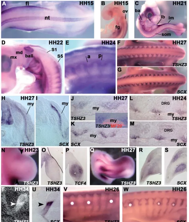
We could not detect TSHZ3 mRNAs by in situ
hybrid-ization prior to HH stage 10 when a faint expression takes
place in the neural plate (not shown). Between HH stage 10
and 15, additional sites of expression are observed. TSHZ3
demarcates the neural tube, the lateral mesoderm (
Fig. 2
A)
and the region of the foregut (
Fig. 2
B). Rostrally,
rhombo-mere r4, anterior to the otic vesicle, constitutes the anterior
limit of expression in the neural tube (
Fig. 2
B). At HH
stage 21 (
Figs. 2
C and D), TSHZ3 is found in the
mesen-chyme of the posterior aspect of the limb buds, in branchial
arches (BA) posterior to BA I, at the level of the foregut
and in the lateral mesoderm between the fore- and the
hindlimb buds. The most striking expression is observed
in the somites. The expression becomes detectable in the
posterior part by HH stage 18, and intensifies as
develop-ment proceeds (
Figs. 2
C and D). No or very weak TSHZ3
expression is observed in the four most anterior somites
(occipital somites,
Fig. 2
D). The staining in the head, not
reproducible, is likely due to the trapping of the
probe/sub-strate. Around HH stage 24 (
Fig. 2
E), TSHZ3 appears in
the anterior part of the somites in addition to the posterior
domain of expression. We focused our analysis on this
interesting expression.
sections (
Figs. 2
H and I) show that TSHZ3 and SCX both
delineate the same region – a narrow stripe of mesenchyme
underlying the myotome. However, frontal sections reveal
that the TSHZ3 domain is broader than the thin, V-shaped
SCX expression domain (
Figs. 2
J and K). TSHZ3 and
SCX do not overlap with the myofibers immunostained
with the anti-myosin heavy chain MF-20 antibody.
Sur-prisingly, in slightly younger embryos (HH stage 24),
TSHZ3 and SCX match more remarkably (
Figs. 2
L and
M). Thus, TSHZ3 expression in the somites follows that
of SCX (
Brent et al., 2003; Brent and Tabin, 2004
), first
in the same domain as SCX (HH stage 24), and
subsequently in a broader domain (HH stage 27).
We also examined the expression in the limbs. At HH
stage 23, TSHZ3 labels the posterior and anterior part of
the hindlimb bud (
Fig. 2
N). TSHZ3 transcripts are
similar-ly distributed in the forelimb bud (not shown). These
expression domains are distinct from the area defined by
the tendons progenitors and the forming muscles, since,
at this stage, both cell types occupy the central region of
the limb bud (
Schweitzer et al., 2001
). On sections, TSHZ3
is separate from TCF4, which is intimately associated with
forming limb muscles and tendons (
Figs. 2
O and P,
Kar-don et al., 2003
). By HH stage 27, TSHZ3 expression
pat-tern becomes more complex (
Fig. 2
Q) and partial
overlapping appears between TSHZ3 and SCX (
Figs. 2
R
and S). In older embryos, TSHZ3 displays the most
pro-nounced staining in the myotendinous junctions, which
are also strongly marked by SCX (HH stage 34,
Figs. 2
T
and U). Astonishingly, while early TSHZ3 expression is
excluded from the myotome and from the limb muscles,
subsequent TSHZ3 transcription is visible in muscles and
in surrounding connective tissues at HH stage 34. Thus,
TSHZ3 marks broader domains than SCX at later stages.
It has been shown that SCX expression is induced by
myotomal FGFs (
Brent et al., 2003
). Here we show that,
as demonstrated for SCX, insertion of FGF8-coated beads
in the trunk somites results in a faint but reproducible
ectopic TSHZ3 expression after 24 h. This upregulation
occurs in cells surrounding the beads that normally do
not express TSHZ3 nor SCX (HH stage 26,
Figs. 2
V
and W). Enhanced TSHZ3 transcription is also detected
upon shorter treatments with FGF8 (after 12 h, not
shown).
1.4. Other places of TSHZ3 expression in the chick embryo
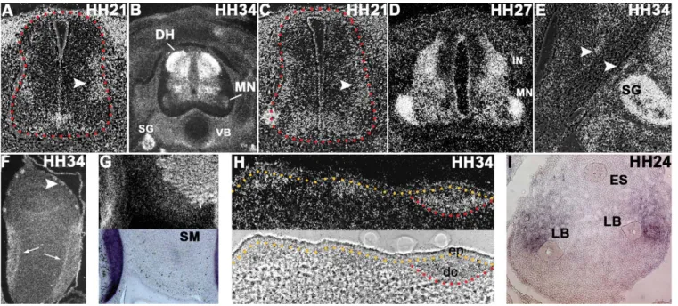
In the neural tube, we noticed dissimilarity between
TSHZ3 expression at brachial and lumbar positions. At
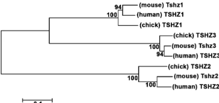
Fig. 1. Phylogenetic tree of Tshz sequences. Tshz proteins fall into three classes. The numbers of the interior branches refer to the bootstrap values with 100 replicates.
the brachial level of a HH stage 21 embryo, TSHZ3
demar-cates a ventral region located dorsal to the floor plate
com-prising motor neurons progenitors. TSHZ3 expression is
also detected in lateral regions of the spinal cord in the
marginal zone (
Fig. 3
A, arrowhead). Later, at HH stage
34, the dorsal half of the spinal cord displays robust
expres-Fig. 2. Expression of TSHZ3 in the chick embryo. (A and B) Whole mount in situ hybridizations at HH stage 15. (C) Additional sites of expression at HH stage 21. (D) Close-up of the branchial arches and the anterior somites at HH stage 22. (E) Close-up, at HH stage 24, of the brachial somites (a: anterior, p: posterior parts of the somites). (F and G) TSHZ3 (F) and SCX (G) expression in the trunk at HH stage 27 (dorsal view). (H and I) Transverse adjacent sections at the brachial level hybridized with TSHZ3 (H) and SCX (I) probes at HH stage 27. (J and K) Frontal sections at the same stage and same A/P axis level hybridized with TSHZ3 (J) and SCX (K) probes. The inset shows TSHZ3 and MF20. (L and M) Transverse sections at the brachial level through a HH stage 24 embryo. (N and Q) Whole mount TSHZ3 expression in a hindlimb bud at HH stage 23 (N), and in a HH stage 27 forelimb (Q). The arrowheads point out the TSHZ3-expressing posterior and anterior regions. (O, P–R, and S) Parasagittal transverse sections through a HH stage 23 hindlimb (O and P) and HH stage 27 forelimb (R and S) stained for TSHZ3 (O and R) and TCF4 (P and S). (T and U) Section through the thigh at HH stage 34 hybridized with35S-labelled TSHZ3 (T) probe or DIG-SCX (U). Arrowheads designate the robust expression in the myotendinous junction. (V
and W) Up-regulated TSHZ3 (V) and SCX (W) expression around FGF8-coated beads. DRG: dorsal root ganglia, fg: foregut, fl: forelimb bud, lb: limb bud, lm: lateral mesoderm, my: myotome, ov: otic vesicle, som: somite, nt: neural tube. Whole mount and frontal sections, except in (B): anterior to the left. Transverse and sagittal sections: dorsal to the top.
sion; the TSHZ3 positive domain covers the subventricular
zone and the dorsal horn (
Fig. 3
B). A very weak staining is
also observed in a lateral population of motor neurons. At
the lumbar level of HH stage 21 embryos, TSHZ3 exhibits
expanded ventral expression compared to the brachial level
(
Fig. 3
C). TSHZ3 is found in the marginal zone as well
(arrowhead). Later, in contrast to the brachial level,
sustained expression is encountered in motor neurons
(
Fig. 3
D). Dorsally, TSHZ3 is prominently expressed in
the mantle layer indicating expression in the alar plate
inter-neurons. In addition to the central nervous system, TSHZ3
is also expressed in the peripheral nervous system. Notable
examples are the sympathetic ganglia (
Figs. 3
B and E), cells
along the axonal tracts where the Schwann cells develop
(
Fig. 3
E) and the enteric nervous system. In the latter,
strong TSHZ3 expression is observed in aggregates of cells
within the outer gut mesenchyme of the gizzard constituting
the ganglia of the myenteric plexus (
Fig. 3
F, arrowhead). In
addition, consistent with TSHZ3 expression in tendons,
TSHZ3 is found in the laminar tendon of the gizzard
(com-pare with SCX in
Figs. 3
F and G). Smooth muscles also
express TSHZ3 (
Fig. 3
G, and in the intestine, not shown).
In addition, TSHZ3 is detected in zones of condensing
mesenchyme forming the cartilage (see the vertebral body
encircling the notochord in
Fig. 3
B). TSHZ3 expression is
also detected in several tissues where epithelio-mesenchymal
interactions take place, such as the feather anlagens
(HH stage 34,
Fig. 3
H, also evident in
Fig. 3
B) and in the
mesenchyme in the area of the lung buds (HH stage 24,
Fig. 3
I).
TSHZ3, like Tshz1 and Tshz2 in mouse, is expressed in
the nervous system and in mesodermal derivatives. A
note-worthy feature of TSHZ3 is its expression in developing
ten-dons and exclusion from the forming muscles at early stages.
Later, TSHZ3 displays enlarged expression in the
myotendi-nous junctions, the muscles (skeletal and smooth muscles)
and connective tissues. We hope this study will improve
our knowledge of the Tshz genes expression patterns for
understanding their specific and redundant functions.
2. Experimental procedures
2.1. Identification of TSHZ3 clones and phylogenetic reconstruction
In a BLAST search with the amino acid sequences of mouse Tshz genes we identified a chick EST clone presenting a high degree of homology with the mouse and human Tshz3 genes (C482, kind gift of Dr. Nat Bumstead). C482 was used to identify a longest chick EST clone, ChEST257k10 (ID BU471594). This clone was used to generate TSHZ3 riboprobe. Sequence alignments were analyzed by Neighbor-joining (NJ) (Gamma model of distances and sites pairwise deletion) with MEGA version 3.0 (Kumar et al., 2004). Confidence estimates included bootstrap analysis with 100 replicates.
Fig. 3. Other places of TSHZ3 expression. (A and C)35S-labelled TSHZ3 transverse sections through the neural tube at HH stage 21 at brachial
(A) and lumbar (C) level. Arrowheads point to the lateral marginal zone. Red dotted lines delineate the spinal cord. (B and D) Same as (A and C) at HH stage 34 (B) and 27 (D). (E) Axonal tract arising from the spinal cord at HH stage 34. The arrowheads indicate TSHZ3-expressing cells at the border. (F and G) Two sections through the HH stage 34 gizzard labelled with TSHZ3 (F and G, top) and SCX (G, bottom) probes. The arrows indicate the laminar tendon and the arrowhead point to one of the forming enteric nervous systems elements (eight ganglia are observed on this section). (H) Transverse section through the skin. The ectoderm is apparent in bright field (demarcated by a yellow dotted line). The red line underlines the condensing mesenchyme (dermal condensates) beneath the ectodermal placode of the feather bud. (I) Zone of the esophagus (dorsal) and of the two lung buds (ventral). Photographs (A, C, E) are bright field/black field composite images. DH: dorsal horn, dc: dermal condensate, ep: ectodermal placode, ES: esophagus, IN: interneurons, LB: lung bud, MN: motoneurons, SG: sympathetic ganglia, SM: smooth muscle, VB: vertebral body.
2.2. Processing of the tissues
Fertilized chicken eggs were purchased from a commercial source. Eggs were routinely incubated, opened and staged according to Hamburg-er and Hamilton (1951). The specimens were fixed in 4% paraformalde-hyde and processed for whole-mount in situ hybridization or cryopreserved in 30% sucrose and embedded in OCT (Tissue-Tek) for freezing and sectioned at 10–15 lm on the cryostat for tissue section in situ hybridization and immunodetection.
2.3. Chick TSHZ3 and SCX probes and in situ hybridizations
Whole mount and on sections in situ hybridizations were performed using digoxigenin (Boehringer)-labelled chick TSHZ3 (DNA linearization: NotI, antisense RNA synthesis: T3), Scleraxis (SCX) (Schweitzer et al., 2001; DNA linearization: EcoRI, antisense RNA synthesis: T3), and TCF4 (Kardon et al., 2003) riboprobes according to Henrique et al. (1995). Embryos were photographed using a Leica MZ8 dissecting microscope with a Canon D30 colour digital camera. Zeiss Axiophot2 microscope equipped with a Nikon DXM1200 Digital Camera. The auto-matic camera tamer software (ACT-1 Version 2.10, Nikon Corporation) was used to allow operation of the Digital Camera Control unit from a net-worked high-performance PC. The SCX cDNA is a kind gift of D. Duprez. Radioactive in situ hybridizations with35S-labelled TSHZ3 riboprobe on sections were performed as described inCaubit et al. (2005).
2.4. Immunohistochemistry staining procedure
Immunodetection of the myosin heavy chain was performed on cryo-sections of embryos previously processed for TSHZ3 whole mount in situ hybridization. 1:20 dilution of an MF-20 hybridoma supernatant directed against the embryonicmyosin heavy chain (Developmental Stud-ies Hybridoma Bank) and was detected by Alexa 546 fluorophor-labelled secondary antibodies (Jackson).
2.5. FGF8-soaked beads procedure
Heparin-immobilized acrylic beads (Sigma) were saturated overnight at 4°C in a solution of 1 lg/ll of FGF8 (R&D systems) diluted in PBS 0.2% BSA. Beads were then implanted in the interlimb somites of a HH stage 23 embryos 24 h prior to dissection.
Acknowledgements
We thank Dr. D. Duprez for providing us with the SCX
and TCF4 probes and Dr. Y. Perez for the phylogenetic
analysis. We are grateful to Lois J. Maltais and the Mouse
Genome Nomenclature Committee (MGNC) and the
HUGO Gene Nomenclature Committee (HGNC) that
have approved the nomenclature for the teashirt genes
fam-ily. We are grateful to M.C. Delphini and members of the
Fasano’s lab for critical reading of the manuscript. We also
wish to thank the members of C. Marcelle’s lab for their
technical assistance and G. Gabella for the analysis in the
gizzard. This work was supported by ‘‘l’Association
Franc¸aise contre les Myopathies’’ (A.F.M.) in contract
with F.L.I. Manfroid was a fellow of the A.F.M.
References
Angelats, C., Gallet, A., Therond, P., Fasano, L., Kerridge, S., 2002. Cubitus interruptus acts to specify naked cuticle in the trunk of Drosophila embryos. Dev. Biol. 241, 132–144.
Bessa, J., Gebelein, B., Pichaud, F., Casares, F., Mann, R.S., 2002. Combinatorial control of Drosophila eye development by eyeless, homothorax, and teashirt. Genes Dev. 16, 2415–2427.
Brent, A.E., Schweitzer, R., Tabin, C.J., 2003. A somitic compartment of tendon progenitors. Cell 113, 235–248.
Brent, A.E., Tabin, C.J., 2004. FGF acts directly on the somitic tendon progenitors through the Ets transcription factors Pea3 and Erm to regulate scleraxis expression. Development 131, 3885–3896.
Caubit, X., Core, N., Boned, A., Kerridge, S., Djabali, M., Fasano, L., 2000. Vertebrate orthologues of the Drosophila region-specific pat-terning gene teashirt. Mech. Dev. 91, 445–448.
Caubit, X., Tiveron, M.C., Cremer, H., Fasano, L., 2005. Expression patterns of the three Teashirt-related genes define specific boundaries in the developing and postnatal mouse forebrain. J. Comp. Neurol. 486, 76–88.
Erkner, A., Gallet, A., Angelats, C., Fasano, L., Kerridge, S., 1999. The role of Teashirt in proximal leg development in Drosophila: ectopic Teashirt expression reveals different cell behaviours in ventral and dorsal domains. Dev. Biol. 215, 221–232.
Fasano, L., Roder, L., Core, N., Alexandre, E., Vola, C., Jacq, B., Kerridge, S., 1991. The gene teashirt is required for the development of Drosophila embryonic trunk segments and encodes a protein with widely spaced zinc finger motifs. Cell 64, 63–79.
Gallet, A., Angelats, C., Erkner, A., Charroux, B., Fasano, L., Kerridge, S., 1999. The C-terminal domain of armadillo binds to hypophosph-orylated teashirt to modulate wingless signalling in Drosophila. EMBO J. 18, 2208–2217.
Gallet, A., Erkner, A., Charroux, B., Fasano, L., Kerridge, S., 1998. Trunk-specific modulation of wingless signalling in Drosophila by teashirt binding to armadillo. Curr. Biol. 8, 893–902.
Hamburger, V., Hamilton, H., 1951. A series of normal stages in the development of the chick embryo. J. Morphol. 88, 49–92.
Henrique, D., Adam, J., Myat, A., Chitnis, A., Lewis, J., Ish-Horowicz, D., 1995. Expression of a Delta homologue in prospective neurons in the chick. Nature 375, 787–790.
Kardon, G., Harfe, B.D., Tabin, C.J., 2003. A Tcf4-positive mesodermal population provides a prepattern for vertebrate limb muscle pattern-ing. Dev. Cell 5, 937–944.
Kumar, S., Tamura, K., Nei, M., 2004. MEGA3: Integrated software for molecular evolutionary genetics analysis and sequence alignment. Brief. Bioinformatics 5, 150–163.
Manfroid, I., Caubit, X., Kerridge, S., Fasano, L., 2004. Three putative murine Teashirt orthologues specify trunk structures in Drosophila in the same way as the Drosophila teashirt gene. Development 131, 1065–1073.
Mathies, L.D., Kerridge, S., Scott, M.P., 1994. Role of the teashirt gene in Drosophila midgut morphogenesis: secreted proteins mediate the action of homeotic genes. Development 120, 2799–2809.
Pan, D., Rubin, G.M., 1998. Targeted expression of teashirt induces ectopic eyes in Drosophila. Proc. Natl. Acad. Sci. USA 95, 15508–15512.
Ro¨der, L., Vola, C., Kerridge, S., 1992. The role of the teashirt gene in trunk segmental identity in Drosophila. Development 115, 1017–1033.
Schweitzer, R., Chyung, J.H., Murtaugh, L.C., Brent, A.E., Rosen, V., Olson, E.N., Lassar, A., Tabin, C.J., 2001. Analysis of the tendon cell fate using Scleraxis, a specific marker for tendons and ligaments. Development 128, 3855–3866.
Soanes, K.H., MacKay, J.O., Core, N., Heslip, T., Kerridge, S., Bell, J.B., 2001. Identification of a regulatory allele of teashirt (tsh) in Drosophila melanogaster that affects wing hinge development. An adult-specific tsh enhancer in Drosophila. Mech Dev. 105, 145–151.
Wu, J., Cohen, S.M., 2000. Proximal distal axis formation in the Drosophila leg: distinct functions of teashirt and homothorax in the proximal leg. Mech Dev. 94, 47–56.