Université de Montréal
Étude neuroanatomique fonctionnelle de l’émoussement
affectif dans la schizophrénie: les implications du
traitement à la quetiapine
Présenté par Cherine Fahim
Vice-décanat à la recherche et aux études supérieures Faculté de médecine
Thèse présentée à la Faculté des études supérieures en vue de l’obtention du grade de
Philosophae Doctor (Ph. D.) en sciences neurologiques
Mars, 2005
2005 uci, n s
© Cherine Fahim, 2005‘I.
Université
de Montréal
Direction des bibliothèques
AVIS
L’auteur a autorisé l’Université de Montréal à reproduire et diffuser, en totalité ou en partie, par quelque moyen que ce soit et sur quelque support que ce soit, et exclusivement à des fins non lucratives d’enseignement et de recherche, des copies de ce mémoire ou de cette thèse.
L’auteur et les coauteurs le cas échéant conservent la propriété du droit d’auteur et des droits moraux qui protègent ce document. Ni la thèse ou le mémoire, ni des extraits substantiels de ce document, ne doivent être imprimés ou autrement reproduits sans l’autorisation de l’auteur.
Afin de se conformer à la Loi canadienne sur la protection des renseignements personnels, quelques formulaires secondaires, coordonnées ou signatures intégrées au texte ont pu être enlevés de ce document. Bien que cela ait pu affecter la pagination, il n’y a aucun contenu manquant.
NOTICE
The author cf this thesis or dissertation has granted a nonexclusive license allowing Université de Montréal to reproduce and publish the document, in part or in whole, and in any format, solely for noncommercial educational and research purposes.
The author and co-authors if applicable retain copyright ownership and moral rights in this document. Neither the whole thesis or dissertation, nor substantial extracts from it, may be printed or otherwise reproduced without the author’s permission.
In compliance with the Canadian Privacy Act some supporting forms, contact information or signatures may have been removed from the document. While this may affect the document page count, it does flot represent any loss cf content from the document.
Université de Montréal faculté des études supérieures
Cette thèse intitulée:
Étude neuroanatomique fonctionnelle de l’émoussement
affectif dans la schizophrénie: les implications du
traitement à la quetiapine
présentéepar: Cherine Fahim
a été évaluée par un jury composé des personnes suivantes:
Louis-Éric Trudeau, président-rapporteur Emmanuel Stip, directeur de recherche
Mario Beauregard, co-directeur Pierre Rainville, membre du jury Robin Mun-ay, examinateur externe Louis-Éric Trudeau, représentant du Doyen
111
AVANT-PROPOS
Cette thèse de Doctorat est présentée sous forme d’articles et a été autorisée par le vice doyen de la faculté des études supérieures Monsieur Femand Roberge. Trois articles scientifiques composent cette thèse, un est publié, le 2ème et le 3ème sont en révision. L’auteur de cette thèse est également le premier auteur des 3 articles.
Le premier article intitulé «Negative socio-ernotional resonance in schizophrenia: a functional magnetic resonance imaging hypothesis» a été publié dans la revue Medical hypotheses, 2004; 63 :467-475.
Le deuxième article intitulé «Brain activity during emotionally negative pictures in schizophrenia with and without flat affect: An fMRI study» est en révision dans le journal Psychiatry Research: Neuroimaging.
Le troisième article intitulé «Differential hernodynamic brain activity in schizophrenia patients with blunted affect during quetiapine treatment» est en révision dans le journal Journal of Clinical Psychopharmacology, 2005 Aug;25(4):367-371.
Résumé
L’objectif de ce travail doctoral est d’examiner des mécanismes cérébraux impliqués dans le traitement d’informations d’ordre émotionnel chez des patients atteints de schizophrénie avec un regard plus particulier sur une éventuelle différence entre ceux qui présentent un érnoussement affectif (FA+) et ceux qui en sont dépourvus (FA-). En second lieu, la thèse propose d’évaluer si la quetiapine (un antipsychotique atypique) peut normaliser ou modifier les modes d’activation cérébrale impliqués dans ces processus de traitement d’information émotionnelle. L’affect émoussé peut être défini en tant que déficit de la spontanéité et de la réactivité affectives; phénomène qui peut être observé et, parfois, subjectivement ressenti. Il constitue un symptôme cardinal de la schizophrénie. De plus, l’affect émoussé fait partie des symptômes négatifs dont l’ensemble constitue le syndrome déficitaire de la schizophrénie, ainsi que le facteur le plus valide de l’évaluation des symptômes négatifs.
À
l’aide de l’imagerie par résonance magnétique nucléaire fonctionnelle (1RMf), nous avons mené trois études.Première étude: Le but de cette étude est d’examiner les mécanismes soutenant la résonance émotive dans deux groupes de patients schizophrènes (FA+ N= 13 et FA- N= 11). Nous tentons d’éclairer ces mécanismes en nous référant aux théories des neurosciences sur le fonctionnement du cerveau basé sur ce que l’on désigne comme les neurones miroirs (MN). Nous prévoyons que le groupe FA+ ne va pas activer les régions préfrontales impliquées dans le traitement émotionnel. Par contre, le groupe FA- va activer ces régions, ce qui témoigne de l’existence d’un système fonctionnel de miroir pour la résonance émotive confirmée par une activation du cortex pré-frontal. Nous avons ainsi comparé les deux groupes en utilisant une banque d’images (JAPS= International Affective Picture System) à l’aide de l’IRMf. Une analyse de type random-efJects, pour les patients FA- a mis en évidence des régions significatives dans le cortex pré-frontal médian. Les analyses de corrélation effectuées entre les estimations individuelles des sentiments négatifs
viii
et des changements du signal BOLD “Blood Oxygenated Level Dependent” ont révélé l’existence d’une corrélation positive dans le cortex pré-frontal médian. Réciproquement, on n’a pas noté d’activation significative dans le cortex pré-frontal dans le groupe fA+. Nous proposons ainsi que la résonance émotive négative induite en regardant passivement des images négatives puisse être une forme de mirroring. Ainsi que l’on pourrait saisir et interpréter les sentiments négatifs par l’intermédiaire d’un mécanisme miroir «empathie émotive en se mettant à la place de l’autre ». Avec les neurones miroirs, l’évolution humaine a franchi un pas de plus dans le monde de la représentation, en facilitant une représentation plus abstraite de l’état interne des autres. Par conséquent, nous proposons que les sujet FA-, relativement au système MN émotionnel (Comportement de résonance) puissent ressentir des images négatives. Inversement, nous proposons que le dysfonctionnement objectivé dans le groupe FA+ soit le résultat d’un échec ou d’une anomalie dans le développement du système MN. Ceci pourrait être dû à des causes génétiques ou d’autres causes endogènes ayant affecté le système MN du cortex pré-frontal impliqué dans la résonance émotive.
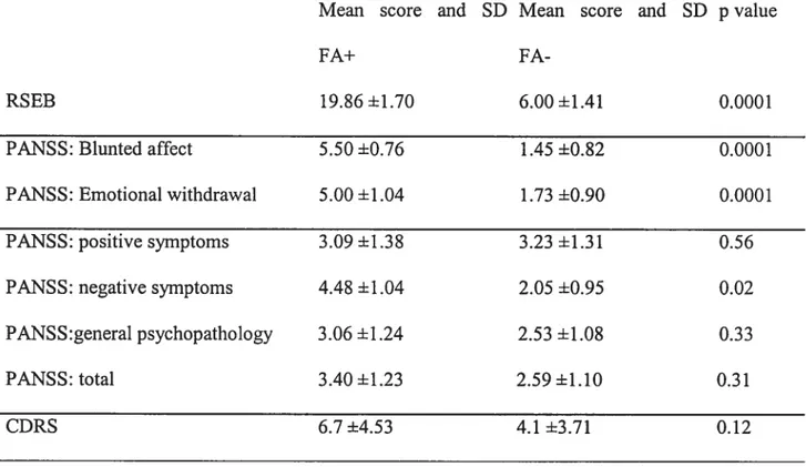
Deuxième étude: pour tenter de comprendre les différences neurobiologiques entre les patients schizophrènes avec (FA+ N= 13) et sans (fA-= 11) émoussement affectif (même patients que la 1êre étude), nous avons scanné pendant un état négatif (neg) puis neutre (neut) au cours d’un visionnement d’images lAPS, en utilisant l’IRMf. Sur une échelle de O à $ correspondant au sentiment négatif ressenti pendant la séance d’imagerie, le groupe de sujets FA+ a exprimé une moyenne de 1±1.52 et le groupe FA- 5.9 ±1.22 (P<O.0001). Ainsi, une analyse de test T à un échantillon (neg —neutre pour FA+ - FA- et vice-versa) a
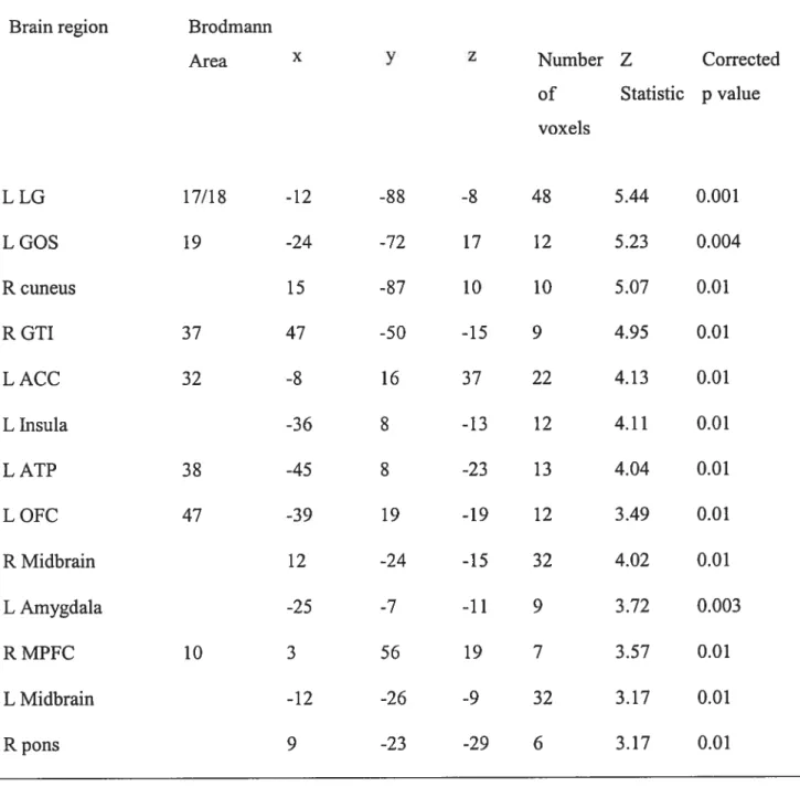
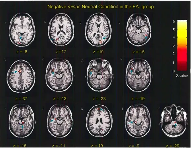
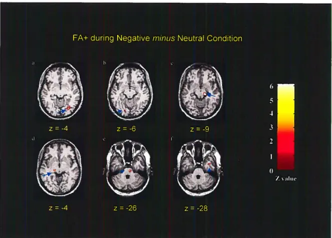
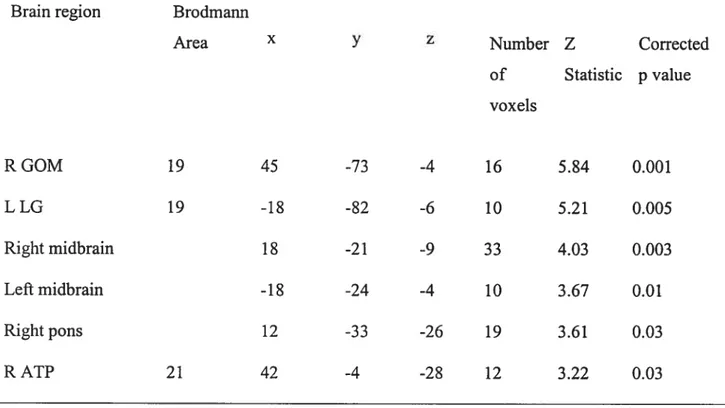
montré une activation significative pour le groupe FA- dans le cortex visuel, visuo temporal, cunéus, pré-frontal médian, orbito-frontal, le cortex cingulaire antérieur, l’amygdale, l’insula, le mésencéphale et la protubérance . Quant à lui, le groupe FA+ a activé de manière significative le cortex visuel, la protubérance et le mésencéphale. L’inactivation relative des régions pré-frontales, orbito-frontales, cingulaires antérieures,
l’amygdale et l’insula pourraient expliquer le fonctionnement émotif altéré dans le groupe FA+. Inversement, l’activation significative de ces mêmes régions chez les parents FA-pourrait signifier que l’altération du fonctionnement émotif soit non pas le symptôme fondamental de la schizophrénie mais plutôt celui du sous-groupe des patients atteints de schizophrénie avec émoussement affectif. Nous proposons que les patients FA+ utilisent une stratégie passive dans la réaction au stimulus négatif en n’activant conséquemment que la région du mésencéphale. Au contraire, les sujets FA- mettent en oeuvre une stratégie active permettant la propagation de l’infonuation au cortex pré-frontal.
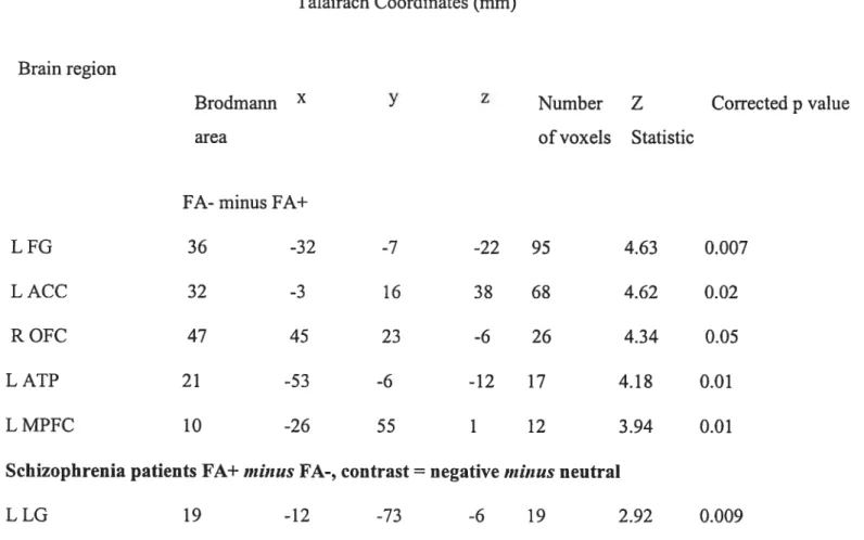
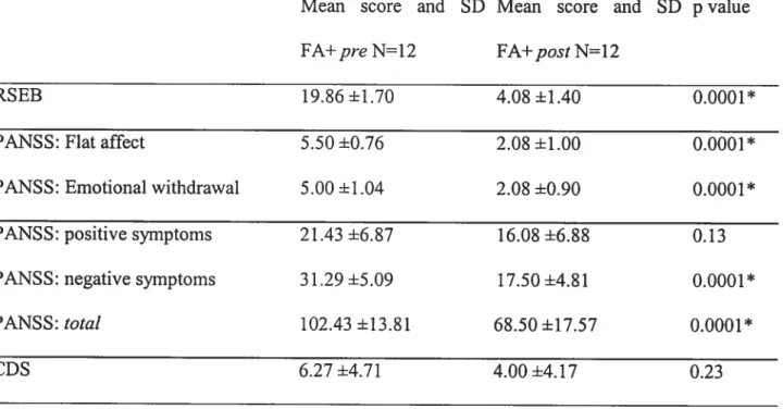
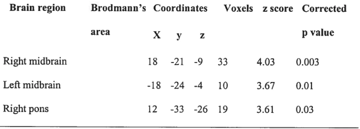
Troisième étude: L’objectif de cette étude en IRMf était de mesurer l’impact d’un traitement à la Quetiapine sur le fonctionnement de la circuiterie neuronale sous-tendant l’expérience de la tristesse chez les sujets schizophrènes souffrant d’un émoussement affectif. Pendant le visionnement passif des extraits tristes et neutres des films, nous avons scanné 12 patients (12 des 13 patients des études 1 et 2) avant et après le traitement à la quetiapine (une médiane de 5,5 mois de traitement avec la quetiapine). Les analyses ‘random-effects’ d’activation de cerveau avant la quetiapine ont indiqué une activation significative dans le tronc cérébral (protubérance). Après une durée suffisamment longue de traitement avec la quetiapine, le même contraste a montré une activation prefrontale (Broadmann “BA” 32, 46) et temporale (BA 38 et l’amygdale) significative. L’activation des régions prefrontales principales impliquées dans le traitement de l’émotion, de plus, l’amélioration, significative de symptômes, rnesureé par l’échelle d’évaluation subjective et le PANSS “Positive and Negative Symptoms Scale” suggèrent un effet potentiel de la quetiapine dans l’amélioration des symptômes relatifs à l’émoussement affectif (c-à-d., retrait passif retrait émotif, évitement) dans la schizophrénie.
L’ensemble des resultats de cette thèse propose que les mécanismes neuronaux sous jacent au traitement de l’information émotionnelle soient différents entre les deux groupes (FA+
X
et FA-). De plus, la quetiapine joue un role dans l’amélioration de l’affect émoussé dans le groupe FA+ en agissant sur le circuit mésocortical.
Mots-clés schizophrénie, symptomes négatifs, émoussement affectif émotion, antipsychotique atypique, quetiapine, résonance magnétique nucléaire fonctionnelle RMNf.
Abstract
Research suggests emotion abnorrnalities to be a highly prevalent symptom of schizophrenia. Such studies argue for a “unitary” based model for schizophrenia. However, we advocate that no “unitary” model is likely to account for the multiplicity of schizophrenia symptomatology. Our study attempts to further test this by comparing schizophrenia patients with (FA+) to without (FA-) flat affect, thus investigating the true neurobiologically based heterogeneity that goes beyond any one dysfunctional circuit (model). Furthermore, we investigate quetiapine as a potential treatment for schizophrenia patients (FA+). To that end, we conducted the following three studies using functional magnetic resonance imaging (fiVIRI). first study: The aim of this study was to use neuroscience theories about brain function (mirror-neurons IVIN) to draw inferences about the mechanisms supporting emotional resonance in two different groups of schizophrenia patients (FA+ n=13 and FA- n=1 1). We hypothesize that FA+ will not activate key brain areas involved in emotional processing. Conversely, FA- will have a functional mirror system for emotional resonance confirmed by activation of the prefrontal cortex and behavioral resuits. To test this hypothesis, we compared the 2 groups using blood oxygenation level dependent (BOLD) fMRI during a passive visual task (44 negative lAPS pictures and 44 neutral pictures). A random-effects ana1ysis for schizophrenia patients FA-, revealed significant loci of activation in the left medial prefrontal (LMPFC), right orbitofrontal (ROFC) and left anterior cingulate cortices (LACC). Correlational analyses carried out between self-report ratings of negative feelings and BOLD signal changes revealed the existence of positive correlation in the LACC, LMPFC and ROFC. Conversely, FA+ did flot show significant activation in the prefrontal cortex. We propose that negative emotional resonance induced by passively viewing negative pictures may be a form of “mirroring” that grounds negative feelings via an experiential mechanism. Hence, it could be argued that FA- were able to ‘feel’ emotions through this resonance behavior.
xii
Conversely, we suggest that the dysfunction seen in the FA+ group is a failure or distortion in the development of the MN system. This could be due to genetic or other endogenous causes, which affected prefrontal cortex Iv1N involved in ernotional resonance. Thesecond study: The aim of this fMRT study was to compare regional brain activity in schizophrenia subjects with (FA+) and without (FA-) flat affect during the viewing of emotionally negative pictures. Thirteen FA+ subjects and eleven FA- subjects were scaimed while being presented with series of emotionally negative and neutral pictures. Experientially, the viewing of the negative pictures induced a negative emotional state whose intensity was significantly greater in the FA- group than in the FA+ group. Neurally, the Negative minus Neutral contrast revealed, in the FA- group, significant loci of activation in the midbrain, pons, anterior cingulate cortex, insula, ventrolateral orbitofrontal cortex, anterior temporal pole, amygdala, media! prefrontal cortex, and extrastriate visual cortex. In the FA+ group, this contrast produced significant loci of activation in the midbrain, pons, anterior temporal pole, and extrastriate visual cortex. When the brain activity measured in the FA+ group was subtracted from that measured in the FA- group, only the lingual gyrus was significantly activated. Perhaps in FA+ subjects an amygdaloid malfunction rendered the amygdala unable to correctly evaluate the emotional meaning of the pictures presented, thus preventing effective connectivity linking the amygdala to the brain regions implicated in the physiological and experiential dimensions of emotion. Alternatively, a disturbance of effective connectivity in the neural networks linking the midbrain and the medial prefrontal system could be responsible for the quasi absence of emotional reaction in FA+ subjects, and the abnormal functioning of the medial prefrontal cortex and anterior cingulate cortex in the FA+ group. The third study: BOLD brain changes underlying response to quetiapine were examined using passive viewing of emotionally aversive and neutral stimuli. Twelve DSM-W schizophrenia patients with flat affect/ernotional withdrawal (FA+) (positive and negative symptoms scale + rating scale for emotional blunting) were scanned before and afier 5.5 months of quetiapine treatment. Whole-brain, voxel-based methods were used to assess response-specific quetiapine effects. A post-hoc comparison to an independent group
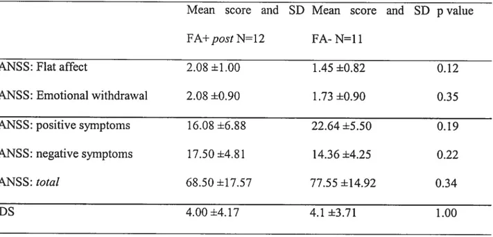
of 11 schizophrenia patients without flat affect/social withdrawal (FA-) was also performed to interpret the specificity of identified quetiapine effects. A 5.5-month treatment with quetiapine resulted in significant clinical improvement in the 12 study completers (mean ±SD post-treatment PANSS flat affect score of 5.50 ±0.76 at baseline to 2.08 ±1.00 at end point (t=7.7$, df=11, p<O.000l). Treatrnent response was associated with significant BOLD changes both in sub-cortical an cortical structures: increases in prefrontal cortex activation right dorsolateral prefrontal (DLPFC, BA 46) and the right anterior cingulate cortex (ACC, BA 32); left putamen, right anterior temporal pole (ATP) and right amygdala. Conversely, before quetiapine, the same subjects activated only sub-corticat structures: the midbrain bilaterally and the right pons. The post-hoc conjunctional analyses demonstrated that FA subjects activated the left ACC, left insula, left ATP (BA 21), left ATP (BA 3$), lefi amygdala and right medial prefrontal cortex. Quetiapine seems to affect clinical recovery by modulating the functioning of specific sites from subcortical to cortical regions (i.e., modulating the mesocortical pathway). Unique 3OLD changes in the putamen and DLPFC with quetiapine, in the FA+ post-quetiapine, relative to other drugs (used by FA-) may reflect modality-specific effects with implications for understanding flic neural correlates underlying different treatment mechanisms. In summary, it should be noted that we do not suggest that there are categorical subtypes of schizophrenia patients based on the with flat affect/without flat affect symptomatology. However, our findings, demonstrating differential haemodynamic flow between schizophrenia patients with and without flat affect, mean that the quest for the symptorn-specific-neuropathology of schizophrenia could help explain the multiplicity of neuro-circuit dysfunction in this heterogeneous disorder. Furthermore, we suggest quetiapine could have a role in improving flat affect/emotional withdrawal symptoms in schizophrenia patients.
Keywords : schizophrenia, flat affect, emotions, functional magnetic resonance imaging, visual cortex, midbrain, amygdala, prefrontal cortex.
xiv
Table des matières
Page titreIdentification dujury.r ii
AVANT-PROPOS iii
Accord des coauteurs des articles iv
Résumé vii
Abstract xi
Table des matières xiv
Liste des tableaux xvi
Liste des figures xvii
Liste des abréviations xviii
Dédicace xx
Remerciements xxi
Introduction 1
1. Schizophrenia 1
2. Diagnosis ofschizophrenia 3
3. Pathogenesis and pathophysiology: focus on abnorrnal brain circuits connectivity
and neurodeveloprnental theories 5
4. Schizophrenia sub-types and negative symptoms: focus on flat affect 11 4. Explanatory models of flat affect (affective deficits) in schizophrenia 15 5. Studies in support of affective deficits in schizophrenia 16 6. Studies contradicting affective deficits in schizophrenia 22 7. Psychopharmacological treatrnents and negative symptoms 26 8. An attempt to refute the hornogeneity of emotion dysfunction in schizophrenia and
the quest for a possible treatrnent 30
Article 1: Negative socio-emotional resonance in schizophrenia: a functional magnetic
resonance imaging hypothesis 35
Article 2: Brain activity during emotionally negative pictures in schizophrenia with and
without flat affect: an fMRI study 65
Article 3: Differential haemodynamic brain activity in schizophrenia patients with blunted
affect during quetiapine treatment 112
Discussion 149
1. Neuroimaging emotional experience and potential treatment in schizophrenia: What
wehavefound 149
1.1. The visual cortex 150
1.2. Midbrain 150
1.3.Thelnsula 153
1.4. The amygdala 155
1.5. The Prefrontal cortex 157
2. Clinical and Biological Implications 159
3. Discussing Methodological Issues 162
3.1. Why choosing the Rating Scale for Ernotional Blunting (RSEB) and the
Calgary Depression Scale (CDS)9 162
3.2. Why Choosing lAPS’? 164
3.3. Why choosing a passive-viewing method’? 165
3.4. Why choosing quetiapine’? 166
4. Limitations 168
xvi
Liste des tableaux
Article 2 Table 1 70 Table 2 77 Table 3 80 Table 4 81 Article 3 Table 1 114 Table 2 121 Table 3 122 Table 4 123 Table 5 125 Table6 126 Table 7 127
Liste des figures
Article 1 Figure 1 45 Aticle 2 Figure 1 78 Figure 2 79 Figure 3 82 Article 3 Figure 1 123 Discussion Figure 1 159xviii
Liste des abréviations
ACC: Anterior cingulate cortex
AC-PC: Anterior commissure-Posterior commissure ATP: Anteriortemporal pole
BA: Brodmann’s area
BOLD: Blood oxygen level dependent
CA-CP: Commissure antérieure-commissure postérieure CDS: Calgary depression scale
D2: Dopamine-2
DSM W: Diagnostic and statistical manual of mental disorders,4th edition
EMG: Electromyography
EPI: Echo planar imaging FA+: With flat affect FA-: Without flat affect
FG: Fusiform gyrus
fMRI: Functional magnetic resonance imaging GOM: Middle occipital gyrus
GOS: Superior occipital gyrus GTI: Inferior temporal gyrus
lAPS: International affective picture system lCD: International classification of disease
IRMf: Imagerie par résonance magnétique fonctionnelle
L: Lefi
LG: Lingual gyrus
MEP: Motor evoked potential
IV1NI: Montreal neurological institute MPFC: Medial prefrontal cortex
OMACC: Orbitofrontal, mesial prefrontal and anterior cingulate cortices PANSS: Positive and negative syndrome scale
PET: Positron ernission tomography PfC: Prefrontal cortex
0fC: Orbitofrontal cortex
R: Right
rCBF: Regional cerebral blood flow ROI: Region of interest
RSEB: Rating scale for emotional blunting
SANS: Scale for Assessment ofNegative Syrnptoms SCD: Structured Clinical Interview for DSM-W 5-HT2A: $erotonin 2A
SPECT: Single single photon emission computed tomography SPEM: Smooth pursuit eye movement
SPM: Statistical Parametric mapping SVC: Small volume correction
T: Tesla
TE: Echo time
xx
Dédicace
À mes chères parents et famille Habachi et Fahim:
Tous mes respects et ma reconnaissance vont àma famille, source de ma motivation et de mes espoirs: votre amour, affection, soutien moral, et gentillesse m’ont donné le goût et l’enthousiasme de faire quelque chose qui ajoute à votre fierté. C’est pour moi un immense plaisir d’écrire vos noms ici, et de vous dédier cette thèse. Ce travail est la reconnaissance des multiples efforts consentis à mon éducation. Je vous dis tout simplement que c’est vous qui avez alimenté l’âme de ce travail de recherche.
À
mon cher époux Adham Mancini-Marïe et mon chèr fils Zein:Adham, tu as pu me supporter, m’encourager et me préparer les conditions parfaites pour mener à bien cette thèse de doctorat. Tu as été très généreux et aimable et tu as consenti beaucoup d’amour pendant des aimées. Permet-moi de te dédier cette thèse en gage de reconnaissance, d’amour et d’affection à toi et à notre fils Zein.
Remerciements
À mes directeurs de recherche, les docteurs Emmanuel Stip et Marïo Beauregard:
A mes Professeurs Emmanuel Stip et Mario Beauregard, dont la rigueur de l’enseignement a nourri mon esprit. Je les remercie surtout pour la confiance qu’ils m’ont gracieusement accordée. Ils m’ont suivi pas à pas en m’ouvrant les portes de leurs laboratoires et m’ont recommandé pour obtenir la bourse de doctorat. Mes chers professeurs,
j
‘ai pu, durant toutes ces aimées sous votre direction, apprécier vos qualités de chercheurs; vos qualités humaines et votre générosité scientifique. Veuillez me permettre de vous exprimer ici ma reconnaissance et vous dire merci pour tout.À
Messieurs Boualem Mensour et Jean-Maxime Leroux du laboratoire d’imagerie fonctionnelle de l’hôpital Notre-Dame, je vous remercie chaleureusement pour votre précieuse aide et disponibilité tout au long de ce travail.1
Introduction
1. Schizophrenia
“The singular indfference of the patients towards their former emotional relations, the extinction of affection for relatives andfriends, of satisfaction in their works and vocation, in recreation andpleasures, is miot setdom thefirst and rnost strlldng symptom ofthe onset of disease. The patients have no real joy of lfe, “no human feelings“, to themn “nothing matters, evetything is the sarne”; theyfeet “no grief and nojoy’ their heart is flot in what they say” (Kraepelin, 1919).
The above words emphasize the fact that schizophrenia is one of the most debilitating brain disease, damaging what we regard as specifically human. It is one of the most important public health problems. Among psychiatric disorders, the combined economic and social costs place schizophrenia among the world’s top ten causes of disability-adjusted life-years (Murray and Lopez, 1996). Schizophrenia typically has its onset between the ages of 16 and 30 years. It usually has a gradual, insidious onset taking place over an average of 5 years (D$M W). Outcomes are currently of interest in health-econornics research; probably no other chronic illness parallels schizophrenia in the potential for poor functional outcome in the absence of a measurable decrease in lifespan, which is a substantial burden ofmorbidity. Noteworthy, approximately 10-15% commit suicide (Ho et al., 1997; Lewis and Lieberman, 2000).
Schizophrenia is a life-long disorder characterized by three broad types of symptoms: (j) Psychotic/positive syrnptoms involving the loss of contact with reality, including false beliefs (delusions), perceptual experiences flot shared with others (visual, auditory, tactile and gustatory hallucinations), hostility, paranoid ideation. (ii) Negative syinptoms including flat affect, emotional withdrawal, passive/apathetic social withdrawal, anhedonia (lack of pleasure), avolitionlapathy (diminished ability to initiate and follow through on plans), and alogia (reduced speech). (iii) Cognitive symptoms including unusual thought content, poor attention, lack of judgment and insight, difficulties in abstract thinking, memory and problem solving (based on the DSM-W description). 0f particular relevance to our study, these various symptoms are present in schizophrenia patients in pattems that may not overlap at all. One person may have delusions without presenting any flat affect symptoms, while another may have hallucinations and anhedonia. From a pathophysiological view, this non-overlapping pattem of symptoms raises some questions: is there a core or single brain circuit that unites schizophrenia as a single disease or different brain circuits depending on the symptom(s) present? In other words, could the brain circuit dysfunction vary according to symptoms? What is the relationship, on a neural circuit level, between a patient who had poor premorbid socioemotional adjustrnent, then develops schizophrenia with a marked flat affect symptom and a patient who had normal socioemotional adjustment, then develops schizophrenia with bouts of positive psychotic symptoms without any trace of flat affect?
3
2. Diagnosis of schizoplirenia
From a criterion-based view, schizophrenia symptoms are grouped into a single disease using a purely descriptive approach using clustering of symptoms. Criterion-based systems have been developed to decrease the complexity and improve the reliability of diagnosis. These systems include the International Classification of Diseases, tenth edition (lCD- 10) (1994), and the Diagnostic and Statisticai Manual of Mental Disorders, fourth edition (DSM-W) (1994), which describe characteristic symptoms of schizophrenia (it is the most widely used). In lCD-10, severe symptoms should have been present for 1 month, whereas in DSM-W, 6 months’ duration is required (ie, including less severe prodromal and residual symptoms). The DSM-W criteria also require deterioration in social and occupational functioning, specified as dysfunction in work, interpersonal relations, or self care. Other diagnoses, such as mood disorders with psychotic features, must be ruled out and symptoms must be shown to be due to no other medical disorder or drug effect, such as steroid-induced or amphetamine-induced psychosis. Noteworthy, diagnostic criteria improve reliability, enable standardization across centers, nationally and internationally, improve clinicai communication, and facilitate research. However, these criteria should not discourage innovative thinking about the fleurai mechanisms of schizophrenia. Rather, they should be combined with symptom-rating scales, ciinical experience and new techniques to better define features ofthis disorder that may respond to various therapeutic interventions.
Unfortunately, with their emphasis on psychotic symptoms (delusions, hallucinations, etc...), criterion-based approaches may have neglected other symptoms. for example, social deterioration commonly becomes prominent before and persists afier the more severe symptoms (positive symptoms) have been controlled with medication. These symptoms resuit in impaired functioning in interpersonal relationships. Most commonly, people who have schizophrenia are unable to continue in employment or education. Fortunately, presently psychosocial impairment is being studied as an important outcome measure, and is becoming the focus of treatment. In addition, psychiatrists are increasingly recognizing the correlation between negative symptoms and loss of social ftmnction arnong schizophrenia patients. Alogia, avolition, apathy, flat affect, etc... are among these negative symptoms, which lead to long-term social and economic burden because patients cannot maintain productive employment (Green, 1996). It should be noted that negative symptoms are the Ieast likely to improve over the course of illness, and resulting cognitive dysfunction in the context of these symptoms is most likely to contribute to unemployment (Pogue Geile and Harrow, 1985). Hence, a better understanding of negative symptoms and the development of effective treatments are required.
Moreover, negative symptoms are known to occur long before the onset of fond psychosis (positive symptoms) in schizophrenia patients (Strauss et al., 1973). However, the emphasis on psychosis as a hallmark of schizophrenia has lcd to conceptualizing the early impairments, i.e. negative symptoms, as premorbid. It is imperative to determine
5
whether such impairments are premorbid, early manifestations of the disease or a schizophrenia subtype, under-diagnosed until the manifestation of positive symptoms. Indeed, schizophrenia, simplistically, couïd be viewed as a whole-brain disease. However, with its extremely diverse but interlocking neural circuit pathologies and symptoms, more research is needed using a true neurobiologically symptom-based models.
3. Pathogenesis and Pathophysiology: focus on abnormal brain circuits connectivity and neurodevelopmental theories
0f the thousands of associative threads that guide our thinking, this disease seerns to interrupt, quite haphazardly, sometimes single threads, sometirnes a whole group, and sornetimes whole segments ofthem... the thousands of associations guiding oui- thought are interrupted by this disease.. .the thought processses, as a resuit, become strange and
illogical and the associations find newpaths” Bleuler, 191].
These words should humble the modem neuroscientists, neuropathologists and neuropsychiatrists investigating the relationship between brain circuits misconnectivity andlor dysfunction and the phenomenology of schizophrenia. Bleuler’s prophetic words have currently their counterparts in modem neuropsychiatry, which have implicated aberrant functional connectivity (miswiring) between different brain regions as the pathophysiological mechanism of psychosis. However, although some insights into the etiology of schizophrenia have been developed, an understanding of the illness remains
elusive. One could say that Bleuler’s (1950) use of the term “group of schizophrenias” was insightful and prophetic.
In an attempt to understand schizophrenia, Weinberger (1995) reviewed the possible causes including: rhesus (Rh) factor incompatibility, perinatal/prenatal viral infections, obstetrical complications, hypoxia at birth, famine and malnutrition, severe environmental stress, the epidemiologic risk factors of urban and winter birth and heritability as demonstrated by twin and adoption studies. A number of abnormalities have been identified and confirmed by meta-analysis, for example, ventricular enlargement, which is accompanied by decreased cortical and association neocortical (prefrontal and superior temporal) and hippocampal volumes (Harrison and Weinberger, 2005).
Noteworthy, these abnormalities are characteristic of schizophrenia as a whole, rather than being restricted to a subtype, and are present in first-episode, unmedicated patients. These morphometric changes are in tum suggestive of alterations in synaptic, dendritic and axonal organization. Generally, it is hypothesized that the interaction of genetic and early neurodevelopmental insuits result in defective connectivity between a number of brain regions, including the midbrain, nucleus accumbens, thalamus, temporolimbic (hippocampus), and prefrontal cortices (Selemon and Goldman-Rakic, 1999) leading to schizophrenia symptomatology. In this context, combinations of genetic and environmental factors may affect various neural circuits within the brain leading to diverse symptoms. The
7
insuit (genetic and!or environmentai) may hit one site and then flow across several fleurai circuits.
These changes tend to link schizophrenia with connectivity and communication disruption within the fleurai circuitry (for a complete review McGlashan and Hofffian, 2000). This defective neural circuitry is then vuinerable to dysfunction when unmasked by the developmental processes and events of adolescence (myelination, synaptic pruning, and hormonal effects of puberty) and/or exposure to stressors as the individual moves through the age ofrisk (Lewis and Lieberman, 2000).
Relatedly, Jones (1995) proposed, based on neuroanatomical and gene expression studies, that a disturbance of migration or in the pattem of preprogrammed celi death in the subplate zone of the developing cerebral cortex causes a failure to establish normal patterns of connections in the overiying cortex. Consequently, this could lead to schizophrenia symptoms and activity-dependent manifestations of altered gene expression for neurotransmitter- and receptor-related molecules. Jones (1997) showed that the number of neurons in the cortical subplate (the white matter irnmediately below layer VI of the cortex, a transitional structure that plays a key role in the formation of connections in the cerebral cortex), is reduced in the preftontai and temporal lobe cortices, whereas their number in the white matter deeper than 3 mm from the cortex is significantly greater compared with normal subjects. In addition, Jones (1997) ffirther suggested that the loss of ceils in the
thalamus may be primary or secondary to cortical or other subcortical pathology. Loss of thalamic celis andlor of corticothalamic inputs could lead to disintegration of thought processes by a failure in functional brain states dependent on collective oscillation of large ensembles of cortical and thalamic fleurons. Structural or functional defects of the thalamus, because it is associated with filtering sensory information and gating mechanisms, may be particularÏy important in the pathogenesis of schizophrenia; indeed, problems of information filtering may underlie disturbed thought processes.
Evidence suggesting that schizophrenia symptoms are flot caused by dysfunction of a single brain region came based on empirical data derived from both magnetic resonance and positron emission tomography. Andreasen et al., (1998), developed a model that implicates the cortico-cerebellar-thalamic-cortical circuit (CCTCC) connectivity in schizophrenia symptomatology. The authors concluded that a dysffinction in this circuitry produces “cognitive dysmetria”, difficulty in prioritizing, processing, coordinating and responding to information, a disruption of the fluid, coordinated sequences of thought and action that are the hallmark of normal cognitive functions.
Furthermore, interesting morphometric findings in the dorsolateral prefrontal cortex (Broadmann areas 9 and 46) have uncovered a form of cortical pathology in schizophrenia in which poor neuronal activity/connectivity appears to correlate with cognitive dysfunction (Selemon and Rajkowska, 2003) and the reduced neuropil hypothesis (Selemon and
9
Goldman-Rakic, 1999). The reduced neuropil hypothesis advances a circuit based model of schizophrenia proposing that a reduction in interneuronal neuropil in the prefrontal cortex is a prominent feature of cortical pathology. The authors suggest that this neuropathology is of subtie changes in cellular architecture and brain circuitry that nonetheless have a devastating impact on cortical function. They report that the increased neuronal density seen on histological examination as reduced neuropil without neuronal loss, point towards loss of connections between neurons.
Recently, a review by McGlashan and Hoffhian (2000) formulated a pathophysiological model of schizophrenia according to postmortem and neuroimaging findings. The authors proposed that schizophrenia resuits from developmentally reduced synaptic connectivity (DRSC). The model posits that schizophrenia arises from critically reduced synaptic connectivity as a result of developmental disturbances of synaptogenesis during gestation and early childhood andlor synaptic pruning during adolescence. The DRSC model identifies reduced synaptic density in prefrontal and other areas of association cortex as the “final common pathway” to the symptoms and course of schizophrenia.
However, before this “final common pathway” mild dysfunctions associated with these early neurobiological lesions occasionally emerge as vulnerability factors or risk markers for schizophrenia. For example, diminished expression of positive and negative emotions, passivity, social withdrawal, and poor relationships (Grimes and Walker, 1994;
Hans et al., 2000), neuromotor abnormalities in the form of poor coordination, poor perceptual motor integration, or abnormal speech (01m and Mednick, 1996), as well as minor physical anomalies (Whitty et al., 2003), and neurocognitive deficits (01m and Mednick, 1996). Noteworthy, such deficits are flot sufficient to produce schizophrenia. further evidence of neuronal circuit dysfunction in schizophrenia, has corne from studies investigating eye tracking. Jndeed, smooth pursuit eye movement (SPEM) abnormalities are associated with liability for schizophrenia (Ross et al., 2002). Relatedly, in a recent article by Hong et al., (2005), the authors stated that understanding the neurophysiologic rnechanisms underlying SPEM deficits is likely to enhance our ability to refine them as endophenotypes for genetic studies. Hong and colleagues have compared pursuit-related brain activation in schizophrenia patients and healthy control subjects, matching subjects on average maintenance pursuit performance. Patients showed decreased activation compared with healthy subjects in the medial superior temporal cortex, frontal eye field, and suplementary eye field areas hypothesized to be involved in extraretinal motion processiiig. However, they have showed increased activation Brodmann areas 19 and 37. These correspond to posterior retinal motion regions and are consistent with the notion that a portion of the individuals who possess liability for schizophrenia rely more on retinal signais to maintain pursuit
Overail, rather than having a distinctive diagnostic neuropathology, schizophrenia seems to consist of quantitative alterations in various normal parameters of neural
11
microcircuitry. In this vein, because of a lack of definite neuropathology, schizophrenia remains a clinical diagnosis based on the presence of positive and negative symptoms. Therefore, and since schizophrenia is a heterogeneous brain disease, understanding the functional abnormalities of different brain circuits and relating this to symptomatology is of primordial importance in our quest to understand schizophrenia.
4. Schizophrenia sub-types and Negative symptoms: focus on Flat affect
In 1857, John Russeil Reynolds stated “Many of the symptoms of disease are merely modified vital actions.. .some symptoms are negative, i.e., they consist in the negation of vital properties...other symptoms are positive, i.e., they consist in the excess of alteration
of vital properties”, hence introducing the concept of negative versus positive symptoms to schizophrenia (Berrios, 1985). Insightfully, John Hughlings Jackson (1875, 1889) described negative symptoms as the diminution or negation of normal processes, resulting from tissue destruction; and positive symptoms as the excess in normal brain processes as disinhibiting consequences ofthis tissue destruction. Going one step further, Kraepelin (1919), reported: • .there are apparently two principal groups of disorders which characterize the malady. On the one hand we observe a weakening of those emotional activities...The result of this
part of the morbid process is ernotional dullness. Bleuler (1911) considered negative symptoms to represent “fundamental” or core psychopathologies in schizophrenia. He defined schizophrenia as essentially a spiitting of thoughts (cognition) from feelings (emotion). More recently, many attempts to subtyping schizophrenia have emanated. I will
briefly outline the main six. First, in 1974, Tsuang and Winokur proposed the paranoid (delusions, hallucinations) versus the nonparanoid (flat affect, disorganized thoughts) subtypes. Then, in 1980 the concept of Type I and Type II schizophrenia was introduced by Crow (1980a). This subdivision put greater emphasis on the presence or absence of negative symptoms, with poverty of speech and flat affect defining the presence of the negative syndrome (Type II) and the prominance of psychotic symptoms for the positive (Type I) syndrome. Third, came Andreasen and Olsen (1982), who conceptualized positive (delusions, hallucinations, hostility) and negative symptoms (affective flatting, alogia, avolition-apathy, anhedonia-asociality, and attentional impairment) as different ends of the same continuum and described patients as either predominantly positive or predominantly negative. Farmer and colleagues (1983) were fourth to propose the P type (delusions, late onset) versus the H type (flat affect, bizarre behavior, incoherent speech and early onset) distinction. fifth, Liddle and collaborators (1987) introduced the concept of the three syndromes: psychomotor poverty (poverty of speech, lack of spontaneous movement and various aspects of blunting of affect); disorganisation (inappropriate affect, poverty of content of speech, and disturbances of the form of thought); and reality distortion (particular types of delusions and hallucinations). Sixth, in an effort to clarify the situation, the deficit/non-deficit distinction was put forth by Carpenter and colleagues (198$). They identified six negative symptoms (i.e., poverty of speech, diminished emotional range, flat affect, diminished sense ofpurpose, curbing ofinterests and diminished social drive) as the deficit syndrome. In this vein, Kirkpatrick and colleagues (2001), stated that the
13
deficit/non-deficit schizophrenia sub-groups differ in their signs and symptoms, course, biological correlates, treatment response, and etiologic factors.
In 1980, Crow hypothesized that negative symptoms (poverty of speech, flat affect) in schizophrenia represent a behavioral syndrome that was the manifestation of unspecified structural brain abnormalities. It should be noted that researchers seeking to establish different etiologies for the positive and negative subtypes struggled with the degree to which the sutypes are truly different because they have found overlap cross-sectionally and longitudinally. Other researchers made the attempt to delineate the defective brain circuits based on biochemical hypotheses. In this vein, the implications of several neurotransmitters have been proposed: GABA, glutamate, serotonin, achetylcholine, noradrenaline (for a full review see Stahl, 2002). However, the classical comerstone theory to explain schizophrenia is the ‘dopamine hypothesis’, proposing that the positive symptoms of schizophrenia are related to dopamine neurons overactivity in the mesolimbic pathway, and that the negative symptoms are related to underactivity of dopamine input to the prefrontal cortex (Weinberger and Wyatt, 1982).
from the above, we should note the emphasis on the role flat affect plays in schizophrenia. Kraepelin (1919) conceptualized affective deficits as fundamental symptoms of schizophrenia. Importantly he gave particular emphasis to diminished emotional experience. More recently, numerous other authors have also advocated the role emotional
deficits play in schizophrenia. For example, Crow (1985) used poverty of speech and flat affect as the key symptoms for the definition of a negative symptom subtype because these are the symptoms that are both primary and enduring.
In support of Kraepelin’s early observation on the role emotional dullness (flat affect) plays in schizophrenia, contemporary research lias provided converging evidence about its diagnostic and prognostic importance (Abrams and Taylor, 1978; Sweet et al., 199$; Gur et al., 2002). flat affect has also been shown to be a relatively enduring symptom that generally responds poorly to treatment (Pogue-Geile and Harrow, 1985). it is important to note that patients with schizophrenia exhibit impairments in emotional discrimination and experience across cultures (Habel et al., 2000). furthermore, the presence of this symptom is related to poor clinical outcomes on several dimensions, including employment (Pogue Geile and Harrow, 1985), social functioning (Breier et al., 1991), and severity of illness (Abrams and Taylor, 1978). Moreover, premorbid affective blunting is associated with an earlier onset of illness and poorer prognosis in schizophrenia (Grimes and Walker, 1994). A great body of evidence suggests that diminshed expression of positive and negative emotions, passivity, social maladjustments, anxiety, withdrawal and poor peer relationships are the clearest prodromal symptoms leading to the onset ofpsychosis (Watt, 1978; Done et al., 1994; Ingraham et al., 1995; Mirsky et al., 1995; 01m and Mednick, 1996). Although research has given greater insights into the impact of flat affect and the nature of the deficit, the pathophysiology and causes are still not clearly understood.
15
4. Explauatory models of flat affect (affective deficits) in schizoplirenia
Explanatory models of flat affect have proposed various etiologies, invoking psychodynamic processes such as repression (Arieti, 1955; Pao, 1979), emphasizing the role of impaired social relations and social rejection (Roff and Knight, 1978). A recent hypothesis has suggested that flat affect is the manifestation of dysfunction in the right hemisphere (Mayer et al., 1985). The potential significance ofright hemisphere dysfunction in flat affect derives from evidence obtained in neurologie populations with cortical lesions. These patients have been described as showing flat affect or emotional indifference that is strikingly similar to the clinical picture of flat affect seen in schizophrenia. Whittaker and colleagues (1985) reported associations between frontotemporal dysfunction and impaired perception of both facially expressed and vocally expressed emotion. Another more recent hypothesis proposed dysfunction of the frontal lobe areas (Gur et al., 2002; Weinberger, 1987; Buchsbaum, 1990; Weddell et al., 1990; Mega and Cummings, 2001), because of their afferent and efferent connections with limbic structures, which have been implicated in affective behavior. Interestinsly, Takahashi et al., (2004) proposed that schizophrenia patients might have relatively intact function of conscious processing of significant emotional information, leading to a categorization of emotional pictures similar to that of controls, however, they have impairment in the rapid, automatic processing of salient stimuli. In other words, patients could assign significance to stimuli through conscious processing, but they might have diminished automatic emotional response to extemal stimuli.
5. Studies in support of affective deficits ïn schïzophrenia
Affective deficits in schizophrenia can be identified in three general domains (reviewed in Limpert and Amador, 2001): 1) deficits in theperception of emotion (i.e., difficulty judging and interpreting the emotional displays of others), 2) deficits in the expression of emotion (i.e., difficulty conveying one’s emotional experience to others) and 3) deficits in the experience of emotion (i.e., difficulty with the subjective feeling of emotion). In this study, we have focused on the latter, with some involvement of the first (i.e., in order to feel we need first to perceive and interpret the emotional displays).
The greatest amount of empirical work, investigating emotion deficits in schizophrenia, has been conducted in the areas of emotion perception and emotion expression, with few studies investigating emotional experience. A number of studies examining interpersonal behaviors in flat affect have focused on either self-report of interpersonal behaviors (Habel et al., 2000; $ison et al., 1996) or have used a behavioral approach to study these behaviors, such as assessing eye contact, verbal comments, and body gestures (Sweet et al., 1998; Kohler et al., 2000; Alpert et al., 2000; Alper et al., 2002). further evidence cornes from studies reporting that schizophrenia patients are impaired in judging various facial expressions depicted in photographs (for example, Cutting, 1981; Heimberg et al., 1992). Several studies also provide evidence for a deficit in judging vocal expressions ofemotion in schizophrenia patients (Borod et al., 1990; Haskins
17
et al., 1995). 0f particular note, the deficits appear to be less consistent, across studies and populations, when using observer ratings. These equivocal findings may be due to the fact that flat affect schizophrenia patients tend to be negatively evaluated andlor negatively evaluate their social skills. Altematively, the faulty interpersonal responses may be too subtie to be reliably detected using a behavioral approach, which usually involves videotaping social responses and later quantifying the responses using a coding system. This approach can be problematic because the behavioral coding systems may flot be those behaviours that are most critical to the social/emotional interaction. furthermore, social/emotional interactions consist of capturing nuanced behaviors (e.g. slight curve of the hp, subtie flare of the nostril) that may be difficuit to capture using behavioral coding systems.
For this reason, a second approach to measuring sociah/emotional disphays was put forth: to employ physiological indices, such as facial electromyography (EMG) (Sison et al., 1996; Iwase et al., 1999; Kring et al., 1999). The authors found that increased bluntedness of affect was associated with longer pauses and reduced dyadic interaction and less zygomatic (cheek) electromyogram activity. The flat affect patients unexpectedly showed more corrugator (brow) electromyogram activity compared with control groups, which perhaps reflects difficuhty in self-expression. Furthermore, schizophrenia patients showed reduced zygomaticus activity (EMG) during both the happy film and the interview, suggesting a reduced ability to express happy facial expressions, such as smiling, in the
lower face (Mattes et al., 1995). However, there remains the question: what happens in the brain?
The answer of this question lies in the third and more recent approach, the one we will be focusing on: functional neuroimaging. Functional magnetic resonance imaging (fiVIRI) is a noninvasive technique for measuring changes in cerebral blood flow and oxygenation that reflect the underlying neuronal activity. This technology offers a powerful method for exploring three ofthe outstanding questions in schizophrenia research (1) neural circuits abnormalities in schizophrenia and their associated symptoms; (2) how these neural circuit dysfunction relate to the heterogeneity of schizophrenia; and (3) how could the widely accepted single unitary mode! for schizophrenia account for the widespread functional deficits. Thus, numerous researchers have also introduced, albeit with different rnethodology, neuroimaging techniques to investigate the neural process of emotional deficits in schizophrenia. Since 1981, more than 100 articles on functional brain imaging in schizophrenia have been published. Positron emission tomography (PET) and single photon emission computed tomography (SPECT), which measure regional cerebral blood flow (rCBF) and/or metabolisrn, reveal a number of abnormalities and deficiencies in people with schizophrenia, compared with healthy subjects. A consistent finding (in two-thirds of the published PET or $PECT studies) is a lower level of prefrontal cortical metabolism in schizophrenia subjects (reviewed by Wu et al., 2000). Furthermore, many studies have reported in schizophrenia patients structural and functional abnormalities in the medial
19
prefrontal cortex, orbitofrontal and anterior cingulate gyrus, regions important in the effortful regulation of affective states and emotional behavior (reviewed in PhiÏlips et al 2003).
Consistent with the theory, these studies have suggested that individuals with schizophrenia may be disturbed not only in their experience andlor expression of affect, but also in their ability to recognize emotions expressed by others (Gur et al., 2002; Schneider et al., 1998; Jobnston et al., 2001; Paradiso et al., 2003). In these studies, the main modality for receiving emotional information was visual, specifically facial expressions. For example, $chneider and coïleagues (Schneider et al., 1998) found that unlike controls, schizophrenia patients have not demonstrated amygdala activation during sadness despite matched ratings to normal controls indicating a similar negative affect. At that time, their results provided new evidence of functional abnormalities in the limbic system of schizophrenia patients processing emotions. Crespo-Facorro and colleagues (Crespo Facorro et al., 2001) PET study reveals an interesting paradox. Schizophrenia: patients appear to have a normal ability to experience unpleasant emotions, coupled with an impairment in the ability to experience pleasant ones, and the more psychotic they are, the greater the acuity of their ability to recognize unpleasantness. At the neural level, patients with schizophrenia who subjectively rated the unpleasant odor the same as did healthy volunteers failed to recruit the subcortical limbic and paralimbic structures that would normally be used for this task. Instead, their normal behavioral response was associated
with abnormally increased rCBf in a widely distributed group of frontal cortical regions. On the other hand, these patients had difficulty identifying the positive emotional valence of the pleasant odors, despite the fact that they gave intensity ratings within the normal range. Accordingly, the authors concluded that abnormalities in the complex functional interactions between mesolimbic and frontal regions may underlie emotional disturbances in schizophrenia. In a more recent fMRI study, Gur and colleagues (2002), found that schizophrenia patients failed to activate limbic regions during emotional valence discrimination tasks. Another fMRI study Kosaka and colleagues (Kosaka et al., 2002), reported that negative face discrimination activated the bilateral amygdalae in the schizophrenia group whereas the right amygdala alone in the control group. The authors argued that exaggerated amygdaïa activation during emotional intensity judgment found in the schizophrenia patients may reflect impaired gating of sensory input containing emotion.
More recently, Takahashi and colleagues (2004), instructed the subjects
(
5 schizophreniall 5 controls), to indicate how each of the presented pictures made them feel. Whole brain activities in response to the affective pictures were measured by fMRI. Controls recruited the neural circuit including amygdaloid-hippocampal region, prefrontal cortex, thalamus, basal ganglia, cerebellum, midbrain, and visual cortex while viewing unpleasant pictures. However, the patients showed less activation in the components of this circuit. Furthermore, an elegant study by loannides and collaborators (2004), provided21
evidence for disturbances in functional connectivity in schizophrenia patients, while processing of facial information.
Together, these studies provide substantial support for a socio-emotional deficit in schizophrenia (Sweet et al., 1998; Sison et al., 1996; Kohler et al., 2000, Cutting, 1981; Feinberg et al., 1986; Gessier et al., 1998; Heimberg et al., 1992; Archer et al., 1992). Reduced facial expression during social interaction (Krause et al., 2003) and diminished expressiveness in response to emotional films (Berenhaum and Oltmanns, 1992) have also been reported. According to Lee and colleagues (Lee et al., 2004) disturbances in socio emotional cognition may represent an abnorrnal interaction between frontal lobe and its functionally connected cortical and subcortical areas.
In summary, substantial evidence supports emotional deficits and, hence, advocates a unitary model to schizophrenia (present in ail schizophrenia patients). However, from a clinical point of view, these symptoms are heterogeneous among schizopbrenia patients. Thus, studies shouid seek to explore the heterogeneity of socio-emotional dysfunction within schizophrenia.
6. Studies contradicting affective deficits in schizophrenia
fi the preceding section, we examined emotional deficits in schizophrenia and reported some of the data that have been pubiished to support it. We now tum to the issue that gave rise to our study. Notwithstanding these studies (see previous section), emotional deficits appear to be inconsistent across studies and populations (Kring et al., 1999; Eamst and Kring 1999). Overail, studies to date have produced contradictory and inconclusive resuits (Edwards et al., 2002). fi their review, Mon-ison and colleagues (1988) reported some conflicting observations among schizophrenia patients in emotion recognition and experience. They proposed that task or rnethodology differences, are likely to explain the inconsistent resuits.
First, many studies include chronic patients without the use of comparably ill psychiatric controls or no psychiatric controïs at ail. Participants have been hospitalized continuously for Iengthy periods, thus being subject to social isolation. Hence, it would be worthwhile to study patients with first-episode psychosis to minimize the influence of variables such as institutionalization. In this vein, Edwards and colleagues (Edwards et al., 2002) demonstrated that the magnitude of the significant differences between first-episode psychosis, assessed as outpatients during the early recovery phase of illness, and non-patients in the recognition of emotion was relatively small, suggesting only a subtie deficit. There are several studies that demonstrate that phase of illness seems to have an
23
effect on emotion recognition, with remitted patients performing better than acute patients (Gessier et al., 1998).
Second, we should note the lack of attention paid to the long-term impact of antipsychotic and anticholinergic medications. Paradiso and colleagues (Paradiso et al., 2003) measured regional rCBF during performance of a task that required unmedicated patients to recognize the emotional valence of visual images and to determine whether they were pleasant or unpleasant. When patients consciously evaluated the unpleasant images, they did flot activate the phylogenetically older fear danger recognition circuit (e.g., the amygdala) used by the healthy volunteers, although they correctly rated them as unpleasant. Likewise, the patients showed no activation in areas of the prefrontal cortex normally used to recognize the images as pleasant and were unable to recognize them as such. Areas of decreased CBF were widely distributed and comprised subcortical regions such as the thalamus and cerebellum. Thus, the authors suggested failure of the neural system used to support emotional attribution consistent with pervasive problems in experiencing emotions.
Third, to our knowledge, very few researchers have attempted to determine whether their findings are indicative of schizophrenia patients with flat affect or manifestations of a more general psychotic process. Investigations would benefit from the inclusion of a psychotic control group (i.e. schizophrenia patients without flat affect), hence, investigating
if this ‘subgroup’ exhibits the same deficits. Especially, if we consider studies reporting that some schizophrenia patients experience emotions (Kring et al., 1999; Berenbaum and Oltmanns, 1992).
In this section, we summarize a series of investigations that separately reftect the heretogeneitv ofschizophrenia.
1) Evidence suggests that the paranoid subtype has more intact cognitive functioning (Morrison et al., 1988), a better premorbid history (Goldstein, 1970), and fewer social deficits (Strauss et al., 1973) than non-paranoid subtypes. These differences would suggest that paranoid patients would perform better on tasks of social perception than non-paranoid patients.
2) Patients with negative symptom schizophrenia have generally been characterized as having poor premorbid adjustment and lower overali social functioning (Andreasen and Olsen, 1982; Crow, 1987; Schuldberg et al., 1999) in comparison to patients with positive symptom schizophrenia. Consistent with this, two studies have found that paranoid schizophrenic subjects are more accurate than non-paranoid subjects in judging facial expression of emotion (Kline et al., 1992; Lewis and Garver, 1995). 3) Bryson et al. (1998) found that deficit patients performed more poorly on an affect
recognition test than non-deficit patients, and within the deficit group, diminished sense of purpose was the deficit symptom most strongly associated with impaired affect
25
recognition. Jnterestingly, the deficit patients demonstrated poorer premorbid adjustment and worse social functioning than non-deficit patients.
4) Blanchard and colleagues (199$) reported less positive affect and significantly greater negative affect in patients with schizophrenia, compared with controls. The authors concluded that their findings indicate that schizophrenia is characterized by both low positive affect and elevated negative affect and that these affective characteristics are a stable feature ofthe illness.
5) A similar result was obtained by Eamst and colleagues (Eamst and Kring, 1999). In this article, the authors exarnined emotional responding in deficit patients, non-deficit patients, and controls, and investigated the importance of the deficit/non-deficit distinction for studies of emotional responding in schizophrenia. They showed that deficit patients exhibited fewer outward facial expressions to the films than both non deficit patients and controls.
6) Noteworthy, in a preliminary report done by our group, we demonstrated no significant difference between schizophrenia patients without flat affect and normal controls in emotion processing, using fMRI (fahim et al., 2003).
7) In a recent study, Cohen and Docherty (2004) examined affective reactivity in the natural speech of schizophrenia patients with and without the deficit syndrome. Nondeficit patients showed greater affective reactivity of speech than deficit patients. Conversely, deficit patients’ speech was not more reactive to emotion than the speech of the control group. These resuits suggest that emotion-related variables mediate the
relationship between stress and language symptom exacerbations insomepatients with schizophrenia.
Although only few studies investigated emotion deficits between two groups of schizophrenia patients, the resuÏts mentioned above nonetheless seem to indicate that whereas schizophrenia patients with severe negative symptoms (flat affect) may have diminished affective experience, those patients with positive symptoms “feel more”. Although the latter group of patients may at times show diminished subjective feeling as a consequence of positive (delusions/hallucinations) or depressive symptoms, this lack of feeling is flot a stable feature of the illness, as it is for the deficit patients. Overail, these findings are consistent with the idea that subgroups of schizophrenia patients may differ in terms of their affective functioning.
7. Psychopli armacological treatments and negative symptoms
The treatment of schizophrenia changed in the 1950s with the discovery of conventional antipsychotics (typical), such as chlorpromazine and haloperidol. However, patients stili had incomplete or partial response, with significant negative, more than positive symptoms remaining despite optimal dosing. In the 1960s, clozapine was introduced as the first ‘atypical’ antipsychotic. Clozapine had a positive effect on both the negative and the positive symptoms. Nonetheless, it was removed from the market because of its agranulocytosis. Following this discoveiy, other atypical antipsychotics have been
27
discovered. However, the major goal of antipsychotic drug development until the 19$Os was, stiil, the treatment of positive symptoms. Nonetheless, Crow’s provocative suggestion (1980a) that chronic schizophrenia patients with negative symptoms were insensitive to neuroieptic treatment, raised interest in the issue oftreating negative symptoms.
Treating positive symptoms mainly relied on the antipsychotics’ ability to bind to dopamine-2 (D2) receptors. Indeed, in vitro studies (Seeman et al., 1976; Seeman et al., 1997) show that ail antipsychotics bind to dopamine-2 (D2) receptors and that the affinity for the D2 receptor is inversely correlated with the clinical dosage. Although the principal brain target that ail antipsychotic drugs attach to is the dopamine D2 receptor, traditional or typical antipsychotics, by attaching to it, induce extrapyramidal signs and symptoms (EPS). They also, by binding to the D2 receptor, elevate serum prolactin. Atypical antipsychotics given in dosages within the clinically effective range do not bring about these adverse clinical effects.
The typical antipsychotics such as trifluperazine, pimozide, chlorpromazine, fluphenazine, haloperidol, and flupenthixol bind more tightly than dopamine itself to the dopamine D2 receptor, with dissociation constants that are lower than that for dopamine. The newer, atypical antipsychotics such as quetiapine, remoxipride, clozapine, olanzapine, sertindole, ziprasidone, and amisuipride ail bind more loosely than dopamine to the dopamine D2 receptor and have dissociation constants higher than that for dopamine (Kapur and Seeman,
2002; Seeman, 2002). Despite the introduction of these several new antipsychotics, quetiapine lias several specificities not shared by other atypical antipsychotics, i.e., lower extrapyramidal symptoms and no hyperprolactinemia (Tauscher et al., 2002). The pharmacological basis of quetiapine’s low propensity to induce extrapyramidal symptoms may be explained by its combined antagonism at 5-HT2A and D2 receptors or its fast dissociation from the D2 receptor (Kapur and Remington, 2001). In this vein, moderate occupancy of D2 receptors has been proposed to be clinically superior to a more complete D2 blockade (Meltzer and Gudelsky, 1992). Tauscher and colleagues (2002) reported, using PET, that in the striatum, the D2 occupancy ranges from 81% for risperidone to 30% for quetiapine, with the following rank order: risperidone > olanzapine > clozapine > quetiapine. Kapur et al., (2000) demonstrated, using PET, that quetiapine does give rise to transiently high (58%-64%) D2 occupancy 2 to 3 hours afier a single dose that then decreases to minimal levels (< 30%) in 12 hours. In other words, quetiapine’s pharmacology lias aspects that are consistent with the atypical pharmacologic profile of an antipsychotic, yet, novel and distinct in its low D2 binding affinity and fast dissociation from the receptor. In vivo functional studies ail provide evidence that quetiapine has a preferential effect on limbic as opposed to striatal D2 receptors. EP$ are associated with D2 occupancy in the striatum, whicli therefore predicts a therapeutic effect with placebo levels ofEPS for quetiapine (Arvanitis et al., 1997).
29
A recent review (Lublin et al., 2005) reported that clinical trials suggest that atypical agents improve negative and affective symptoms, and cognitive functioning more than typical antipsychotics. 0f particular relevance to our study, quetiapine has a direct effect on the negative symptoms of schizophrenia (Tandon, 2004). In this study, the author evaluated, quetiapine’s effect on negative symptoms, from four randomized, controlled clinical studies involving 1106 patients employing a path analysis model. The total effect of quetiapine on negative symptoms was measured using the Scale for Assessment of Negative Symptoms (SANS) total score. Indirect effects on negative symptoms via positive, depressive and EPS were assessed using appropriate instruments. Effect sizes were calculated by path analysis for the difference between treatment groups in change from baseline to endpoint in SANS total score. Analysis confirmed that quetiapine produced a greater overali improvement in negative symptoms than placebo (effect size 1.96); this was explained by a significant direct effect (p = 0.001; 44.2% of total improvement), and a secondary effect of improved
positive symptoms (p <0.001; 47.5% of total improvement), but was not a consequence of changes in depressive symptoms or EPS. Thus, the author concluded that quetiapine has a substantial direct effect on improving the negative symptoms of schizophrenia. In addition, in animal studies, quetiapine has been shown to reverse amphetamine-induced social isolation in monkeys (an animal model for the negative symptoms (Ellenbroek et al., 1996). In this vein, chronic administration of quetiapine reverses the inhibitory effects of amphetamine on midbrain dopamine neurons and produces depolarization inactivatioin of limbic-related AlO dopamine neurons (Goldstein et al., 1993).
finally, worth mentioning, a fundamental problem in schizophrenia treatment is the traditional view of schizophrenia as a disease entity rather than a syndrome comprising several pathologic domains. This is what we will attempt to explain in the next section.
8. An attempt to refute the homogeneity of emotion dysfunction in schizophrenia and the quest for a possible treatment
At this point, we should have noted that despite extensive research for many years and many promising hypotheses, we do not, yet, have a clear understanding of either the causes of schizophrenia or how the causative factors lead to the clinical features. Previous researcli has demonstrated abnormalities in most of the brain regions in schizophrenia. However, these ultimately convincing findings have yielded disappointing conclusions in our quest to understand schizophrenia as a disease. Hence, instead of advocating a “Gestalt” approach (the whole equals the sum of its parts) to schizophrenia, researchers should investigate the parts (symptomatology) then put them together to understand the whole (schizophrenia). Thus, any attempts to understand it requires explanation of its various guises and components. furthermore, although the general concept of negative symptoms has been of considerable heuristic value and was successful in generating research findings leading to six major theories of sub-typing (section 4 in the introduction), the time has corne for abandoning its general construct and focus on its discrete components. This approach may yield findings that have potential significance for an understanding of etiologic and