HAL Id: hal-02467119
https://hal.archives-ouvertes.fr/hal-02467119
Submitted on 4 Feb 2020HAL is a multi-disciplinary open access
archive for the deposit and dissemination of sci-entific research documents, whether they are pub-lished or not. The documents may come from teaching and research institutions in France or abroad, or from public or private research centers.
L’archive ouverte pluridisciplinaire HAL, est destinée au dépôt et à la diffusion de documents scientifiques de niveau recherche, publiés ou non, émanant des établissements d’enseignement et de recherche français ou étrangers, des laboratoires publics ou privés.
Density functional theory-assisted 31P and 23Na
magic-angle spinning nuclear magnetic resonance study
of the Na3V2(PO4)2F3–Na3V2(PO4)2FO2 solid
solution: unraveling Its local and electronic structures
Long H. B. Nguyen, Paula Sanz-Camacho, Thibault Broux, Jacob Olchowka,
Christian Masquelier, Laurence Croguennec, Dany Carlier-Larregaray
To cite this version:
Long H. B. Nguyen, Paula Sanz-Camacho, Thibault Broux, Jacob Olchowka, Christian Masquelier, et al.. Density functional theory-assisted 31P and 23Na magic-angle spinning nuclear magnetic reso-nance study of the Na3V2(PO4)2F3–Na3V2(PO4)2FO2 solid solution: unraveling Its local and elec-tronic structures. Chemistry of Materials, American Chemical Society, 2019, 31 (23), pp.9759-9768. �10.1021/acs.chemmater.9b03546�. �hal-02467119�
1
DFT-assisted
31
P and
23
Na MAS-NMR Study of
the Na
3
V
2
(PO
4
)
2
F
3
– Na
3
V
2
(PO
4
)
2
FO
2
Solid
Solution: Unravelling its Local and Electronic
Structures
Long H. B. Nguyen a,b,c, Paula Sanz Camacho a,c, Thibault Broux a,b,c,d, Jacob Olchowka a,c,d,
Christian Masquelier b,c,d, Laurence Croguennec a,c,d, and Dany Carlier a,c,d *
a CNRS, Univ. Bordeaux, Bordeaux INP, ICMCB UMR CNRS #5026, F-33600, Pessac, France.
b Laboratoire de Réactivité et de Chimie des Solides, Université de Picardie Jules Verne, UMR CNRS #7314,
80039 Amiens Cedex 1, France.
c RS2E, Réseau Français sur le Stockage Electrochimique de l’Energie, FR CNRS #3459, F-80039 Amiens
Cedex 1, France.
d ALISTORE-ERI European Research Institute, FR CNRS #3104, Amiens, F-80039 Cedex 1, France.
Key words: Na3V2(PO4)2F3, Na3V2(PO4)2FO2, solid-state NMR, Fermi contact, electron spin transfer,
first-principles DFT calculations, structural distortion.
2
Abstract
The local and electronic structures of the Na3V2(PO4)2F3 – Na3V2(PO4)2FO2 electrode
materials have been investigated by a combination of 23Na and 31P magic-angle spinning NMR spectroscopy and Density Functional Theory calculations. The spins distribution and the 31P NMR Fermi contact shifts in these materials were calculated based on the projector augmented wave approach implemented in the VASP code. Upon oxygen substitution, some V4+ ions are formed and involved in highly covalent vanadyl bond. We show that they exhibit a very specific electronic structure with a single electron on the 3dxy orbital perpendicular to the bi-octahedra axis. The V3+ ions, on the other end, exhibit a partial occupation of the t2g orbitals by two
electrons. The peculiar electronic structure of the V ions is at the origin of the complex spin transfer mechanisms observed in the Na3V2(PO4)2F3 – Na3V2(PO4)2FO2 materials and results
in the existence of several 23Na and 31P MAS NMR resonances. We also demonstrate here that
a 31P MAS NMR signal close to 0 ppm can also be observed in paramagnetic materials if there is no proper orbital overlap for the electron spin transfer to occur. Thanks to the proper signal assignment achieved using DFT calculations, we could estimate the degree of substitution of oxygen for fluorine in the materials and discuss the local distribution of V3+/V4+ ions.
3
INTRODUCTION
Over the last few decades, rechargeable lithium-ion batteries (LIBs) have been one of the most dominant means of energy storage for portable devices due to their high operating voltage and high energy density.1,2 Nevertheless, the unequal distribution of lithium resources
and its high price have led to a call on the development of alternative systems which can be used in parallel to LIBs.3 Among the prototypes that have been recently developed, sodium-ion batteries (SIBs) are the most promising ones.4 In most of the cases, the active materials used in positive electrodes in LIBs and SIBs contain transition metal ions possessing one or several unpaired electron(s). The presence of these unpaired electrons usually modifies significantly the magnetic field surrounding the nuclei in the crystal structure and results in highly shifted resonant frequencies on the recorded nuclear magnetic resonance (NMR) spectra of the nucleus of interest. The interpretation of NMR resonances of paramagnetic materials is often challenging and requires the help from density functional theory (DFT) calculations. Nevertheless, the use of paramagnetic NMR spectroscopy has been increasing significantly in recent years and is applied widely in the field of batteries. An insightful analysis of paramagnetic NMR spectra can provide important information on the atomic local environment as well as the electronic structure of the observed nuclei, such as the coordination number, the oxidation state and the number of unpaired electrons of the transitional metal ions which are adjacent to it.5
6/7Li, 23Na, and 31P are the three most common NMR active nuclei encountered in the
field of batteries. 6/7Li, 23Na, and 31P solid-state NMR (ss-NMR) spectroscopies were used extensively to study local environments in LiCoO2-related materials, LiMn2O4, LiFePO4, and
Na2FePO4F.6–12 Furthermore, 7Li and 23Na ss-NMR were used as key tools to probe the layer
4
to the atomic local environment, ss-NMR was also used to detect atomic defects that can exist in various crystal structures: Na3V2(PO4)2F3 has long been reported to be a pure phase
containing only V3+; however, by the use of 31P ss-NMR our group has recently explained the origin of the structural and electrochemical discrepancies reported in literature for Na3V2(PO4)2F3. Indeed, most of Na3V2(PO4)2F3 phases reported in the literature appeared to be
slightly oxidized by the partial substitution of oxygen for fluorine leading to the formation of Na3V2(PO4)2F3-O ( ~ 0) with highly covalent vanadyl-type bonds (V4+=O).14 The use of 7Li
ss-NMR to detect the formation of oxygen defects in LiVPO4F1-yOy (0 ≤ y ≤ 1) phases was also highlighted in our recent studies.15–17 Although with weaker spectral resolution, Operando NMR technique has also been developed to investigate processes occurring in batteries upon cycling.18–23
For paramagnetic materials, the observed resonances are usually broad and highly shifted due to the electron-nuclear spin interaction, which is also known as the hyperfine shift.5 The hyperfine shift can occur (i) through space by the electron-nuclear dipolar coupling, which broadens the line shape of the observed resonances and can be partially suppressed using Magic Angle Spinning (MAS), and (ii) through chemical bonds (Fermi contact), which results in a highly shifted resonant value.5,24 The Fermi contact can further be classified into spin
delocalization and spin polarization mechanisms. The former contact leads to a positive Fermi contact shift while the latter induces a negative value. A schematic description of the spin delocalization and spin polarization mechanisms in transition metal oxides was reported by some of us,24 which could also be used to predict the sign of the observed resonances in similar structures. In paramagnetic polyanionic materials, the spin transfer can still occur through the Fermi contact, however the spin interaction pathways are usually not obvious and it is difficult to make a general formalism due to the variety of the polyanionic frameworks.
5
Na3V2(PO4)2F3 was recently reported to be a prospective positive electrode material for
SIBs as a result of its high operating voltage, high energy density, excellent cycling stability and remarkable rate capability.25–28 One of the first 31P ss-NMR measurements on Na3V2(PO4)2F3 was reported by Liu et al.29 The recorded spectrum revealed the presence of two
resonances at 6000 ppm and 4500 ppm, which were then assigned to the non-equivalent crystallographic P(1) and P(2) sites of the crystal structure described in the P42/mnm space
group. Recently, some of us revisited the crystal structure of Na3V2(PO4)2F3 and discovered
that the structure should rather be described in the Amam space group with a single phosphorus site.30 Park et al. discovered that the terminal fluorine atoms in Na
3V2(PO4)2F3 can easily be
replaced by oxygen leading to the formation of a family of materials with the general formula Na3V3+2-y’V4+y’(PO4)2F3-y’Oy’ (0 ≤ y’ ≤ 2), where Na3V2(PO4)2F3 and Na3V2(PO4)2FO2 are the
two end members of this solid solution. The partial oxygen substitution for fluorine leads to the oxidation of some V3+ to V4+ ions and to the formation of very covalent vanadyl-type bonds. The crystal structure of this family of materials was indexed in the P42/mnm space group.26 Our
group has recently revisited the crystal structure of these compositions by the use of high resolution synchrotron X-ray powder diffraction (SXRPD) and showed that Amam was in fact the most appropriate space group that should be used to describe their structure, except for Na3V2(PO4)2FO2 where a modulated vector in the P21/m space group is required to take into account the Na+-vacancy ordering in the structure.14,31 The 31P ss-NMR spectra of several
members of the Na3V2(PO4)2F3 – Na3V2(PO4)2FO2 family were also reported and revealed a
more complicated scenario with five resonances at 6000 ppm, 4500 ppm, 3000 ppm, 1500 ppm, and 0 ppm in all the intermediate phases of this solid solution.14
In this paper, we first aim to confirm the 23Na and 31P NMR signal assignments proposed previously for the Na3V3+2-y’V4+y’(PO4)2F3-y’Oy’ (0 ≤ y’ ≤ 2) material series,14 using DFT
6
combination of DFT calculations and the concept of orbital overlap, hoping this will lead to a better understanding of both local atomic and electronic structures of these materials. Finally, as a local probe and with a proper signal assignment, the NMR results allow a discussion on the V3+/V4+ ionic repartition in the lattice.
METHODS
Material Preparations. The oxygen-substituted Na3V3+2-y’V4+y’(PO4)2F3-y’Oy’ (0 ≤ y’ ≤ 2) materials were obtained by following the solid-state synthesis route described in details elsewhere.14,26 The as-obtained materials were washed in distilled water at ambient temperature
during four nights to eliminate impurities traces. Their in-depth structural and electrochemical characterizations are reported in 14,26. Some of us had earlier reported the 23Na and 31P ss-NMR characterization of the Na3V3+2-yV4+y(PO4)2F3-yOy phases for 0 ≤ y ≤ 0.5 14; however, we have recently found out that the degree of oxygen substitution in these phases was in fact two times more extended than reported 31 and the proper chemical formula of all the compositions reported in 14 should be re-written as Na3V3+2-y’V4+y’(PO4)2F3-y’Oy’ (0 ≤ y’ ≤ 1.0 and y’ = 2y) with y being the value of oxygen-substitution reported in 14. The synchrotron X-ray diffraction patterns of all the synthesized Na3V3+2-y’V4+y’(PO4)2F3-y’Oy’ (0 ≤ y’ ≤ 2.0) compositions are given in Figure
S1. An Al-substituted phase, Na3V1.5Al0.5(PO4)2F3, was obtained by a sol-gel synthesis method
as reported in 32.
Solid-State Nuclear Magnetic Resonance Measurement. 31P ss-NMR spectra were acquired on a Bruker Avance III spectrometer with a 2.35T magnetic field at a resonance frequency of 40.6 MHz by using a standard Bruker 2.5 mm magic-angle spinning (MAS) probe with a 30 kHz spinning frequency. A Hann echo sequence was applied with a 90o pulse 1.1 s
7
and a recycle delay of 1 s. H3PO4 85% (Sigma-Aldrich) was used as external reference for 0
ppm chemical shift.
23Na ss-NMR spectra were acquired using a Bruker Avance 500 MHz spectrometer,
equipped with a 11.7 T widebore magnet (operating at the Larmor frequency of 132.3 MHz for
23Na). Experiments were performed using a conventional 2.5 mm MAS probe, with 30 kHz
MAS rate. Chemical shifts are referenced relative to an aqueous 0.1M NaCl solution at 0 ppm. In each case, a short pulse length of 1.1 μs corresponding to a selective π/8 pulse determined by using a 0.1 M NaCl aqueous solution was employed. The spectral width was set to 1 MHz and the recycle delay of 0.5 s, which was long enough to avoid T1 saturation effects. The
baseline distortions resulted from the spectrometer dead time (5-10 μs) were removed computationally by using a polynomial baseline correction routine. The obtained data was processed by TopSpin and DM-fit softwares.33
Density Functional Theory Calculations. First-principles calculations were performed
within the DFT framework, and the calculations using the projector augmented wave (PAW) method were computed with the Vienna ab initio simulation package (VASP) code.34 A wave plane cutoff energy of 600 eV and a k-mesh of 2x2x2 were applied. The experimental crystal structure of the materials was used as the input model and the calculations were spin polarized type with ferromagnetic ordering, which is appropriate to take into account the Fermi contact interaction as discussed in 24. The energy minimization and electron density on each atom were
calculated by using the generalized gradient approximation (GGA) and GGA+U approaches, where the Hubbard type interaction is added to the latter method to localize d electrons. In this study, the U values of 3.0, 4.0, and 5.0 eV were tested.
The isotropic shift of a specific nucleus due to Fermi contact can then be quantified from the electron spin density:
8
𝛿𝑖𝑠𝑜𝑖 = 1
3𝑆𝑁𝐴 𝜌
𝑖(0)𝜒
𝑀(𝑇) (3)
where S is the spin quantum number of the paramagnetic ion, 𝜌𝑖(0) is the computed spin density on the i nucleus as given in ref. 15, and 𝜒𝑀(𝑇) is the molar magnetic susceptibility at the temperature where the NMR spectrum was recorded. The temperature (T) was chosen to be 320K, which is the approximate temperature inside the rotor at a spinning rate of 30 kHz.
The following nuclear gyromagnetic ratio (MHzT-1) were used for the calculations: 11.215 (51V), 17.2538 (31P), 11.271 (23Na), 40.078 (19F), and 5.775 (17O). The Curie
temperature was assumed to be 0K for all compounds and the theoretical value of molar magnetic susceptibility calculated by Curie law was used for the Fermi contact shift calculations.
The partial density of states on vanadium ions were calculated by considering the ionic radius of V3+ equals to 0.55 Å.35 The 3D charge density and electronic spin density distribution
maps were visualized utilizing VESTA software.36
RESULTS AND DISCUSSIONS
31P ss-NMR Measurements in the Na3V2(PO4)2F3 - Na3V2(PO4)2FO2 solid solution
Figure 1 shows the 31P ss-NMR spectra already reported in 14 for the Na3V3+
2-y’V4+y’(PO4)2F3-y’Oy’ compositions in the range of 0 < y’ ≤ 1, and a comparison with those
recently recorded for the more oxidized phases Na3V2(PO4)2F1.5O1.5 (y’ = 1.5) and
Na3V2(PO4)2FO2 (y’ = 2). This set of spectra allows us to get an insight into the origin of the 31P ss-NMR spectra of the whole composition range. For Na
3V2(PO4)2F3 (y’ = 0), the main 31P
9
phosphorus local environment. In addition to the main signal, a second resonance around 4500 ppm was also detected due to the presence of oxygen defects in the structure leading to the formation of the P(OV3+)3(OV4+) local environment.14 In the Na3V3+2-y’V4+y’(PO4)2F3-y’Oy’ (0 <
y’ < 1) series, in addition to the 6000 ppm signal, four new resonances were observed at 4500
ppm, 3000 ppm, 1500 ppm and ~ 0 ppm, whose intensity varied as a function of the O-content. We had earlier speculated that these signals could be originated from different phosphorus local environments in the structure by considering the first vanadium coordination sphere: P(OV3+)3(OV4+), P(OV3+)2(OV4+)2, P(OV3+)(OV4+)3, and P(OV4+)4, respectively.14 The
broadening and the asymmetry of some signals can be resulted from various 31P environments
if one considers also the second vanadium coordination sphere, i.e. the second vanadium site of a given bi-octahedron. Among those signals, it was really surprising to observe a very narrow signal at ~ 0 ppm corresponding to the P(OV4+)4 local environment, as V4+ ions are
10
Figure 1: Hahn echo 31P MAS NMR spectra of the Na3V3+2-y’V4+y’(PO4)2F3-y’Oy’ (0 ≤ y’ ≤ 2)
samples (B0= 2.35T; R= 30 kHz). The spinning sidebands are marked with asterisks. The
PO4 tetrahedral units are in green while the V3+O4F2 and V4+O5F1 octahedral units are in red
and blue, respectively.
In fact, for the Na3V2(PO4)2FO2 (y’ = 2) composition, where P(OV4+)4 is the only
permitted phosphorus environment, the main and highly intense resonance is recorded nearby 0 ppm (Figure 1). This latest finding thus confirms the hypothesis that some of us had proposed earlier in 14, and proves that there is no electron-nuclear spin interaction between the unpaired
electron of V4+ and phosphorus nuclei in the P(OV4+)4 local environment in this structural
framework, which gives rise to a 31P NMR shift value in the diamagnetic region. First-principles calculations were performed on Na3V3+2(PO4)2F3, Na3V4+2(PO4)2FO2, and
11
ions as well as the mechanism of the spin interaction between the unpaired electron(s) of V3+/V4+ and the phosphorus nuclei in these structures.
Local electronic structure around the vanadium ions in Na3V2(PO4)2F3 and in
Na3V2(PO4)2FO2
In order to perform DFT calculations, we aimed at using the experimental crystal structure of Na3V2(PO4)2F3 as input model. However, it contains three different Na
crystallographic sites, Na1, Na2, and Na3, in which Na1 is fully occupied whereas the other two have partial occupancies (Figure S2).30 An approximate model structure had thus to be
considered in our DFT calculations. Considering the close proximity (0.90 Å) between the two partially occupied Na2 and Na3 sites, an approximate input model was created by placing a fully occupied Na2’ site on the barycenter between Na2 and Na3. This input model was then used in the structural relaxation step and the DFT optimized structure is compared to the experimental one in Figure S2. Since here we focus on the Fermi contact shift calculations between vanadium ions and 31P nuclei, we do not expect a strong influence of Na positions on the strength of this Fermi contact. The structure was first relaxed using GGA or GGA+U methods. The optimized structure of Na3V2(PO4)2F3 crystallizes in an orthorhombic system,
which is in good agreement with the experimental Amam space group (Table S1). The optimized cell parameters (a, b, and c) calculated by the GGA method are in the error range of 1% in comparison to those obtained experimentally by diffraction. Using GGA+U (here U = 3
eV, 4 eV, and 5 eV), a greater deviation from the experimental data is observed (Table S1)
suggesting that the GGA method is a better choice to study this material.
For Na3V2(PO4)2FO2, the situation is more complex. The crystal structure of
12
in the P42/mnm space group, which contained a fully occupied Na1 site and a half occupied
Na2 site with a distance of 2.374 Å between the two closest Na2 sites (Figure S3).37 We have recently revisited the crystal structure of this material by high resolution SXRPD 31 and we have in fact shown that the structure is even more complex: it should be described in a monoclinic cell with a vector of modulation q = ½ b* + ½ c* in the P21/m space group that takes into account a long-range ordering of Na+ ions on three different partially occupied sites Na1, Na2 and Na3.31 As in the case of Na3V2(PO4)2F3, partially occupied sites cannot be taken into
account in DFT calculations, we therefore retained the structural description reported by Tsirlin
et al. where a fully occupied Na3’ site was placed at the barycenter between two Na2 sites
despite the great distance between the two actual sites (Figure S3). The input model relaxes into a tetragonal cell whose unit cell parameters are in agreement with those reported by Tsirlin
et al.37 As for Na3V2(PO4)2F3, upon increasing the U values, the cell parameters deviate from
the experimental ones (Table S2).
The optimized V–O, V–F distances and the density of states (DOS) calculation of vanadium ions in Na3V2(PO4)2F3 both agree with the presence of V3+ ion a symmetric
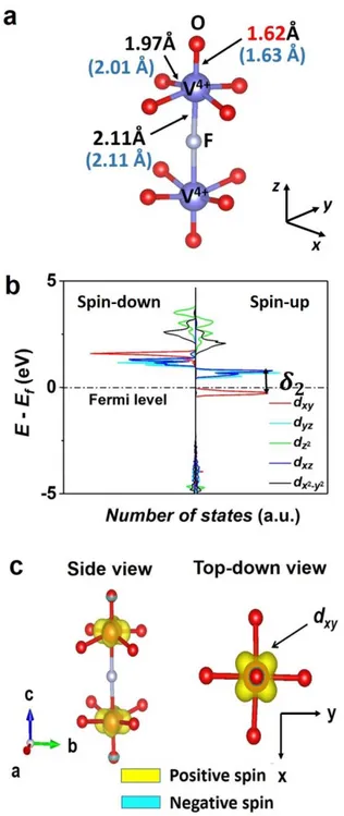
octahedral site formed by four oxygen atoms in the basal plane of the octahedron and two fluorine atoms on the axial positions, one of which being shared with the neighboring V3+ site (Figure 2a), as reported experimentally.30,38 The difference between the experimental and the calculated V–O, V–F, and P–O distances is less than 1% with GGA (Figure 2a). Despite the difference in the nature of fluorine and oxygen, the V3+ site is ‘quasi’-symmetric as the six surrounding bond lengths are nearly similar (~ 2.0 Å) (Figure 2a).
13
Figure 2: (a) Local environment of V3+ in Na3V2(PO4)2F3, with the optimized
distances compared to the experimental ones given in parentheses ; (b) Calculated partial DOS around the vanadium ions in Na3V2(PO4)2F3 using the GGA method; (c) Calculated spin
distribution map surrounding a V2O8F3 bi-octahedral unit. An iso-surface value of 810-3
electron.Å-2 was used for this plot. The positive and negative electron spin are represented in
14
The partial DOS around vanadium ions in Na3V2(PO4)2F3 indicates that the two
unpaired electrons of V3+ ions are partially located in the three dxy, dxz, and dyz (t2g) orbitals (Figure 2b). The x, y, and z directions used to define the orientation of the orbitals were chosen to run along the ‘ligand – Vn+’ interatomic axes: x and y axes running along the V–O bonds in
the basal plane while the z axis orienting along the bi-octahedron axis, all of them are independent of the a, b, and c crystallographic directions of the unit cell. Figure 2c shows the 3D electronic spin distribution map calculated for Na3V2(PO4)2F3, which allows to visualize the
local spin distribution in the t2g orbitals. The two V3+ ions in the bi-octahedral units of
Na3V2(PO4)2F3 exhibit similar spin distribution, with a maximum spin concentration in lobes
pointing in-between the two V–O bonds in the basal plane or in-between the V–O and V–F bonds in the (xz) and (yz) planes, thus confirming the partial occupancy of the three t2g orbitals.
For Na3V2(PO4)2FO2, the vanadium ions reside in octahedral sites formed by four equal
V–O bonds (1.97 Å) in the basal plane, a very short V=O bond (1.62 Å), and a long V–F bond (2.1 Å), where the fluorine is shared with another vanadium ion in the axial position (Figure
3a). The calculated partial DOS on V shows that vanadium ions are in the +4 oxidation state,
as expected, and that the single unpaired electron is located only in the dxy orbital which is perpendicular to the bi-octahedron axis (Figure 3b). Due to the presence of a short vanadyl bond, a distortion occurs on all VO5F sites, lowering the Oh symmetry to C4v locally. As a
consequence, the t2g orbitals of V4+ are split into b2 (dxy) and e (dxz, dyz) sub-groups while eg
orbitals will be split further into b2 (𝑑𝑥2−𝑦2) and a1 (𝑑𝑧2). The splitting scheme and the relative
energy of the orbitals in this case are shown in Figure S4. The orbital 𝑑𝑥𝑦 has therefore the
lowest energy value and is occupied by the single electron of V4+, with the 2 splitting energy
of 0.426 eV being higher than kT at ambient temperature (25 meV) (Figure 3b).The 3D spin density map plotted for a bi-octahedron in Na3V2(PO4)2FO2 confirms the localization of the
15
spin density only in lobes pointing in-between two V–O bonds in the basal plane, which is perpendicular to the bi-octahedron axis (Figure 3c).
Figure 3 : (a) Local environment of V4+ in Na3V2(PO4)2FO2 with the optimized distances
compared to the experimental ones given in parentheses; (b) DOS diagram of V4+ in
Na3V2(PO4)2FO2 calculated by GGA method. Only the ‘spin-up’ state of dxy orbital is
occupied; (c) Calculated spin distribution map surrounding a V2O10F bi-octahedral unit. An
iso-surface value of 210-2 electronÅ-2 was used for this plot. The single electron of V4+ occupies the dxy orbital uniquely. The positive and negative electron spin are represented in
16
Our DFT calculations also provide approximate values of electron spin densities on the nuclei in the structures, thus allowing to compute the expected Fermi contact shifts for 31P and
23Na nuclei by using equations (1), (2) and (3) given in the experimental part.
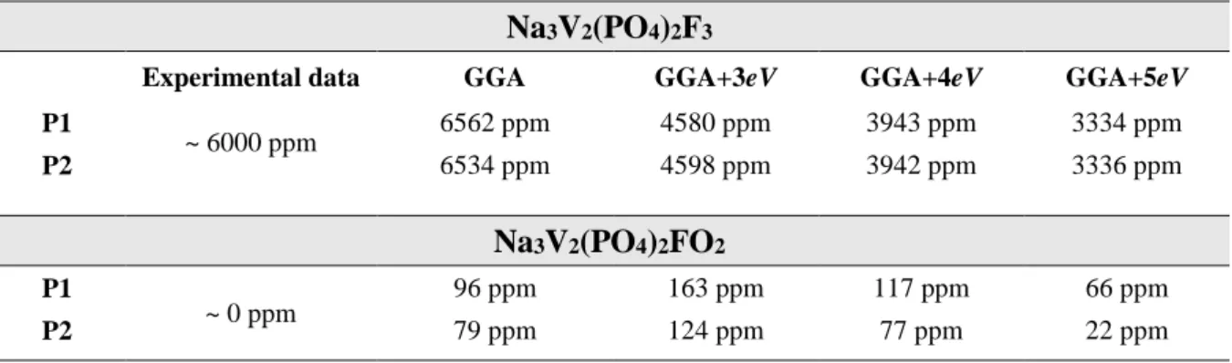
Table 1 : Calculated 31P Fermi contact NMR shifts (in ppm) in Na
3V2(PO4)2F3 and
Na3V2(PO4)2FO2 using GGA and GGA+U methods, compared to the experimental ones.
Na3V2(PO4)2F3
Experimental data GGA GGA+3eV GGA+4eV GGA+5eV
P1 ~ 6000 ppm 6562 ppm 4580 ppm 3943 ppm 3334 ppm P2 6534 ppm 4598 ppm 3942 ppm 3336 ppm Na3V2(PO4)2FO2 P1 ~ 0 ppm 96 ppm 163 ppm 117 ppm 66 ppm P2 79 ppm 124 ppm 77 ppm 22 ppm
Each unit cell of Na3V2(PO4)2F3 or Na3V2(PO4)2FO2 contains two groups of 31P nuclei
with different Fermi contact shifts. As seen in Table 1, the difference in the calculated shifts for these two groups is however negligible. For Na3V2(PO4)2F3, the 31P ss-NMR Fermi contact
shifts computed either by GGA or GGA+U are positive and large, in agreement with the strong interaction expected between V3+ ions and P nuclei. As also expected, as the U value increases, the unpaired electrons are more localized on the t2g orbitals of V3+ and therefore less delocalized
to the neighboring phosphorus nuclei, leading to lower computed shifts.39 Fermi contact shifts
computed within the GGA are in better agreement with the experimental ones. For Na3V2(PO4)2FO2, whatever the method: GGA or GGA+U, all the calculated 31P ss-NMR Fermi
contact shifts are very weak and fall in the typical range of shifts for 31P in diamagnetic compounds, which is also in agreement with the experimental observation (Table 1). This implies that there should be almost no electron spin transfer from the paramagnetic V4+ ions to
17
obtained using the GGA method are in good agreement with the experimental data, and in much better agreement than the GGA+U methods, hence only the GGA results will further be discussed in the following parts.
V3+-O-P / V4+-O-P Spin Transfer Mechanisms
In order to investigate the spin transfer mechanism in Na3V2(PO4)2F3, 3D electronic spin
distribution maps were plotted at different iso-surface values and for different local orientations.
The electron spin transfer occurs through the so-called spin delocalization mechanism, which results from the overlap between the t2g orbitals of the V3+ ions (Figure 4a) and the p
orbitals of its neighboring O, which is in turn hybridized with the sp3 orbitals of P (Figure 4a). Among the three partially filled t2g orbitals of V3+ ions, it appears that dxz and dyz are the two orbitals mostly involved in the spin transfer, as properly oriented for the -overlap with O p orbital that is also overlapping with sp3 orbital of P. This results in a dxz (V3+) – pπ (O) – sp3 (P) hybridization with a 135.8o V–O–P angle as schematized in Figure 4b. In Na
3V2(PO4)2F3,
each phosphorus is surrounded by four V3+ ions and each of them contributes to a Fermi contact shift of around 1500 ppm through the dxz/dyz (V3+) – pπ (O) – sp3 (P) interaction. In Figure 4c, the overall electron spin density map viewed along the c direction shows a strong and positive spin density on the P site in this structure, which is the origin of the highly shifted resonance (6000 ppm) observed on the31P ss-NMR.
18
Figure 4: (a) 3D calculated spin density map showing the electron spin transfer mechanism from a V3+ ion to a neighboring Phosphorus (810-4 electronÅ-2 iso-surface value); (b)
Schematic representation of the spin transfer mechanism from V3+ to P by orbital hybridization model; (c) 3D calculated spin density map (810-4 electronÅ-2 iso-surface
value) showing that each phosphorus nucleus in Na3V2(PO4)2F3 receives the electron spin
from four neighboring V3+ ions. The positive and negative electron spin are represented in yellow and blue, respectively.
19
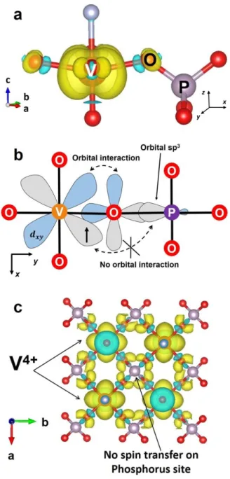
The spin transfer mechanism was also analyzed for Na3V2(PO4)2FO2. As previously
discussed, the two V4+ ions of the bi-octahedral units are similar and exhibit one unpaired electron localized only in the dxy orbital perpendicular to the bi-octahedron axis (Figure 3c and
Figure 5a). As seen in Figure 5b, no orbital overlap is involving this V4+ 3d
xy orbital and the P sp3 one. The V4+ 3dxy orbital is only -hybridized with one O 2p orbital that does not overlap with the P orbitals and the V–O–P angle is equal to 131.9o. However, as can be seen also in
Figure 5b another O 2p orbital, perpendicular to this -bond, is negatively polarized as there is no overlap with V4+ 3d
xy orbital. One could have actually expected a negative Fermi contact shift for 31P in Na3V2(PO4)2FO2, but the participation of the P sp3 orbitals in the negatively deep
polarized level should be very weak. This observation explains why we recorded a 31P signal nearby 0 ppm for Na3V2(PO4)2FO2 despite the presence of paramagnetic V4+ ions around P
20
Figure 5 : (a) 3D calculated spin density map showing the electron spin density surrounding a V4+ ion in Na3V2(PO4)2FO2 (210-3 electronÅ-2 iso-surface value); (b) A 2D projection of
the V–O–P chemical bond on the (xy) plane of the octahedral unit. The single electron of V4+ occupies the dxy orbital, which orients along the diagonal lines of the (xy) plane while the V–
O–P bond is formed along the x/y direction; (c) The spin distribution map calculated at 810-4 electronÅ-2 revealing that there is no spin transfer between V4+ ions and phosphorus nuclei in Na3V2(PO4)2FO2 due to the lack of orbital overlap. The positive and negative electron spin
21
We further calculated the spin distribution map for some intermediate compositions in the Na3V2(PO4)2F3 – Na3V2(PO4)2FO2 system. In Na3V3+V4+(PO4)2F2O (y’ = 1.0), for example,
half of the vanadium ions are expected to be +3 and half +4. The terminal oxygen ions could be distributed differently in the crystal structure leading to different distribution of V3+/V4+ ions
over the vanadium sites. In order to study the relationship between the V3+/V4+ distribution in the structure and the observed 31P ss-NMR shifts, we generated four different input models for Na3V2(PO4)2F2O with four different terminal O-distributions. We assumed that the Na+
ordering in this case was similar to the one used for Na3V2(PO4)2F3. The structures of the input
models are given in Figure S5. The as-mentioned models were relaxed until they reach their minimum energy. As no significant difference in the total energies (E) of all the calculated structural models were spotted, we conclude that they exhibit similar stability; hence, from DFT calculations, we do not predict any V3+ or V4+ ions segregation in the same bi-octahedron. The oxygen distribution on the terminal positions and hence the V3+/V4+ distribution should be controlled solely by statistics leading to a random formation of V3+–F–V3+, V4+–F–V4+ and
V3+–F–V4+ bi-octahedra. This aspect will be further discussed in the following part. Nevertheless, our different models allowed to compute the expected Fermi contact shifts for different V3+/V4+ phosphorus local environments. In all models, the vanadium ions are found in the +3 state, with the same local electronic structure as in Na3V3+2(PO4)2F3, when localized
in VO4F2 octahedral sites, whereas they are found in the +4 oxidation state, with the same local
electronic structure as in Na3V4+2(PO4)2FO2, when localized in VO5F octahedral sites.
Therefore, one expects the same spin transfer mechanisms as those described above for V3+ in Na3V3+2(PO4)2F3 and V4+ in Na3V4+2(PO4)2FO2. As shown in Figure 6 for model 1, where only
the P(OV3+)2(OV4+)2 environment is detected, a spin transfer mechanism does occur only from
the two V3+ ions leading to computed 31P Fermi contact shifts in the range of 3282 - 3285 ppm
22
supplementary information (Figure S6). In Table 2, we summarize the computed Fermi contact shifts depending on the phosphorus environments. DFT calculations thus allowed us to rationalize that in the Na3V2(PO4)2F3 - Na3V2(PO4)2FO2 solid solution, each V3+ ions located in
the proximity of P contributes for ~1500 ppm to the Fermi contact shift whereas a V4+ ion does
not lead to a significant shift. This confirms the signal assignment proposed earlier.14
Figure 6 : The spin distribution map showing P(OV3+)2(OV4+)2 local environment in Model 1
of Na3V2(PO4)2F2O. The positive and negative electron spin are represented in yellow and
blue, respectively.
Table 2 : Phosphorus local environments generated from DFT calculations and their
respective Fermi contact shifts in comparison to those observed experimentally.
DFT calculations Experimental shift (ppm)
Local environment Calculated shift (ppm)
Model 3 P(OV3+)
4 5800 – 5900 6000
Model 2 P(OV3+)
3(OV4+) 4200 – 4600 4500
Model 1/ Model 4 P(OV3+)
2(OV4+)2 3282 – 3285 3000
Model 2 P(OV3+)(OV4+)
3 1700 – 2100 1500
Model 3 P(OV4+)
4 665 ~ 0
In order to further confirm the absence of spin transfer from V4+ ions to the phosphorus nuclei in the Na3V2(PO4)2F3 - Na3V2(PO4)2FO2 system, we chose to compare these results with
those obtained for the partially aluminum-substituted composition Na3V1.5Al0.5(PO4)2F3,
23
Fermi contact with the neighboring P nuclei in the structure. In Na3V1.5Al0.5(PO4)2F3
composition, new phosphorus local environments are generated, i.e. P(OV3+)3(OAl3+),
P(OV3+)2(OAl3+)2, P(OV3+)(OAl3+)3 and P(OAl3+)4, and they should exhibit similar shifts to
those observed in Na3V3+1.5V4+0.5(PO4)2F2.5O0.5. The 31P ss-NMR spectrum of
Na3V1.5Al0.5(PO4)2F3 is shown in Figure 7 and indeed exhibits five different resonances at 6000
ppm, 4500 ppm, 3000 ppm, 1500 ppm, and 0 ppm, very similar to the spectra of the Na3V3+1.6V4+0.4(PO4)2F2.6O0.4 composition. DFT calculations performed for the
Na3V1.5Al0.5(PO4)2F3 composition further confirm that solely the V3+ ions contribute to the 31P
Fermi contact shifts for about 1500 ppm per V3+ through a delocalization mechanism (Figure
7).
Figure 7 : (Top) 3D calculated electronic spin map around P if one of the surrounding V3+ is substituted by Al3+. (Bottom) 31P MAS NMR spectra of Na3V2(PO4)2F2.6O0.4 and
Na3V1.5Al0.5(PO4)2F3.
23Na ss-NMR Measurements in the Na
24
The 23Na ss-NMR signatures of Na3V3+2-y’V4+y’(PO4)2F3-y’Oy’ (0 ≤ y’ ≤ 2) phases have
been studied in details by Park et al 26 who proposed that there were eight different paths to transfer the electron spin from the neighboring vanadium sites to Na+; however, only two of them can give rise to a significant Fermi contact shift.26 There are three different Na sites in the
crystal structure of Na3V3+2-y’V4+y’(PO4)2F3-y’Oy’ phases 14,30 and the Na local environments of Na3V2(PO4)2F3 (y’ = 0) are described in Figure S7. Depending on the positions of the Na+ ions
in the channel, the spin diffusion paths can slightly vary; nevertheless, the two most dominant spin transfer mechanisms always come from the t2g (V) – pπ (F1) – s (Na) orbital overlaps, with F1 as the sharing fluorine atom in the bi-octahedral units. The 23Na ss-NMR Fermi contact shift
depends on the number of V3+/V4+ in its closest bi-octahedral unit.26 In this previous study, the authors ignored the local distortion and the peculiar local electronic structure of the V4+ ions involved in a vanadyl bond. Actually, by considering the partial occupation of t2g orbitals for
V3+ ions (Figure 2b and c) and the dxz occupation for V4+ ions as described previously (Figure
3b and c), the electronic spin transfer mechanism can be described differently. We here
re-describe the spin transfer mechanism between V3+/V4+ and the Na nuclei by considering the peculiar electronic structure of the V4+ ion. In Na3V3+2(PO4)2F3, among all the partially
occupied t2g orbitals, the dxz one is properly oriented to form a π bond with F 2px that is pointing towards the 3s orbital of a nearby Na+ in the channel, leading to a 3dxz (V3+) – 2px (F1) – 3s (Na) hybridized orbital suitable for the spin transfer through a delocalization mechanism (Figure 8a). As a result, a positive Fermi contact shift is observed for 23Na. In Na3V4+2(PO4)2FO2, the single occupied orbital, 3dxy, of the V4+ ions (Figure 3b and c) cannot strongly overlap with the 2px orbital of F1 and the 3s orbital of Na (Figure 8b), resulting in a weaker Fermi contact shift. In Na3V2(PO4)2F3-y’Oy’ oxygen-substituted phases, V3+ and V4+
co-exist in the structure and are distributed randomly over all the vanadium sites. This random distribution thus creates a third Na local environment, in which the Na nucleus resides next to
25
a bi-octahedron containing one V3+ and one V4+. This Na nucleus is exposed to a strong interaction with V3+, and to a weaker one with V4+. The value of the 23Na NMR shift in this case is expected to be lie between the values obtained in Na3V2(PO4)2F3 and in
Na3V2(PO4)2FO2.
Figure 8 : (a) The strong delocalization interaction between the electronic spin on orbital 3dxz (V3+) and Na nuclei in Na3V2(PO4)2F3; (b) The weak delocalization interaction between
26
Figure 9 : Selective single pulse 23Na MAS NMR spectra of Na3V3+2-y’V4+y’(PO4)2F3-y’Oy’ (0 ≤
y’ ≤ 2) samples (B0 = 11.7 T; R= 30 kHz). The Na sites are in yellow and V3+O4F2 and
V4+O5F1 octahedral units in red and blue, respectively.
Note that in our study, we had to consider different Na+-Vacancy orderings or intermediate positions in order to model compounds with partial site occupancies. Therefore, the calculated Fermi contact shifts summarized in Tables S3 and Table S4 do not reflect the exact positions observed experimentally for the 23Na ss-NMR signals and will thus not be further discussed here. Nevertheless, our approach allows to confirm the signals assignment presented in Figure 9.
V3+/V4+ distribution and the relative intensities of 23Na and 31P NMR resonances
As the 23Na and 31P NMR signals assignment was confirmed by our DFT calculations,
one can analyze the relative intensities of the 31P and 23Na signals in order to discuss the local atomic and electronic structures of the series. If the V3+ and V4+ are distributed randomly over the whole structure, the probability of occurrence of each phosphorus or sodium local environment can be estimated using a binomial distribution:
𝐶(𝑛, 𝑘)𝑝𝑘(1 − 𝑝)(𝑛−𝑘) = ( 𝑛!
𝑘! (𝑛 − 𝑘)!) 𝑝
𝑘(1 − 𝑝)(𝑛−𝑘)
In the case of phosphorus, n = 4, i.e. the number of surrounding vanadium sites, k = 0, 1, 2, 3, or 4, i.e, the number of V4+ among the vanadium sites, and p is the percentage of V4+ for a given composition. For Na3V2(PO4)2F3-y’Oy’ compositions, p = y’/2. In the case of sodium,
n = 2 as the shift of each sodium is influenced by two neighboring vanadium sites, and k = 0,
27
Figure 10a compares the experimental and theoretical relative intensities between the
five 31P ss-NMR resonances observed for Na3V2(PO4)2F2O, characterized by half vanadium
ions in the +4 state and half in the +3 state. The experimental values are in good agreement with those expected from binomial distribution supporting the assumption that F/O are distributed randomly in the whole structure.
Figure 10 : Comparison betweeen theoretical and experimental relative intensities of (a) 31P ss-NMR and (b) 23Na ss-NMR resonances recorded on Na3V2(PO4)2F2O.
28
In our previous publication, we stated that “the experimental intensities distribution does not follow the theoretical one if the repartition of V3+ and V4+ ions would have been statistical around each phosphorus: phosphorus nuclei appear to be experimentally surrounded by more V4+ than expected” 14; in fact, the chemical formulas announced in 14 were not correct and we
indeed had more V4+ than we had expected. With a correction of the chemical formula of these compounds in 31, the theoretical and experimental relative intensities determined for the 31P NMR resonances of Na3V2(PO4)2F2O are in good agreement with each other. Note that the 31P
ss-NMR spectrum of Na3V2(PO4)2F2O is quite broad with resonances ranging from 0 ppm to
6000 ppm (Figure 10a), and depending on the position of the off-set of the excitation pulse, their relative intensities can vary significantly. This observation is demonstrated in Figure S8, where the off-set of the excitation was placed at 800 ppm, 2978 ppm, and 5000 ppm: the intensities of resonances close to the off-set are enhanced while those far away are underestimated. Furthermore, the resonances observed in Na3V2(PO4)2F2O have different
relaxation times. When the refocalization delay of the echo acquisition is varied, the intensity of some peaks can be underestimated or overestimated (Figure S9). Therefore, the 31P ss-NMR experimental measurements on Na3V2(PO4)2F2O are just semi-quantitative.
The theoretical relative intensities between 23Na NMR resonances can also be calculated by the binomial equation considering a random distribution of V3+/V4+ on two vanadium positions. The relative intensity of each 23Na NMR signal can be extracted from the peak area
calculated using the DM-fit software that uses different Pseudo-Voigt peak shape functions. All the observed 23Na NMR resonances in the Na3V2(PO4)2F3-y’Oy’ compositions can be fitted with
a Pseudo-Voigt peak shape function, except the resonance at 80 ppm in Na3V2(PO4)2FO2
composition which requires to be described by two peak functions. This could imply a Na ordering in the structure leading to two Na local environments which are slightly different from each other.26 Nevertheless, the intensity of this resonance at 80 ppm was considered to be equal
29
to the sum of the area of these two signals. The theoretical and observed relative intensities between 23Na NMR lines are given in Figure 10b as an example for the Na3V2(PO4)2F2O
composition, the results for the other compositions being reported in Table S5.
The resonant region of the 23Na ss-NMR signals is quite narrow; therefore, there is no
influence of the off-set position on the relative intensities of the 23Na ss-NMR signals, whose determination is thus quantitative. In this case, the binomial equation gives results which are in good agreement with the experimental ones proving that V3+/V4+ are randomly distributed in these structures. Therefore, the relative intensities between 23Na resonances can be used to estimate the y’ value in a Na3V2(PO4)2F3-y’Oy’ composition.
CONCLUSIONS
The local atomic and electronic structures of the compositions belonging to the solid solution Na3V2(PO4)2F3 – Na3V2(PO4)2FO2 have been investigated by a combination of 23Na
and 31P MAS NMR and DFT calculations. The experimental 31P NMR Fermi contact shift values are in agreement with those calculated using the PAW approach implemented in the VASP code considering a Curie-Weiss law. The V3+ ions in Na3V2(PO4)2F3 reside in a
‘quasi’-symmetric octahedral site with its electron spins spreading out to the four neighboring phosphorus nuclei through the orbital overlap between its 3dxz/3dyz orbitals and the sp3 hybridized orbital of phosphorus atoms via the O 2p orbital. Once oxygen substitution occurs, the vanadium ion bonded to this oxygen is oxidized to V4+ and form a vanadyl type bond with a very peculiar electronic structure: (dxy1). This highly covalent bond distorts the local structure of the vanadium ion, imposes further orbital splitting, and suppresses the spin transfer through 3dxz and 3dyz orbitals. A combination of these effects results in the formation of a P(OV4+)4
30
assignment was also confirmed, and based on the relative intensity distribution of the signals properly assigned for both 31P and 23Na we conclude that no segregation of V4+ ions are observed in the Na3V2(PO4)2F3 – Na3V2(PO4)2FO2 electrode materials unlike what was
suspected for the LiVPO4F – LiVPO4O series.15
ASSOCIATED CONTENT
Supporting Information
Synchrotron XRD of Na3V2(PO4)2F3-y’Oy’ (0 ≤ y’ ≤ 2) (Figure S1); Experimental structure and the input model used in the DFT calculations for Na3V2(PO4)2F3 (Figure S2); Experimental and
optimized unit cell parameters of Na3V2(PO4)2F3 calculated by GGA and GGA+U methods
(Table S1); Experimental structure and the input model used in the DFT calculations for Na3V2(PO4)2FO2 (Figure S3); Experimental and optimized unit cell parameters of
Na3V2(PO4)2FO2 calculated by GGA and GGA+U methods (Table S2); The splitting scheme
of the five 3d orbitals of V3+ in Na3V2(PO4)2F3 and those of V4+ in Na3V2(PO4)2FO2 (Figure
S4); Different optimized structures obtained for Na3V2(PO4)2F2O by DFT calculations (Figure
S5); Different spin transfer pathways observed in Na3V2(PO4)2F2O by considering different
V3+:V4+ distributions (Figure S6); Na local environments in Na
3V2(PO4)2F3 (Figure S7); 23Na
NMR Fermi contact shifts calculated by GGA and GGA+U methods for Na3V2(PO4)2F3 and
Na3V2(PO4)2FO2 (Table S3); 23Na NMR Fermi contact shifts calculated by GGA method for
Na3V2(PO4)2F2O (Table S4); 31P ss-NMR spectra of Na3V2(PO4)2F2O recorded at different
off-set positions (Figure S8); 31P ss-NMR spectra of Na3V2(PO4)2F2O recorded at different
refocalization delays (Figure S9). The Supporting Information is available free of charge on the ACS Publications website at DOI: .
31
AUTHOR INFORMATION
Corresponding Author:
*E-mail: Dany.Carlier@icmcb.cnrs.fr
Notes
The authors declare no competing financial interest.
ACKNOWLEDGEMENT
The authors acknowledge the French RS2E Network for the funding of LHBN’s PhD thesis, the RS2E and Alistore-ERI networks for the funding of TB’s postdoctoral fellowship, as well as the financial support of Région Nouvelle Aquitaine, of the French National Research Agency (STORE-EX Labex Project ANR-10-LABX-76-01 and SODIUM Descartes project ANR-13-DESC-0001-02) and of the European Union’s Horizon 2020 research and innovation program under the Grant Agreement No. 646433-NAIADES. The Mésocentre de Calcul Intensif Aquitain (MCIA) and the modelling center of ISM are acknowledged for computing facilities. The authors want to thank Yohan BIECHER (ICMCB-CNRS) for his help with DMfit.
32
REFERENCES
(1) Armand, M.; Tarascon, J.-M. Building Better Batteries. Nature 2008, 451 (7179), 652– 657. (DOI: 10.1038/451652a).
(2) Tarascon, J.-M. and Armand, M. Issues and Challenges Facing Rechargeable Lithium Batteries. Nature 2001, 414 (November), 359–367. (DOI: 10.1038/35104644)
(3) Goodenough, J. B.; Kim, Y. Challenges for Rechargeable Li Batteries. Chem. Mater.
2010, 22, 587–603. (DOI: 10.1021/cm901452z).
(4) Hwang, J.-Y.; Myung, S.-T.; Sun, Y.-K. Sodium-Ion Batteries: Present and Future.
Chem. Soc. Rev. 2017, 46 (12), 3529–3614. (DOI: 10.1039/C6CS00776G).
(5) Pell, A. J.; Pintacuda, G.; Grey, C. P. Progress in Nuclear Magnetic Resonance Spectroscopy Paramagnetic NMR in Solution and the Solid State. Prog. Nucl. Magn.
Reson. Spectrosc. 2019, 111, 1–271. (DOI: 10.1016/j.pnmrs.2018.05.001).
(6) Murakami, M.; Noda, Y.; Koyama, Y.; Takegoshi, K.; Arai, H.; Uchimoto, Y.; Ogumi, Z. Local Structure and Spin State of Cobalt Ion at Defect in Lithium Overstoichiometric LiCoO2 As Studied by 6/7Li Solid-State NMR Spectroscopy. J. Phys. Chem. C 2014, 118
(28), 15375–15385. (DOI: 10.1021/jp5039909)
(7) Carlier, D.; Cheng, J.-H.; Pan, C.-J.; Ménétrier, M.; Delmas, C.; Hwang, B. DFT+U Calculations and XAS Study: Further Confirmation of the Presence of CoO5
Square-Based Pyramids with IS-Co3+ in Li-Overstoichiometric LiCoO
2. J. Phys. Chem. C 2013,
117 (50), 26493–26500. (DOI: 10.1021/jp409850q)
(8) Levasseur, S.; Ménétrier, M.; Shao-Horn, Y.; Gautier, L.; Audemer, A.; Demazeau, G.; Largeteau, A.; Delmas, C. Oxygen Vacancies and Intermediate Spin Trivalent Cobalt Ions in Lithium-Overstoichiometric LiCoO2. Chem. Mater. 2003, 15 (1), 348–354. (DOI:
10.1021/cm021279g)
(9) Ménétrier, M.; Shao-Horn, Y.; Wattiaux, A.; Fournès, L.; Delmas, C. Iron Substitution in Lithium-Overstoichiometric “Li1.1CoO2”: Combined 57Fe Mössbauer and 7Li NMR
Spectroscopies Studies. Chem. Mater. 2005, 17 (18), 4653–4659. (DOI: 10.1021/cm0504384)
(10) Lee, Y. J.; Wang, F.; Grey, C. P. 6Li and 7Li MAS NMR Studies of Lithium Manganate
Cathode Materials. J. Am. Chem. Soc. 1998, 120 (48), 12601–12613. (DOI: 10.1021/ja9817794)
(11) Kim, J.; Middlemiss, D. S.; Chernova, N. A.; Zhu, B. Y. X.; Masquelier, C.; Grey, C. P. Linking Local Environments and Hyperfine Shifts: A Combined Experimental and Theoretical 31P and 7Li Solid-State NMR Study of Paramagnetic Fe(III) Phosphates. J.
Am. Chem. Soc. 2010, 132 (47), 16825–16840. (DOI: 10.1021/ja102678r)
(12) Li, Q.; Liu, Z.; Zheng, F.; Liu, R.; Lee, J.; Xu, G.; Zhong, G.; Hou, X.; Fu, R.; Chen, Z.; et al. Identifying the Structural Evolution of the Sodium Ion Battery Na2FePO4F
Cathode. Angew. Chemie Int. Ed. 2018, 57 (37), 11918–11923. (DOI: 10.1002/anie.201805555)
(13) Xu, J.; Lee, D. H.; Clément, R. J.; Yu, X.; Leskes, M.; Pell, A. J.; Pintacuda, G.; Yang, X.; Grey, C. P.; Meng, Y. S. Identifying the Critical Role of Li Substitution in P2– Nax[LiyNizMn1–y–z]O2 (0 < x , y , z < 1) Intercalation Cathode Materials for High-Energy
33
Na-Ion Batteries. Chem. Mater. 2014, 26 (2), 1260–1269. (DOI: 10.1021/cm403855t) (14) Broux, T.; Bamine, T.; Fauth, F.; Simonelli, L.; Olszewski, W.; Marini, C.; Ménétrier,
M.; Carlier, D.; Masquelier, C.; Croguennec, L. Strong Impact of the Oxygen Content in Na3V2(PO4)2F3–yOy (0 ≤ y ≤ 0.5) on Its Structural and Electrochemical Properties. Chem.
Mater. 2016, 28 (21), 7683–7692. (DOI: 10.1021/acs.chemmater.6b02659)
(15) Bamine, T.; Boivin, E.; Boucher, F.; Messinger, R. J.; Salager, E.; Deschamps, M.; Masquelier, C.; Croguennec, L.; Ménétrier, M.; Carlier, D. Understanding Local Defects in Li-Ion Battery Electrodes through Combined DFT/NMR Studies: Application to LiVPO4F. J. Phys. Chem. C 2017, 121 (6), 3219–3227. (DOI:
10.1021/acs.jpcc.6b11747)
(16) Boivin, E.; David, R.; Chotard, J.-N.; Bamine, T.; Iadecola, A.; Bourgeois, L.; Suard, E.; Fauth, F.; Carlier, D.; Masquelier, C.; et al. LiVPO4F1−yOy Tavorite-Type Compositions : Influence of the Concentration of Vanadyl-Type Defects on the Structure and Electrochemical Performance. Chem. Mater. 2018, 30, 5682–5693. (DOI: 10.1021/acs.chemmater.8b02138)
(17) Boivin, E.; Chotard, J.-N.; Ménétrier, M.; Bourgeois, L.; Bamine, T.; Carlier, D.; Fauth, F.; Masquelier, C.; Croguennec, L. Oxidation under Air of Tavorite LiVPO4F: Influence
of Vanadyl-Type Defects on Its Electrochemical Properties. J. Phys. Chem. C 2016, 120 (46), 26187–26198. (DOI: 10.1021/acs.jpcc.6b07342)
(18) Zhou, L.; Leskes, M.; Ilott, A. J.; Trease, N. M.; Grey, C. P. Paramagnetic Electrodes and Bulk Magnetic Susceptibility Effects in the in Situ NMR Studies of Batteries: Application to Li1.08Mn1.92O4 Spinels. J. Magn. Reson. 2013, 234, 44–57. (DOI:
10.1016/j.jmr.2013.05.011)
(19) Shimoda, K.; Murakami, M.; Takamatsu, D.; Arai, H. Electrochimica Acta In Situ NMR Observation of the Lithium Extraction / Insertion From. Electrochim. Acta 2013, 108, 343–349. (DOI: 10.1016/j.electacta.2013.06.120)
(20) Pecher, O.; Bayley, P. M.; Liu, H.; Liu, Z.; Trease, N. M.; Grey, C. P. Automatic Tuning Matching Cycler (ATMC) in Situ NMR Spectroscopy as a Novel Approach for Real-Time Investigations of Li- and Na-Ion Batteries. J. Magn. Reson. 2016, 265, 200–209. (DOI: 10.1016/j.jmr.2016.02.008)
(21) Poli, F.; Kshetrimayum, J. S.; Monconduit, L.; Letellier, M. Electrochemistry Communications New Cell Design for In-Situ NMR Studies of Lithium-Ion Batteries.
Electrochem. commun. 2011, 13 (12), 1293–1295. (DOI: 10.1016/j.elecom.2011.07.019)
(22) Pecher, O.; Carretero-Gonzalez, J.; Griffith, K. J.; Grey, C. P. Materials’ Methods: NMR in Battery Research. Chem. Mater. 2017, 29 (1), 213–242. (DOI: 10.1021/acs.chemmater.6b03183)
(23) Ogata, K.; Salager, E.; Kerr, C. J.; Fraser, A. E.; Ducati, C.; Morris, A. J.; Hofmann, S.; Grey, C. P. Revealing Lithium-Silicide Phase Transformations in Nano-Structured Silicon-Based Lithium Ion Batteries via in Situ NMR Spectroscopy. Nat. Commun. 2014,
5, 1–11. (DOI: 10.1038/ncomms4217)
(24) Carlier, D.; Ménétrier, M.; Grey, C. P.; Delmas, C.; Ceder, G. Understanding the NMR Shifts in Paramagnetic Transition Metal Oxides Using Density Functional Theory Calculations. Phys. Rev. B 2003, 67 (17), 174103(1)–174103(14). (DOI: 10.1103/PhysRevB.67.174103)
34
(25) Gover, R. K. B.; Bryan, A.; Burns, P.; Barker, J. The Electrochemical Insertion Properties of Sodium Vanadium Fluorophosphate, Na3V2(PO4)2F3. Solid State Ionics
2006, 177 (17–18), 1495–1500. (DOI: 10.1016/j.ssi.2006.07.028)
(26) Park, Y. U.; Seo, D. H.; Kim, H.; Kim, J.; Lee, S.; Kim, B.; Kang, K. A Family of High-Performance Cathode Materials for Na-Ion Batteries, Na3(VO1-xPO4)2F1+2x (0 ≤ x ≤ 1):
Combined First-Principles and Experimental Study. Adv. Funct. Mater. 2014, 24 (29), 4603–4614. (DOI: 10.1002/adfm.201400561)
(27) Qi, Y.; Mu, L.; Zhao, J.; Hu, Y.; Liu, H.; Dai, S. Superior Na-Storage Performance of Low-Temperature-Synthesized Na3(VO1−xPO4)2F1+2x (0 ≤ x ≤ 1) Nanoparticles for
Na-Ion Batteries. Angew. Chemie 2015, 127 (34), 10049–10054. (DOI: 10.1002/ange.201503188)
(28) Broux, T.; Fauth, F.; Hall, N.; Chatillon, Y.; Bianchini, M.; Bamine, T.; Leriche, J.; Suard, E.; Carlier, D.; Reynier, Y.; Simonin, L.; Masquelier, C.; Croguennec, L. High Rate Performance for Carbon‐Coated Na3V2(PO4)2F3 in Na‐Ion Batteries. Small Methods
2019, 3 (4), 1800215(1)–1800215(12). (DOI: 10.1002/smtd.201800215)
(29) Liu, Z.; Hu, Y.; Dunstan, M. T.; Huo, H.; Hao, X.; Zou, H.; Zhong, G.; Yang, Y.; Grey, C. P. Local Structure and Dynamics in the Na Ion Battery Positive Electrode Material Na3V2(PO4)2F3. Chem. Mater. 2014, 26 (8), 2513–2521. (DOI: 10.1021/cm403728w)
(30) Bianchini, M.; Brisset, N.; Fauth, F.; Weill, F.; Elkaim, E.; Suard, E.; Masquelier, C.; Croguennec, L. Na3V2(PO4)2F3 Revisited: A High-Resolution Diffraction Study. Chem.
Mater. 2014, 26 (14), 4238–4247. (DOI: 10.1021/cm501644g)
(31) Nguyen, L. H. B.; Broux, T.; Camacho, P. S.; Denux, D.; Bourgeois, L.; Belin, S.; Iadecola, A.; Fauth, F.; Carlier, D.; Olchowka, J.; Masquelier, C.; Croguennec, L. Stability in Water and Electrochemical Properties of the Na3V2(PO4)2F3 –
Na3(VO)2(PO4)2F Solid Solution. Energy Storage Mater. 2019, 20, 324–334. (DOI:
10.1016/j.ensm.2019.04.010)
(32) Olchowka, J.; Nguyen, L. H. B.; Broux, T.; Sanz Camacho, P.; Petit, E.; Fauth, F.; Dany, C.; Masquelier, C.; Croguennec, L. Aluminum Substitution for Vanadium in the Na3V2(PO4)2F3 and Na3V2(PO4)2FO2 Type Materials. Chem. Commun. 2019, 55, 11719–
11722. (DOI: 10.1039/C9CC05137F)
(33) Massiot, D.; Fayon, F.; Capron, M.; King, I.; Le Calvé, S.; Alonso, B.; Durand, J.; Bujoli, B.; Gan, Z.; Hoatson, G. Modelling One- and Two-Dimensional Solid-State NMR Spectra. Magn. Reson. Chem. 2002, 40 (1), 70–76. (DOI: 10.1002/mrc.984)
(34) Kresse, G.; Furthmüller, J. Efficiency of Ab-Initio Total Energy Calculations for Metals and Semiconductors Using a Plane-Wave Basis Set. Comput. Mater. Sci. 1996, 6 (1), 15–50. (DOI: 10.1016/0927-0256(96)00008-0)
(35) Shannon, R. D.; Prewitt, C. T. Effective Ionic Radii in Oxides and Fluorides. Acta
Crystallogr. Sect. B Struct. Crystallogr. Cryst. Chem. 1969, 25 (5), 925–946. (DOI:
10.1107/S0567740869003220)
(36) Momma, K.; Izumi, F. VESTA: A Three-Dimensional Visualization System for Electronic and Structural Analysis. J. Appl. Crystallogr. 2008, 41 (3), 653–658. (DOI: 10.1107/S0021889808012016)
35
M.; Krug Von Nidda, H. A.; Loidl, A.; Geibel, C.; Rosner, H. Phase Separation and Frustrated Square Lattice Magnetism of Na1.5VOPO4F0.5. Phys. Rev. B - Condens. Matter
Mater. Phys. 2011, 84 (1), 2–13. (DOI: 10.1103/PhysRevB.84.014429)
(38) Le Meins, J.-M.; Crosnier-Lopez, M.-P.; Hemon-Ribaud, A.; Courbion, G. Phase Transitions in the Na3M2(PO4)2F3 Family (M = Al3+, V3+, Cr3+, Fe3+, Ga3+): Synthesis,
Thermal, Structural, and Magnetic Studies. J. Solid State Chem. 1999, 148, 260–277. (DOI: 10.1006/jssc.1999.8447)
(39) Castets, A.; Carlier, D.; Zhang, Y.; Boucher, F.; Marx, N.; Croguennec, L.; Ménétrier, M. Multinuclear NMR and DFT Calculations on the LiFePO4·OH and FePO4·H2O
Homeotypic Phases. J. Phys. Chem. C 2011, 115 (32), 16234–16241. (DOI: 10.1021/jp204767c)