HAL Id: hal-01831754
https://hal.archives-ouvertes.fr/hal-01831754
Submitted on 11 Jul 2018
HAL is a multi-disciplinary open access
archive for the deposit and dissemination of
sci-entific research documents, whether they are
pub-lished or not. The documents may come from
teaching and research institutions in France or
abroad, or from public or private research centers.
L’archive ouverte pluridisciplinaire HAL, est
destinée au dépôt et à la diffusion de documents
scientifiques de niveau recherche, publiés ou non,
émanant des établissements d’enseignement et de
recherche français ou étrangers, des laboratoires
publics ou privés.
Loss of Iroquois homeobox transcription factors 3 and 5
in osteoblasts disrupts cranial mineralization
Corey J. Cain, Nathalie Gaborit, Wint Lwin, Emilie Barruet, Samantha Ho,
Carine Bonnard, Hanan Hamamy, Mohammad Shboul, Bruno Reversade,
Hülya Kayserili, et al.
To cite this version:
Corey J. Cain, Nathalie Gaborit, Wint Lwin, Emilie Barruet, Samantha Ho, et al.. Loss of Iroquois
homeobox transcription factors 3 and 5 in osteoblasts disrupts cranial mineralization. Bone Reports,
2016, Equipe 7, 5, pp.86–95. �10.1016/j.bonr.2016.02.005�. �hal-01831754�
Loss of Iroquois homeobox transcription factors 3 and 5 in osteoblasts
disrupts cranial mineralization
Corey J. Cain
a, Nathalie Gaborit
b,c,d, Wint Lwin
a, Emilie Barruet
a, Samantha Ho
a, Carine Bonnard
e,
Hanan Hamamy
f, Mohammad Shboul
e, Bruno Reversade
e, Hülya Kayserili
g,h,
Benoit G. Bruneau
i,j, Edward C. Hsiao
a,⁎
aDepartment of Medicine, Division of Endocrinology and Metabolism, Institute for Human Genetics, Program in Craniofacial Biology, University of California, San Francisco, San Francisco, CA
94143-0794, USA
b
Inserm, UMR 1087, l’institut du thorax, Nantes, France
c
CNRS, UMR 6291, Nantes, France
d
Université de Nantes, France
e
Human Embryology and Genetics Laboratory, Institute of Medical Biology, A*STAR, Singapore 138648, Singapore
f
Department of Genetic Medicine and Development, Geneva University, Geneva 1211, Switzerland
gMedical Genetics Department, Koc University School of Medicine, Rumelifeneri Yolu, Sarıyer, Istanbul 34450, Turkey h
Medical Genetics Department, Istanbul Medical Faculty, Istanbul University Topkapi, Fatih, 34093 lstanbul, Turkey
i
Gladstone Institute for Cardiovascular Disease, San Francisco, CA 94158, USA
j
Department of Pediatrics, University of California, San Francisco, San Francisco, CA 94143, USA
a b s t r a c t
a r t i c l e i n f o
Article history: Received 25 January 2016 Accepted 2 February 2016 Available online 13 April 2016
Cranial malformations are a significant cause of perinatal morbidity and mortality. Iroquois homeobox transcrip-tion factors (IRX) are expressed early in bone tissue formatranscrip-tion and facilitate patterning and mineralizatranscrip-tion of the skeleton. Mice lacking Irx5 appear grossly normal, suggesting that redundancy within the Iroquois family. However, global loss of both Irx3 and Irx5 in mice leads to significant skeletal malformations and embryonic lethality from cardiac defects. Here, we study the bone-specific functions of Irx3 and Irx5 using Osx-Cre to drive osteoblast lineage–specific deletion of Irx3 in Irx5−/−mice. Although we found that the Osx-Cre transgene
alone could also affect craniofacial mineralization, newborn Irx3flox/flox/Irx5−/−/Osx-Cre+mice displayed
additional mineralization defects in parietal, interparietal, and frontal bones with enlarged sutures and reduced calvarial expression of osteogenic genes. Newborn endochondral long bones were largely unaffected, but we observed marked reductions in 3–4-week old bone mineral content of Irx3flox/flox/Irx5−/−/Osx-Cre+mice. Our
findings indicate that IRX3 and IRX5 can work together to regulate mineralization of specific cranial bones. Our results also provide insight into the causes of the skeletal changes and mineralization defects seen in Hamamy syndrome patients carrying mutations in IRX5.
© 2016 The Authors. Published by Elsevier Inc. This is an open access article under the CC BY license (http://creativecommons.org/licenses/by/4.0/). Keywords: Osteoclast/osteoblast biology Osteoporosis Osteoblast mineralization 1. Introduction
Craniofacial development requires tight coordination of cell migra-tion, proliferamigra-tion, and mineralization of osteogenic lineages (Wilkie & Morriss-Kay, 2001; Franz-Odendaal, 2011). Osteoblast dysfunction is thought to be a major contributor to diseases that affect craniofacial bones and mineralization (Rice, 2008). The complex genetic and spatial interactions that occur during craniofacial development pose major challenges to understanding mineralization during the development of the skull.
Iroquois homeobox domain transcription factors (IRX) are highly conserved proteins that regulate neural, cardiac, and bone develop-ment (Kerner et al., 2009; Cavodeassi et al., 2001; Kim et al., 2012; Li et al., 2014). IRX proteins all contain two highly conserved do-mains. The homeodomain is postulated to regulate interactions be-tween transcriptional regulators by binding to genomic regions to regulate target gene expression, and the IRO Box is involved in protein-protein binding (Cavodeassi et al., 2001; Hiroi et al., 2001). Six IRX transcription factors have been identified (IRX1-IRX6), of which Irx1, Irx2, and Irx4 cluster to chromosome 5 in humans (chro-mosome 13 in mice) and Irx3, Irx5, and Irx6 cluster to chro(chro-mosome 16 (chromosome 8 in mice) (Gaborit et al., 2012; Houweling et al., 2001). Irx3 and Irx5 expression is strikingly similar in developing mouse tissues (Houweling et al., 2001).
⁎ Corresponding author at: University of California, San Francisco, 513 Parnassus Ave., HSE901G, San Francisco, CA 94143-0794, USA.
E-mail address:edward.hsiao@ucsf.edu(E.C. Hsiao).
Contents lists available atScienceDirect
Bone Reports
j o u r n a l h o m e p a g e :w w w . e l s e v i e r . c o m / l o c a t e / b o n r
http://dx.doi.org/10.1016/j.bonr.2016.02.005
IRX proteins are required for the formation of limbs and skeletal tissues. Irx1 is important for the specification of individual placodes through BMP signaling (Glavic et al., 2004). IRX3 has been shown to bind to the Bmp10 promoter, which is important for ventricular septation, while IRX5 can bind to GATA3 and TRPS1 to regulate CXCL12 during bone progenitor migration in Xenopus embryos (Gaborit et al., 2012; Bonnard et al., 2012). Irx1 and Irx2 have been shown to regulate vertebrate digit formation, while Irx3 and Irx5 mediate early mouse limb bud specification by regulating Gli3 expression (Li et al., 2014; Becker et al., 2001; McDonald et al., 2010). Surprisingly, loss of Irx5 alone leads to a grossly normal mouse (Gaborit et al., 2012), while loss of both Irx3 and Irx5 to-gether result in an embryonic lethal phenotype from cardiac defects and skeletal malformations (Li et al., 2014; Gaborit et al., 2012). Unfortu-nately, the early embryonic lethality of Irx3−/−/Irx5−/−mice contributes to our incomplete understanding of the role of Irx3 and Irx5 in osteoblast function (Li et al., 2014; Gaborit et al., 2012).
Two mutations in human IRX5, Ala150Pro and Asn166Lys occur in pa-tients with Hamamy syndrome (OMIM MIM611174;Bonnard et al., 2012), who present with craniofacial dysmorphism, osteopenia, tooth eruption defects, and hip dysplasia, along with cardiac defects and micro-cytic hypochromic anemia (Bonnard et al., 2012; Hamamy et al., 2007a, 2007b). The function of IRX5 seems to differ in mice and humans as the human phenotype is not observed in Irx5−/−mice (Costantini et al., 2005). Iroquois homeodomains helix two and helix three are completely conserved in IRX1-IRX6 (Bonnard et al., 2012). The Hamamy mutations Ala150Pro and Asn166Lys occur in the IRX5 helix two or three respective-ly, indicating the importance of these residues in IRX homeodomain func-tion. Irx5 was previously shown to form homodimers and heterodimers with Irx3 and Irx4 in transfected 10T1/2 cells (He et al., 2009). Previous meta-analysis of IRX3, IRX5, GATA3, and TRPS1 also indicated these pro-teins were involved in a regulatory network (Bonnard et al., 2012). Inter-estingly, patients with a 3.2 Mb deletion at 16q12.2–13, which includes IRX3, IRX5, and IRX6, show similar craniofacial features to those of Hamamy patients (Bonnard et al., 2012; Chang et al., 2010).
Irx3−/−and Irx5−/−mice do not show any Hamamy syndrome characteristics (Costantini et al., 2005; Smemo et al., 2014), but several aspects of the Hamamy syndrome phenotype are recapitulated in Irx3 and Irx5 double knockout mice (Irx3−/−/Irx5−/−mice), including the cardiac defects and craniofacial dysmorphisms (Hiroi et al., 2001). Furthermore, the hind limb skeletal hypoplasia reported in Irx3−/−/ Irx5−/−mouse embryos and the femoral fragility observed in Hamamy syndrome patients suggest that IRX3 and IRX5 are important for limb development in vertebrates (Gaborit et al., 2012; Hamamy et al., 2007a). Although the Irx3−/−/Irx5−/−mice have a complete loss of protein in comparison to the point mutations found in humans with Hamamy syndrome, thefinding that multiple features of Hamamy syndrome phenotype are recapitulated in Irx3−/−/Irx5−/−mice indi-cates that this mouse model may be useful for understanding the role of Iroquois proteins in vivo. Here, we avoid the embryonic lethality from cardiac malformations in Irx3−/−/Irx5−/−mice by using the Osx-Cre transgene (Rodda & McMahon, 2006) to drive osteoblast-specific deletion of Irx3 in Irx5−/−mice.
2. Results
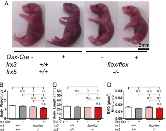
2.1. Body size and craniofacial bone mineralization are reduced in newborn Irx3flox/flox/Irx5−/−/Osx-Cre+mice
We focused our studies on newborn Irx3flox/flox/Irx5−/−/Osx-Cre+
mice (Supplementary Fig. 1A) to understand the role of Irx3 and Irx5 in early bone mineralization. The allelic separation of Irx3 and Irx5 was a rare event (approximately 0.5–2%, Supplementary Fig. 1B) and thus single allele mice were excluded from further analysis. Newborn Irx3flox/flox/Irx5−/−/Osx-Cre+mice appeared grossly normal but slightly smaller than control littermates (Fig. 1A). 43% of Irx3flox/flox/Irx5−/−/ Osx-Cre+were viable at birth, which appeared to be an effect of the
Osx-Cre allele as Irx3+/+/Irx5+/+/Osx-Cre+ also showed similar
decreased neonatal viability (Supplementary Fig. 1C). However, Irx3flox/flox/Irx5−/−/Osx-Cre+mice rarely survived to 4 weeks of age.
This early lethality was not observed in 3–4-week-old Irx3flox/flox/
Irx5−/−/Osx-Cre−mice (Supplementary Fig. 1D) suggesting that the loss of Irx3 and Irx5 contributed to this decrease in survival in the neonatal stage. We observed small but statistically significant reduc-tions in newborn total body weight and length in mice with a loss of Irx5 independent of Osx-Cre expression (Fig. 1B, C and Supplementary Fig. 2A and B). Lower limbs of Irx3flox/flox/Irx5−/−/Osx-Cre+assessed by
Dual energy X-ray absorptiometry (DEXA) showed no differences in bone mineral density (Fig. 1D and Supplementary Fig. 2C). These data indicate that global deletion of Irx5 alone was sufficient to affect body size, but that additional deletion of Irx3 in osteoblastic cells did not have further effects on body size.
During the course of our studies, it became evident that Irx3+/+/
Irx5+/+/Osx-Cre+mice had an unexpected basal phenotype with
reduc-tions in the mineralized area of the frontal bone. This was confirmed by recent reports of effects by the Osx-Cre transgene alone on craniofacial mineralization (Wang et al., 2015; Huang & Olsen, 2015). Micro-computed tomography (microCT) 3D image reconstruction showed both Irx3+/flox/Irx5+/−/Osx-Cre+and Irx3flox/flox/Irx5−/−/Osx-Cre+mice had reduced mineralization in the frontal, parietal, and interparietal bones (Fig. 2and Supplementary Figs. 2D, E, and 3). The long bones from both Irx3+/flox/Irx5+/−/Osx-Cre+and Irx3flox/flox/Irx5−/−/Osx-Cre+ newborn mice also appeared unaffected by microCT.
We next performed whole body skeletal staining of newborn Irx3flox/flox/Irx5−/−/Osx-Cre+mice with alizarin red and alcian blue.
Irx3+/flox/Irx5+/−/Osx-Cre+ and Irx3flox/flox/Irx5−/−/Osx-Cre+ mice
showed reduced mineralization in frontal, parietal, and interparietal bones (Fig. 3A and3B). We measured the frontal bone total mineralized area, using the orbits as landmarks; the suture width in the same area; the length of the parietal and frontal bones along the suture; and the interparietal bone width (Fig. 3C). As noted above, we found reductions in the mineralized area of the frontal bone in Irx3+/+/Irx5+/+/Osx-Cre+ mice; however, Irx3+/flox/Irx5+/−/Osx-Cre+and Irx3flox/flox/Irx5−/−
/Osx-Cre+mice showed an even greater reduction in frontal bone
minerali-zation (Fig. 3D and Supplementary Fig. 2F-I). Additionally, the width of the suture was significantly greater in Irx3flox/flox/Irx5−/−/Osx-Cre−
mice than in Irx3+/+/Irx5+/+/Osx-Cre+and wildtype mice (Fig. 3E and
F). The parietal and frontal bone total length showed a reduction caused by the Osx-Cre but not with Irx3 or Irx5 loss (Fig. 3G). We noted no sig-nificant differences between Irx3+/flox/Irx5+/−/Osx-Cre−and Irx3+/flox/
Irx5+/−/Osx-Cre+mouse cranial measurements (Supplementary
Fig. 2F-L). These data indicate that the presence of the Osx-Cre trans-gene alone can reduce cranial mineralization, but the absence of Irx3 and Irx5 in osteoblastic cells leads to a greater reduction in bone mineralization.
2.2. Irx3flox/flox/Irx5−/−/Osx-Cre+skulls have reduced osteoblastic mineralization
We next used hematoxylin and eosin staining to identify alterations to the bone architecture and mineralization in Irx3flox/flox/Irx5−/− /Osx-Cre+mice. We found that bone accumulation was less in Irx3flox/flox/
Irx5−/−/Osx-Cre+mice in the parietal and interparietal bones than in
Irx3flox/flox/Irx5−/−/Osx-Cre−mice (Fig. 4A). Closer inspection of the frontal, parietal, and interparietal bones revealed a reduction in the thickness of mineralized bone in Irx3flox/flox/Irx5−/−/Osx-Cre+mice
with no major change in cuboidal osteoblasts adjacent to bony surfaces (Fig. 4B, green arrows). Occipital bones showed no significant differences among the genotypes (data not shown). These histological analyses suggest that there is not a major change in osteoblasts mor-phology and numbers at the bone surfaces, but decreased osteoblast mineralization caused by loss of Irx3 and Irx5.
2.3. Irx3flox/flox/Irx5−/−/Osx-Cre+skulls have reduced expression of genes
that regulate osteoblastic mineralization
We next examined whole calvarial expression of genes involved in osteoblast mineralization and maturation. Early markers of osteoblast
lineage specification such as Runx2 (Fig. 5A) and Osx (Fig. 5B) were not significantly altered; however, the mature osteoblast markers Col1a1 (Fig. 5C) and Osteocalcin (Bglap) (Fig. 5D) were significantly reduced in Irx3flox/flox/Irx5−/−/Osx-Cre+calvaria, even in relation to the reductions
in Bglap expression in Irx3+/+/Irx5+/+/Osx-Cre+and Irx3flox/flox/Irx5−/−/
Fig. 2. Irx3flox/flox/Irx5−/−/Osx-Cre+mice have reduced skull mineralization. Representative skull microCT imaging of newborn Irx3flox/flox/Irx5−/−/Osx-Cre+and control littermate skulls.
(A) Right lateral view of the skull. (B) Posterior view of the skull. Green arrows denote the interparietal bone, red arrows denote the parietal bones, and yellow arrows denote the frontal bones. Images are representative of n = 2 of each genotype. (For interpretation of the references to color in thisfigure legend, the reader is referred to the web version of this article.)
Fig. 1. Osteoblastic specific deletion of Irx3 in newborn Irx5−/−mice results in smaller mice. (A) Representative photos of newborn mice. (B) Newborn total body weight, (C) newborn
body length, and (D) newborn BMD of lower right limb of indicated genotypes. Data are from n = 16 Irx3+/+
/Irx5+/+
/Osx-Cre−, n = 5 Irx3+/+
/Irx5+/+
/Osx-Cre+
, n = 27 Irx3flox/flox/ Irx5−/−/Osx-Cre−, n = 9 Irx3flox/flox/Irx5−/−/Osx-Cre+
for body weight and length measurements. For BMD, n = 3 Irx3+/+
/Irx5+/+
/Osx-Cre−, n = 3 Irx3+/+
/Irx5+/+
/Osx-Cre+
, n = 3 Irx3flox/flox/Irx5−/−/Osx-Cre−, n = 3 Irx3flox/flox/Irx5−/−/Osx-Cre+
Osx-Cre−control calvaria. Enam and Tifip11, two genes implicated in min-eralization, were not changed in expression in Irx3flox/flox/Irx5−/− /Osx-Cre+calvaria (data not shown).
We next examined calvarial gene expression of chondrogenesis using Sox9, Col2a1, Acan, and Mmp9, since mesenchymal cells from intermembranous bones maintain chondrogenic gene expression (Aberg et al., 2005). There were no observable differences in Sox9 (Fig. 5E) and Col2a1 (Fig. 5F) levels, although we observed significant reductions of Acan (Fig. 5G) in Irx3flox/flox/Irx5−/−/Osx-Cre−calvaria that were not present in Irx3flox/flox/Irx5−/−/Osx-Cre+calvaria. We
ob-served a significant reduction in Mmp9 expression in Irx3flox/flox/
Irx5−/−/Osx-Cre+calvaria that was not observed in the other
geno-types, even though Irx3+/+/Irx5+/+/Osx-Cre+and Irx3flox/flox/Irx5−/−/
Osx-Cre−calvaria were modestly reduced in Mmp9 expression (Fig. 5H). We observed no differences in expression of apoptosis related genes Bcl2 and Bcl-xl in Irx3flox/flox/Irx5−/−/Osx-Cre+calvaria (Fig. 5I-J).
This indicated that loss of Irx3 and Irx5 together in osteoblastic lineage cells affects later osteogenic genes more significantly than chondrocyte genes and does not result in increased apoptosis of osteogenic cells in the calvaria.
In studies using Xenopus laevis embryos, IRX5 interacted with GATA3 and TRPS1, forming a complex that down regulated CXCL12 production (Bonnard et al., 2012), although IRX3 and IRX5 did not directly influence Gata3 transcription (data not shown). We examined the expression of Cxcl12 and Trps1 in order to determine if the reduced bone mineraliza-tion in Irx3flox/flox/Irx5−/−/Osx-Cre+mice was through downstream
Fig. 3. Irx3flox/flox/Irx5−/−/Osx-Cre+
display reduced mineralization in frontal, parietal, and interparietal bones. (A) Left lateral view and (B) superior view of representative alizarin red and alcian blue stained newborn mice. Green arrows denote the interparietal bone, red arrows denote the parietal bones, and purple arrows denote the frontal bones. (C) Superior view images were measured for (D) the width of mineralized frontal bone, (E) sagittal suture width, (F) suture width divided by the width between the eye sockets, and (G) length of the frontal and parietal bones, measured along the sagittal suture. Photos and measurements are from n = 5 Irx3+/+/Irx5+/+/Osx-Cre−, n = 4 Irx3+/+/Irx5+/+/Osx-Cre+, n = 7 Irx3flox/flox/Irx5−/−
/Osx-Cre−, n = 6 Irx3flox/flox/Irx5−/−/Osx-Cre+
. Statistical differences were determined by a Student's t-test, *pb 0.05, **p b 0.01, ***p b 0.001. N.S., not significant. (For interpretation of the references to color in thisfigure legend, the reader is referred to the web version of this article.)
mediators of Gata3, Irx3, and Irx5. Cxcl12 expression was not signi ficant-ly altered in Irx3flox/flox/Irx5−/−/Osx-Cre+mice (Fig. 5K). Interestingly, there was a significant reduction in Trps1 in Irx3flox/flox/Irx5−/−
/Osx-Cre+calvaria (Fig. 5L), a gene that can reduce Bglap expression in vitro
and is required for proper osteoblast mineralization (Piscopo et al., 2009; Kuzynski et al., 2014). Thesefindings suggest a role for Irx3 and Irx5 in the regulation osteoblast mineralization gene expression and suggest that in mineralization, Irx3 and Irx5 may function through a pathway that is distinct from Gata3.
2.4. Older Irx3flox/flox/Irx5−/−/Osx-Cre+mice have reduced bone
mineralization
We next looked at whole body and skeletal mineralization of Irx3flox/
flox/Irx5−/−/Osx-Cre+
mice that survived to 3–4 weeks of age to identify if there were similar reductions in bone mineralization described in Hammy patients (Hamamy et al., 2007a). 3–4-week-old Irx3flox/flox/
Irx5−/−/Osx-Cre+mice had significant reductions in body size and length (Fig. 6A-C), while body weights remained comparable in all the other genotypes through 12 weeks of age, with the exception of a signif-icant decrease in bodyweight in 12 week old Irx3+/flox/Irx5+/−/Osx-Cre+ mice (Fig. 6D-E). Alizarin red and alcian blue staining of 3–4-week-old Irx3flox/flox/Irx5−/−/Osx-Cre+mice revealed an overall reduction bone
mineralization with gross skeletal abnormalities and spontaneous frac-tures in 3 out of the 10 mice that were analyzed (Fig. 6F). Additionally, cranial bone mineralization appeared reduced in Irx3flox/flox/Irx5−/−/ Osx-Cre+ mice, mineralized by 3.5 weeks of age (Supplementary
Fig. 6G-H). Irx3flox/flox/Irx5−/−/Osx-Cre+mice whole body bone mineral density (BMD) were unchanged compared to control littermates but there was a significant decrease in bone mineral content (BMC), consistent with the reduced bone size (Supplementary Fig. 6I-L). Unfor-tunately, the high lethality of Irx3flox/flox/Irx5−/−/Osx-Cre+mice limited
our ability to generate sufficient numbers to adequately analyze the 4 week phenotype further. These data indicate that deletion of Irx3 and Irx5 in osteoblastic cells can influence both body size and bone min-eralization in both newborn and older mice.
2.5. Bone density is reduced in Hamamy syndrome patients
Hamamy patient mutations in IRX5 result in craniofacial dysmorphisms and mineralization defects while loss of Irx5 in mice results in no detectable bone abnormalities (Li et al., 2014). Since the global loss of both Irx3 and Irx5 leads to cardiac phenotypes similar to those seen in Hamamy patients, we wanted to see if the decreased min-eralization we found in Irx3flox/flox/Irx5−/−/Osx-Cre+mice also reflected
the clinical presentation of Hamamy syndrome patients.
Fig. 4. Osteoblastic bone mineralization is reduced in Irx3flox/flox/Irx5−/−/Osx-Cre+
skulls. (A) Representative sagittal sections of Irx3flox/flox/Irx5−/−/Osx-Cre−and Irx3flox/flox/Irx5−/− /Osx-Cre+
skulls stained with hematoxylin and eosin. Frontal, parietal, and interparietal bones are shown in the boxes and taken at higher magnification to observe cellular morphology. (B) Irx3+/+
/Irx5+/+
/Osx-Cre−, Irx3+/+
/Irx5+/+
/Osx-Cre+
, Irx3flox/flox/Irx5−/−/Osx-Cre−, and Irx3flox/flox/Irx5−/−/Osx-Cre+
enlarged photos of frontal, parietal, interparietal, and occipital bones. Green arrows denote regions with mineralized bone in Irx3flox/flox/Irx5−/−/Osx-Cre+
mice and control littermates. Data is representative n = 3 Irx3+/+
/Irx5+/+
/Osx-Cre−, n = 3 Irx3+/+
/Irx5+/+
/Osx-Cre+
, n = 5 Irx3flox/flox/Irx5−/−/Osx-Cre−, n = 4 Irx3flox/flox/Irx5−/−/Osx-Cre+
. (For interpretation of the references to color in thisfigure legend, the reader is referred to the web version of this article.)
Patients with Hamamy syndrome at 8 and 9 years of age displayed reduced bone mineral density with spine lumbar Z-scores of−3.7 and −1.5 (Table 1). Bone mineral density improved with age in these patients to−2.7 and −1.4 at ages 19 and 20 years of age, respectively (Table 1). The femoral Z-score was determined at 9 years of age for one Hamamy patient (femoral Z-score of−2.2), but both patients fem-oral Z-scores remained above−1.0 at 19 and 20 years of age (Table 1). We noted dramatic reductions in bone mineral content in 3–4 week old Irx3flox/flox/Irx5−/−/Osx-Cre+mice with spontaneous fractures in 3 out of
10 mice, indicating that 3–4 week old Irx3flox/flox/Irx5−/−/Osx-Cre+mice
have similarities to Hamamy patient bone mineralization. Furthermore, the spontaneous fractures observed in 3–4 week old Irx3flox/flox/Irx5−/−/
Osx-Cre+mice resembles the bone fragility reported in femora and
other long bones of 8–10 year old Hamamy patients (Hamamy et al., 2007a).
3. Discussion
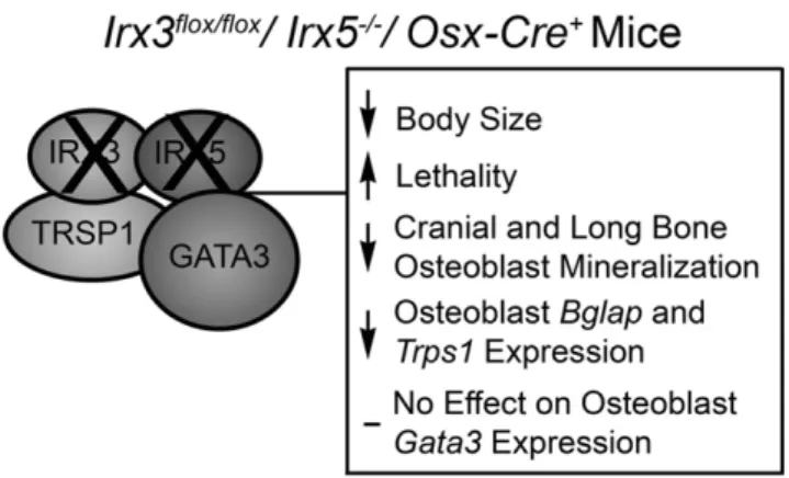
Proper craniofacial development requires control of bone minerali-zation by osteoblastic cell lineages (Mackie et al., 2008; Percival & Richtsmeier, 2013). Our studies show that osteoblast-specific loss of Irx3 and Irx5 leads to impaired mineralization in a very specific subset of cranial bones, possibly by blocking their expression of mature osteoblast mineralization genes (Fig. 7).
During the course of our study, we unexpectedly discovered that Osx-Cre mice have a newborn mineralization defect independent of the Irx3 and Irx5 mutation status. Our studies are consistent with recent reports that Osx-Cre mice alone have a newborn bone mineralization defect, specifically in intramembranous bones (Wang et al., 2015; Huang & Olsen, 2015). Interestingly, the absence of both Irx3 and Irx5 in osteoblastic cells caused an even more dramatic defect in intramembranous mineralization. Furthermore, Osx-Cre mice can sur-vive past weaning and later stages of bone development occur normally,
whereas Irx3flox/flox/Irx5−/−/Osx-Cre+ mice experience premature
lethality around 3.5–4 weeks of age with bone fragility and spontaneous fractures. This indicates that the absence of both Irx3 and Irx5 in osteoblastic cells can influence neonatal survival at later stages of devel-opment. Ourfindings also emphasize the importance of using Osx-Cre+
littermates as controls for studies involving skeletal development. Fur-thermore, our results suggest that other Cre drivers, such as Runx2-Cre or Bglap-Cre mice, may be useful for future studies to confirm early skeletal mineralization phenotypes (Elefteriou & Yang, 2011).
Irx3flox/flox/Irx5−/−/Osx-Cre+mice that survive to 3–4 weeks of age
have smaller femora and tibiae and appeared to have signs of bone fragility, which is consistent with reports of Hamamy syndrome patients developing bone fragility and long bone fractures later in life (Hamamy et al., 2007a). Hamamy syndrome patients also had reduced BMD that was not observed in either newborn or 3–4 week old Irx3flox/ flox/Irx5−/−/Osx-Cre+mice. BMD measurements in mice are not
particu-larly sensitive and more detailed analysis of Irx3flox/flox/Irx5−/−/Osx-Cre+
bones may be warranted. Our data demonstrate that IRX3 and IRX5 are important for both early osteoblast mineralization function and later skeletal mineralization, which also will help in understanding the bone fragility that occurs in Hamamy patients.
Our use of osteoblast specific deletion of Irx3 in Irx5−/−mice differs
from previous models that have germ-line deletions of Irx3 and Irx5 or deletions of Irx1 and Irx2 in chick embryos, all of which showed severe limb defects (Li et al., 2014; Diaz-Hernandez et al., 2013). We were sur-prised tofind that use of the Osx-Cre to delete Irx3 in Irx5−/−mice did
not lead to significant limb malformations in newborn Irx3flox/flox/
Irx5−/−/Osx-Cre+mice; this is likely due to the fact that endochondral bone patterning is determined much earlier in development and may involve a different subset of cell types (Mackie et al., 2008; Knothe Tate et al., 2008). Furthermore, Irx3 and Irx5 germline deletion resulted in increased sonic hedgehog signaling sensitivity through upregulation of Ptc1 and Gli1, which are important for early establishment of limb
Fig. 5. Mineralization and chondrocyte gene expression is reduced expression in Irx3flox/flox/Irx5−/−/Osx-Cre+
calvaria. Relative expression levels of osteoblastic genes (A) Runx2, (B) Osx, (C) Col1a1, and (D) Bglap. Relative expression levels of chondrogenic genes (E) Sox9, (F) Col2a1, (G) Acan, and (H) Mmp9. Relative expression levels of apoptosis related genes (I) Bcl2 and (J) Bcl-xl. Relative expression levels of Irx3 and Irx5 related genes (K) Cxcl12 and (L) Trps1. All samples are from whole newborn calvaria, n = 5 Irx3+/+/Irx5+/+/Osx-Cre−, n = 3 Irx3+/+/
Irx5+/+
/Osx-Cre+
, n = 4 Irx3flox/flox/Irx5−/−/Osx-Cre−, n = 5 Irx3flox/flox/Irx5−/−/Osx-Cre+
. Whole calvaria samples were normalized to Irx3+/+
/Irx5+/+
/Osx-Cre−mice. Statistical differences were determined by a Student's t-test, *pb 0.05, **p b 0.01, ***p b 0.001. N.S., not significant.
Fig. 6. Postnatal Irx3flox/flox/Irx5−/−/Osx-Cre+
mice show reductions in body weight and body length. (A) Representative photos of Irx3 and Irx5 genotypes Irx3flox/flox/Irx5−/−/Osx-Cre−, and Irx3flox/flox/Irx5−/−/Osx-Cre+mice at 3.5 weeks of age. (B) Body weight and (C) body length of 3–4-week-old Irx3flox/flox/Irx5−/−/Osx-Cre+
mice and littermate controls. Body weight of (D) 8- and (E) 12-week-old mouse littermate controls, without Irx3flox/flox/Irx5−/−/Osx-Cre+mice due to lethality at 4 weeks of age. (F) Ventral, (G) superior, and (H) lateral views of
alizarin red and alcian blue stained Irx3flox/flox/Irx5−/−/Osx-Cre+
and control littermates. Red arrow in (F) denotes spontaneous fracture in Irx3flox/flox/Irx5−/−/Osx-Cre+
tibia. 3–4-week old whole body (I) bone mineral density (BMD) and (J) bone mineral content (BMC) of Irx3flox/flox/Irx5−/−/Osx-Cre+
and control littermates. Femur (K) and tibia (L) length from 3 to 4 week old Irx3flox/flox/Irx5−/−/Osx-Cre+and control littermates. 3–4-week old body weight and length old data is from n = 15 (n = 10 for length measurement) Irx3+/+
/Irx5+/+
/Osx-Cre−, n = 10 (n = 9 for length measurement) Irx3+/+
/Irx5+/+
/Osx-Cre+
, n = 15 (n = 12 for length measurement) Irx3+/flox/Irx5+/−/Osx-Cre−, n = 12 (n = 13 for length measurement) Irx3+/flox/Irx5+/−/Osx-Cre+
, n = 3 (n = 3 for length measurement) Irx3flox/flox/Irx5−/−/Osx-Cre−, and n = 4 (n = 4 for length measurement) Irx3flox/flox/Irx5−/− /Osx-Cre+
mice. For body mass measurements at 8 and 12 weeks of age, data are from n = 7 Irx3+/+
/Irx5+/+
/Osx-Cre−, n = 5 Irx3+/+
/Irx5+/+
/Osx-Cre+
, n = 10 Irx3+/flox/Irx5+/−/Osx-Cre− , n = 7 Irx3+/flox/Irx5+/−/Osx-Cre+, and n = 2 Irx3flox/flox/Irx5−/−/Osx-Cre−mice, pooled sexes. For BMD and BMC, data are from n = 8 Irx3+/+/Irx5+/+/Osx-Cre−, n = 5 Irx3+/+/Irx5+/ +
/Osx-Cre+
, n = 9 Irx3+/flox/Irx5+/−/Osx-Cre−, n = 8 Irx3+/flox/Irx5+/−/Osx-Cre+
, n = 2 Irx3flox/flox/Irx5−/−/Osx-Cre−mice, and n = 2 Irx3flox/flox/Irx5−/−/Osx-Cre+
mice, pooled sexes. For femur and tibia measurements, data are from n = 6 (n = 5 for tibia) Irx3+/+
/Irx5+/+
/Osx-Cre−, n = 6 Irx3+/+
/Irx5+/+
/Osx-Cre+
, n = 5 Irx3flox/flox/Irx5−/−/Osx-Cre−mice, and n = 11 Irx3flox/flox/Irx5−/−/Osx-Cre+
formation and osteoblast proliferation, but the contribution to the ex-pression of bone mineralization genes was minimal (Li et al., 2014; Pan et al., 2013). This further supports the notion that Irx3 and Irx5 play roles in both limb and cranial development, and in cranial and limb mineralization by osteoblast lineage cells.
While it is clear that IRX3 and IRX5 can regulate cranial bone mineralization, the mechanism for how IRX3 and IRX5 control bone mineralization remains unclear. IRX5 might bind to GATA3 and TRPS1 proteins in a complex that regulates cranial neural crest cell migration (Bonnard et al., 2012). However, our results did not show differential expression of Cxcl12 (Gaborit et al., 2012), a target of IRX3 and IRX5 which is thought to be regulated by GATA3. In addition, the affected bones in Irx3flox/flox/Irx5−/−/Osx-Cre+mice were largely derived from
the mesenchyme, rather than neural crest derivatives. Furthermore, Trps1 levels were significantly reduced in Irx3flox/flox/Irx5−/−/Osx-Cre+
mice suggesting that loss of mineralization is occurring through a Trps1 specific pathway (Piscopo et al., 2009). Indeed, previous studies have shown that murine Irx5 co-immunoprecipitated with Gata3 and Trps1. When co-immunoprecipitation was done with these proteins with the Irx5 Asn166Lys mutation, there was markedly less binding to Trps1 and Gata3, demonstrating that Irx5 binding is reduced by Hamamy mutations and that Trps1 binding to Irx3 and Irx5 is likely affected in Irx3flox/flox/Irx5−/−/Osx-Cre+mice (Bonnard et al., 2012).
Gata3−/− mice are embryonically lethal from noradrenaline deficiency (Lim et al., 2000) and Gata3−/−rescued embryos display cranial bone development defects, but Gata3+/−mice appear to have no bone developmental abnormalities, which suggests that Gata3 is im-portant for the development of skeletal tissues, but may not be involved in the regulation of bone mineralization gene expression (Lim et al., 2000; Pandolfi et al., 1995). Unfortunately, the expression of Trps1 has not been reported in Gata3+/−and Gata3−/−mice (Lim et al., 2000). Future studies to understand how Trps1 is regulated at specific stages of osteoblast mineralization, and if this defect is associated with the early lethality prior to puberty, will help us to determine the role of Trps1 in the reduced mineralization of Irx3flox/flox/Irx5−/−/Osx-Cre+
mice.
Finally, why mice require loss of both IRX3 and IRX5 to develop reductions in mineralization similar to Hamamy patients who carry a nonsense mutation in the IRX5 gene remains unclear (Gaborit et al., 2012). One notion to explain the similar reductions in bone mineraliza-tion between Irx3flox/flox/Irx5−/−/Osx-Cre+mice and Hamamy patients is
that deletion of both IRX3 and IRX5 in osteoblasts removes the ability to compensate for the loss of these proteins, whereas Hamamy IRX5 non-sense mutations influences IRX3 and IRX5 heterodimer formation and downstream mineralization function (Gaborit et al., 2012; He et al., 2009). More detailed analysis of Hamamy syndrome patients cells using a human induced pluripotent stem cell model may help demonstrate the dynamics of IRX3 and IRX5 interactions in Hamamy patient cells.
In conclusion, we identified a novel role for IRX3 and IRX5 in early cra-nial mineralization of osteoblastic cells. Furthermore, Irx3flox/flox/Irx5−/−/ Osx-Cre+mice displayed reduced bone mineralization without affecting
early osteogenic gene expression. Finally, ourfinding that Irx3flox/flox/
Irx5−/−/Osx-Cre+mice have reduced osteoblastic mineralization
indi-cates that IRX3 to IRX5 binding maintains an important role in Hamamy syndrome and understanding the role of IRX3 and IRX5 together will help provide insight into the roles of IRX proteins in other organs. 4. Methods
4.1. X-ray analysis of Jordanian and Turkish patients with Hamamy Syndrome
The Jordanian patients were originally described by Hanan Hamamy at Jordan University Hospital (Hamamy et al., 2007a). The Turkish family was diagnosed by Hülya Kayserili at the Medical Genetics Department of the Istanbul Medical Faculty (Bonnard et al., 2012; Hamamy et al., 2007a). Both sets of patients provided informed consent for radiographs to be published, and all studies have been approved by the local ethic commissions as described (Bonnard et al., 2012). Radio-graph analysis was done on IRX5 Asn166Lys and IRX5 Ala150Pro patients and compared to control to assess osteopenia and craniofacial dysmorphisms. DEXA was used to measure area, BMD, and BMC of the lumbar spine and femoral neck, and to calculate Z-score. DEXA were performed just before puberty age (8–9 years old) and at adult age (19–20 years old).
4.2. Mice
All transgenic mouse studies were approved by and performed in ac-cordance with the Institutional Animal Care and Use Committee at the University of California, San Francisco. Irx3flox/flox/Irx5−/−mice were gen-erated as described (Gaborit et al., 2012). To create tissue specific Irx3 knockout in osteoblast-lineage cells, we crossed Irx3flox/flox/Irx5−/− mice with Osx-Cre hemizygous transgenic mice (Jackson Laboratory; strain: B6·Cg-Tg(Sp7(Osx)-tTA,tetO-EGFP/cre)1Amc/J Jackson ID: 6361) to generate Irx3flox/flox/Irx5−/−/Osx-Cre+ mice (Rodda &
McMahon, 2006). Osx-Cre+transgenic mice were found to have early
le-thality (Supplemental Fig. 1C) and a modest newborn bone phenotype that subsided by 3–4 weeks of age, which has been previously reported (Wang et al., 2015). We obtained 43% viability of Irx3flox/flox/Irx5−/−/ Osx-Cre+at birth. Since the Irx3 and Irx5 loci are close together, these
alleles segregate independently only at low frequency (Supplemental Fig. 1) and so these genotypes were not analyzed. In addition, we found that Osx-Cre+mice have a skeletal mineralization deficiency at
birth, consistent with recent reports (Wang et al., 2015; Huang & Olsen, 2015). Thus, all experiments include these mice as a control in our analyses. For gene expression analysis, breeding pairs of Irx3flox/+/ Irx5+/−/Osx-Cre+crossed with Irx3flox/+/Irx5+/−/Osx-Cre+mice were
used with no detectable phenotypic differences observed in Osx-Cre sin-gle and double transgenic mice. All data are from both male and female mice.
Table 1
Hamamy Patient Z-scores.
IRX5 Asn166Lys Patient 1 Patient 2 Age (years) 9 20 8 19 Z-score lumbar spine −1.5 −1.4 −3.7 −2.7 Z-score femoral neck −2.2 Greater than−1 N.D. Greater than−1
Fig. 7. Model for the effect of Irx3 and Irx5 on bone mineralization. IRX3 and IRX5 are required for proximal and anterior limb development and maintain a role in osteoblast mineralization. In the absence of Irx3 and Irx5 in osteoblast lineage cells, there is a reduction in osteoblast mineralization, most notably in bones that undergo intramembranous ossification and later in the general skeleton. Osteoblastic cells that lack IRX3 and IRX5 display reduced Col1a1, Bglap, Mmp9, and Trps1 and there is reduced bone formation in intramembranous bones, specifically in the frontal, parietal, and interparietal bones.
4.3. Alizarin red and alcian blue staining of skeletons
Newborn mice and 3–4 week old mice of both sexes were eutha-nized and prepped for alizarin red and alcian blue skeletal staining (Ovchinnikov, 2009) byfixing in 100% ethanol for 24 h. Samples were then switched to acetone (Sigma-Aldrich) for an additional 24 h. Once fixed, samples were stained with final concentration of 5% glacial acetic acid, 0.5% alizarin red S Aldrich), 0.9% alcian blue 8GX (Sigma-Aldrich) in ethanol for 3 h at 37 °C and then at room temperature for 24 h. Samples were then placed in 1% KOH (Amresco) for 3 h and replaced with fresh KOH until non-bone tissue was transparent. Samples were then replaced with increasing concentrations of glycerol and photographed with a Leica MZFLIII dissection microscope with Diagnostic Instruments 14.2 Color Mosaic camera for newborn samples. 3–4 week old samples were photographed with a Nikon E5200 without a microscope.
4.4. Histology
Newborn skulls were skinned andfixed in neutral buffered formalin for at least 48 h and then replaced with 70% ethanol for at least 24 h. Skull tissues were paraffin embedded and sectioned. Skulls were then cut at the midline and then stained with hematoxylin & eosin, using standard protocols (J. David Gladstone Institutes Histology Core). 4.5. Bone densitometry and microCT imaging
DEXA was used to measure mouse whole-body BMD and BMC. Mice were anesthetized with inhaled isofluorane (1.5% to 2% in oxygen) and scanned on a GE Lunar Piximus2 (Piximus). Newborn mice that underwent whole-mouse microCT scans were sacrificed and stored in 70% ethanol before scanning. Ex vivo images were obtained on a Scanco vivaCT-40 microCT scanner (SCANCO) at an X-ray energy of 55 kV, with sigma 0.8/support 1/threshold 120 (103.7 mg HA/cm3), a
voxel size of 76μm, and integration times of 200 ms for whole-body images.
4.6. RNA isolation, cDNA synthesis, and qPCR
Whole calvaria or dissected calvarial tissues were placed in Trizol (Invitrogen) and homogenized using a Powergen 125 homogenizer (Fisher). RNA was isolated using chloroform extraction for whole calvaria or by Picopure RNA isolation columns for dissected calvaria (Life Technologies). Purified mRNA was then used as a template to syn-thesize cDNA with oligo dT primers with the Superscript III (Invitrogen) kit as described (Cain & Manilay, 2013). qPCR expression analysis was performed using TaqMan primers for qPCR reactions (Supplementary Table 1) on a Viia7 real-time thermocycler (Applied Biosystems) run in 5μl sample volumes in triplicate or preamplified using Fludigm preamplication qPCR mix and assayed using Fluidigm dynamic array IFC qPCR plates (Fludigm). All expression values were normalized to Gapdh levels.
4.7. Statistics
Differences between the means of biological replicates for all analy-ses were calculated using two tailed Student's T-test (GraphPad Prism. La Jolla, CA). Analyses were considered statistically significant if p ≤ 0.05.
Supplementary data to this article can be found online athttp://dx. doi.org/10.1016/j.bonr.2016.02.005.
Conflict of interest statement
Edward Hsiao receives funding from Clementia Pharmaceuticals for an unrelated clinical trial.
Acknowledgments
The authors would like to thank J. Bush and R. Nissenson for their valuable comments and discussion; A. Li and Z. Chang with the San Francisco VA Medical Center Bone Histomorphometry Core, T. Alliston with the Department of Orthopaedic Surgery, and C. Miller with the Gladstone Histology Core and Aaron Mattingly and Kate Jordan for tech-nical assistance. B. Reversade is a fellow of the Branco Weiss Foundation, an EMBO Young Investigator and a recipient of the A*STAR Investigator award. This work was supported in part by a Strategic Positioning Fund on Genetic Orphan Diseases from the Biomedical Research Council, A*STAR, Singapore (SPF2012/005), the National Institutes of Health (NIH) Fellowship Training Grants (5T32GM007085-35, 5T32DK007418-34, and 5T32DK007161-41 to C. Cain), NIH K08 (AR056299 to E. Hsiao), NIH R01 (HL93414 ARRA to B. Bruneau), the UCSF Department of Medicine (to E. Hsiao), and by the Marie Curie European Actions (PIIF-GA-2012-331436 to N. Gaborit).
References
Aberg, T., Rice, R., Rice, D., Thesleff, I., Waltimo-Siren, J., 2005.Chondrogenic potential of mouse calvarial mesenchyme. J. Histochem. Cytochem. 53, 653–663.
Becker, M.B., Zulch, A., Bosse, A., Gruss, P., 2001.Irx1 and Irx2 expression in early lung development. Mech. Dev. 106, 155–158.
Bonnard, C., Strobl, A.C., Shboul, M., Lee, H., Merriman, B., Nelson, S.F., Ababneh, O.H., Uz, E., Guran, T., Kayserili, H., Hamamy, H., Reversade, B., 2012.Mutations in IRX5 impair craniofacial development and germ cell migration via SDF1. Nat. Genet. 44, 709–713.
Cain, C.J., Manilay, J.O., 2013.Hematopoietic stem cell fate decisions are regulated by Wnt antagonists: comparisons and current controversies. Exp. Hematol. 41, 3–16.
Cavodeassi, F., Modolell, J., Gomez-Skarmeta, J.L., 2001.The Iroquois family of genes: from body building to neural patterning. Development 128, 2847–2855.
Chang, C.F., Li, L.H., Wang, C.H., Tsai, F.J., Chen, T.C., Wu, J.Y., Chen, Y.T., Tsai, A.C., 2010.
Identification of a submicroscopic 3.2 Mb chromosomal 16q12.2-13 deletion in a child with short stature, mild developmental delay, and craniofacial anomalies, by high-density oligonucleotide array-a recognizable syndrome. Am J Med Genet A 152A, 2365–2371.
Costantini, D.L., Arruda, E.P., Agarwal, P., Kim, K.H., Zhu, Y., Zhu, W., Lebel, M., Cheng, C.W., Park, C.Y., Pierce, S.A., Guerchicoff, A., Pollevick, G.D., Chan, T.Y., Kabir, M.G., Cheng, S.H., Husain, M., Antzelevitch, C., Srivastava, D., Gross, G.J., Hui, C.C., Backx, P.H., Bruneau, B.G., 2005.The homeodomain transcription factor Irx5 establishes the mouse cardiac ventricular repolarization gradient. Cell 123, 347–358.
Diaz-Hernandez, M.E., Bustamante, M., Galvan-Hernandez, C.I., Chimal-Monroy, J., 2013.
Irx1 and Irx2 are coordinately expressed and regulated by retinoic acid, TGFbeta and FGF signaling during chick hindlimb development. PLoS One 8, e58549.
Elefteriou, F., Yang, X., 2011.Genetic mouse models for bone studies—strengths and limitations. Bone 49, 1242–1254.
Franz-Odendaal, T.A., 2011.Induction and patterning of intramembranous bone. Front. Biosci. (Landmark Ed) 16, 2734–2746.
Gaborit, N., Sakuma, R., Wylie, J.N., Kim, K.H., Zhang, S.S., Hui, C.C., Bruneau, B.G., 2012.
Cooperative and antagonistic roles for Irx3 and Irx5 in cardiac morphogenesis and postnatal physiology. Development 139, 4007–4019.
Glavic, A., Maris Honore, S., Gloria Feijoo, C., Bastidas, F., Allende, M.L., Mayor, R., 2004.
Role of BMP signaling and the homeoprotein Iroquois in the specification of the cranial placodalfield. Dev. Biol. 272, 89–103.
Hamamy, H.A., Teebi, A.S., Oudjhane, K., Shegem, N.N., Ajlouni, K.M., 2007a.Severe hypertelorism, midface prominence, prominent/simple ears, severe myopia, border-line intelligence, and bone fragility in two brothers: new syndrome? Am. J. Med. Genet. A 143, 229–234.
Hamamy, H.A., Masri, A.T., Al-Hadidy, A.M., Ajlouni, K.M., 2007b.Consanguinity and genetic disorders. Profile from Jordan. Saudi Med. J. 28, 1015–1017.
He, W., Jia, Y., Takimoto, K., 2009.Interaction between transcription factors Iroquois proteins 4 and 5 controls cardiac potassium channel Kv4.2 gene transcription. Cardiovasc. Res. 81, 64–71.
Hiroi, Y., Kudoh, S., Monzen, K., Ikeda, Y., Yazaki, Y., Nagai, R., Komuro, I., 2001.Tbx5 associates with Nkx2-5 and synergistically promotes cardiomyocyte differentiation. Nat. Genet. 28, 276–280.
Houweling, A.C., Dildrop, R., Peters, T., Mummenhoff, J., Moorman, A.F., Ruther, U., Christoffels, V.M., 2001.Gene and cluster-specific expression of the Iroquois family members during mouse development. Mech. Dev. 107, 169–174.
Huang, W., Olsen, B.R., 2015.Skeletal defects in Osterix-Cre transgenic mice. Transgenic Res. 24, 167–172.
Kerner, P., Ikmi, A., Coen, D., Vervoort, M., 2009.Evolutionary history of the iroquois/Irx genes in metazoans. BMC Evol. Biol. 9, 74.
Kim, K.H., Rosen, A., Bruneau, B.G., Hui, C.C., Backx, P.H., 2012.Iroquois homeodomain transcription factors in heart development and function. Circ. Res. 110, 1513–1524.
Knothe Tate, M.L., Falls, T.D., McBride, S.H., Atit, R., Knothe, U.R., 2008.Mechanical modu-lation of osteochondroprogenitor cell fate. Int. J. Biochem. Cell Biol. 40, 2720–2738.
Kuzynski, M., Goss, M., Bottini, M., Yadav, M.C., Mobley, C., Winters, T., Poliard, A., Kellermann, O., Lee, B., Millan, J.L., Napierala, D., 2014.Dual role of the Trps1 tran-scription factor in dentin mineralization. J. Biol. Chem. 289, 27481–27493.
Li, D., Sakuma, R., Vakili, N.A., Mo, R., Puviindran, V., Deimling, S., Zhang, X., Hopyan, S., Hui, C.C., 2014.Formation of proximal and anterior limb skeleton requires early function of irx3 and irx5 and is negatively regulated by shh signaling. Dev. Cell 29, 233–240.
Lim, K.C., Lakshmanan, G., Crawford, S.E., Gu, Y., Grosveld, F., Engel, J.D., 2000.Gata3 loss leads to embryonic lethality due to noradrenaline deficiency of the sympathetic nervous system. Nat. Genet. 25, 209–212.
Mackie, E.J., Ahmed, Y.A., Tatarczuch, L., Chen, K.S., Mirams, M., 2008.Endochondral ossification: how cartilage is converted into bone in the developing skeleton. Int. J. Biochem. Cell Biol. 40, 46–62.
McDonald, L.A., Gerrelli, D., Fok, Y., Hurst, L.D., Tickle, C., 2010.Comparison of Iroquois gene expression in limbs/fins of vertebrate embryos. J. Anat. 216, 683–691.
Ovchinnikov, D., 2009.Alcian blue/alizarin red staining of cartilage and bone in mouse. Cold Spring Harb Protoc 2009 (pdb prot5170).
Pan, A., Chang, L., Nguyen, A., James, A.W., 2013.A review of hedgehog signaling in cranial bone development. Front. Physiol. 4, 61.
Pandolfi, P.P., Roth, M.E., Karis, A., Leonard, M.W., Dzierzak, E., Grosveld, F.G., Engel, J.D., Lindenbaum, M.H., 1995.Targeted disruption of the GATA3 gene causes severe abnormalities in the nervous system and in fetal liver haematopoiesis. Nat. Genet. 11, 40–44.
Percival, C.J., Richtsmeier, J.T., 2013.Angiogenesis and intramembranous osteogenesis. Dev. Dyn. 242, 909–922.
Piscopo, D.M., Johansen, E.B., Derynck, R., 2009.Identification of the GATA factor TRPS1 as a repressor of the osteocalcin promoter. J. Biol. Chem. 284, 31690–31703.
Rice, D.P., 2008.Developmental anatomy of craniofacial sutures. Front. Oral Biol. 12, 1–21.
Rodda, S.J., McMahon, A.P., 2006.Distinct roles for hedgehog and canonical Wnt signaling in specification, differentiation and maintenance of osteoblast progenitors. Develop-ment 133, 3231–3244.
Smemo, S., Tena, J.J., Kim, K.H., Gamazon, E.R., Sakabe, N.J., Gomez-Marin, C., Aneas, I., Credidio, F.L., Sobreira, D.R., Wasserman, N.F., Lee, J.H., Puviindran, V., Tam, D., Shen, M., Son, J.E., Vakili, N.A., Sung, H.K., Naranjo, S., Acemel, R.D., Manzanares, M., Nagy, A., Cox, N.J., Hui, C.C., Gomez-Skarmeta, J.L., Nobrega, M.A., 2014.Obesity-associated variants within FTO form long-range functional connections with IRX3. Nature 507, 371–375.
Wang, L., Mishina, Y., Liu, F., 2015.Osterix-cre transgene causes craniofacial bone development defect. Calcif. Tissue Int. 96, 129–137.
Wilkie, A.O., Morriss-Kay, G.M., 2001. Genetics of craniofacial development and malformation. Nat Rev Genet 2, 458–468.