HAL Id: inserm-01644778
https://www.hal.inserm.fr/inserm-01644778
Submitted on 22 Nov 2017HAL is a multi-disciplinary open access archive for the deposit and dissemination of sci-entific research documents, whether they are
pub-L’archive ouverte pluridisciplinaire HAL, est destinée au dépôt et à la diffusion de documents scientifiques de niveau recherche, publiés ou non,
IL-34 and M-CSF form a novel heteromeric cytokine and
regulate the M-CSF receptor activation and localization
Aude Segaliny, Régis Brion, Bénédicte Brulin, Mike Maillasson, Céline
Charrier, Stéphane Téletchéa, Dominique Heymann
To cite this version:
Aude Segaliny, Régis Brion, Bénédicte Brulin, Mike Maillasson, Céline Charrier, et al.. IL-34 and M-CSF form a novel heteromeric cytokine and regulate the M-CSF receptor activation and local-ization: IL-34/M-CSF: a new heteromeric cytokine. Cytokine, Elsevier, 2015, 76 (2), pp.170 - 181. �10.1016/j.cyto.2015.05.029�. �inserm-01644778�
IL-34 and M-CSF form a novel heteromeric cytokine and regulate the M-CSF receptor activation and localization
SÉGALINY Aude I. 1,2, BRION Régis1,2,3, BRULIN Bénédicte1,2, MAILLASSON
Mike4, CHARRIER Céline1,2, TELETCHEA Stéphane5, HEYMANN Dominique1,2,3
1INSERM, UMR 957, Equipe Ligue 2012, Nantes F-44035, France
2Université de Nantes, Laboratoire de Physiopathologie de la Résorption Osseuse et
Thérapie des Tumeurs Osseuses Primitives, France
3 Centre hospitalier universitaire de Nantes, France
4INSERM, U892; CNRS, U6299, Centre de Recherche en Cancérologie Nantes-Angers,
Plateforme IMPACT, SFR Bonamy, Université de Nantes, France.
5 FIP, UMR CNRS 6286, Faculté des Sciences et Techniques,Université de Nantes, France.
Running title: IL-34/M-CSF: a new heteromeric cytokine
*Corresponding authors:
Prof. Dominique Heymann
INSERM UMR 957, Faculty of Medicine, 1 rue Gaston Veil, 44035 Nantes cedex, France Phone: 33 (0) 272 641 132; Fax: 33 (0) 240 412 860 E-mail: dominique.heymann@univ-nantes.fr
ABSTRACT
Interleukin-34 (IL-34) is a newly-discovered homodimeric cytokine that regulates, like
Macrophage Colony-Stimulating Factor (M-CSF), the differentiation of the myeloid
lineage through M-CSF receptor (M-CSFR) signaling pathways. To date, both cytokines
have been considered as competitive cytokines with regard to the M-CSFR. The aim of the
present work was to study the functional relationships of these cytokines on cells
expressing the M-CSFR. We demonstrate that simultaneous addition of M-CSF and IL-34
led to a specific activation pattern on the M-CSFR, with higher phosphorylation of the
tyrosine residues at low concentrations. Similarly, both cytokines showed an additive effect
on cellular proliferation or viability. In addition, BIAcore experiments demonstrated that
M-CSF binds to IL-34, and molecular docking studies predicted the formation of a
heteromeric M-CSF/IL-34 cytokine. A proximity ligation assay confirmed this interaction
between the cytokines. Finally, co-expression of the M-CSFR and its ligands differentially
regulated M-CSFR trafficking into the cell. This study establishes a new foundation for the
understanding of the functional relationship between IL-34 and M-CSF, and gives a new
vision for the development of therapeutic approaches targeting the IL-34/M-CSF/M-CSFR
axis.
Keywords: Interleukin-34; Macrophage-Colony Stimulating Factor; Heteromeric cytokine;
1. INTRODUCTION
Interleukin-34 (IL-34), discovered six years ago, acts as a “twin” cytokine for
Macrophage Colony-Stimulating Factor (M-CSF), exerting its roles through a common
receptor, the M-CSF receptor (M-CSFR) 1. M-CSF and IL-34 redundancy is illustrated by studies of M-CSFR knockout mice that exhibit a more severe phenotype 2 than op/op
mice which only have an inactivation of the M-CSF gene 3,4 . In addition, expression of the 34 gene under the M-CSF promoter rescues the phenotype of the op/op mice 5. IL-34 supports the proliferation and survival of the myeloid lineage, but also drives monocyte
differentiation into macrophages 6. More specifically, macrophage polarization induced by M-CSF and IL-34 is similar, as both cytokines lead to immunosuppressive macrophages
M2 7. In addition, IL-34 promotes osteoclastic differentiation with almost the same efficiency as M-CSF 8. However, IL-34 may also display singular functions, as supported by a different expression pattern of M-CSF and IL-34 during brain development, thus
suggesting complementary activities for these “twins” 9. The differential expression of M-CSF and IL-34, with respect to M-CSFR expression, may be related to their own
specific activities. The recent establishment of IL-34 knockout mice showed a specific role
of the cytokine in the development of Langerhans cells and microglia and is consequently
in favor of specific activities for both cytokines 10. Consistent with these findings, Chihara et al. observed notable differences in human immunodeficiency virus (HIV)
replication and activation of the MAPK pathway in macrophages differentiated with
Compared to M-CSF, IL-34 is reported to have better affinity for the M-CSFR,
which displays intrinsic tyrosine kinase activity and activates intracellular signaling
cascades after ligand binding 12. Recently, structural studies of M-CSFR activation following M-CSF or IL-34 binding have shed light on their overlapping and independent
roles. IL-34 and M-CSF remain surprising glycoproteins as they do not share any
homology in their primary protein sequences, despite similar folding as seen by the
tridimensional structure of their homodimers. Both cytokines are effectively produced as
homodimeric glycoproteins, with a helical structure. M-CSF’s tertiary structure is
composed of two small β-sheets and four α-helices paired with intra-chain disulfide bonds
13. IL-34 shares M-CSF’s four-helical bundle core fold, but the β-strands are shorter and partially substituted by three other short helix. Hence, IL-34 belongs to the short-chain
helical cytokine family, despite the singular localization of its intra-molecular disulfide
bonds 14,15. After translation and N-glycosylation in the endoplasmic reticulum, M-CSF proteins are rapidly dimerized thanks to inter-chain disulfide bonds 16. However, the main and original characteristic of IL-34 remains its structural plasticity, associated with its
small and hydrophobic dimerization interface in its non-covalent dimeric form 15. These two related “twin” cytokines use a similar bivalent mode for binding to the M-CSFR,
leading to homotypic M-CSFR/M-CSFR interactions through their D4 domain 17. The M-CSFR belongs to the class III receptor-type tyrosine kinases, exhibiting five Ig-like
extracellular domains 18. Like M-CSF, IL-34 binds to the first three D1-D3 extracellular domains of the receptor with similar structural and mechanistic features 19. Thus, IL-34/M-CSFR and M-CSF/M-CSFR complexes are highly similar in geometry and molecular
assembly 19. Although murine IL-34 and M-CSFR target and cover the same areas on the M-CSFR, distinct interactions are implemented for each ligand 12. Hydrogen-bonding interactions between M-CSF and M-CSFR are replaced by hydrophobic interactions in the
IL-34/M-CSFR complex. Consequently, these differences in the interface composition
result in a rearrangement of the receptor domains in the IL-34/M-CSFR complex,
explaining the higher affinity of IL-34 for the M-CSFR compared to M-CSF 15.
The present study focuses on the interactions between these non-homologous
ligands, and on their functional consequences on M-CSFR-expressing cells. Here, we
demonstrate that simultaneous addition of M-CSF and IL-34 led to a specific activation
pattern on the M-CSFR, with higher phosphorylation of the tyrosine residues at low
concentrations. Similarly, both cytokines showed an additive effect on cellular proliferation
or viability. The absence of the previously reported competitive effects between the “twins”
can be explained by the formation of a heteromeric M-CSF/IL-34 cytokine predicted by
molecular docking studies. This interaction between M-CSF and IL-34 was confirmed by
surface plasmon resonance and proximity ligation assays. In addition, co-expression of the
M-CSFR and its ligands differentially regulates the receptor’s glycosylation state and
localization in the cell. This is the first report demonstrating the direct interaction between
IL-34 and M-CSF and their ability to form a new heteromeric cytokine that may play a part
2. MATERIAL AND METHODS 2.1. Reagents
Recombinant human glycosylated interleukin-34 (IL-34), Macrophage Colony-Stimulating
Factor (M-CSF), Macrophage Colony-Stimulating Factor Receptor (M-CSFR) and
Receptor activator of nuclear factor kappa-B ligand (RANKL) were obtained from R&D
Systems (Abingdon, UK). Anti-human M-CSFR for immunofluorescence, flow cytometry
and Western blot were respectively from eBiosciences (Paris, France), R&D and Cell
Signaling (Ozyme, Saint Quentin Yvelines, France). Antibodies for Western blot directed
against the phospho-tyrosines 708, 723 and 923 of M-CSFR, phospho-Erk1/2, and total
Erk1/2 were purchased from Cell Signaling, and -actin from Sigma-Aldrich (Saint Quentin Fallavier, France). Horseradish peroxidase-conjugated secondary antibodies were
obtained from Santa-Cruz (CliniSciences, Nanterre, France). Polyclonal anti-human
M-CSF antibodies were purchased from R&D Systems for blocking activities and from Life
Span Biosciences (CliniSciences) for immunofluorescence. Monoclonal anti-human
antibodies against IL-34 used for immunofluorescence and flow cytometry were
respectively obtained from Diaclone (Besançon, France) and R&D Systems. The
Alamar-Blue® cell viability assay was purchased from Life Technologies (Villebon sur Yvette,
France) and the Duolink® in situ PLA Technology from Olink Bioscience. The Tartrate
Resistance Phosphatase Acid assay (TRAP), cycloheximide, tunicamycin, Brefeldin A,
Dimethyl sulfoxide (DMSO), saponin and other biochemical reagents were purchased from
Sigma-Aldrich. Cell culture products were obtained from Lonza (Levallois-Perret, France).
Alexa fluor 488 anti-rat, alexa fluor 568 Phalloidin and DAPI
(4',6-Diamidino-2-Phenylindole, Dihydrochloride) were obtained from Invitrogen (Life Technologies) and
Draq 5 from Eurobio/Biostatus (Courtaboeuf, France).
2.2. Cloning of the human M-CSFR gene
The human M-CSFR gene (c-fms, Accession number NM_005211.3) was cloned in a
pCDNA3.3 TOPO TA vector (Life Technologies) from the cDNA of CD14+ cells from a
healthy donor (Etablissement Français du Sang, Nantes, France). RT-PCR was carried out
using the following primers: Forward CACCATGGGCCCAGGAGTTCTGCTGCT and
Reverse AACTCCTCAGCAGAACTGATAGTTGTTGGGCTGCA. Denaturation,
hybridation and elongation cycles were done with the MiniBiorad (Biorad,
Marnes-la-Coquette, France). The M-CSFR gene was ligated in the pCDNA3.3 TOPO TA vector, then
competent DH5-α Max efficiency cells (Life Technologies) were transformed. Minipreps
were prepared from the colonies obtained using the Nucleospin Plasmid kit (Macherey
Nagel, Duren, Germany). Plasmids containing the gene of interest were then fully
sequenced to check for the presence of mutations compared to the initial sequence (SFR
Bonamy, Genomic facility, University of Nantes).
2.3. Cell cultures
HEK293 (ATCC® Number: CRL-1573™), osteosarcoma MG-63 (ATCC® Number:
CRL-1427™) and MNNG/HOS (ATCC® Number: CRL-1547™) cells were cultured in DMEM
(Dulbecco's Modified Eagle's Medium, Lonza) supplemented with 10% fetal bovine serum
Number: TIB-202™) were maintained in RPMI (Roswell Park Memorial Institute, Lonza)
medium supplemented with 10% FBS. The TF-1 cell line (ATCC® Number: CRL-2003™)
was also cultured in RPMI medium with 10% FBS, and 3 ng/mL of GM-CSF following
ATCC recommendations. The modified HEK, HOS and TF-1 cell lines (Mock or
M-CSFR), as well as MG-63 (clone 2A8), were respectively cultured in a selective medium
with 0.5 mg/mL of G418 or 4 µg/mL of puromycin.
CD14+ were obtained from peripheral blood mononuclear cells isolated by
centrifugation over Ficoll gradient (Sigma) from the blood of five healthy donors
(Etablissement Français du Sang, Nantes, agreement referenced NTS 2000-24, Avenant
n°10). CD14+ cells were magnetically labeled with CD14 microbeads and positively
selected by MACS technology (Miltenyi Biotec, Bergisch Gladbach, Germany). The purity
of the cells was assessed by flow cytometry (Cytomics FC500; Beckman Coulter,
Villepinte, France) and was > 95%. The CD14+ were cultured in -MEM medium
(Minimum Essential Medium, Lonza) supplemented with 10% FBS.
2.4. Stable and transient modified cell lines
Embryonic HEK293 cells and osteosarcoma MNNG/HOS cells were transfected as
described below with the pCDNA3 empty plasmid or the pCDNA3 plasmid containing the
M-CSFR gene. To obtain a polyclonal population expressing the M-CSFR, 5x10⁶ cells were
then stained with phycoerythrin (PE) conjugated antibodies directed against the M-CSFR
and sorted out on a FACSAria III (BD Biosciences, Le Pont de Claix Cedex, France). The
Cells that only transfected the empty pCDNA3 vector were called HEK Mock and HOS
Mock. Erythroblastic TF-1 cells were transfected using an Amaxa® Cell Line
Nucleofector® Kit from Lonza and the M-CSFR-expressing cells were isolated by flow
cytometry. Cells transfected with the empty pCDNA3 vector or the vector containing the
M-CSFR gene were respectively named TF-1 Mock and TF-1 M-CSFR. Osteosarcoma
MG-63 cells expressing human IL-34 were obtained after cationic liposome-mediated
transfection with a vector pEZ-Lv105 (GeneCopoeia, Rockville, USA) containing the
human IL-34 gene (Accession Number BC029804). Clones were then obtained after serial
dilutions in a selective medium containing puromycin (Sigma-Aldrich).
The pEZ-Lv105 vector (GeneCopoeia, Rockville, USA) containing either the
human IL-34 gene (Accession Number BC029804) or the human M-CSF gene (Accession
number NM_000757) were used for transient transfections. Transfections were performed
with either jetPEI® (Polyplus-transfection, Illkirch, France) or Lipofectamine 2000™ (Life
Technologies) according to the manufacturers’ instructions.
Several siRNA directed against the M-CSF gene were designed using Reynolds’
criteria 20 and Naito’s recommendations 21. The efficiency of the siRNA designed (Eurogentec, Angers, France) was assessed after transfection at 2.5, 5, 10 and 20 nM with
interferin® (PolyPLus transfection, Saint Quentin Yvelines, France) into the
M-CSF-expressing HOS cells. Down-regulation of the M-CSF gene expression was measured at 24,
48 and 72 hours by RT-qPCR and various other genes were also tested to check for
off-target effects. Three validated siRNAs, siM-CSF621 (sense strand: GTA GAC CAG GAA
siM-CSF952 (sense strand: GCC AAG ATG TGG TGA CCA A) were then transfected into HOS
M-CSFR cells. An siRNA targeting the luciferase gene (named siLucF, sense strand: CUU
ACG CUG AGU ACU UCG A) was used as the negative control.
2.5. Proliferation assays
The effects of IL-34 and M-CSF on CD14+ survival/proliferation were determined by
measuring metabolic activity using an Alamar Blue® assay. Forty thousand cells per well
were put into 96-well plates with -MEM and 5 or 10 ng/mL of M-CSF or IL-34 (10 wells per condition for each donor). After 3 days, Alamar blue® reagent was added and the
fluorescence produced was read in the linear range (excitation 530 nm/emission 600 nm).
For the TF-1 M-CSFR cell line, 104 cells per well were put (in quadruplicate) into 48-well
plates with RPMI medium, 1% FBS and 5 or 10 ng/mL of cytokines. Fresh medium was
added every two days, cells were harvested after 7 days of culture and counted manually.
Experiments were performed three times. A similar assay was also performed on TF-1
M-CSFR cells with 25 ng/mL of cytokines in the presence of 2 µg/mL of a M-CSF blocking
antibody, where viability was measured after 7 days with an Alamar blue® assay.
2.6. Osteoclastogenesis assay
Forty-five thousand CD14+ cells per well were seeded into 96-well plates in α-MEM
medium containing 10% FBS, 100 ng/mL of human RANKL and 10 or 25 ng/mL of human
every 3 days. After 10 days of culture, TRAP+ multinucleated cells containing more than 3
nuclei were considered to be osteoclasts and counted manually.
2.7. Western blot analysis
The cells were collected in a RIPA buffer (10 mM Tris pH8, 1 mM EDTA, 150 mM NaCl,
1% NP40, 0.1% SDS) containing a cocktail of protease and phosphatase inhibitors: 1 mM
of sodium orthovanadate, 1 mM of phenylmethylsulforyl fluoride and 1X of Protease
Inhibitor Cocktail (Roche). The protein concentration was determined using a BCA
(bicinchoninic acid) protein assay (Sigma Aldrich). 40 μg of total protein extracts were
prepared in a Laemmli buffer (62.5 mM Tris–HCl, pH 6.8, 2% SDS, 10% glycerol, 5%
2-mercaptoethanol, 0.001% bromophenol blue) and then separated by SDS-polyacrylamide
gel electrophoresis. After electrophoretic transfer, the immobilon-P membranes (Millipore,
Molsheim, France) were blotted with the antibodies referenced in the reagents section. The
membranes were then probed with secondary antibodies coupled with horseradish
peroxidase. Antibody binding was visualized with a Pierce enhanced chemiluminescence
(ECL) kit (Thermo Scientific, Illkirch, France). The luminescence detected with a Charge
Couple Device (CCD) camera was quantified using the Gene Tools image analysis software
(Syngene, Cambridge, United Kingdom).
2.8. Flow cytometry experiments
Human IL-34 expression was assessed by flow cytometry (Cytomics FC500; Beckman
Coulter, Villepinte, France) after fixation with 4% paraformaldehyde, permeabilization
min on ice. IL-34 expression was analyzed using the CXP Analysis software 2.2 (Beckman
Coulter).
2.9. ELISA assay
M-CSF released into the culture medium was quantified using a Duoset ELISA assay
(R&D Systems, Abingdon, UK) according to the manufacturer’s instructions. All
measurements were performed in triplicate and the mean values were used in this study.
2.10. RNA isolation and real-time PCR
Total RNA was extracted using NucleoSpin® RNAII (Macherey Nagel, Duren, Germany)
or TRIzol reagent (Life Technologies) with a step of DNase I treatment (25 units, 15 min)
to prevent genomic contamination. One microgram of total RNA was used for first strand
cDNA synthesis using the ThermoScript RT-PCR System (Invitrogen). Real-time PCR was
performed from 20 ng of reverse transcribed total RNA (cDNA), 300 nM of primers and 2x
SYBR Green Supermix (Biorad, Marnes-la-Coquette, France). Quantitative PCRs (qPCR)
were carried out on a Bio-Rad CFXTM System (Biorad, Marnes-la-Coquette, France).
Analyses were performed according to the method described by Hellemans and
Vandesompele 22 and Bustin et al. 23 using both human and mouse hypoxanthine guanine phosphoribosyl transferase 1 (HPRT1) as invariant controls. Standard / calibration
curves were produced using decreasing quantities of cDNA to validate the primers and
determine their efficiency, according to the MIQE guidelines 24. The sense and antisense primers used are as follows (human HPRT forward:
TGACCTTGATTTATTTATTTTGCATACC, reverse: CGAGCAAGACGTTCAGTCCT;
mouse HPRT forward TCCTCCTCAGACCGCTTTT, reverse
CCTGGTTCATCATCGCTAATC; human IL-34 forward:
AATCCGTGTTGTCCCTCTTG, reverse: CAGCAGGAGCAGTACAGCAG; human
M-CSF forward: GTTTGTAGACCAGGAACAGTTGAA, reverse
CGCATGGTGTCCTCCATTAT; mouse IL-34 forward GGACACACTTCTGGGGACA,
reverse: CCAAAGCCACGTCAAGTAGG; mouse M-CSF forward
ACACCCCAATGCTAACG, reverse TGGAAAGTTCGGACACAGG; mouse M-CSFR
forward ATGCTAGGACCCAGCCTGA, reverse CCTGACTGGAGAAGCCACTG).
2.11. Surface plasmon resonance assay
Experiments were carried out on a BIAcore 3000 instrument (Biacore, Uppsala, Sweden).
Recombinant human M-CSF (5 µg/mL, in sodium acetate buffer pH=4.0) was covalently
immobilized at a flow rate of 5 µl/min in the dextran matrix of a CM5 sensor chip
(BIAcore) previously activated with an ethyl(dimethylaminopropyl) carbodiimide/
N-hydroxysuccinimide mixture. M-CSF was immobilized in a range of 200 RU (Resonance
Units) and residual reactive sites were inactivated with ethanolamine pH=8.5 for 7 min.
Binding assays were performed at 25°C in 10 mM Hepes buffer, pH 7.4, containing 0.15M
NaCl and 0.005% P20 surfactant (HBS-P buffer, BIAcore) at a flow rate of 30 µl/min for
all steps. A 1:2 serial dilution of recombinant human M-CSFR was done (from 50 nM to
0.78 nM) to validate the chip. Then, an increasing dose of recombinant human IL-34 (15.6;
31.25; 62.5; 125; 250; 500 nM) was tested for M-CSF/IL-34 binding. A regeneration step
recombinant human M-CSFR was assessed at the end to test whether the chip was still
functional. The resulting sensorgrams were fitted using BiaEval 4.1 software.
2.12 Molecular docking studies of IL-34 binding to M-CSF
The three-dimensional coordinates for IL-34 were extracted from the structure published by
Ma et al. (PDB ID: 4DKD) 15. The coordinates for M-CSF and its receptor were extracted from the crystal structure proposed by Chen et al. (PDB ID: 3EJJ) 25. The charges and atom types were assigned using the CHARMm force field 26. IL-34 and M-CSF binding were assessed using the ZDOCK 27, 28 protein-protein docking software. The best 2000 poses were re-scored using ZRANK, a scoring function with detailed and
weighted electrostatics, van der Walls and desolvation terms 29, and then clustered with an RMSD cutoff of 1 nm. As recommended, starting structures for the ligand protein were
displaced from the near-native structure 27. A representative member of the most populated cluster with the highest ZSCOREs was selected to illustrate the most favorable
binding mode predicted. All calculations were executed on Pipeline Pilot 8.5 and analyses
were performed using Accelrys Discovery Studio 3.5 (San Diego, CA, USA).
2.13. Confocal microscopy experiments
Cells were cultured in a plastic chamber on microscope glass slides (Millicell EZ Slide,
Millipore, Billerica, MA, USA) as described above. Slides were coated using type I
collagen for better spreading of the HEK cells. The cells were washed in PBS, fixed in 4%
for 20 min and incubated with a blocking solution [BSA 1% with 1% of non-immune goat
serum (Dako, Les Ulis, France) and 0.05% triton] for 30 min at room temperature. The
cells were incubated with either the primary antibody against M-CSFR (1/200) or the
blocking solution as the negative control for 90 min at 37°C. After washings, Alexa Fluor
488 secondary antibody (1/200) was added for 60 min at room temperature. Actin filaments
were stained using 546-conjugated phalloidin and nuclei stained with DAPI or Draq5.
For the PLA assay, the blocking solutions used were BSA 1% with 5% of
non-immune goat or donkey serum, or a mixture of both (Dako) and 0.1% triton. The cells were
incubated with a rabbit anti-hM-CSF antibody (1/30) and a mouse anti-hIL-34 antibody
(1/50) for 90 min at 37°C. The experiment was performed following Olink’s instructions.
Several negative controls were carried out: (i) PLA probes alone (no primary antibodies,
addition of the blocking solution); (ii) a mixture of the two PLA probes (rabbit plus and
mouse minus or rabbit minus and mouse plus) after incubation with only one of the primary
antibodies to check for any cross-reactivity and non-specific signals; (iii) incubation of all
antibodies on the MG-63 parental cells. All the controls were perfectly negative. Two
positive controls were used: MG-63 cells (parental or 2A8 clone) incubated with the rabbit
anti-hM-CSF or mouse anti-hIL-34 antibody before the two rabbit/mouse PLA probes (plus
and minus). These two controls gave a signal in agreement with the results obtained with
conventional immunocytostaining. Slides were mounted with liquid Prolong Gold antifade
reagent (Life technologies). The slides were then observed under a confocal Nikon A1 R Si
microscope using a 60X NA 1.4 oil objective and the acquisitions were obtained with NIS
Element (Nikon, Champigny Sur Marne, France) at room temperature. Data were then
Bethesda, Maryland, USA). A Z-project was made with a maximal intensity projection.
Smooth and sharpened functions were used, and brightness and contrast were adjusted in a
same way for all conditions. The channels were then merged using the FIJI function.
2.14. Statistical analysis
Results were analyzed using GraphPad Prism 4.0 software (GraphPad Software, La Jolla,
CA, USA). Non-parametric one-way analysis of variance (Kruskal–Wallis) was done,
followed by a Dunn’s post hoc test. Results are given as mean ± SEM, and results with
3. RESULTS
3.1. M-CSF and IL-34 induced dual additive and competitive biological effects
As M-CSF and IL-34 were considered as competitive cytokines in the literature 6, we first investigated the effects of M-CSF and IL-34 alone or in combination on the M-CSFR
signaling pathways. As expected, CSF and IL-34 induced phosphorylation of the
M-CSFR in a dose-dependent manner in HEK M-M-CSFR as well as the downstream signaling
protein ERK1/2 (Figure 1A, B). The combination of both cytokines differentially regulated
the M-CSFR signaling compared to a single dose of M-CSF or IL-34. Interestingly, when
10 ng/mL of IL-34 or M-CSF were mutually added to low doses of both cytokines, we
observed an additive effect as revealed by an increased level of M-CSFR phosphorylation
compared to the cytokines alone (Figure 1A, B). Similarly, this additive effect was
observed for ERK1/2 in the presence of 25 ng/mL of M-CSF and 10 ng/ mL of IL-34
(Figure 1A) or 10 ng/mL of M-CSF and 25 ng/ mL of IL-34 (Figure 1B) compared to
single treatments. The competitive effects previously described between M-CSF and IL-34
were observed at the higher concentrations, as shown for instance by decreased
phosphorylation on M-CSFR tyrosine residues and on ERK1/2 when 10 ng/mL of M-CSF
were added to 100 or 200 ng/mL IL-34 on HEK M-CSFR cells (Figure 1B).
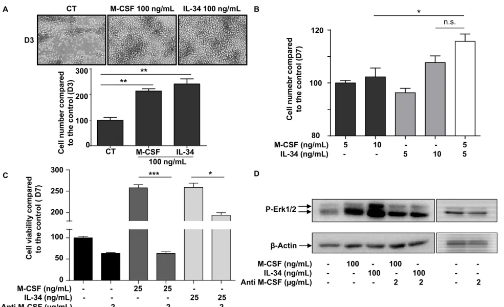
In the light of these observations, we analyzed the biological impact of the addition
of both cytokines on human CD14+ monocytes (Supplementary Figure 1) and TF-1
M-CSFR cell viability (Figure 2). Consistent with previous findings 6, IL-34 and M-CSF similarly increased CD14+ cell viability in a dose-dependent manner (Supplementary
led to higher viability than an equivalent cumulated dose (10 ng/mL) of IL-34 but not of
M-CSF (Supplementary Figure 1B). In addition, as shown previously 8, M-M-CSF and IL-34 similarly induced RANKL-associated osteoclastogenesis in a dose-dependent manner and
the combination of M-CSF and IL-34 did not reveal any competitive or additive activity
between these cytokines (Supplementary Figure 2). Both cytokines significantly
up-modulated the proliferation of M-CSFR overexpressing TF-1 cells (TF-1 M-CSFR) (Figure
2A). As with the experiment conducted with CD14+ cells, a combination of 5 ng/mL of
each cytokine induced significantly higher proliferation compared to 10 ng/mL of M-CSF
alone, whereas this difference was not significant compared to 10 ng/mL of IL-34 (Figure
2B, p<0.05). Addition of a blocking anti-M-CSF antibody reduced the proliferation of TF-1
M-CSFR cells, thus revealing autocrine M-CSF production by the cells (Figure 2C). This
autocrine expression of M-CSF was confirmed by RT-qPCR and ELISA (Data not shown).
Interestingly, this blocking anti-M-CSF antibody also decreased IL-34-induced cell
proliferation with 25 ng/mL of the cytokine (Figure 2C, p < 0.05), and strongly reduced the
modulation of ERK1/2 phosphorylation induced by IL-34 (Figure 2D). As with the TF-1
cells, HOS M-CSFR produced M-CSF in an endogenous manner (Data not shown). We
then studied the contribution of autocrine M-CSF on IL-34-induced M-CSFR activation by
a silencing RNA approach targeting M-CSF (Figure 3, Supplementary Figure 3A). M-CSF
expression was down-regulated by around 50% 48 hours after transfections of siRNA
targeting the M-CSF gene (Supplementary Figure 3A), and the silencing of M-CSF
expression decreased M-CSFR phosphorylation induced by 50 ng/mL of IL-34 (Figure 3).
cell-derived M-CSF in M-CSFR activation (Supplementary Figure 3B). Overall, these data demonstrated that combinations of both cytokines are not restricted to competitive
activities, and that IL-34 and M-CSF can exhibit additive effects on various cells at certain
concentrations. In addition, M-CSF is able to modulate intracellular signaling induced by
IL-34 through the M-CSFR.
3.2. IL-34 can interact with the M-CSF to form a heteromeric cytokine
Because the modulation of autocrine production of M-CSF affects the M-CSFR activation
induced by IL-34, we analyzed the potential molecular interaction between both cytokines
by surface plasmon resonance. The sensorgrams shown in Figure 4A demonstrate the
specific binding of IL-34 to immobilized M-CSF, characterized by rapid dissociation. Thus,
IL-34 bound M-CSF with a low affinity (KD=114nM) (Figure 4B). Molecular modeling
based on the crystal structures published for the M-CSF, M-CSFR 13 and IL-34 15 illustrated the conventional binding of the homodimeric cytokines on the M-CSFR (Figure
4C). A docking binding analysis was performed using the human dimer of IL-34 against the
humanized structure of M-CSF derived from the mouse crystal structure. The docking
strategy consisted in defining M-CSF as the rigid molecule, thus keeping its position fixed,
and applying rotations and translations in the three-dimensional space for a total of 54000
docking energy evaluations, called poses. The best 2000 poses according to ZSCORE
(determined using ZDOCK) were clustered to regroup similar orientations. In these
clusters, the most common dimer-dimer interface was found to be a parallel orientation of
first and most populated cluster was made of 82 structures and contained the best pose
(Figure 4D, left panel). The binding interface was made of 56 amino acids on M-CSF and
47 on IL-34, for a total interface surface of 1764 Å. For each dimer, the loss in accessible
solvent was of 1680 Å and 1845 Å for M-CSF and IL-34 respectively, indicating the
stronger binding role of IL-34 in the resulting binding interface. The binding interface
consisted of 14 hydrogen bonds between dimers and 9 salt bridges, and a detailed energy
analysis of their binding mode revealed a balanced contribution of electrostatic interactions
and hydrophobic contacts (data not shown). To see whether the heteromeric cytokine
identified in this study can engage M-CSFR pathway activation, we explored whether the
already-described binding sites for IL-34 and M-CSF could still be accessible for binding to
the M-CSFR. As presented in Figure 4D (right panel), despite a steric hindrance due to its
large size, the heteromeric cytokine was able to bind to the M-CSFR, leading to a different
conformation of the intracellular M-CSFR chains. In this conformation, one of the two
cytokine binding sites remained accessible to the M-CSFR and the two free sites were
opposite each other on the tetrameric cytokine. If we superimposed two M-CSFR
monomers on the predicted dimer of dimers, the resulting orientation for each M-CSFR
monomer was compatible with the estimated distance between the missing D4 and D5
domains of the two M-CSFR monomers. This distance was a critical step for receptor
activation as it allowed contact between the two D4 monomers via homotypic contacts 19. M-CSF and IL-34 can thus form a heteromeric cytokine able to bind to the M-CSFR.
3.3. Endogenous M-CSF and IL-34 can interact together within the cell cytoplasm
As M-CSF and IL-34 were predicted to form a heteromeric cytokine, we further
investigated by means of a PLA assay whether such interaction could be relevant in cells
producing both cytokines (Figure 5). We developed from the parental MG63 cell line
(MG63 NT) that intrinsically expressed M-CSF but not IL-34, a transfected MG63 IL-34
cell line that expressed both M-CSF and IL-34 at the protein level, as demonstrated by the
pink staining around the nuclei compared to the negative controls (Figure 5A,B). The PLA
assay demonstrated molecular interaction between the two cytokines as shown by pink
fluorescent points within the cytoplasm in Figure 5C, characterized by a close proximity of
the protein epitopes (30-40nm).
3.4. Endogenous M-CSF and IL-34 expression differentially control intracellular trafficking and maturation of the M-CSFR
M-CSFR half-life was analyzed in the presence of M-CSF or IL-34 in HEK cells
expressing the CSFR. As expected, HEK CSFR cell lysates revealed two forms of
M-CSFR, the higher molecular form corresponding to the membrane form of the receptor
(Figure 6). Adding M-CSF or IL-34 decreased the membrane expression of M-CSFR, thus
reflecting the receptor’s internalization and degradation as protein synthesis was blocked by
cycloheximide. At the same dose, M-CSF was more efficient in reducing the half-life of the
M-CSFR compared to IL-34 (Figure 6A). When M-CSF was overexpressed concomitantly
to the M-CSFR, the cytokine markedly down-regulated the expression of the membrane
form of its receptor in favor of the intracellular form (Figure 6B). On the contrary, the
with HEK M-CSFR cells, overexpression of M-CSF in HOS M-CSFR cells quantitatively
increased the intracellular form and decreased the membrane form of the receptor in
contrast to IL-34 overexpression, which maintained receptor expression at the cell
membrane (Supplementary Figure 4). Non-transfected HOS M-CSFR and HEK M-CSFR
cells or cells transfected with an empty vector (Mock) mainly expressed the receptor at the
membrane (Figure 6B, Supplementary Figure 4). A trypsin treatment of HEK M-CSFR
cells was associated with the formation of degradation products of the M-CSFR in contrast
with the M-CSF co-expressing cells (Figure 6C). These results demonstrated that the upper
band (175 kDa) corresponded well to the membrane form of the M-CSFR and the lower
band (150 kDa) to an intracellular form. Adding tunicamycin to HEK M-CSFR cells for 24
hours or 48 hours resulted in a lower molecular weight form of the receptor, around 100
kDa corresponding to its non-N-glycosylated form, whereas brefeldin A treatment induced
a total shift toward the 150 kDa M-CSFR form, similar to M-CSF co-expression (Figure
6C). As tunicamycin is known to prevent proteins’ N-glycosylation, and brefeldin A to both
interfere with transport from the endoplasmic reticulum to the Golgi apparatus and block
protein secretion, the present data suggest that M-CSF treatment blocked M-CSFR
trafficking in the endosomal network, leading to the accumulation of a less N-glycosylated
form of 150 kDa (Figure 6C). However, treatment of HEK M-CSFR cells with M-CSFR
ligands for 48 hours did not induce any shift between the two forms of M-CSFR,
demonstrating that only intrinsic expression of M-CSF and IL-34 can impact receptor
maturation (Figure 6D).
intracellular sequestration of the M-CSFR that appeared located in the perinuclear region
(Figure 7, Supplementary Figure 5). Overexpression of IL-34 led to minor intracellular
sequestration of the receptors as they were still localized at the cell membrane. Moreover,
co-expression of M-CSF and IL-34 mainly led to intracellular expression of the M-CSFR
clustered in the endosomal network around the nuclei (Figure 7, Supplementary Figure 5),
confirming the Western blot observations (Data not shown). Similar investigations were
performed in the murine osteosarcoma cells K7M2 that spontaneously expressed M-CSF
and IL-34 (Supplementary Figure 6). Supplementary Figure 6 shows a main intracellular
localization of the M-CSFR with slight expression of its membrane form. Western blot
analysis confirmed the expression of both M-CSFR forms with a relatively high expression
of the intracellular form compared to HEK M-CSFR (Figure 6) or HOS M-CSFR
(Supplementary Figure 4). Overall, these data demonstrate that intrinsic IL-34 and M-CSF
4. DISCUSSION
Despite common features, previous studies described specific activities for M-CSF
and IL-34, especially in their ability to modulate the expression of various
chemokines/chemokine receptor expression (monocyte chemoattractant protein-1 MCP-1,
eotaxin-2, C-C chemokine receptor type 2 CCR2), and the migration of myeloid cells
through their shared M-CSFR chains 11, 30. In the present work, we report for the first time the molecular interaction between M-CSF and IL-34, forming a new heteromeric
cytokine, and the differential role of M-CSFR ligands in controlling M-CFSR trafficking.
Furthermore, the M-CSF/IL-34 heteromer may play a specific biological role by
differentially phosphorylating the M-CSFR due to the tridimensional conformation of the
receptor chains adopted when the cytokines bind. ZDOCK docking software was used to
look for an interaction between M-CSF and IL-34, as it is particularly efficient on dimeric
proteins, while finding the correct interface from two monomers to form a dimer. We first
explored whether the docking followed by clustering strategy could be useful for studying
existing binding interfaces of the M-CSFR ligands by isolating each cytokine from its
receptor or by splitting dimers into monomers. Each docking assessment found the correct
position and orientation in the first populated and most energy favorable cluster, even in the
case of the M-CSF molecule where the disulfide bridge could not be created during the
docking process. Without the N-glycosylations present in the IL-34 structures, we also
observed aggregation-like motifs, a phenomenon already identified experimentally 12. There was no defined orientation or specific binding interfaces that could be deduced from
aggregation pattern, which was also a good indicator of the sensitivity of the methods used.
These validations reinforced our confidence in the docking protocol used for studying the
putative binding mode of the cytokines. The in silico experiments revealed a potential
binding mode for M-CSF and IL-34 dimers (Figure 4D), arranged parallel to each other
with their beta-strands pointing in the same direction. This striking binding mode was
unexpected so we first decided to rule out the possibility that the results obtained were only
due to the docking strategy or algorithm employed. We therefore used another popular rigid
protein docking algorithm called PIPER via its web-based interface ClusPro, which best
performed recently in the latest CAPRI contest 31, 32. By using dimers of dimers as starting structures we also found a parallel orientation for both cytokines. Consistent with
this docking study, surface plasmon resonance assays experimentally validated the binding
ability of IL-34 to the M-CSF characterized by low affinity and rapid dissociation (Figure
4A). The proximity ligation assay confirmed their interaction. Indeed, this technique made
it possible to detect closed epitopes separated by a maximum distance of around 30-40 nm
33, and finally strengthened the binding between both in their physiological context (Figure 5). This heteromeric cytokine may be stabilized by its binding to the M-CSFR, as
the molecular docking study proposed a new binding mode for this heteromeric cytokine to
two isolated M-CSFR chains (Figure 4D). This recruitment could bring each M-CSFR
domain D3 to a distance of about 60Å, close enough to bring domains D4 and D5 into
contact, a step known to be critical for receptor activation 17-19.
The oligomerization process is widespread in biology and it is estimated that
possible by better stability, amplification of their affinity or the protective mechanisms of
the proteins are amongst the main benefits of this oligomerization process. By forming
homodimers, M-CSFR, M-CSF and IL-34, like numerous other cytokines/receptors, do not
break this oligomerization rule 35. To increase the complexity, diversity and regulation of cell signaling events, several cytokines are able to form heteromeric structures. This is the
case of interleukin-12 (IL-12) related cytokines which differentially regulate the
maintenance or activation of the Th1 immune response 36. Similarly to IL-12, IL-17A and IL-17F, two members of the IL-17 family, also interact to form a heterodimer able to
bind to the IL-17RA/IL-17RC receptor complex 37.
Based on our data, M-CSF and IL-34 now belong to the cytokine families forming
heteromeric entities and the M-CSF/IL-34 cytokine plays a part in increasing the functional
diversity of these molecules. By interfering with M-CSFR trafficking, more specifically in
the balance between the intracellular and membrane forms of the receptor, IL-34, M-CSF
and M-CSF/IL-34 may tightly and specifically control the biological activities associated
with the M-CSFR-dependent signaling pathways. In M-CSFR expressing cells,
overexpression of M-CSF led preferentially to maintaining the M-CSFR in an N–
glycosylated form clusterized into the endosomal network. Western blot carried out in these
experimental conditions showed a shift in M-CSFR expression from the high molecular
weight of 175 kDa, corresponding to the membrane mature form of the receptor, to the 150
kDa intracytoplasmic form. These results were consistent with previous studies which
demonstrated that the 175 kDa membrane form was highly processed and modified with
modifications, and accumulated in the Golgi apparatus, a process called receptor maturation
arrest 38-40. The tunamycin treatment, which is known to prevent the glycosylation process 41, and brefeldin A treatment, which blocks anterograde transport from the endoplasmic reticulum to the Golgi apparatus and protein secretion 42, 43, confirm that the M-CSFR form processed when M-CSF is highly expressed by the cells is less
glycosylated compared to the mature form, and that the receptor is retained between the
endoplasmic reticulum and the Golgi apparatus. In contrast, co-expression of IL-34 did not
induce massive intracellular clustering of M-CSFR and maintained the receptor at the cell
surface, suggesting a different regulatory role for the twin cytokines in M-CSFR
trafficking. Similar functional differences can be observed on the receptor half-life after
adding exogenous cytokines (Figure 6), but also at the transcriptional level as an increase in
IL-34 expression has a moderate impact on M-CSF or M-CSFR expression levels
(unpublished data). In contrast, overexpression of M-CSF induces significant changes in
the M-CSFR or IL-34 levels. Indeed, overexpression of M-CSF induces a significant
increase in IL-34 expression, whereas overexpression of IL-34 does not modify M-CSF
expression (unpublished data). In addition, knock-down of M-CSF expression using siRNA
enhances IL-34 expression, suggesting a compensatory mechanism between these two
cytokines. Nandi et al. recently published that CSFR and its two ligands IL-34 and
M-CSF exhibit distinct expression patterns during brain development and that IL-34 exhibits a
broader regional expression than M-CSF, mostly without overlap 44. Their observations suggest a key regulatory role for IL-34 in the M-CSF/M-CSFR activation pathway, which
show that IL-34 may modulate M-CSF effects through the M-CSFR, explained in part by
the formation of a heteromeric cytokine M-CSF/IL-34. In addition to the already identified functions of IL-34 and M-CSF in exacerbated macrophage activation (bowel diseases, liver fibrosis, chronic skin inflammation, arthritis, etc), this new heteromeric cytokine M-CSF/IL-34 which may differentially regulates the activation/localisation of M-CSFR strengthens the global therapeutic approaches for blocking all biological functions coming from these molecules 7,8, 45-51.
In contrast to IL-17A or IL-17F that form either disulfide-linked homodimers or
heterodimers through their cysteine knot motif, M-CSF and IL-34 probably interact in their
homodimeric form to constitute a heterotetrameric cytokine. Our molecular docking studies
predicted a 2:2 interaction without any disulfide bond, which is able to bind to the
M-CSFR, unlike a single M-CSF/IL-34 heterodimer. The exact functional implication of this
new oligomeric cytokine still needs to be explored and must now be analysed in the light of
the receptor protein tyrosine phosphatase β/ζ (RPTP β/ζ) identified as a new receptor for
IL-34 44, not expressed in the CD14+ and myeloid cells analysed in the present study
51. The involvement of these oligomeric cytokine should be also investigated in light of syndecan-1which has been recently identified as new IL-34 effector 51. As with the IL17A/IL17F and IL-17RA/IL-17RC systems, the heteromeric M-CSF/IL-34 may regulate
IL-34 and M-CSF functions through one receptor or another and the heteromer may also
interact with both M-CSFR and RPTP β/ζ receptors. This new M-CSFR cooperative
binding mode needs further exploration, and the crystal structure of M-CSF/IL-34 should
be determined as well as co-crystal studies with the receptors to better characterize this new
mechanism is more beneficial than the already described one-to-one competition. In
conclusion, the present work demonstrated that simultaneous addition of M-CSF and IL-34
led to a specific activation pattern on the M-CSFR, with higher phosphorylation of the
tyrosine residues at low concentrations. Similarly, both cytokines showed an additive effect
on cellular proliferation or viability that can be explained by the formation of a heteromeric
M-CSF/IL-34 cytokine predicted by molecular docking studies. This interaction between
M-CSF and IL-34 was confirmed by surface plasmon resonance and proximity ligation
assays. In addition, co-expression of the M-CSFR and its ligands differentially regulates the
receptor’s glycosylation state and localization in the cell. This is the first report
demonstrating the direct interaction between IL-34 and M-CSF and their ability to form a
new heteromeric cytokine that may play a part in the tissue homeostasis and development.
ACKNOWLEDGEMENTS: The authors would like to thank Juliette Desfrançois-Noel
from the cell sorting facility (Plateau de cytométrie SFR Bonamy/INSERM U892, Nantes, France) for her help in cell sorting the cells expressing the M-CSFR, and Mike Maillasson from the Plateforme IMPACT (SFR Bonamy, FED 4203/ INSERM UMS 016/CNRS 3556, Nantes, France) where the surface plasmon resonance experiments were carried out. We are especially grateful to Julien Jardin and Valérie Briard from the analytical facility at the UMR 1253 INRA/Agrocampus-Ouest (STLO Science et Technologie du Lait et de l'œuf, Rennes, France), who helped us to perform spectrometry analysis to determine M-CSF/IL-34 stoichiometry. We would also like to thank Dr Valérie Trichet (INSERM U957, Nantes, France) for her technical and rewarding advice regarding molecular cloning of the human M-CSFR gene. This study was supported by the Region des Pays de la Loire (CIMATH II research project) and by the Ligue Nationale Contre le Cancer (Equipe LIGUE 2012).
AUTHORSHIP: A.I.S., R.B. and C.C. performed cell biology and biochimistry studies.
A.S. and B.B. performed P.L.A. and confocal microscopy experiments. M.M. perfomed Surface plasmon resonance experiments. S.T. performed molecular docking. D. H. and A.S. planned the work and D.H., A.S. and S.T. contributed to the manuscript.
REFERENCES
1 Stanley ER, Chitu V. CSF-1 receptor signaling in myeloid cells. Cold Spring Harbor Pespect Biol 2014 ;6 : a021857.
2 Dai XM, Ryan GR, Hapel AJ, Dominiguez MG, Russel RG, Kapp S, Sylverstre V, Stanley ER. Targeted disruption of the mouse colony-stimulating factor 1 receptor gene results in osteopetrosis, mononuclear phagocyte deficiency, increased primitive progenitor cell frequencies, and reproductive defects. Blood 2002;99 :111–120.
3 Wiktor-Jedrzejczak W, Bartocci A, Ferrante AW Jr, Ahemd-Ansari A, Sell KW, Pollard JW, Stanley ER. Total absence of colony-stimulating factor 1 in the macrophage-deficient osteopetrotic (op/op) mouse. Proc Natl Acad Sci USA 1990 ;87 : 4828–4832. 4 Yoshida H, Hayashi S, Kunisada T, Ogawa M, Nishikawa S, Okamura H, Sudo T,
Shultz LD, Nishikawa S. The murine mutation osteopetrosis is in the coding region of the macrophage colony stimulating factor gene. Nature 345, 442–444.
5 Wei S, Nandi S, Chitu V, Yeung YG, Yu W, Huang M, Williams LT, Lin H, Stanley ER. Functional overlap but differential expression of CSF-1 and IL-34 in their CSF-1 receptor-mediated regulation of myeloid cells. J Leukoc Biol 2010 ;88 : 495 –505. 6 Lin H, Lee E, Hestir K, Leo C, Huang M, Bosch E, Halenbeck R, Wu G, Zhou A,
Berhens D, Hollebaugh D, Linnemann T, Qin M, Wong J, Chu K, Doberstein SK, Williams LT. Discovery of a Cytokine and Its Receptor by Functional Screening of the Extracellular Proteome. Science 2008 ; 320 :807–811.
7 Foucher ED, Blanchard S, Preisser L, Garo E, Ifrah N, Guardiola P, Delneste Y, Jeannin P. IL-34 Induces the Differentiation of Human Monocytes into Immunosuppressive Macrophages. Antagonistic Effects of GM-CSF and IFNγ. PloS One 2013 ;8 : e56045.
8 Baud’Huin M, Renault R, Charrier C, Riet A, Moreau A, Brion R, Gouin F, Duplomb L, Heymann D. Interleukin-34 is expressed by giant cell tumours of bone and plays a key role in RANKL-induced osteoclastogenesis. J Pathol 2010 ; 221 : 77–86.
9 Nandi S, Gokhan S, Dai XM, Wei S, Enikolopov G, Lin H, Mehler MF, Stanley ER. The CSF-1 receptor ligands IL-34 and CSF-1 exhibit distinct developmental brain expression patterns and regulate neural progenitor cell maintenance and maturation. Dev Biol 2012 ; 367 : 100–113.
10 Wang Y, Szretter KJ, Vermi W, Gilfillan S, Rossini C, Cella M, Barrow AD, Diamond MS, Colonna M. IL-34 is a tissue-restricted ligand of CSF1R required for the development of Langerhans cells and microglia. Nat Immunol 2012 ; 13 : 753-760.
11 Chihara T, Suzu S, Hassan R, Chutiwitoonchai N, Hiyoshi M, Motoyoshi K, Kimura F, Okada S. 2010 L-34 and M-CSF share the receptor Fms but are not identical in biological activity and signal activation. Cell Death Differ. 17, 1917–1927.
12 Liu H, Leo C, Chen X, Wong BR, Williams LT, Lin H, He X. The mechanism of shared but distinct 1R signaling by the non-homologous cytokines IL-34 and CSF-1. Biochim Biophys Acta 2012 ;1824 : 938–945.
13 Pandit J, Bohm A, Jancarik J, Halenbeck R, Koths K, Kim SH. Three-dimensional structure of dimeric human recombinant macrophage colony-stimulating factor. Science 1992 ;258 : 1358–1362.
14 Garceau V, Smith J, Paton IR, Davey M, Fares MA, Sester DP, Burt DW, Hume DA. Pivotal Advance: Avian colony-stimulating factor 1 (CSF-1), interleukin-34 (IL-34), and CSF-1 receptor genes and gene products. J Leukoc Biol 2010 ; 87 : 753–764.
15 Ma X, Lin WY, Chen Y, Stawicki S, Mukhyala K, Wu Y, Martin F, Bazan JF, Staronasnik MA. Structural Basis for the Dual Recognition of Helical Cytokines IL-34 and CSF-1 by CSF-1R. Structure 2012;20 : 676–687.
16 Price K, Choi HU, Rosenberg L, Stanley ER. The predominant form of secreted colony stimulating factor-1 is a proteoglycan. J. Biol. Chem 1992 ; 267, 2190-2199.
17 Felix J, Elegheert J, Gutsche I, Shkumatov AV, Wen Y, Bracke N, Pannecouke E, Vandenberghe I, Devreese B, Svergun DI, Pauwels E, Vergauwen B, Savvides SN.
Human IL-34 and CSF-1 establish structurally similar extracellular assemblies with their common hematopoietic receptor. Structure 2013 ;21 : 528-39.
18 Verstraete K, Savvides SN. Extracellular assembly and activation principles of oncogenic class III receptor tyrosine kinases. Nat Rev Cancer 2012 ;12:753–766.
19 Elegheert J, Desfosses A, Shkumatov AV, Wu X, Bracke N, Verstrate K, Van Craenenbroeck K, Brooks BR, Svergun DI, Vergauwen B, Gutsche I, Savvides SN. Extracellular complexes of the hematopoietic human and mouse CSF-1 receptor are driven by common assembly principles. Structure 2011 ; 19 : 1762–1772.
20 Reynolds A, Leake D, Boese Q, Scaringe S, Marshall WS, Khvorova A. Rational siRNA design for RNA interference. Nat Biotechnol 2004 ; 22 : 326–330.
21 Naito Y, Ui-Tei K. Designing functional siRNA with reduced off-target effects. Methods Mol Biol 2013 ; 942 : 57–68.
22 Hellemans J, Vandesompele J. Selection of reliable reference genes for RT-qPCR analysis. Methods Mol Biol 2014 ; 1160 : 19–26.
23 Bustin SA, Benes V, Garson J, Hellemans J, Huggett J, Kubista M, Mueller R, Nolan T, Pfaffl MW, Shipley G, Wittwer CT, Schjerling P, Day PJ, Abreu M, Aguado B, Beaulieu JF, Beckers A, Bogaert S, Browne JA, Carrasco-Ramiro F, Ceelen L, Ciborowski K, Cornillie P, Coulon S, Cuypers A, De Brouwer S, De Ceuninck L, De Craene J, De Naeyer H, De Spiegelaere W, Deckers K, Dheedene A, Durinck K, Ferreira-Teixeira M, Fieuw A, Gallup JM, Gonzalo-Flores S, Goossens K, Heindryckx F, Herring E, Hoenicka H, Icardi L, Jaggi R, Javad F, Karampelias M, Kibenge F, Kibenge M, Kumps C, Lambertz I, Lammens T, Markey A, Messiaen P, Mets E, Morais S, Mudarra-Rubio A, Nakiwala J, Nelis H, Olsvik PA, Pérez-Novo C, Plusquin M, Remans T, Rihani A, Rodrigues-Santos P, Rondou P, Sanders R, Schmidt-Bleek K, Skovgaard K, Smeets K, Tabera L, Toegel S, Van Acker T, Van den Broeck W, Van der Meulen J, Van Gele M, Van Peer G, Van Poucke M, Van Roy N, Vergult S, Wauman J, Tshuikina-Wiklander M, Willems E, Zaccara S, Zeka F, Vandesompele J. The need for transparency and good practices in the qPCR literature. Nat Methods 2013 ; 10 : 1063– 1067.
24 Huggett JF, Foy CA, Benes V, Emslie K, Garcon JA, Haynes R, Hellemans J, Kubista M, Mueller RD, Nolan T, Pfaffl MW, Shipley GL, Vandesompele J, Wittwer CT, Bustin
SA. The digital MIQE guidelines: Minimum Information for Publication of Quantitative Digital PCR Experiments. Clin Chem 2013 ; 59 : 892–902.
25 Chen R, Weng Z. Docking unbound proteins using shape complementarity, desolvation, and electrostatics. Proteins 2002 ; 47 : 281–294.
26 Momany FA, Rone R. Validation of the general purpose QUANTA ®3.2/CHARMm® force field. J Comput Chem 1992 ; 13 : 888–900.
27 Chen X, Liu H, Focia PJ, Shim AHR, He X. Structure of macrophage colony stimulating factor bound to FMS: diverse signaling assemblies of class III receptor tyrosine kinases. Proc Natl Acad Sci USA 2008 ; 105 : 18267–18272.
28 Li L, Chen R, Weng Z. RDOCK: refinement of rigid-body protein docking predictions. Proteins 2003 ;53 :693–707.
29 Pierce B, Weng Z. ZRANK: reranking protein docking predictions with an optimized energy function. Proteins 2007 ;67 : 1078–1086.
30 Barve BA, Zack MD, Weiss D, Song RH, Beidler D, Head RD. 2013 Transcriptional profiling and pathway analysis of CSF-1 and IL-34 effects on human monocyte differentiation. Cytokine 63, 10–17.
31 Kozakov D, Beglov D, Bohnuud T, Mottarella SE, Xia B, Hall DR, Vajda S. 2013 How good is automated protein docking? Proteins 81, 2159–2166.
32 Kozakov D, Brenke R, Comeau SR, Vajda S. IPER: an FFT-based protein docking program with pairwise potentials. Proteins 2006 ;65 : 392–406.
33 Söderberg O, Gullberg M, Jarvius M, Ridderstråle K, Leuchowius KJ, Jarvius J, Wester K, Hydbring P, Bahram F, Larsson LG, Landegren U. Direct observation of individual endogenous protein complexes in situ by proximity ligation. Nat Methods 2006 ;3 : 995–1000.
34 Goodsell DS, Olson AJ. Structural symmetry and protein function. Annu Rev Biophys Biomol Struct 2000 ; 9 : 105–153.
mechanisms. Crit Rev Biochem Mol Biol 2012 ; 47 : 502–530.
36 Gately MK, Renzetti LM, Magram J, Stern AS, Adorini L, Gubler U, Presky DH. The interleukin-12/interleukin-12-receptor system: role in normal and pathologic immune responses. Annu Rev Immunol 1998 ;16 : 495–521.
37 Toy D, Kugler D, Wolfson M, Vanden Bos T, Hurgel J, Derry J, Tocker J, Peschon J. Interleukin 17 Signals through a Heteromeric Receptor Complex. J Immunol 2006;177; 36–39.
38 Kawasaki ES, Ladner MB. Molecular biology of macrophage colony-stimulating factor. Immunol Ser 1990;49: 155–176.
39 Hassan R, Suzu S, Hiyoshi M, Takahashi-Makise N, Ueno T, Agatsuma T, Akari H, Komano J, Takebe Y, Motoyoshi K, Okada S. Dys-regulated activation of a Src tyroine kinase Hck at the Golgi disturbs N-glycosylation of a cytokine receptor Fms. J Cell Physiol 2009; 221: 458–468.
40 Hiyoshi M, Suzu S, Yoshidomi Y, Hassan R, Harada H, Sakashita N, Akari H, Motoyoshi K, Okada S. Interaction between Hck and HIV-1 Nef negatively regulates cell surface expression of M-CSF receptor. Blood 2008;111: 243–250.
41 Price NPJ, Tsvetanova B. Biosynthesis of the tunicamycins: a review. J Antibio 2007; 60: 485–491.
42 Nebenführ A, Ritzenthaler C, Robinson DG. Brefeldin A: Deciphering an Enigmatic Inhibitor of Secretion. Plant Physiol 2002;130: 1102–1108.
43 Verbert A, Cacan R. Trafficking of oligomannosides released during N-glycosylation: a clearing mechanism of the rough endoplasmic reticulum. Biochim Biophys Acta 1999; 1473: 137–146.
44 Nandi S, Cioce M, Yeung YG, Nieves E, Tesfa L, Lin H, Hsu AW, Halenbeck R, Cheng HY, Gokhan S, Mehler MF, Stanley ER. Receptor-type protein-tyrosine phosphatase ζ is a functional receptor for interleukin-34. J Biol Chem 2013; 288: 21972– 21986.
45 Masteller EL, Wong BR. Targeting IL-34 in chronic inflammation. Drug Discov Today, 2014 19 (8), 1212-1216.
46 Yamane F, Nishikawa Y, Matsui K, Asakura M, Iwasaki E, Watanabe K, Tanimoto H, Sano H, Fujiwara Y, Stanley ER, Kanayama N, Mabbott NA, Magari M, Ohmori H. CSF-1 receptor-mediated differentiation of a new type of monocytic cell with B cell-stimulating activity: its selective dependence on IL-34. J Leukoc Biol, 2014 95(1):19-31.
47 Chemel M, Le Goff B, Brion R, Cozic C, Berreur M, Amiaud J, Bougras G, Touchais S, Blanchard F, Heymann MF, Berthelot JM, Verrecchia F, Heymann D. Interleukin 34 expression is associated with synovitis severity in rheumatoid arthritis patients. Ann Rheum Dis, 2012 71(1):150-154.
48 Preisser L, Miot C, Le Guillou-Guillemette H, Beaumont E, Foucher ED, Garo E, Blanchard S, Frémaux I, Croué A, Fouchard I, Lunel-Fabiani F, Boursier J, Roingeard P, Calès P, Delneste Y, Jeannin P. IL-34 and macrophage colony-stimulating factor are overexpressed in hepatitis C virus fibrosis and induce profibrotic macrophages that promote collagen synthesis by hepatic stellate cells. Hepatology, 2014 60(6):1879-1090.
Ortenzi A, Laudisi F, Sica G, Sileri P, Pallone F, Monteleone G. Interleukin-34 sustains inflammatory pathways in the gut. Clin Sci, in press.
50 Fend L, Accart N, Kintz J, Cochin S, Reymann C, Le Pogam F, Marchand JB, Menguy T, Slos P, Rooke R, Fournel S, Bonnefoy JY, Préville X, Haegel H. Therapeutic effects of anti-CD115 monoclonal antibody in mouse cancer models through dual inhibition of tumor-associated macrophages and osteoclasts. PLoS One, 2013 8(9):e73310.
51 Ségaliny AI, Brion R, Mortier E, Maillasson M, CherelM, Jacques Y, Le Goff B, Heymann D. Syndecan-1 regulates the biological activities of interleukin-34. Biochemica Biophysica Acta Mol Cell Res, 2015 1853(5):1010-1021
Figure Legends
Figure 1: CSF and IL-34 alone or in combination differentially activate the M-CSFR. Tyrosine phosphorylation patterns (P-Tyr708, P-Tyr723, and P-Tyr923) for M-CSFR
and Erk1/2 phosphorylation were investigated by Western blot after 5 min of cytokine
addition to the cells. (A) HEK M-CSFR cells were cultured in serum-free conditions for
12h before stimulation for 5 min at 37°C with increasing doses of M-CSF (10, 25, 50, 100
and 200 ng/mL) combined or not with 10 ng/mL of IL-34. (B) Similarly, HEK M-CSFR
cells were incubated with increasing concentrations of IL-34 (10, 25, 50, 100 and 200
ng/mL) with or without 10 ng/mL of M-CSF. -actin was used as the loading control. Bar graphs show relative densitometric values of pTyr-708 an pERK1/2 normalised to β-actin
and to the control (CT) condition.
Figure 2: Autocrine production of M-CSF modulates the biological effects of IL-34. (A) TF-1 M-CSFR cells were cultured in the presence or absence of M-CSF or IL-34 for 3
days. Cell number was counted manually and histograms represent the percentage of
proliferation compared to the control group. Original magnification: X100. (B) TF-1
M-CSFR cells were cultured in the presence or absence of M-CSF or IL-34 or both cytokines
at 5 ng/mL for 7 days. Cell proliferation was quantified by manual counting and histograms
illustrate the percentage of proliferation compared to D0 and normalized to the 5 ng/mL
without M-CSF or IL-34 in the presence or absence of a blocking anti-M-CSF antibody.
After 3 days of culture, cell viability was determined by an Alamar blue assay. (D) TF-1
M-CSFR cells were cultured in serum free conditions for 12 hours before incubation with a
blocking anti-M-CSF antibody. The cells were then stimulated with M-CSF or IL-34 for 5
min and the level of P-ERK1/2 was studied by Western blot. -actin was used as the loading control. Error bars show the SEM for three different experiments. *p < 0.05, ** p <
0.01, *** p < 0.001.
Figure 3: Blockade of M-CSF cell expression decreases IL-34-induced M-CSFR activation. HOS M-CSFR cells were transfected with siRNAs directed against the human
M-CSF gene (siM-CSF621, siM-CSF778, siM-CSF952). An siRNA directed against the
luciferase gene (siLucF) was used as a control. Forty-eight hours after transfection, the cells
were cultured in serum-free conditions for 12 hours and then stimulated with 50 ng/mL of
M-CSF or IL-34 for 5 min. Tyrosine708 and tyrosine723 phosphorylation of the M-CSFR
were studied by Western blot. -actin was used as the loading control. The phosphorylation density of the tyrosine723 was quantified using GeneTools (Syngene) and normalized to the
-actin density.
Figure 4: IL-34 binds M-CSF to form a heterodimeric protein able to interact with the M-CSFR. (A) M-CSF was immobilized on the dextran matrix of a sensor chip (BIAcore)
and increasing doses of IL-34 (from 15.575 to 500 nM) were loaded on to the chip to
binding in RU and shows dissociation constant (KD) determination. (C) Molecular
modeling was performed on the homodimeric cytokines in complex with the M-CSFR
using the three-dimensional coordinates extracted from the crystal structure previously
published. (D) The interactions between M-CSF and IL-34 were assessed by molecular
modeling using three-dimensional cytokine structures. The left panel shows the most
favorable binding mode between the two homodimeric cytokines, and the right panel
illustrates interaction between the heteromeric cytokine and the M-CSFR. M-CSF in
brown, IL-34 in green and M-CSFR in blue.
Figure 5: M-CSF and IL-34 interact within the cytoplasm of cells expressing both cytokines. (A) First, the proximity ligation assay (PLA) from Olink was used to detect
M-CSF and IL-34 expression (far red, represented in pink) in MG63 cell lines (MG63 NT only
express M-CSF, and MG63 IL-34 cells express both cytokines). Nuclei were stained with
DAPI (cyan). (B) Several negative controls were performed on MG63 IL-34 cells to
validate the PLA assays: cells incubated with only one primary antibody (either against
M-CSF or IL-34) and both secondary antibodies, or with only secondary antibodies; MG63
NT cells incubated with all the antibodies. (C) In situ PLA showed the co-localization of
M-CSF and IL-34 in the cytoplasm of MG63 IL-34 cells (pink staining). Cell morphology
was analyzed using digital interference reflection images captured using the reflection
mode (gray).
Figure 6: M-CSF and IL-34 differentially control the intracellular trafficking of the M-CSFR. (A) HEK M-CSFR cells were treated with 4 µg/mL of cycloheximide to block