HAL Id: tel-02012151
https://pastel.archives-ouvertes.fr/tel-02012151
Submitted on 8 Feb 2019
HAL is a multi-disciplinary open access archive for the deposit and dissemination of sci-entific research documents, whether they are pub-lished or not. The documents may come from teaching and research institutions in France or abroad, or from public or private research centers.
L’archive ouverte pluridisciplinaire HAL, est destinée au dépôt et à la diffusion de documents scientifiques de niveau recherche, publiés ou non, émanant des établissements d’enseignement et de recherche français ou étrangers, des laboratoires publics ou privés.
Dynamique interne des flavoprotéines étudiée par
spectroscopie femtoseconde
Lipsa Nag
To cite this version:
Lipsa Nag. Dynamique interne des flavoprotéines étudiée par spectroscopie femtoseconde. Biological Physics [physics.bio-ph]. Université Paris Saclay (COmUE), 2018. English. �NNT : 2018SACLX121�. �tel-02012151�
Th
`ese
de
doctor
at
NNT
:2018SA
CLX121
Internal dynamics of flavoproteins studied
using femtosecond spectroscopy
Th`ese de doctorat de l’Universite´ Paris-Saclay pr´epar´ee a` l'École polytechnique Ecole doctorale n 573 Ecole Doctorale INTERFACES (Approches interdisciplinaires /
fondements, applications et innovation)
Sp´ecialite´ de doctorat : Physique
Th`ese pr´esent´ee et soutenue a` Palaiseau, le 10 Decembre 2018, par
L
IPSAN
AGComposition du Jury : Stephen Meech
School of Chemistry, University of East Anglia Pr´esident Thomas Gustavsson
LIDYL,CEA Saclay Rapporteur
Catherine Berthomieu
LIPM, CEA Cadarache Rapporteur
Joanna Brazard
IPCMS, University of Strasbourg Examinateur
Marten H. Vos
LOB, Ecole Polytechnique Directeur de th`ese
Ursula Liebl
যিদ েতার ডাক শুেন েকউ না আেস তেব একলা চেলা ের॥ রবীন্দৰ্নাথ ঠাকুর
Acknowledgements
“Clouds come floating into my life, no longer to carry rain or usher storm, but to add colour to my sunset sky.”- Rabindranath Tagore
Firstly, my sincere gratitude to Fran¸cois Hache for accepting my application to work towards a PhD at the LOB. I am forever indebted to my supervisor Marten Vos for his steadfast scientific direction and support and without whom this thesis would not have been possible. My sincere thanks to Ursula Liebl for co-supervising my thesis and for many interesting biology discussions. I would also like to give special thanks to my jury: my reviewers Thomas Gustavsson and Catherine Berthomieu, examiner Johanna Brazard and the jury’s president Stephen Meech. Their insightful reports and questions made the joy of finishing my dissertation even sweeter.
Our work on TrmFO progressed with immense help from Pierre Sournia and Mayla Salman, who were always available to answer all my questions. I am grateful to Jean-Christophe Lambry for molecular dynamics simulations which helped us understand this work in depth. I am also thankful to Hannu Myllykalio for his valuable suggestions throughout this work.
I am glad to have had the opportunity to work on an interesting and rewarding scientific project for three years. My scientific journey became more vibrant thanks to my wonderful colleagues at LOB who filled my life with colour. Thank you Emilie, Laura, Marco, Mayla, Olga, Pierre and Xiujun. Thank you also to H´el`ene, Jos´ephine, Guillaume, Lamiae, Thuy, Paul and Lien for making our office so lively! I’m grateful for the many friends I have made here, which makes this parting bittersweet.
No amount of words are enough to express my gratitude towards my parents. It wouldn’t have been possible to embark upon my scientific adventure without their unfailing support. Thank you to my sister and brother, who are both my strength and the cause of my undoing. Lastly, thank you to Dhruv for always living up to his name.
Abstract
Nature employs charge transfer reactions in many biological functions. Redox-active cofactors like flavins (FAD and FMN) are often implicated in such reactions. Long-range charge transfer in proteins often proceeds via formation of radical intermediates. The aromatic amino acids tyrosine (TyrOH) and tryptophan radicals are thought to play important roles as intermediates in intra- and interprotein charge transfer reactions. Tryptophanyl radicals, in their protonated cation and deprotonated neutral form, have been observed and characterized before. However, tyrosyl radicals had only been characterized in the neutral form, and were thought to be formed by concerted electron extraction and deprotonation of tyrosine. Short-lived intermediates are often difficult to observe directly in biochemical reactions, but may be populated when they can be photochemically formed using short light pulses.
In this work, we have characterized intermediates in non-functional charge transfer reactions in flavoproteins using femtosecond time-resolved fluorescence and absorption spectroscopy. With the initial aim to study active site flexibility, excited states and product states formed in the wild type and mutant forms of the methyltransferase flavo-enzyme TrmFO from Thermus thermophilus were investigated. In the active site of this enzyme, a tyrosine (Tyr343), is closely stacked on the flavin isoalloxazine ring and a cysteine (Cys51) can form a highly fluorescent adduct with the flavin. In the mutant C51A fluorescence of FADox is strongly quenched by electron transfer from the
tyrosine in ⇠1 ps. The resulting product state displayed a distinct spectral feature, a strong absorption band at ⇠490 nm that did not correspond to any previously characterized radical species. It was assigned to the radical cation of tyrosine (TyrOH+) which had never been observed before. The FAD– TyrOH +intermediate, is very short-lived as it decays in ⇠3 ps, back to the initial state by charge recombination. An important general conclusion of the work on this model system is that, despite the very low pKa of TyrOH+, electron transfer from tyrosine can take place without concomitant proton transfer.
We also performed polarization photoselection experiments to estimate the dipole moment direction for this new transition. The resultant angle between the excited FADox transition and the probed TyrOH+ transition in C51A TrmFO was 31° ± 5°.
This result sets limits to the orientation of the dipole moment of the transition in the molecular frame of the phenol ring. Moreover, the finding of distinct directions for the excited flavin transition band and the 490 nm transition provides a valuable confirmation of their origin in di↵erent molecular enitities.
Based on the above results from TrmFO, we reinvestigated the photochemistry in the model flavoprotein glucose oxidase (GOX) with transient absorption and fluorescence spectroscopy. Here, both tryptophan and tyrosine residues are located in the vicinity of FAD and the photoproduct evolution on the picosecond timescale is more complex. Distinct phases of excited state decay with time constants of 1 ps and ⇠4 ps were observed, as well as phases of ⇠4 ps, ⇠37 ps and a longer-lives phase for product state evolution. Analysis of these phases resulted in a comprehensive model for the involvement of tyrosine and tryptophan radicals, as well as the di↵erent FAD redox states, in the light-induced charge separation and recombination in GOX. To account for the ensemble of data, partial involvement of the TyrOH+ radical cation, showing similar spectral properties as in C51A TrmFO, was required for the 4-ps and 37-ps phases. This result explains previous enigmatic features and indicates the involvement of TyrOH+ in a variety of protein systems.
So far, only the deprotonated tyrosyl radical TyrO has been observed as a functional intermediate in systems such as ribonucleotide reductase, photosystem II and certain photolyases. The visualization of a TyrOH+ radical cation in TrmFO C51A and GOX suggests the possibility of its intermediate formation as a precursor of TyrO in functional biochemical reactions, light-driven or not-light driven.
Finally, in TrmFO the construction of variants altered at several positions with site-directed mutagenesis was initiated with the aim to identify systems suitable for studying active-site flexibility using electron transfer rates as conformational markers. Further experimental and modeling work is required to pursue this goal.
R´
esum´
e en fran¸cais
La nature utilise des r´eactions de transfert de charge (TdC) dans de nombreuses fonctions biologiques. Les cofacteurs `a activit´e redox, comme les flavines (FAD et FMN), sont souvent impliqu´es dans ces r´eactions. Le TdC `a longue distance dans les prot´eines s’e↵ectue souvent par la formation d’interm´ediaires radicalaires. Les acides amin´es aromatiques tyrosine (TyrOH) et tryptophane sont consider´es `a jouer un rˆole important en tant qu’interm´ediaires. Les radicaux tryptophanyle, avaient ´et´e observ´es et caract´eris´es auparavant dans leurs formes cation proton´e et neutre d´eproton´e, Cependant, les radicaux tyrosyles n’avaient ´et´e caract´eris´es que dans la forme neutre et on pensait qu’ils ´etaient form´es par extraction ´electronique et d´eprotonation concert´ee de la tyrosine. Les interm´ediaires `a courte dur´ee de vie sont souvent difficiles `a observer directement dans les r´eactions biochimiques, mais peuvent ˆetre peupl´es s’ils sont form´es photochimiquement par de courtes impulsions.
Ici, nous avons caract´eris´e des interm´ediaires dans des r´eactions non-fonctionnelles de transfert de charge dans des flavoprot´eines en utilisant la spectroscopie femtoseconde de fluorescence et d’absorption. Dans le but initial d’´etudier la flexibilit´e du site actif, des ´etats excit´es et produits form´es dans le type sauvage et des formes mutantes de la flavo-enzyme m´ethyltransf´erase TrmFO de Thermus thermophilus ont ´et´e ´etudi´es. Dans le site actif de cette enzyme, une tyrosine (Tyr343) est empil´ee sur le cycle isoalloxazine de la flavine, et une cyst´eine (Cys51) peut former un adduit avec la flavine tr`es fluorescent. Dans le mutant C51A, la fluorescence du FADoxest fortement quench´ee par
transfert d’´electrons de la tyrosine dans ⇠1 ps. L’´etat produit r´esultant pr´esentait une caract´eristique spectrale distincte, une forte bande d’absorption `a ⇠490 nm qui ne correspondait `a aucune esp`ece radicalaire pr´ec´edemment caract´eris´ee. Il a ´et´e attribu´e au cation radical de la tyrosine (TyrOH +) qui n’avait jamais ´et´e observ´e auparavant. L’interm´ediaire FAD– TyrOH+, est de tr`es courte dur´ee car il retombe, en ⇠3 ps, vers l’ ´etat initial par recombinaison de charge. Une conclusion g´en´erale importante de ces travaux est que, malgr´e le tr`es bas pKa de TyrOH +, le transfert d’´electrons `a partir de la tyrosine peut avoir lieu sans transfert concomitant de proton.
Nous avons aussi r´ealis´e des exp´eriences de photos´election par polarisation pour estimer l’orientation du moment dipolaire de la nouvelle transition. L’angle ´etabli entre les transitions FADox et TyrOH+ dans le TrmFO C51A ´etait de 31° ± 5°. Ce r´esultat
pose des limites pour l’orientation du moment dipolaire au sein du cycle ph´enolique. De plus, la d´ecouverte de directions distinctes pour la bande de transition de la flavine excit´ee et la transition `a 490 nm confirme leur origine dans di↵´erentes entit´es mol´eculaires.
Sur la base des r´esultats de TrmFO, nous avons r´eexamin´e la photochimie dans la flavoprot´eine mod`ele glucose oxydase (GOX). Ici, des r´esidus de tryptophane et de tyrosine sont situ´es proche du FAD et l’´evolution du photoproduit `a l’´echelle picosecondes est plus complexe. Des phases distinctes de d´eclin de l’´etat excit´e avec des constantes de temps de 1 et ⇠4 ps ont ´et´e observ´ees, ainsi que des phases pour l’´evolution de l’´etat produit de ⇠4 ps, ⇠37 ps et une phase plus longue. L’analyse a abouti `a un mod`ele complet de la s´eparation des charges et la recombinaison photo-induites dans GOX implicant, des radicaux de tyrosine et de tryptophane, ainsi que des di↵´erents ´etats redox du FAD. Pour tenir compte de l’ensemble des donn´ees, pour les phases de 4 ps et de 37 ps une implication partielle du cation radical TyrOH+, avec des propri´et´es spectrales similaires `a celles du C51A TrmFO, ´etait requise. Ce r´esultat explique des caract´eristiques ´enigmatiques connues et indique l’implication de TyrOH+ dans divers syst`emes prot´eiques.
Jusqu’`a pr´esent, seul le radical tyrosyle d´eproton´e TyrO a ´et´e observ´e comme interm´ediaire fonctionnel dans des syst`emes tels que la ribonucl´eotide r´eductase, le photosyst`eme II et certaines photolyases. La visualisation d’un radical cation TyrOH+ dans TrmFO C51A et GOX sugg`ere la possibilit´e de sa formation interm´ediaire en tant que pr´ecurseur de TyrO dans des r´eactions biochimiques fonctionnelles, d´eclench´e par la lumi`ere ou non.
Finalement, dans TrmFO, la construction de variantes modifi´ees `a plusieurs endroits par mutag´en`ese dirig´ee a ´et´e initi´e dans le but d’identifier des syst`emes appropri´es pour ´etudier la flexibilit´e du site actif en utilisant la vitesse de TdC comme marqueur conformationnel. D’autres travaux sont n´ecessaires pour poursuivre ce voie.
Contents
Abstract 1
1 Introduction 8
2 Charge transfer in biological systems 11
2.1 Overview . . . 11
2.2 Electron transfer theory . . . 13
2.3 Well-studied biological charge transfer systems . . . 19
3 Flavoprotein photochemistry and aromatic amino acid radicals 26 3.1 Flavins and flavoproteins . . . 26
3.2 Charge transfer intermediates involving amino acid residues . . . 29
4 Motivation for this work 35 4.1 Conformational flexibility in ThyX . . . 35
4.2 Motivation and aim of the thesis . . . 38
5 Experimental methods 40 5.1 Protein procurement and handling . . . 40
5.2 Expression of TrmFO variants . . . 40
5.3 Purification of TrmFO variants . . . 40
5.4 Pump-probe spectroscopy . . . 41
5.5 Femtosecond time-resolved fluorescence spectroscopy . . . 42
5.6 Time-resolved Absorption Spectroscopy . . . 44
5.7 Raw Data and Analysis . . . 46
6 Femtosecond spectroscopy of wild type TrmFO and mutants 51 6.1 Experiments on Wild type TrmFO and mutation of Cys51 . . . 51
6.2 Experiments on TrmFO mutants . . . 58
6.2.1 Results from ultrafast fluorescence measurements . . . 58
6.2.2 Results from time-resolved absorption measurements . . . 60
6.3 Identification of the TyrOH+intermediate . . . 63
CONTENTS
6.5 Secondary quencher of fluorescence . . . 67
6.6 Flexibility studies on TrmFO C51A/Y343F . . . 72
7 Identification and characterization of TyrOH+ of wild type Glucose Oxidase 74 7.1 Wild type glucose oxidase from Aspergilus niger . . . 74
7.2 Previous studies on photochemistry of glucose oxidase . . . 75
7.3 Pump energy dependency of GOX spectrum . . . 78
7.4 Excited state kinetics from time-resolved fluorescence . . . 78
7.5 Time-resolved absorption of GOX . . . 79
7.6 Characterization of product states . . . 81
8 Conclusions and perspectives 87
References 91
A Identification of the TyrOH + Radical Cation in the Flavoenzyme
Chapter 1
Introduction
Charge transfer reactions involve the movement of electrons and/or protons from donor to acceptor molecules. Many biological functions rely greatly on charge transfer reactions. Such processes include photosynthesis, mitochondrial respiration (cytochrome c oxidase), DNA repair (photolyase), light sensing (BLUF domain, cryptochrome), among many others. Charge transfer proteins routinely employ specialized redox cofactor molecules. Biochemical charge transfer can be inter- or intraprotein in nature and often involves intermediates to form bridges in the charge transfer pathway between the redox centres. Visualization and characterization of intermediates formed during charge transfer reactions can shed light on their role in the function of proteins.
As charge transfer reactions involve the movement of small entities, they are often very rapid in nature. Short-lived intermediates in functional biochemical reactions can therefore be challenging to characterize. In this work, we have studied non-physiological charge transfer reactions triggered by impulsive perturbation with light using time-resolved spectroscopy in non-light activated proteins. Here, the electronic excited state created by the absorption of a photon by a coloured cofactor, flavin, acts as an electron acceptor for close-lying electron donors. This allows for the real-time characterization of the ensuing radical pair intermediates prior to their recombination.
Tyrosine (Tyr) and tryptophan (Trp) residues frequently act as reaction intermediates in biological charge transfer reactions. While tryptophanyl radicals have been characterized in both their protonated and neutral form, at the start of this thesis tyrosyl radicals had only been characterized in the neutral form. Moreover, the protonated tyrosyl radical (TyrOH +) had been suggested to not form due to concomitant proton transfer. The main aim of this work was to characterize short-lived radical intermediates involving Tyr. Light-triggered charge transfer reactions were therefore investigated in the flavoproteins TrmFO (from the bacterium Thermus thermophilus, TtTrmFO) and glucose oxidase (from the fungus Aspergillus niger, GOX), both of which harbour a Tyr residue close to the flavin active site. For the first
CHAPTER 1. INTRODUCTION time, the TyrOH+ radical has been observed to form and has been characterized in both systems.
Chapter 2 starts with a description of the evolution of studies on biological charge transfer. The roles of di↵erent protein components involved in these reactions are also explained. The use of light to trigger charge transfer reactions in proteins has been justified as well as its role as a probe to observe reaction intermediates. The prevailing theory of charge transfer, used in the context of biochemical reactions is also summarized in this chapter. Finally, some relatively well-studied charge transfer systems are briefly introduced to the reader as examples of the role of intermediates biological charge transfer reactions.
This thesis describes studies performed on flavoproteins. Chapter 3 introduces the reader to flavoprotein photochemistry. The di↵erent redox and protonation states of flavins have been elucidated. The advantages of specifically flavoproteins in observing intermediates of Tyr and Trp is explained. Furthermore, this chapter describes the current state of the art in the characterization of radicals of Tyr and Trp.
Chapter 4 briefly summarizes the early motivation for this work, which was based on flexibility studies performed on the active site of the flavoprotein ThyX from Thermotoga maritima. Since TrmFO is a functional homologue of ThyX and both proteins were discovered by members of the Laboratoire d’Optique et Biosciences, studies on the flexibility of the active site of TtTrmFO were a natural progression. This chapter also describes the change in the primary aim of this work to the identification and characterization of the properties of the FAD* quencher Tyr343 in TtTrmFO.
The experimental techniques used in this work have been described in Chapter 5. The molecular biological and biochemical techniques to express and purify wild type and genetically modified TrmFO proteins are briefly introduced. Time-resolved pump-probe spectroscopy is used extensively in our measurements and a brief general description of this method has been provided. Subsequently, the time-resolved fluorescence and absorption setups have been described extensively.
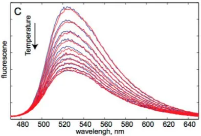
Figure 1.1 shows a graphical representation of the visualization of light-triggered electron transfer in TtTrmFO- the major focus of chapter 6. In this chapter, the main results obtained on electron transfer in TtTrmFO have been presented in detail. The reasons for focussing on mutant TrmFO C51A are elucidated. Time-resolved fluorescence was performed to visualize the excited state FAD in TrmFO and its mutants. Time-resolved absorption was performed in tandem to visualize both excited and product states formed in TrmFO mutants. Subsequently, deconvolution of a decay
CHAPTER 1. INTRODUCTION
Figure 1.1: Light-triggered electron transfer in TrmFO from Thermus thermophilus.1
associated di↵erence spectrum leads to the identification and characterization of the TyrOH+ intermediate. Furthermore, the direction of the transition dipole moments were studied using polarization photoselection experiments.
Chapter 7 describes similar experiments (as above) on the model flavoprotein GOX. TyrOH+ was observed in two decay phases of in the transient absorption spectra of GOX. Through spectral deconvolution, the spectrum of TyrOH + was calculated for each phase and compared with that observed for TrmFO C51A. Finally, a kinetic model was proposed for the charge transfer processes in light-triggered GOX.
Chapter 2
Charge transfer in biological systems
2.1
Overview
Redox charge transfer reactions are ubiquitous in biological systems. They involve the movement of electrons and protons, which are sometimes coupled. They can play an important role in energy transduction pathways in living organisms like photosynthesis2
and respiration.3 They are also associated with processes like signalling4 or catalysis.5
Charge transfer proteins are of interest to a wide community of researchers as understanding them can shed light on fundamental biological phenomena which are relevant for designing biocatalysts6 and artificial photosynthesis systems7,8 among
others.
Movement of small charged entities (electrons and protons) is often very rapid. High speed of charge-transfer steps can often be advantageous in biological systems because it prevents them from being rate-limiting in protein-substrate interaction processes. Conversely, this often makes pure redox intermediates elusive and difficult to characterize in enzymatic catalysis.
A major reason for the initiation of the wide range of studies in the field of charge transfer came from the development of instrumentation which enabled the study of rates of rapid chemical reactions. Electron transfer reactions are among the fastest to occur in nature. Some of the earliest experiments were performed using rapid mixing technology (stopped flow) on inorganic chemical model systems and charge transfer was observed with a time-resolution in the order of milliseconds.9 With the advent of lasers
and the concurrent development of newer spectroscopic technologies, it has become possible to visualize faster electron transfer processes, eventually down to the picosecond and sub-picosecond regime, for cases where the reaction can be initiated by light. This has proven to be particularly useful in the case of light-driven biological systems, as electron transfer has been observed to occur over a wide range of timescales:
CHAPTER 2. CHARGE TRANSFER IN BIOLOGICAL SYSTEMS starting from microseconds/milliseconds (timescale typical for catalytic turnover) going down to femtoseconds.
In mixing methods, the protein is modified by binding of external molecules. Here, the technology-limited mechanical mixing speeds, or intrinsic binding rates, limit the local perturbation created in the protein. Hence, the first electron transfer steps are often too fast to be observed by stopped flow techniques. Laser-based technologies have proved to be especially useful for observing charge transfer in biological systems. Pulsed lasers are able to create flash perturbations (down to femtosecond duration since the 1980s) within the protein. This can induce electron transfer in the protein which is often orders of magnitude slower than the impulsive light perturbation, making its signal easy to resolve. Electron transfer reactions can be characterized by measuring various parameters like rates, activation energy, temperature dependence. In order to determine rates, we measure changes in the physical properties of the donor and acceptor moieties, in particular their optical absorption and emission spectrum. It is therefore convenient to also use light for this purpose, as it can monitor changes in the redox state of the sample with high time resolution. Incidentally, many biological redox partners are coloured as they consist of cyclic extended ⇡-conjugated systems that absorb in the visible region. As will be emphasized below, such approaches are limited to (not necessarily naturally) light-activatable proteins.
Productive long-range biological electron transfer can occur either between di↵erent proteins or between multiple redox centres located within the same protein. Electron transfer proteins appear to be naturally designed to contain robust redox centres to facilitate their function (even when exposed to limited thermal fluctuations and genetic modifications).10 In 1951 Michaelis, in the context of oxidoreductase protein design,
wrote that “proteins must bring redox centres together”.11 At the time, initial models
to describe electron transport within proteins were still under development. In 1956, Chance and Williams realized that a rigid structure with immobilized redox centres would require the encapsulating polypeptides to act as electron conductors or semiconductors.12,13 As these requirements could not be reconciled with the actual
properties of enzymes, they suggested that thermally promoted conformational changes could lead to the encounter of redox cofactors. Also in 1956, Marcus first wrote about electron transfer theory. Initially, this was in the context of pure chemical reactions. Ultimately the theory has been widely used in various forms for decades in biochemical studies.14,15
CHAPTER 2. CHARGE TRANSFER IN BIOLOGICAL SYSTEMS
2.2
Electron transfer theory
As interest in reaction kinetics and electron transfer grew after 1945, the field was characterized by a strong cooperation between theory and experiments.16 With
increased availability of isotopes after the second world war,17 electron transfer
reactions were studied extensively using isotope exchange reactions. This approach proved useful in observing simple electron transfer in homogeneous inorganic chemical systems.9,17,18 Such reactions were of the type:
Fe2++ Fe*3+⌦ Fe3++ Fe*2+ (2.1) where * designates the labelled isotope.
These reactions are relatively straightforward to interpret as the reactants are the same as the products (but for the isotope substitution) thus eliminating the need to account for the relative thermodynamic stability of reactants and products. More generally, electron transfer reactions represent the simplest class of reactions in chemistry as no bonds are broken or made in an elementary electron transfer process.
Several theories have been proposed to describe such reactions.14,19,20 The most
successful among them appeared to be the one proposed by Marcus for calculating rates in outer-sphere electron transfer reactions in 1956,14 coincidentally at the same time as Chance and co-workers (section 2.1) were experimentally studying electron transfer. In later years, Marcus extended the application of this theory to include biological systems.15 This is perhaps the simplest among the theories to describe electron transfer. For this reason, it has also gained a wide-ranging support from researchers, including biologists.
The initial molecular events in light-based perturbations, the absorption of photons by coloured entities, are extremely fast. They consist of electronic transitions within the molecule from the ground state to an excited state. According to the Franck-Condon principle, these transitions can be regarded as occurring in a stationary nuclear framework i.e., they would be vertical in energetic diagrams such as those of Fig.2.1.
Marcus implemented a slight variation to the Franck-Condon principle. He proposed that thermal fluctuations (including those for surrounding solvent molecules) of the nuclei of reactants and products bring the system in a configuration favourable for barrier crossing to occur on a much longer timescale than the electron displacement from donor to acceptor itself. Fig. 2.1 shows the reactant and product Gibbs energy surfaces represented as parabolas characteristic of harmonic oscillators as a function of reaction coordinate (which accounts for nuclear coordinates). The geometry of these parabolas can be used
CHAPTER 2. CHARGE TRANSFER IN BIOLOGICAL SYSTEMS
Figure 2.1: Potential energy curves assuming harmonic oscillators of the reactants (D + A) and products (D++A–), where D is donor and A is acceptor. In the plot, q0
DA
and q0
D+A– represent the position of the potential energy minima of the reactants and
products respectively. The two parabolas intersect at q*. Gibbs energy of activation ( G‡),
standard Gibbs energy ( G°) and reorganizational energy ( ) have also been represented. to predict reaction rates and kinetic barriers. The electronic and nuclear terms can then be regarded separately while calculating the rate:
kET =
2⇡
~ (EC)(F C) (2.2)
where EC is the electronic coupling term and FC is the (nuclear) Franck Condon term. Electron transfer can occur when thermal fluctuations bring the reaction coordinates of the reactants to q* (Fig. 2.1), where the parabolas for reactants and products intersect. The potential energy minima of the reactants and products are represented by q0
DA and q0D+A– respectively (Fig. 2.1). Gibbs energy of activation ( G‡) is the
energy required by the reactants to go from q0
DA to q*, and is the energy required for
self-exchange reactions ( described in Equation 2.1).
It can be inferred from Equation 2.2 that the probability of electron transfer depends on the probability of the reactants to reach the q⇤ configuration and on the probability of moving to the product energy surface after this, which itself depends on the electron coupling between the DA state and the D+A– state. The reorganizational energy ( ) is an energy change resulting from the molecular rearrangement that occurs as the charge distribution is altered in the donor-acceptor complex in the (protein)
CHAPTER 2. CHARGE TRANSFER IN BIOLOGICAL SYSTEMS medium as well as in the surrounding solvent molecules. The driving force of the reaction, G° , has also been indicated in Fig. 2.1.
For one-step reactions, the main mechanism of electron transfer is tunnelling through the potential energy barrier. For efficient tunneling, there needs to be an overlap between the wavefunctions of the reactants and products. The electronic coupling term, EC, within a homogeneous medium has been described by Hopfield21 in 1974 as proportional
to the square of the coupling hamiltonian energy HDA which can be written as
EC =hHDAi2 =hHDA0i 2
e r (2.3)
where r is the edge-to-edge distance between the donor and acceptor and is a constant reflecting the speed at which the exponential wavefunctions of donor and acceptor decrease within the medium. hHDA0i stands for the electronic coupling matrix
element when the donor and acceptor are in contact (r = 0). In this formalism, the electronic coupling between donor and acceptor decreases exponentially as the distance (r) between them increases.
In the classical limit, the rate of electron transfer from donor to acceptor can be described by kET = 2hHDAi2 h ⇡3 4 RT !1/2 e G‡/RT (2.4)
Furthermore, from Equation 2.4, the Gibbs energy of activation ( G‡) can also be represented in terms of the driving force ( G°) and the reorganizational energy as ( )
G‡ = ( G° + )
2
4 (2.5)
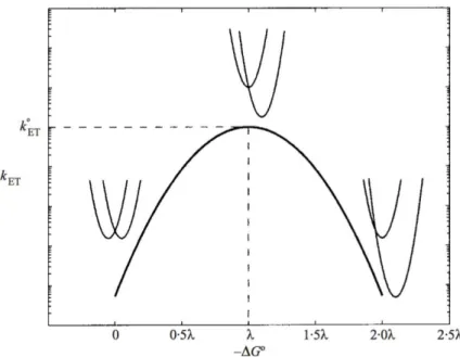
It can be seen intuitively that with the disposition of the parabola in Fig. 2.1, the rate increases as the driving force ( G°) increases. Fig. 2.2 shows the dependency of the rate of electron transfer on the driving force ( G°). It can be inferred that electron transfer becomes more efficient when G° becomes negative in the normal Marcus region. At G° ⇡ , G‡ ⇡ 0 and electron transfer is barrierless. In the third case, as the driving
force is increased further, electron transfer enters the Marcus inverted region and the rate decreases again. The three regions have been indicated in Fig. 2.2. All this indicates that for optimal electron transfer, nuclear rearrangements accompanying electron transfer must be compensated for by the reaction driving force. Gray and Winkler suggest that this balance between G° and is a direct consequence of protein structure.13 They
suggest that biological electron transfer over long distances is possible because protein folds create a suitable balance between G° and , as well as adequate coupling between redox centres.
CHAPTER 2. CHARGE TRANSFER IN BIOLOGICAL SYSTEMS
Figure 2.2: Dependence between the rate of electron transfer kET and driving force G°
as derived from equations 2.4 and 2.5. Rates keep increasing (from left to right) with driving force until they reach the barrierless electron transfer regime (where G° ⇡ ). Following this, electron transfer rates keep decreasing as G° continues to increase. This is the inverted region. (Source: Gray and Winkler13)
It must be noted that the above relations are at the semi-classical limit of Fermi’s Golden rule and are valid only when nuclear motions can be treated classically. This is possible at high temperatures and when electronic coupling between the donor and acceptor is weak (below a few tens of cm–1).22 At low temperatures, in principle,
quantization of the energy levels has to be taken into account. At the adiabatic limit, coupling between DA and D+A– states is very strong and surface hopping at q* has a high probability. Thus, for strongly coupled systems, electron transfer depends on the rate of reaching q⇤.
The coupling term proposed by Hopfield21 is applicable for weakly coupled systems.
The formalism implies that the donor and acceptor are separated by a homogeneous medium, and therefore electronic coupling decays exponentially with distance (Equation 2.3). As mentioned earlier, the decay of this electronic coupling is represented by the constant in Equation 2.3. On representing the medium by a single constant , it is assumed that it is homogeneous. However, the nature of the medium of electron transfer proteins has long been a subject of debate.
The simplest view advocated by Dutton and co-workers is to assume homogeneity of the protein medium through the uniform-barrier model. In this analysis, they take to be the same for all electron transfer reactions at am empirical value of = 1.4˚A 1.10,23
CHAPTER 2. CHARGE TRANSFER IN BIOLOGICAL SYSTEMS To a first order approximation, they described the rate of electron transfer at room temperature is given by:10
log kET = 13 0.6(r 3.6) 3.1
( G° + )2
(2.6) The assumption of a uniform presupposes that biological electron transfer is independent of the nature and structure of the biological medium. This view is likely quite valid for very long-range reactions, where the atom-level details of the intervening media are e↵ectively averaged. However, at shorter distances, small variations from this average value of can lead to large changes in rate constants of biological electron transfer. Hence, structural dependencies of have been invoked.13 The packing density model introduced by Page, Dutton et al.10 is another way to introduce structure
dependent variation of .
Interpreting the protein medium as heterogeneous, overall electronic coupling has been described by Gray and Winkler in the tunneling pathway model as arising from a combination of parallel pathways each consisting of distinct steps through covalent bonds, through hydrogen bonds and through space. The total coupling of a single pathway is given as a repeated product of coupling decays of individual links in the pathway.
Interpretations of by uniform- barrier and tunneling pathway models are at odds for specific systems.24 In many of these cases, there is a debate concerning the usage of
edge-to-edge distances for calculation of electron transfer rates. As mentioned earlier, small changes in can lead to larger changes in rates. In other cases, di↵erences arise due to the method by which was deduced. Ideally, values of can be determined from electron transfer reactions at several donor-acceptor distances in the barrierless regime (See Fig. 2.2). The barrierless regime is not always determined, as it is not straightforward to construct a Marcus curve. Consequently, is extracted from electron transfer reactions that are activated. This makes it difficult to disentangle the distance dependence of tunneling from that of the activated ET processes. Therefore, in our studies and interpretations, the edge-to-edge distances between donor and acceptor are linked qualitatively to the rate of electron transfer.
As the earlier work of Chance and Williams (see section 2.1), Page, Dutton et al. have described the fundamental design of electron transfer in proteins as “two catalytic sites connected by redox chains”.23 Catalytic sites have been described as multi-electron
redox centres or groups of single-electron redox centres that communicate with substrates and behave as donors and/or acceptors of electrons. Chains are described as groups of single-electron redox centres containing wider separation among them, with fewer near-neighbours. Catalytic sites can be located within a single protein or placed
CHAPTER 2. CHARGE TRANSFER IN BIOLOGICAL SYSTEMS sufficiently close to each other to allow electron transfer without a connecting chain. Frequently, proteins contain one catalytic site linked to a redox cofactor chain. These chains link the catalytic site to those in other proteins or to the solvent. Common examples of “catalytic sites” are oxidizable or reductible metal cations like Cu2+, Mg2+, Fe2+ etc. They can also include organic cofactors of proteins like FAD, FMN, heme, chlorophyll, etc. some of which also harbour metal atoms. In the last decades it has also become apparent that examples of participants in the redox chain also include radical-forming amino acids such as tyrosine and tryptophan.
It has been observed that the edge-to-edge distance between the donor and acceptor in many natural electron transfer proteins extends from the point of van der Waal’s contact to about 14 ˚A10 for rates to be in a physiologically relevant regime. Generally
for distances greater than 14˚A, functional “long-range” charge transfer reactions between donor and acceptor redox centres are thought to be facilitated by such redox chains (bridges). The role played by the bridge entities has been considerably debated as the bridge-state energy is expected to have a marked influence on the ET mechanism.25 Two electron transfer mechanisms have been suggested - superexchange
and hopping. According to the superexchange mechanism, electron transfer proceeds directly from the donor to the acceptor without creating real intermediates among the bridge molecules.26 Contrarily, when the orbital wavefunctions of the bridge molecules
overlap with the donor and acceptor, the bridge molecules are directly involved as intermediates in electron transfer between the donor and acceptor. This mechanism is called hopping. Electron hopping involves multiple tunneling steps, often to promote charge separation within and between proteins, which is essential for instance in energy storage and conversion.27
In several studies, it has also been observed that electron transfer over very long distances occurs with the formation of charged bridge molecules as reaction intermediates28,29 (lending credence to the hopping mechanism). An evaluation of the
redox potentials of prospective chain elements is helpful in determining the possibility of formation of oxidized or reduced bridge intermediates. In proteins with a single redox cofactor, side-chains provide opportunities for multistage tunneling. While the redox potentials required to oxidise and reduce polypeptide side chains are generally high, the side-chains of certain amino acids (tyrosine and tryptophan) have redox potentials which are potentially suited to them30–33 (See chapter 3).
In our study, we have performed fluorescence and absorption time-resolved spectroscopy on several flavoproteins. Through these techniques, we have deduced the rates of electron transfer from the neutral ground state amino acid donor to excited state acceptor. We have also discussed these rates in relation to the distance between
CHAPTER 2. CHARGE TRANSFER IN BIOLOGICAL SYSTEMS donor and acceptor entities. Analysis of electron transfer rate and distance relationships is facilitated by a consistent definition of the donor-acceptor distance. As FAD is a ⇡ aromatic molecule, we have taken edge-to-edge distances using the shortest distance between the ⇡ conjugated rings of the donor and acceptor.
2.3
Well-studied biological charge transfer systems
Figure 2.3: Z-scheme of oxygenic photosynthesis showing the electron flow and the potentials of redox partners involved. Note: the tyrosine residue is involved in a charge transfer relay between P680 and Mn in the oxygen evolving complex of PSII.34,35
Conversion of light energy to electrical energy to chemical energy has been famously studied in photosynthetic reaction centres.2 In the initial stages for this process,
photons are acquired from sunlight to induce charge separation in a membrane-protein complex. This creates an electric potential and a proton gradient over the membrane which ultimately drives ATP synthesis. Photon absorption is enhanced by well-optimized antenna systems which funnel the excitation energy to the reaction centres (as seen in Fig. 2.3). This phenomenon occurs widely in green plants, algae and cyanobacteria. In plants, two reaction centres are used in series - photosystem II and photosystem I (Fig. 2.3). Among these, photosystem II (PSII) is unique as it catalyses the four-electron oxidation of two water molecules to O2 in the oxygen evolving complex
(Fig. 2.3). In fact, PSII is the only enzyme known to perform this function. In the oxygen-evolving complex, tyrosine plays an important role. Each charge separation event here proceeds via the reduction of the photo-oxidized chlorophyll dimer primary electron donor P680 by the nearby redox-active tyrosine (⇠ 20-200 ns)36 and the
CHAPTER 2. CHARGE TRANSFER IN BIOLOGICAL SYSTEMS subsequent re-reduction of the tyrosyl by Mn (30µs ⇠ 1ms) range.
Oxygenic photosynthesis is the phenomenon in which water is oxidized using photons to yield O2, four electrons and four protons. Reversely, respiration is the (non
light-active) process in which O2 is reduced using four electrons and four protons.
Cytochrome c oxidase (CcO) is another prototypical protein in the study of biological charge transfer, and is an enzyme implicated in the mitochondrial (and bacterial) respiration process. During this process, it accepts electrons from one side of the mitochondrial membrane and protons from the other, thus creating an electrochemical proton gradient across the membrane. Although CcO is not functionally light-active, it has been studied using light to visualize the electron transfer pathways involved.37 CcO
contains four redox-active cofactors, two “a”-type hemes, and two copper centres, and accommodates four electrons in its fully reduced form.37 The complex mechanism of O
2
reduction to H2O in CcO mobilizes a number of redox carriers and is notably thought
to involve a Tyr structuring the active site. This Tyr is cross-linked to a histidine residue, itself coordinating another redox carrier, a Cu atom. The formation of transient product states is linked to the proton pumping function of the enzyme.
Apart from photosynthetic and signalling proteins, there is a small group of “true” enzymes where the catalytic reactions are driven by light. These so-called photoenzymes use light as a substrate and therefore,require a continuous supply of light (of a certain energy) for their function. They contain cofactors which are chromophores and absorb the light. There are only three known classes of “true” photoenzymes -Photolyases,38 Light-Dependent NADPH: Protochlorophyllide Oxidoreductases
(LPORs) and the recently discovered fatty acid photodecarboxylase (FAP).39 The
light-activation of photolyase has been well studied and is described below.
DNA photolyase is an enzyme implicated in the light-induced repair of UV-damaged DNA in bacteria, plants and certain animals. Apart from photocatalysis involving electron transfer from the reduced flavin FADH– to the substrate, the protein performs a second photoreaction: preparing the catalytic state FADH– from the resting state FADH . The latter reaction involves a triple tryptophan chain, which constitutes one of the best examples of redox-active components bridging a donor and acceptor. Here, the two redox centres are the solvent-exposed terminal tryptophan and the flavin radical cofactor FADH . Electron transport through the protein is initiated by light absorption which creates the excited state of the flavin radical. An electron is then extracted from a nearby Trp (W382, Fig. 2.4) in ⇠ 30ps. Subsequently, electrons move along the bridge of tryptophans (as shown in Fig 2.4) to stabilize the catalytically active FADH– in ⇠ 30ps. The latter processes occurs faster than the initial charge separation, leading to an e↵ective overall trans-protein charge transfer in 30 ps.40
CHAPTER 2. CHARGE TRANSFER IN BIOLOGICAL SYSTEMS
Figure 2.4: Scheme of electron and proton transfer reaction chain in DNA photolyase from E. coli by Lukacs et. al..40 The FADH cofactor is excited and charge separation
occurs as electrons hop through a chain of tryptophans.
This reaction itself is challenging to study as the three bridge entities are chemically identical tryptophans. Complementary molecular biology and biophysical techniques such as site-directed mutagenesis and polarization photoselection41 have aided in the
experimental study of such systems. In the particular case of DNA photolyase, the reaction was studied by changing each tryptophan one-by-one in the chain to redox-inert phenylalanine.40–43 It has subsequently been demonstrated that this
tryptophan chain in DNA photolyase (Fig. 2.4) e↵ectively acts as a “wire” within the protein to facilitate long-range electron transfer.40
While photoenzymes require continuous exposure to light to function, there are other enzymes which use light for signalling. They are called photoreceptors as they “receive” photons to signal a reaction. Many organisms in nature have developed light-sensing proteins to optimize certain functions. Mechanisms utilizing blue light have been observed to be involved in various biological functions44 and are frequently mediated by proteins
containing a flavin cofactor (see chapter 3). Flavin-binding blue-light photoreceptors can be categorized based on their structure and photochemistry into three distinct groups:
• light-oxygen-voltage (LOV) photoreceptors,
• sensors of blue-light using flavin adenine dinucleotide (FAD) (BLUF photoreceptors) and
• cryptochromes (CRYs)
Other classes of photoreceptors (not involving flavins) include rhodopsins (with the chromophore retinal),45 phytochromes (with phytochromobilin), photoactive yellow
CHAPTER 2. CHARGE TRANSFER IN BIOLOGICAL SYSTEMS protein (with p-coumarin) and the UV sensor UVR8 (with 14 tryptophan residues).46
LOV domains are blue-light photoreceptors that contain a non-covalently bound flavin mononucleotide (FMN) or in some cases flavin adenine dinucleotide (FAD), which acts as a sensor for environmental stimuli such as light or oxygen.47,48 This
non-covalently bound flavin chromophore senses blue light in the 400-470 nm range. Upon light activation, it undergoes a reversible photocycle, what can be observed looking at the steady state spectra: under blue light illumination the structured 447 nm peak of the flavin disappears and a single non-structured band will rise around 390 nm. Due to the blue light irradiation flavin is promoted to the excited singlet state, which undergoes intersystem crossing to the excited triplet state. This is followed by adduct formation between the C4a atom of the flavin cofactor and the sulphur atom of the neighbouring cysteine.49,50 In the dark, the flavin-cysteinyl adduct degrades slowly, on a
time-scale from minutes to hours. This reverts the LOV domain to the ground state and concludes its photocycle. Natural systems utilize blue-light sensing of LOV domains to pass this information to the nearby e↵ector domain that they are linked to.51 This mechanism, that does not involve an electron transfer reaction, is used in
phototropism,52 stomatal opening and temporal circadian rhythms among others.
Proteins containing BLUF domains are responsible for photoadaptive responses of many prokaryotes and a few eukaryotes.53,54 They are implicated in various reactions
including phototaxis55,56 and photosynthetic gene regulation57,58 in phototrophic
organisms, biofilm formation59 along with virulence in pathogenic bacteria.60,61 BLUF
domains contain a non-covalently bound FAD as the light-absorbing cofactor. Like the LOV domain, the BLUF domain passes through a light and dark state during its photocycle. The light state of BLUF has been observed to be almost identical to the dark state, with spectra resembling that of oxidized FAD, except for a 10-15 nm shift of the 447nm peak to the red.57,62 This red-shifted photoproduct relaxes to the dark state
within seconds to minutes.62,63
Mechanisms of the BLUF domain proteins can di↵er from one organism to another. In AppA – the most studied BLUF domain protein – the protein initially forms a complex with a transcription factor (PpsR). Absorption of blue light would lead to larger structural change resulting in the release of PpsR, preventing the biosynthesis of the photosynthetic genes.64 The mechanism which leads to this large structural
movement and to the dissociation of the AppA-PpsR complex is still not completely elucidated and the mechanism of photoactivity changes in di↵erent BLUF domain proteins. In Pixd (Slr1694), which controls phototaxis in the cyanobacterium Synechocystis sp. PCC 6903 a proton coupled electron transfer was observed between the well conserved Y8 tyrosine and FAD.65,66 Light absorption initially triggers the
CHAPTER 2. CHARGE TRANSFER IN BIOLOGICAL SYSTEMS formation of the anionic flavin radical, which is protonated in a few picoseconds (most probably by the tyrosine) forming the neutral flavin radical (see chapter 3 for flavin redox states). As the close tyrosine residue is well conserved – replacement with other amino acid abolishes the photoactivity of the protein – in all BLUF domain proteins it was proposed that a similar mechanism is responsible for the function of these photoactive flavoproteins. A similar mechanism was found to be present indeed in PapB where formation of the neutral semiquinone decays to the red shifted signaling state, in AppA however, the role of radical intermediates in the photocycle is debated and continues to be explored.67 It has been suggested that the formation of radical intermediates is not necessary for BLUF domain activity.67 Lukacs et al. have proposed a non-radical pathway for BLUF domain function which proceeds via keto-enol tautomerization.67
Figure 2.5: Active site of cryptochrome from Arabidopsis thaliana68 (in blue, PDB:
1U3D) overlaid with that of E. coli photolyase (in red, PDB: 1DNP). The electron is transported through a triad of tryptophans in cryptochrome similar to the mechanism in DNA photolyase. It has been proposed that magnetic sensitivity in cryptochrome is induced by radical formation of the FAD and the terminal tryptophan residue WC.
Electron transfer proved to be crucial in the function of E. coli photolyase which repairs the UV induced lesions in the DNA strand. In catalytically active photolyase the flavin is in its fully reduced form and upon blue light absorption an electron is ejected to the pyrimidine dimer thus breaking the bond. It is interesting that after purification in the majority of photolyases, FAD is in the neutral semireduced radical form, but it can be reduced by using visible light. In this process also ET takes place via a well conserved tryptophan triad.69 Despite being homologous to photolyases, cryptochromes
are involved in very di↵erent processes like the regulation of the hypocotyl growth in plants and entrainment of circadian rhythm in animals.70 They have also been
suggested to be sensors for the geomagnetic field to assist many animals in long-range navigation.71,72 The formation of the signaling state in cryptochrome involves blue
light-induced electron transfer to the FAD cofactor from a tryptophan triad (W400, W377, W324). These tryptophans are highly conserved among cryptochromes from many species and bridge the space between flavin and the protein surface (Fig. 2.5). The sequence of electron transfer steps result in an increased separation among the
CHAPTER 2. CHARGE TRANSFER IN BIOLOGICAL SYSTEMS radical pair, eventually leading to the formation and stabilization of the signaling state of cryptochrome (reported to be FADH–). It is interesting to note the similarity between the mechanisms of electron transfer in the homologous proteins DNA photolyase and cryptochrome, as they both proceed through a conserved tryptophan triad (Fig. 2.5).
Redox intermediates have also been observed, or inferred to form, in catalytic proteins which are photo-inactive. Class 1a ribonucleotide reductase (Class 1a-RNR) is an example of such a system. Ribonucleotide reductases (RNR) catalyse the reduction of nucleotides to 2’-deoxynucleotides, which are essential for the de novo synthesis of deoxyribonucleotides. As deoxyribonucleotides are precursors to DNA, RNRs are important for maintaining the precision of DNA replication and repair. They are found in all organisms and are classified based on the metal cofactor used to initiate catalysis. Class 1a RNR contains a diferric-tyrosyl radical (TyrO ) as “cofactor”. The prototypical class 1a RNR from E. coli is comprised of two subunits, ↵2 and 2, and functions actively as an ↵2 2 complex.73 The 2 subunit contains the diferric-TyrO
122
cofactor. The TyrO122 species is observed to show high stability, atypical for redox
intermediates of amino acids, as it has a lifetime of 4 days. TyrO in solution has a lifetime of milliseconds, but the longer lifetime of TyrO122 in class 1a RNR is likely due
to the fact that it is positioned in a hydrophobic pocket within the protein along with its position between the two Fe atoms. Additionally, it has also been proposed that a specific charge transfer pathway exists across the ↵2 2 complex which contains four tyrosines involved in long-range charge transfer over 35 ˚A.73 The individual charge
transfer steps are linked to the RNR function of nucleotide reduction, and several TyrO intermediates have been visualized for these steps aided by protein engineering.
Figure 2.6: Relative orientation of hemes and the intervening tryptophan residue (PDB: 3L4M) in the diheme center of MauG. The edge-to-edge distances between the indole side chain of Trp93 and each heme porphyrin ring, and between the two heme porphyrin rings have been indicated.74
CHAPTER 2. CHARGE TRANSFER IN BIOLOGICAL SYSTEMS MauG is another protein whose main catalytic function is enabled by the formation of redox intermediates involving an aromatic amino acid residue. MauG is a diheme enzyme that catalyzes the final steps in the biosynthesis of the catalytic tryptophan tryptophenylquinone (TTQ) cofactor of methylamine dehydrogenase. It has been observed to contain an electron transfer chain containing two protein-bound hemes and a tryptophan residue.74 Fig. 2.6 shows the diheme centre of MauG. The distances
between the two hemes as well as their distances from Trp93 have been shown. This Trp-93 resides midway between the hemes, and each Fe(IV) heme (as shown in Fig. 2.6) has been noted to be a sufficiently strong oxidant for Trp .
It has been proposed by Geng et al. that the charge equilibration proceeds through hole hopping via Trp93 without Trp radicals getting accumulated.74 Their model supposes that rapid and reversible electron transfer occurs between the two hemes and the intervening Trp93 in the hopping site. It can be inferred that Trp93 intermediates form and decay rapidly within the MauG system. Such a “charge resonance” system between the two hemes has been suggested to be the basis of the stability of the bis-Fe(IV) species in MauG which is an intermediate in its catalytic reaction.
The systems that have been studied in this work, the t-RNA methyltransferase TrmFO from Thermus thermophilus and glucose oxidase (GOX) from Aspergillus niger, are also not naturally photoactive. They are flavoproteins involved in the catalysis of di↵erent biological reactions (see chapter 6 and chapter 7). In both systems, like the flavoprotein systems described above, FAD excitation with blue light leads to subsequent charge transfer steps involving neighbouring residues. This has allowed us to visualize previously unobserved intermediate states in both systems, particularly those of close-lying redox active residues tyrosine and tryptophan. In several examples mentioned above, tyrosine (TyrOH in the neutral ground state) is actively involved in bridging electron transfer. The nature of the intermediates formed by tyrosine has been debated, generally assumed to include the deprotonated TyrO neutral radical. The radical cation form TyrOH+ has also been proposed to be involved in charge transfer, but had never been characterized.75 A major aim of this thesis is to investigate this
issue. The subsequent chapter will introduce the background of this work in greater detail.
Chapter 3
Flavoprotein photochemistry and
aromatic amino acid radicals
3.1
Flavins and flavoproteins
Proteins containing flavins as redox cofactors have unique properties, as they are able to participate in both 1 e– and 2 e– transfer reactions and/or hydride exchange reactions. This enables them to participate in a wide range of redox reactions. They are found abundantly in nature as they are estimated to account for up to 2% of all proteins.76 As
seen from the examples given in the previous chapter, the optical properties of flavoproteins enable a limited number of them to act as “photoswitches” through light-triggered reactions. However, a large proportion of flavoproteins, whilst participating in functional electron transfer processes, are not naturally photoactive. In this section, we will discuss the general redox properties of flavins to understand their role in electron transfer processes.
Riboflavin, isolated from cow milk by Blyth in 1879,78 was the first flavin to be
discovered. Fig. 3.1 shows the structure of riboflavin. It contains a conjugated ring system (isoalloxazine) which is linked to a ribityl chain. Riboflavin is present in various food items including milk, eggs and cereal. While bacteria, fungi and plants can produce riboflavin, other organisms like humans do not have the ability to synthesize it.79 Interestingly, flavins in animal tissues are rarely present as free riboflavin. They are
mostly present as flavin mononucleotide (FMN) or flavin adenine dinucleotide (FAD) bound to flavoenzymes. Riboflavin is a precursor to FMN and FAD in such systems. As seen in Fig. 3.1, the structure of FMN is similar to that of riboflavin with the addition of a phosphate group. On addition of an adenosine group to FMN, it becomes FAD. Riboflavin is an important dietary component and is known commonly today as vitamin B2 which is a popular dietary supplement. Its deficiency has been linked to
CHAPTER 3. FLAVOPROTEIN PHOTOCHEMISTRY AND AROMATIC AMINO ACID RADICALS
Figure 3.1: Structures of riboflavin, flavin mononucleotide (FMN) and flavin dinucleotide adenine (FAD). The isoalloxazine ring has been shown in its oxidised and two-electron reduced state. Additionally, the numbering scheme for the isoalloxazine ring is indicated. Image taken from ref [77].
infection.80,81
Figure 3.2: Di↵erent redox states of FAD : the oxidized form FADox (designated FAD
here), the anionic semiquinone form FAD–, the neutral semiquinone form FADH , the hydroquinone form FADH– and the fully reduced form FADH2. These are the
five physiologically relevant states of FAD. Other states can also be produced outside physiological conditions. Image taken from ref [82].
More than 90% of the known flavin-dependent enzymes are classified as oxidoreductases, and the remaining enzymes are classified as transferases, lyases, isomerases and ligases.77 The immense biochemical utility of FMN and FAD is due to
one-CHAPTER 3. FLAVOPROTEIN PHOTOCHEMISTRY AND AROMATIC AMINO ACID RADICALS and two-electron, proton and/or hydride transfer reactions. Consequently, FMN and FAD can exist in several physiologically relevant redox states: oxidized (FADox),
one-electron reduced anionic semiquinone (FAD –), one-electron reduced protonated semiquinone (FADH ), two-electron reduced protonated (FADH–) and two-electron reduced bi-protonated (FADH2) (as shown in Fig. 3.2). In addition, the oxidized flavin
molecule is susceptible to nucleophilic attack at positions N5 and C4a of the isoalloxazine ring. The ability of FAD and FMN to exist in various redox states makes them versatile enzyme cofactors for numerous biochemical reactions, most of all in oxidoreductases, which participate in biological redox reactions.
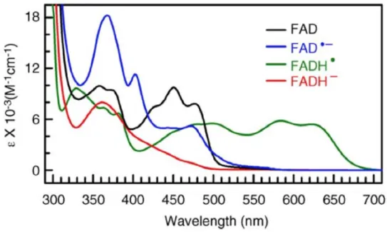
Figure 3.3: Spectra of four di↵erent redox states of FAD.83 FAD here refers to oxidized
FAD. The FADH2 spectrum, not included here, slightly resembles that of FADH– with a
more gradual incline and lower extinction in the blue region. The FAD and FAD–spectra are from mosquito cryptochrome AgCRY1. The FADH and FADH– spectra are from E. coli photolyase.
As stated before, flavins (FAD and FMN) are among the few known redox coenzymes (like quinones) that can participate in both one-electron and two-electron transfer processes.84 Furthermore, they prove to be suited to spectroscopic studies as all
five redox states of flavins absorb in the visible region with distinctly di↵erent spectral signals. Fig. 3.3 shows the steady state absorption spectra of four relevant redox states of FAD.83 The oxidized form FAD
ox, the resting state for many flavoproteins, has a
distinct absorption band at ⇠ 450nm. This yellow form is the one with the highest extinction in the visible part of the spectrum. The reduction of FADoxcan be visualized
spectrally by the absorption decrease in the 450 nm region. For the one-electron reduced forms, this is additionally accompanied by an absorption increase in the red for FAD–, a small signal in the ⇠ 510 nm region and for FADH , a stronger signal
CHAPTER 3. FLAVOPROTEIN PHOTOCHEMISTRY AND AROMATIC AMINO ACID RADICALS extending over the entire visible spectrum (it is the only FAD redox state to absorb significantly beyond 530 nm). The two-electron reduced forms absorb weakly in the visible region; the FADH2 spectrum resembles that of the FADH– spectrum in Fig. 3.3,
with a more gradual incline in the blue region and with lower absorptivity (extinction coefficients).
Flavins have lower extinction coefficients compared to many of the coloured cofactors which participate in charge transfer reactions like chlorophyll and hemes (See Fig. 3.3). The molar absorption coefficient for the long-wavelength absorption maximum of chlorophyll a ("419) is ⇠ 90mM 1cm 185 and for hemes up to
⇠ 200mM 1cm 186 whereas the corresponding value for oxidized flavin (for example,
FAD87) at 450 nm is only 12 mM 1cm 1. Intermediates formed during biological redox
reactions are of considerable interest (see section 2.3). Using flavoenzyme systems to monitor such intermediates may facilitate characterizing them and evaluating their role in biological charge transfer reactions. In this context, flavoenzymes provide a unique advantage as their extinction coefficients are lower than that of other systems where intermediates have been proposed to participate. They have extinction coefficients of the same order of magnitude as the Tyr and Trp redox intermediates (see section 3.2) making it easier to distinguish the latter’s signal. In systems containing chlorophyll or hemes as cofactors, the absorption signal of these Tyr and Trp redox intermediates would be much smaller than the signal of the corresponding cofactor redox state. This makes flavoproteins especially suited to visualize charge transfer intermediates.
3.2
Charge transfer intermediates involving amino
acid residues
Biological systems are often naturally optimized to conduct long-range charge transfer between redox sites. The mechanism of charge transfer between a donor and acceptor frequently involves electron hopping via bridging amino acid residues (see section 2.2). These bridge amino acid residues have in turn been observed to form functionally relevant radical intermediates in many systems such as photosystem II,36 cytochrome c
oxidase,88 DNA photolyase,40,42,89 cryptochrome,71,72 among others.
In most biological systems where charge transfer has been studied, amino acid residues involved in redox reactions and formation of intermediates are tyrosine and tryptophan. The redox potentials of the constituents of the amino acid side chains are generally extremely high.13 However, tyrosine (Tyr) and tryptophan (Trp) have been
CHAPTER 3. FLAVOPROTEIN PHOTOCHEMISTRY AND AROMATIC AMINO ACID RADICALS
Figure 3.4: Schematic representation of the electron transfer from a photoexcited FAD to a nearby Tyr/Trp and the subsequent intermediates formed. The 2.7eV represents the excitation of FAD from ground state to FAD*. Free energies have been indicated for the formation of product states FAD–TyrOH+(1.08 eV) and FAD –TrpH+ (1.33 eV) which have been calculated using redox potentials.90,91 The pK
a values for the deprotonation of
radical cations have also been given. TyrOH+ has been proposed to be less likely to be observed as it is highly unstable (pKa ⇠ 2).92 On the other hand, TrpH+ is a more
CHAPTER 3. FLAVOPROTEIN PHOTOCHEMISTRY AND AROMATIC AMINO ACID RADICALS illustrated in the scheme in Fig. 3.4 for the case of light-induced reactions with FAD. The relative free energies of product states formed FAD –TyrOH+ (1.08 V) and FAD–TrpH+ (1.33 V) have also been indicated. The free energies have been calculated using the redox potentials for the pairs FAD/FAD– (-0.22 V),91 TyrOH+/TyrOH (1.4
V)90 and TrpH+/TrpH (1.15 V).90 The pK
a values for TyrOH+and TrpH+ are ⇠ 292
and ⇠ 493 respectively. TyrOH +has been proposed to be less likely to be observed75 as
it is highly unstable (pKa ⇠ 2)92 and is expected to undergo rapid proton transfer to
form TyrO . On the other hand, TrpH+ is a more stable radical cation as it has a pKa
⇠ 4.93
In DNA photolyase, both protonated (TrpH +) and deprotonated (Trp ) radicals of tryptophan have been observed to form sequentially at di↵erent timescales.69 On the
other hand, in a model compound containing a Trp-analogue, the latter species was inferred to be formed by concerted proton and electron transfer in aqueous solution.95
Together, these studies indicate that while Trp radicals can be formed by concerted electron and proton transfer, this is not always the case.
Tyrosine radicals have markedly di↵erent characteristics compared to tryptophan radicals. Since the pKa of TyrOH + is ⇠ 2, it is expected to be highly unstable if it is
formed at all. At the start of this thesis, it had not yet been observed as a reaction intermediate and was spectrally uncharacterised. In the prototypical photosynthetic system PSII (described in section 2.3), the secondary electron donor to the photo-oxidized chlorophyll dimer P680 is a tyrosine. Here, the electron transfer
(⇠ 20 200ns) occurs concomitant to proton transfer and therefore, no TyrOH+ intermediates have been observed. For several of the examples discussed in section 2.3, like cytochrome c oxidase, BLUF domain and ribonucleotide reductase, Tyr was indicated to form a TyrO intermediate, a form that has been characterized.96 Tyr has
also been observed to form radical intermediates in photolyase from the cyanobacterium Anacystis nidulans.89 For tyrosine-containing photoactivatable model compounds in
aqueous solution, a stepwise mechanism with electron transfer preceding proton transfer has been invoked.97 However, in this case TyrOH+ deprotonation is much faster than
its formation, prohibiting its visualization and characterization.
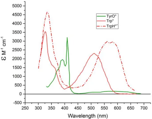
Tyr and Trp radicals have been characterized spectroscopically in many systems. Interestingly, in the neutral ground state, both Tyr and Trp absorb in the UV region of the spectrum. However, their characterized intermediates have distinctive spectra in the near-UV to the visible spectral range. Fig. 3.5 shows the characteristic spectra of known Tyr and Trp intermediates. Both the Trp radicals - neutral and cation - have been spectrally characterised. While the spectrum of TrpH+ contains a broad band centred ⇠ 570 nm, the spectrum of Trp contains a similarly broad band but centred at
CHAPTER 3. FLAVOPROTEIN PHOTOCHEMISTRY AND AROMATIC AMINO ACID RADICALS
Figure 3.5: Spectra of known intermediates of tyrosine and tryptophan. The tyrosine intermediate has been shown in green.96 The tryptophan intermediates have been shown