HAL Id: tel-03027465
https://tel.archives-ouvertes.fr/tel-03027465
Submitted on 27 Nov 2020
HAL is a multi-disciplinary open access
archive for the deposit and dissemination of sci-entific research documents, whether they are pub-lished or not. The documents may come from teaching and research institutions in France or abroad, or from public or private research centers.
L’archive ouverte pluridisciplinaire HAL, est destinée au dépôt et à la diffusion de documents scientifiques de niveau recherche, publiés ou non, émanant des établissements d’enseignement et de recherche français ou étrangers, des laboratoires publics ou privés.
commensaux promeut les poussées de maladies
inflammatoires cutanées
Charlotte Hurabielle-Claverie
To cite this version:
Charlotte Hurabielle-Claverie. L’immunité adaptative contre les champignons commensaux promeut les poussées de maladies inflammatoires cutanées. Médecine humaine et pathologie. Université de Paris, 2019. Français. �NNT : 2019UNIP7059�. �tel-03027465�
Université de Paris
Ecole doctorale Hématologie, Oncogenèse, Biothérapies (HOB) 561 Laboratoire: National Institutes of Health (NIH), NIAID, LPD then LISB, Metagenome
Immunology Section / INSERM U976
Adaptive immunity to commensal skin
fungi promotes inflammatory flares
Par Charlotte Hurabielle-Claverie
Thèse de doctorat en Immunologie
Dirigée par Yasmine Belkaid et Et par Armand Bensussan
Présentée et soutenue publiquement le 27 septembre 2019
Devant un jury composé de :
Président du jury : Pr. Bouaziz Jean-David, MD, PhD, Hopital Saint-Louis, Paris, France Rapporteurs : Dr. Segre Julie, PhD, National Institutes of Health, Bethesda, USA
Dr. Lionakis Michail, PhD, National Institutes of Health, Bethesda, USA Examinateurs : Pr. Caillat-Zucman Sophie, MD, PhD, Hopital Saint-Louis, Paris, France
Pr. Musette, Philippe, MD, PhD, CHU de Rouen, France
Directeur de thèse : Dr. Belkaid Yasmine, PhD, National Institutes of Health, Bethesda, USA Co-directeur de thèse : Dr. Bensussan Armand, PhD, INSERM U976, Paris, France
Titre : L’immunité adaptative contre les champignons commensaux promeut les poussées de
maladies inflammatoires cutanées
Résumé: Le microbiote cutané joue un rôle fondamental dans les réponses immunitaires physiologiques de l'hôte. Cependant, la façon dont les réponses immunitaires aux commensaux cutanés peuvent influencer la physiopathologie des maladies inflammatoires cutanées est encore mal comprise. Les champignons font partie du microbiote cutané normal et ils induisent de fortes signatures IL-17 (interleukine 17), une cytokine qui a également un rôle fondamental dans la promotion du psoriasis. Nous démontrons ici que les réponses cellulaires T aux champignons commensaux augmentent la dermatose psoriasiforme imiquimod-induite chez la souris. Lorsque la peau des souris était colonisée avec des champignons avant d’induire une inflammation cutanée avec de l'imiquimod, le phénotype de la maladie psoriasique humaine était reproduit, avec une augmentation des réponse TH17
induites par les commensaux, ainsi que de neutrophil extracellular traps (NETs). Les réponses mémoires TH17 aux champignons commensaux étaient suffisantes pour induire une nouvelle
poussée de la maladie après la résolution de l'inflammation initiale. En conclusion, nos données démontrent qu'une réponse TH17 aux champignons cutanés commensaux peut
aggraver l'inflammation cutanée et que la reconnaissance des commensaux cutanés dans un contexte inflammatoire peut induire une cicatrice inflammatoire permanente chez les lymphocytes T conduisant à de nouvelles poussées inflammatoires lors de la réexposition aux commensaux initiaux.
Mots clefs : microbiote cutané, champignons, psoriasis, TH17, imiquimod
Title: Adaptive immunity to commensal skin fungi promotes inflammatory flares
Abstract: Skin microbiota plays a fundamental role in host physiological immune responses. However, how immune responses to skin commensals can alter the pathogenesis of inflammatory disorders remains unclear. Fungi are part of the normal microbiota of the skin and they induced strong interleukin-17 signatures, a cytokine that is also critical in psoriasis inflammation. Here we demonstrate that T cell responses to canonical fungal commensals increased the pathology of murine imiquimod-induced psoriasiform dermatitis. Skin colonization with fungi prior to inflammation triggering with imiquimod further resumed the human phenotype, with increased in the commensal-induced Th17 responses and neutrophil extracellular traps. Notably, memory Th17 responses to commensal fungi were sufficient to induce disease flare after resolution of the initial inflammation. Together, our data demonstrate that a Th17 response to commensal skin fungi can worsen skin inflammation and that sensing of the commensals in an inflammatory manner induce a permanent inflammatory scar in the T cells leading to a pro-inflammatory transcription signature.
Acknowledgements
A really big thanks to Yasmine. It has been awesome working under your supervision for the last few years. I have learnt a lot and really enjoyed my time at the NIH. I understand now better the science world! Your guidance and support have been precious to drive my project. I really enjoyed working around all the wonderful, smart and fun people you hired. I also really enjoyed to learn how to present data and create a ‘story’ out of it, it has been an incredibly helpful lesson for my future career.
I am infinitely grateful to have received your support in my career choices, your help navigating immigration issues and your advice going forward.
Thank you to Saeko who started this project and with whom I hope we can collaborate again in the future!
Thank you to Julie and Verena helping with the last experiments and analysis!
Thanks a lot, to Ollie, Samira, Seong-Ji, NickNeck, Michael George, Ai-Ing, Ivan, Juliana, Nico, Verena, Julie, Apollo, Michel, Taylor, Djalma, Pete, Sidd, Hugh, for all of your feedback and advice over the last few years. Being able to regularly present in front of amazing young scientists has been incredibly valuable to learn how to think of results and next steps. I also really enjoyed learning to know each of you, spending time with you, improving my English (or destroying it depending on the days) and receiving support whenever I needed some!
Thank you Jackie!! I don’t know how I would have done anything without you, you have helped so much to solve any single problem I encountered with lab issues.
Thank you Teresa for the massive amount of sort you did for me! It was so helpful that you were able to accommodate any of the date I needed one, and it made those long days so pleasant because I knew I would laugh with you and share some passion about the science!
Thank you to Mihalis Lionakis and his postdocs, to allow me to get fundamental strains of
Candida albicans and for the mice you kindly offered!
Thanks to my committee Julie Segre, Mihalis Lionakis, and David Sacks for listening to my presentation and providing guidance each year.
Thank you to Armand Bensussan, Martine Bagot, and Jean-David Bouaziz to make my stay in the US a possibility, and to have been so understanding of my decision to stay there for my personal life and career!
Importantly, thanks to my family! Maman, Papa, Adele et Marc, de m’avoir accompagnée, soutenue, d’etre venus me voir et d’avoir accueilli Deanou pendant cette période!
Thank you ma choupinette chérie Marie, ma TouyTouy, Hélène Chérie, Marie J, and Chaton for the long hours of phone support while I was in the mouse house, allowing this to be a pleasant experience!
It has definitely been a challenge being away of all my family and friends but I’m infinitely grateful for your support and to have the feeling that you are always with me, to receive texts and calls almost every day, making the distance way more sustainable.
Thank you to my family in law, Nancy and Scott, Brit and Trevor, Libby and Ross, for welcoming me warmly in your family.
Thank you to the GIGOGY 3.4 team, Nick Neck, Ivan, SJ, Durant, M George, and Dean for helping balancing the hard work with balance, lift weight, squats, a lot of inappropriate comments, river dogs, sprints and going out. It was definitely a strong part of my daily wellbeing and I really appreciated all the laughs and snacks we shared together.
Thanks to the amazing women in science, Samira, Ai-Ing, SeongJi, Taylor (and Ollie obviously), for sharing inspiration together!
And … Thank you to my love, Dean, my biggest discovery of this Ph.D. and probably of my lifetime, to agree to spend the rest of your life with me, to help making it possible, to help me every day, including during the difficult times, to (sometimes) help me with my English, to break my stereotypes on America, to have learnt French, to go on all this crazy French trips with me and my friends and family, to have welcomed them, to help me being able to develop my career here and lift me up every day. Merci pour les ‘rigoles’ et les bisous et tous les jours passés ensemble. I feel very honored to be your wife!
Table of contents
ACKNOWLEDGEMENTS 3 TABLE OF CONTENTS 6 PREFACE 8 ABBREVIATIONS 9 INTRODUCTION 111. The immune system 11
1.1. Introduction 11
1.2. Innate immunity 11
1.3. Receptors of innate immunity 13
1.4. Adaptive immunity 14
2. The skin 26
2.1 Skin structure 26
2.2 Skin immune system 27
3. The microbiome 30
3.1 Introduction 30
3.2 The gut microbiota 31
3.3 The skin microbiota 32
4. Psoriasis 37 5. Thesis aims 41 MATERIAL AND METHODS 42 Mice 42 Microbes 42 Conditioned medium 43 Topical association 43 Imiquimod treatment 43
In vivo treatment with monoclonal antibodies 44
Tissue processing 44
Histology 44
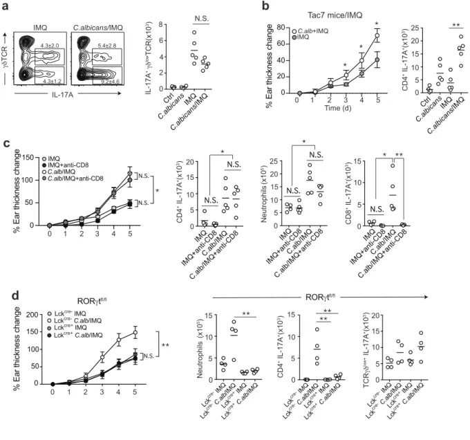
Confocal microscopy of ear pinnae 45 Phenotypic analysis 46 FUNGAL SKIN COLONIZATION DRIVES A POLARIZED IL-17A SIGNATURE AND EXACERBATES PSORIASIFORM SKIN INFLAMMATION 48 C. ALBICANS PROMOTES TISSUE INFLAMMATION IN A TH17 DEPENDENT MANNER 56 C. ALBICANS COLONIZATION PROMOTES NEUTROPHIL INFILTRATION AND NETS FORMATION IN THE CONTEXT OF SKIN INFLAMMATION 59 LANGERHANS CELLS ARE INDISPENSABLE TO INDUCE C. ALBICANS DERIVED EXACERBATION OF THE INFLAMMATION 64 MEMORY RESPONSES TO C. ALBICANS CAN BE SUFFICIENT TO INDUCE DISEASE FLARE 68 CONCLUSION 72 BIBLIOGRAPHIE 75
Preface
The work presented in this thesis was conducted at the National Institutes of Health, National Institute of Allergy and Infectious Diseases (NIAID), Laboratory of Immune System Biology (LISB), Metagenome Immunology Section (MIS) of Doctor Yasmine Belkaid. The project was funded by NIH grants and the National Psoriasis Foundation.
Charlotte Hurabelle-Claverie was funded by Fondation pour la Recherche Médicale (FRM), Fondation La Roche-Posay, Collège des Enseignants en Dermatologie de France, Société Française de Dermatologie (SFD), Philippe Foundation, Fondation Groupe Pasteur Mutualité.
Abbreviations
A
AF Alexa-fluor
APC Antigen Presenting Cell
B
BCR B cell receptor
C
C. albicans, C. alb Candida albicans
CD Cluster of differentiation
CDR Complementarity Determining Regions
CFU Colony forming unit
CLA Cutaneous Lymphocyte Antigen
CLEC7A C-type lectin domain family 7 member A
CLP Common Lymphoid Progenitor
D
DC Dendritic cell
DETCs Dendritic epidermal T cells
G
GWAS Genetic-wide association studies
H
HSC Hematopoietic Stem Cell
I
IFN Interferon
IL Interleukin
L
LCs Langerhans cells
M
MHC Molecular histocompatibility complex
N
NETs Neutrophil extracellular traps
NK cells Natural Killer cells
P
PAMPs Pathogen-associated molecular patterns
PRR Pattern-recognition receptor
S
SLO Secondary lymphoid organs
SNPs Single Nucleotide Polymorphisms
T
TCR T cell receptor
TCM Central memory T cells
TEM Effector memory T cells
TH T helper
Treg Regulatory T cell
W
Introduction
1. The immune system
1.1. Introduction
While all the interfaces with the environment represent both a physical and chemical barrier against the invasion of the host by pathogens, they are also the home for a myriad of micro-organisms such as bacteria, viruses, fungi and archea, commonly referred to as the microbiota. Each of the body barriers co-evolved with the microbes they are surrounded with and developed a relationship that is mutually beneficial for the microbes and their host. These microbes exist on a spectrum ranging from symbiotic to pathogenic and form complex relationships with the host immune system. The skin, oral and intestinal mucosa are examples of the anatomic barriers that prevent intrusion of the host by microbes. In case of a breach or an invasion of these barriers, the host developed an efficient immune system to defend against these micro-organisms, broadly divided into two branches: the innate and the adaptive immune system. While those two branches are traditionally separated from each other, there is a constant crosstalk between innate and adaptive immunity, allowing to provide the best protection against infections.
1.2. Innate immunity
The innate immune system is evolutionarily conserved and allows rapid sensing and responses to infections.
There are three types of phagocytes in the immune system, represented by macrophages, granulocytes (such as neutrophils, eosinophils, basophils and mast cells), and dendritic cells (DCs). They share the ability to engulf and kill pathogens.
Among granulocytes, neutrophils represent the majority of the circulating white blood cells (50 to 75% of the blood WBC). They are one of the first responders to infection and injury
sites, and they play a key role in first line defense against pathogens, using different methods to clear pathogens: phagocytosis, degranulation of antimicrobial agents (such as myeloperoxidase, reactive oxygen species, cathepsin, cathelicidin etc.) and neutrophil extracellular traps (NETs). The neutrophil extracellular traps are a recently described mechanism of neutrophil death where they extrude webs of fibers composed of chromatin and serin proteases (Brinkmann et al., 2004a). Mastocytes, eosinophils and basophils are also involved in defense against pathogens, especially parasites, but have also a prominent role in allergy.
Macrophages and DCs are also professional antigen presenting cells (APCs), that are able to uptake antigens, process them, and present them to adaptive immune cells, as well as producing inflammatory mediators such as cytokines and chemokines. As such, they can be considered as bridges between innate and adaptive immunity. Antigen presentation is done through the major histocompatibility complex (MHC). MHC-I molecules are expressed by almost all the body cells with the exception of non-nucleated cells (such as mature red blood cells). MHC-I molecules mainly bind peptides derived from the cytosolic compartment, such as host-derived peptides or peptides generated from an intracellular pathogen. More recently, it has been discovered that MHC-I molecules can present peptides generated from exogenous proteins, a process called cross-presentation. In contrast, MHC-II molecules expression is largely confined to APCs. MHC-II molecules present extracellular proteins that have been internalized in the APCs in phagolysosomes.
In addition to the myeloid cells described above, innate lymphoid cells (ILCs) and natural killer cells are cells that do not recognize a specific antigen and are therefore not considered part of the adaptive immune system but share some similarities with lymphoid lineages. Innate lymphoid cells (ILCs) are a recently discovered class of immune cells that are defined by a classic lymphoid cell morphology, but lack lineage markers and rearranged antigen receptors. The ILC family comprises classic cytotoxic natural killer cells but also three distinct groups of non-cytotoxic ILCs based on their transcription factor expression profile and expression of effector cytokines: ILC1s, ILC2s, and ILC3s, the latter including lymphoid tissue inducer cells (Vivier et al., 2018). ILCs are enriched at barrier surfaces. Natural killer cells (NK cells) are effector cells that are able to recognize and kill tumor cells and virus infected cells. This can happen because of lack of expression of MHC-I-peptide complex – a process known as ‘missing
self’. In this case, they release the contents of their granules (perforin, granzymes), which leads to induction of apoptosis in the target cell or to cell lysis. NK cells also play a role in antibody-dependent cell-mediated toxicity (ADCC), which is the release of cytolytic granules upon recognition of infected cells opsonized with antibodies.
All these cells provide rapid response to pathogens, taking only a few minutes to hours to be recruited on the site of injury, and are therefore indispensable to provide an immediate response. However, their response may not be powerful enough to clear the pathogen, they can create a lot of collateral damages because of their untargeted mode of action, and they mostly lack memory (with the exception of NK cells that display a type of memory). In contrast, the adaptive immune cells respond slower, but they are maintaining long term memory.
1.3. Receptors of innate immunity
When a pathogen is able to cross the physical and chemical barriers of the host, they are immediately detected by the immune system. Indeed, during an infection, conserved pathogen-associated molecular patterns (PAMPs) are recognized by pattern recognition receptors (PRR) (Takeuchi and Akira, 2010). PRRs include toll-like receptors (TLRs), nod-like receptors (NLRs), Retinoic acid-inducible gene (RIG)-I-like receptors (RLRs) and C-type lectin receptors (CLR). TLR and CLR are transmembrane proteins while RIG-I-like receptors and NLD are cytoplasmic. PRRs are expressed mainly in innate immune cells, but also in various cells not traditionally thought of as immune cells such as keratinocytes. Activation of the PRRs lead to intracellular signaling cascades such as nuclear factor-κB (NF-κB)-dependent and interferon (IFN)-regulatory factor (IRF)-dependent signaling pathways, leading to expression of inflammatory mediators (Trinchieri and Sher, 2007).
TLRs collectively recognize lipids, carbohydrates, peptides and nucleic-acid structures that are broadly expressed and conserved by the different categories of microbes. Some TLRs are expressed at the cell surface, whereas others are expressed on the membrane of intracellular endosomes and lysosomes, allowing responses to both extra- and intra-cellular pathogens. At least 10 TLRs exist in humans and 12 in mice. The table below shows the ligand and specificity of each TLR.
TLR PAMPs recognized Origin of the PAMP Localization of the TLR
1 Triacyl lipoprotein Bacteria Plasma membrane
2 Lipoprotein Bacteria, virus,
parasite, self, fungi
Plasma membrane
3 Double-stranded RNA (dsRNA) Virus Endolysosome
4 Lipopolysaccharides (LPS) Bacteria, virus, self Plasma membrane
5 Flagellin Bacteria Plasma membrane
6 Diacyl lipoprotein Bacteria, virus Plasma membrane
7 Single-stranded RNA (ssRNA), imidazoquinoline compound
Bacteria, virus, self Endolysosome 8 Single-stranded RNA (ssRNA),
imidazoquinoline compound
Bacteria, virus, self Endolysosome (human)
9 CpG-DNA Bacteria, virus,
parasite, self
Endolysosome
10 Unknown Unknown Endolysosome
11 Profilin-like molecule Protozoa Plasma membrane
C-type lectin receptors such as Dectin-1 and Dectin-2 have an important role in the recognition of b-glucan from fungi.
NLRs encompass NOD receptors and NALPs (NACHT-, LRR- and pyrin-domain-containing proteins) and are important for the recognition of intracellular PAMPs.
RLRs comprise RIG-I, melanoma differentiation-associated gene 5 (MDA5), and LGP2 and play a role in the recognition of viruses.
1.4. Adaptive immunity
The B and T cells are the two mainstays of the adaptive immune system. B cells represent the humoral component of adaptive immunity, while T cells represent the cellular component. B cells
B cells develop in the bone marrow in mammals and migrate to the secondary lymphoid organs (SLO) such as the spleen and lymph nodes when they are mature. They are able to bind
directly to a specific antigen through the B cell receptor (BCR) at their surface. The BCR is composed of a membrane-bound immunoglobulin that has a unique specificity for an antigen, and of CD79, a transmembrane molecule allowing for signal transduction after antigen recognition. B cells can be activated directly after the interaction with their specific antigen through the BCR, which is called T-cell independent activation. In this case, the antigen is usually a polysaccharide or an unmethylated CpG DNA. B cells can also be activated with the help from T helper cells, typically follicular helper T cells (TFH). These responses take a longer
time to develop compared to the T-independent activation but they allow responses to a broader spectrum of antigens with more precise responses and longer memory. In T-dependent B cell activation, the antigen is internalized and processed in small peptides and then presented to the T cells on the MHC II of the B cell. T helper cells that are able to recognize the MHC II – antigen complex through their T cell receptor (TCR) will then express co-stimulatory molecules such as CD40L, as well as cytokines such as IL-4 and IL-21, leading to B cell activation. Activated B cells undergo clonal expansion and some differentiate into plasma cells while other become memory B cells for long-lived protection. Plasma cells are able to secrete immunoglobulins in high quantities. B cells activated through T cells help also undergo immunoglobulin class switching (a mechanism allowing to change from one immunoglobulin type to another such as from IgM to IgG or IgA), and somatic hypermutation which is a mechanism allowing for the development of the highest affinity of the immunoglobulin for its antigen.
Antibodies are Y-shaped proteins that can bind to antigens through their 2 variable regions (Fab) and can communicate with other components of the immune system through their constant region (Fc region). Immunoglobulins work by
- neutralizing pathogens, preventing their interaction and entry with host cells - antibody-dependent cell-mediated cytotoxicity which is the process of
opsonization of a pathogen with antibodies, resulting in activation of phagocytosis by scavenger cells that will destroy the pathogens
- complement activation, which leads to lysis of the target cell and chemoattraction of other components of the immune system
Naïve T cells and antigen recognition
All T cells originate from a common lymphoid progenitor (CLP) itself developing from hematopoietic stem cells (HSC) from the bone marrow. CLP migrate and engraft to the thymus, where they start the maturation process as early thymic progenitors until they become naïve T cells. The maturation process starts with division of the early thymic progenitor and downregulation of c-kit. These cells are double negative for CD4 and CD8 co-receptor and are called double negative (DN) 1. Several steps allow the maturation into a naïve T cell:
- T cell receptor (TCR)-b rearrangement and selection: the TCR is a heterodimeric surface protein consisting of an a and a b chain. Each of these chains is made up of a constant and a highly variable region, the latter allowing the recognition of a different epitope for each of the TCR chains (Bentley and Mariuzza, 1996). Most of the variation in the variable region is due to differences in the three complementarity determining regions (CDRs). Diversity in the TCR repertoire is created through the random rearrangement of the DNA encoded segment of the V-J chains and V-D-J chains respectively in a and a b chain. RAG1 and RAG2 recombinases are the enzymes responsible of these rearrangements (Davis and Bjorkman, 1988). After both chains rearrange and successfully pair, the TCR is expressed at the cell surface, alongside with both CD4 and CD8 receptors and the cell is then referred to as double positive (DP).
- Positive selection: this step allows the survival of the thymocytes with a TCR recognizing with a strong enough affinity a self-antigen presented by the thymus cortical epithelial cells. Thymocytes selected on their ability to recognize an antigen presented on an MHC-I molecule will become CD8+ T cells and downregulate their
expression of CD4, while thymocytes recognizing MHC-II-antigen complex will become CD4+ T cells.
- Negative selection: this step removes the majority of the thymocytes with a too strong affinity with a self-peptide. The rest of the ones with high affinity then become regulatory T cells (Treg). This process is called central tolerance.
Naïve T cells then leave the thymus. They express the chemokine receptor CCR7 and L-selectin (CD62L), two molecules allowing second lymphoid organs homing. Priming of naïve T cells occur when APCs in peripheral tissues process an antigen and migrate to the draining lymph node and present the antigen molecule on their MHC.
CD8+ T cells recognize peptides presented on MHC-I molecules of APCs through their TCR,
while the TCR of CD4+ T cells recognizes peptides presented on MHC-II molecules of APCs.
CD8+ T cells are also called cytotoxic T cells as they are able to recognize and kill cells that are
infected, cancerous, or damaged in another way. They can do so through the secretion of perforin, a molecule that forms pores in the membrane of the target cell, and granzymes, a cytotoxic molecule. Cytotoxic T cells can also produce pro-inflammatory cytokines such as IFNγ and TNFα. The role of CD4+ T cells is described further below. Briefly, their main role is to
product cytokines and enhance other cell types responses to infections, such as B cells and innate cells.
In addition to the classic ab T cells, other subpopulations of T cells exist:
- Mucosal-associated invariant T (MAIT) cells are a population characterized by a semi-invariant TCRa and restricted by the MHC class I-related protein 1 (MR1) (Adams and Luoma, 2013). They represent around 5% of the human T cells
- gd T cells have a TCR made of a g chain and a d chain instead of the ab TCR. They are also way less diverse than the ab T cells. They recognize non-classical antigens such as lipid antigens presented by CD1 molecules (in particular CD1d). They also represent only 5% of the T cells in the blood but are more abundant in the gut.
- Natural killer T cells (NKT) share properties of both NK and T cells. They represent around 0.1% of the circulating T cells and are also restricted by CD1d molecules.
Priming of naïve CD4+ T cells into effector T cells
When naïve CD4+ T cells encounter an antigen, TCR stimulation via MHC class II on APCs and
distinct TH effector subsets: TH1, TH2, TH9, TH17, TH22, TFH and inducible regulatory T cells. The
lineage commitment depends upon the nature of the environing cytokines signals, activating various signal transducers and activator of transcription (STAT) proteins, ultimately leading to the expression of master transcription factors (Christie and Zhu, 2014; Loo et al., 2018). The master transcription factors then lead to the downstream expression of a differentiation program and the repression of other TH effector subsets developmental programs. After
stimulation of the TCR in presence of IFNg and IL-12, CD4+ T cells differentiate in T
H1 cells
through the activation of STAT1, leading to an upregulation of T-bet which is the master transcription for TH1 cells (Szabo et al., 2000). This increases downstream expression of the
IFN-g gene and upregulation of the inducible chain of the IL-12 receptor (IL-12Rb2), while suppressing TH2-associated factors. TH1 cells have a central role in immune responses against
intracellular pathogens (intracellular bacteria, viruses, and some protozoa) (Hsieh et al., 1993; Mosmann et al., 1986). In the presence of IL-4, T cells differentiate into TH2 CD4+ T cells: TH2
cells produce IL-4, IL-5 and IL-13, which are potent activators of B cell IgE production and eosinophil recruitment, and target helminths. TH9 cells develop in the presence of TGFb and
IL-4, which induces the expression of the transcription factors PU.1, IRF4, BATF, Foxo1. They have a role in anti-helminths and anti-tumor immunity (Kaplan et al., 2015). TH17 cells produce
pro-inflammatory cytokines such as IL-17A, IL-17F, IL-21 and IL-22 and have a role in protection against extracellular bacteria and fungi. As explained in the B cells chapter, TFH cells are
important in the formation and maintenance of B cell germinal centers by providing CD40-CD40L interactions and producing IL-4 and IL-21. TH22 cells produce IL-22, IL-13 and TNF-a and
The table below describes the lineage commitment of each of the known TH subsets after
receiving their respective cytokine stimuli, and the master transcription factor and cytokines expressed after initiation of the commitment process:
T helper cell subset Cytokines in the microenvironment triggering the lineage commitment Signal transducers and activators of transcription (STAT) Master transcription factor Cytokine(s) produced Protective function
TH1 IFN-g, IL-12 STAT1, STAT4 T-bet IFN-g Intracellular
pathogens
TH2 IL-2, IL-4 STAT5, STAT6 Gata3 4, 5,
IL-13
Helminths TH9 TGFb, IL-2, IL-4 SMADs, STAT5,
STAT6 PU.1, IRF4, BATF, Foxo1 IL-9 Helminths TH17 TGFb, IL-6, IL-21, IL-23
SMADs, STAT3 RORgt IL-17, IL-21,
IL-22
Extracellular pathogens TH22 IL-6, TNF-a, IL-23,
IL-1b Aryl hydrocarbon receptor (AhR), T-bet IL-22, IL-13, TNF-a Tissue repair, epidermal immunity
TFH IL-6 STAT3 Bcl6 IL-21 Humoral
immunity
Treg TGFb, IL-2 SMADs, STAT5 Foxp3 IL-10, TGFb
Immuno-regulation
Tolerance mechanisms and regulatory T cells (Treg)
To be efficient but not harmful, the immune system must avoid to mount deleterious responses against environmental, food, and commensal antigens. One of the important way the immune system achieves tolerance is to negatively select self-reactive T cells in the thymus, a process leading to elimination or inactivation of self-reactive T cell clones and known as central tolerance (Starr et al., 2003). In the periphery, other mechanisms of tolerance avoid autoimmunity. Anergy is a state of inactivation of the T cells that happen when there is chronic engagement of TCRs by self-antigens, without simultaneous engagement of T cell co-stimulatory receptor CD28 by CD80 and CD86 on APCs.
However, circulation of reactive T cells escaping those mechanisms of regulation are a threat to the host as they can result in autoimmunity. Therefore, another important mechanism
allowing immunological homeostasis is the development of regulatory T cells as they help to suppress excessive and destructive immune responses against self and foreign antigens. Importantly, in absence of Treg, severe inflammation and autoimmunity develop, with failure
to thrive, lymphadenopathy, splenomegaly, immune dysregulation, polyendocrinopathy, enteropathy etc., demonstrating the importance of Treg in providing proper immune
homeostasis (Bennett et al., 2001; Kim et al., 2006).
Natural Treg (nTreg) develop in the thymus through selection of naïve T cells, while induced Treg
(iTreg) differentiate in the peripheral tissues. Thymic selection of Treg starts with recognition of
a self-peptide-MHC2 complex by the TCR, with co-stimulation and IL-2-STAT5 signaling. iTreg
develops in the periphery in a mechanism similar to the T helper cells: TGFb and IL-2 cytokines signal through SMADs, STAT5 (Josefowicz et al., 2012). The master transcription factor for all Treg is Foxp3, leading to the downstream expression of immunosuppressive cytokines such as
IL-10 and TGF-b. All Treg lack expression of the IL-7 receptor and express high levels of the IL-2
receptor alpha chain (CD25).
Memory T cells
After clearance of an infection, the majority of effector T cells die via apoptosis, but a small percentage of the T cells persist and differentiate into long-lived memory cells (Badovinac et al., 2002; Pepper and Jenkins, 2011). Memory T cells respond more rapidly and more efficiently to secondary infections than naïve T cells. Memory cells express markers of antigen-experience such as CD44. They lack expression of CD45RA but express CD45RO. In addition, two main subsets of memory T cells were initially described depending on their expression of CD62L and CCR7 (Sallusto et al., 1999). Central memory T cells (TCM) express these lymph node
homing receptors and are therefore mostly found in the SLO and in the circulation. On the other hand, effector memory T cells TEM don’t express these receptors, and are mostly found
in the peripheral tissues and in the circulation. TEM therefore migrate rapidly to secondary
infection sites where they have immediate and potent effector functions. TCM lack this
immediate effector function in the periphery but have a high proliferative capacity and efficiently stimulate dendritic cells.
Other memory T cells subsets have been more recently discovered, such as resident memory T cells (TRM), which remain poised in the previously infected tissue and don’t re-enter in the
circulation (Schenkel and Masopust, 2014).
TH1 and TH2 cells in inflammatory diseases
Adaptive immune responses are essential for fighting against invasion of the host with pathogens. Indeed, in absence of the main transcription factors for the different effector T cell subsets, or if there are mutations of STATs or cytokines genes, various primary immunodeficiency diseases can occur. Many functions of different T cell effector subsets have been learnt through the discovery of these primary human immunodeficiencies and by creating mouse models specifically deleting or overexpressing TH subsets associated factors
such as transcription factors, STATs, or cytokines. For example, T-bet transcription factor deletion are more susceptible to several intracellular pathogens, such as Salmonella
typhimurium or Mycobaterium tuberculosis (Ravindran et al., 2005; Sullivan et al., 2005).
On the other hand, dysregulated immune responses may happen and lead to autoimmune and chronic inflammatory disorders. These responses can be targeted against self or commensal floral antigens.
Figure: transcriptional regulation f CD4+ T helper (TH) cells that mediate tissue inflammation. From Loo TT et al, J Leuk Biol.
Copyrighted material.
Dysregulated TH1 responses can promote tissue destruction and chronic inflammation
(Lazarevic and Glimcher, 2011). In human, genetic studies have shown associations between single nucleotide polymorphisms (SNPs) in T-bet binding sites with inflammatory bowel diseases (Soderquest et al., 2017), or with asthma (Munthe-Kaas et al., 2008). In mice, lack of T-bet prevents the development of inflammatory bowel disease (Neurath et al., 2002), experimental autoimmune encephalomyelitis (Bettelli et al., 2004), diabetes (Juedes et al., 2004), arthritis (Wang et al., 2006), and systemic lupus erythematosus (Peng et al., 2002). On
bet, mice can develop more severe asthma-like disease. This can happen both independently of allergic responses, due to lack of repression of pro-fibrotic cytokines, and due to unregulated TH2 responses (Finotto et al., 2002).
While TH2 responses are primarily aiming at expulsing parasitic infections, dysregulated Th2
responses in human can lead to atopy and allergic reactions (Walker and McKenzie, 2017). Indeed, damaged tissue induces the release of alarmins, such as TSLP and IL-33, which triggers the TH2 cytokine production, itself leading to the stimulation of IgE production by B cells,
recruitment of eosinophils and mast cells, and increased mucus production, smooth muscle contractility, and ultimately leading to the expulsion of the parasites (Anthony et al., 2007). Helminths invasion also triggers tissue repair by increasing angiogenesis, myofibroblast activation and extracellular matrix deposition, as well as promoting the differentiation of regulatory M2 macrophages (Gause et al., 2013). However, these responses are also amplified in response to allergens in atopic patients, and lead to the development of pathologies such as asthma, atopic dermatitis, or chronic allergic sinusitis. Genome-wide associated studies have identified polymorphisms in genes encoding alarmins, cytokines and epithelial barrier proteins in asthma or atopic dermatitis (Moffatt et al., 2010). Sputum and blood cytology in asthma patients demonstrate evidence of TH2 involvement, with eosinophils and IL-5
producing CD4+ T cells, hyper-IgE production, while mouse models lacking expression of T H2
cytokines often have improved asthma-associated features (Lambrecht et al., 2019; Papi et al., 2018). Asthma patients also exhibit airway remodeling, with epithelial damage, goblet cell hyperplasia, and increased myofibroblast and fibrocyte proliferation, leading to smooth muscle hyperplasia. Monoclonal antibodies targeting IL-5, or IL-4 and IL-13 are used in clinic to reduce exacerbation frequency in patients with asthma (Rabe et al., 2018; Yancey et al., 2017). (Of note, only half of the patients with asthma have responses strongly skewed towards TH2, while the rest of the patients have asthma that are described as ‘non-eosinophilic’, with
involvement of TH1, TH17 responses and neutrophils). Atopic dermatitis is also considered a
TH2 disease, and monoclonal antibody blocking IL-4 and IL-13 signaling are also improving this
disease (Beck et al., 2014; Gittler et al., 2012).
At steady state, TH17 cells are mostly found at barrier sites such as the gastrointestinal tract,
the skin and the lungs, where they provide protection against extracellular bacteria and fungi as well as homeostatic functions. These functions are achieved thanks to the production of the signature cytokines IL-17A, IL-17F, IL21 and IL-22. They have an essential role in preventing invasion of the host by extracellular microbes. Indeed, TH17 signature cytokines induce the
expression of antimicrobial peptides (lipocalin-2, Reg3!, Reg3", S100A8, S100A9), chemokines (CXCL1, CXCL2, CXCL8, CXCL10) and metalloproteases helping wound healing and tissue remodeling, and pro-inflammatory cytokines (IL-1b, IL-6, TNF-a, GM-CSF) (Stockinger and Omenetti, 2017). IL-17A also promotes the expression of tight junction proteins (Lee et al., 2015). All these responses help to contain potential pathogens outside of the host. Interestingly, commensal microbes themselves promote the differentiation of TH17 cells,
which in turn control the burden of these bacteria and prevent infiltration of the host : in the gut, one single commensal microbe, segmented filamentous bacterium, is able to induce TH17
cells (Ivanov et al., 2009).
Importantly, TH17 cells have a fundamental role in the defense against fungi, especially Candida albicans, and extracellular bacteria such as Staphylococcus aureus, especially at
mucocutaneous barrier sites. This is highlighted by the development of chronic and / or severe infections with those microbes in patients lacking the cytokines IL-17A, IL-17F, their receptor IL-17 receptor A (IL-17RA) or with mutations in the gene coding for the TH17 master
transcription factor, RORc (Okada et al., 2015; Puel et al., 2011). Loss of function mutations in STAT3 also result in recurrent Candida albicans and Staphylococcus aureus infections and in increased colonization with Candida species (Abusleme et al., 2018; Holland et al., 2007). Several murine models recapitulate the importance of TH17 cells and signature cytokines for mucocutaneous
protection against Candida albicans infection (Khader et al., 2009). For example, mice deficient in IL-17 receptor A (IL-17RA) have significantly less survival compared to control mice in a model of systemic candidiasis and have significantly more severe infection in models of oral candidiasis, with increased fungal burden and decreased mobilization of neutrophils at the sites of infection (Conti et al., 2009; Huang et al., 2004). To be able to clear potential pathogens, TH17 induce rapid recruitment of granulocytes and macrophages through the
production of IL-17. IL-17 cytokines are potent inducer of neutrophils expansion and survival, as well as chemoattraction (Onishi and Gaffen, 2010). TH17 cytokines also induce the
up-regulation of antimicrobial peptides, which directly contribute to host defense by direct killing of invading organisms.
On the other hand, several human diseases have been associated with dysregulated TH17 cells.
TH17 cells master transcription factor is RORgt, which is induced in response to cytokines such
as TGF-b and IL-6, with or without additional IL-23 and IL-1b (Ivanov et al., 2006). However other transcription factors are necessary to specify the full differentiation of TH17 cells (Ciofani
et al., 2012). The basic leucine zipper transcriptional factor ATF-like (BATF) and interferon-regulatory factor 4 (IRF4) complexes are essential to remodel chromatin accessibility, allowing the RORgt-dependent upregulation of TH17 signature genes. Other transcription factors
regulate the fate of TH17, leading to maintenance or repression of TH17 signature genes, such
as AP-1, Fosl2, c-Maf, or Ahr.
Interestingly, the cytokine TGF-b is important both for the development of Treg and TH17
(Bettelli et al., 2006; Weaver and Hatton, 2009). Depending on the relative abundance of TGF-b and IL-6 in the environment, a CD4+ T cell can develop either toward a Treg or a TH17 cell,
with an initial and transient co-expression of Foxp3 and RORgt. If there is a high TGF-b, Foxp3 inhibits RORgt activity, and the cell will develop as a Treg, while at low TGF-b concentrations,
TGF-b and IL-6 act synergistically to induce RORgt transcription of TH17 program (Veldhoen et
al., 2006). IL-23 acts synergistically with IL-6, IL-21, and TGF-b and also induces differentiation of TH17 cells (Zhou et al., 2007).
The mechanisms described above are important because they show that TH17 lineage is plastic
and therefore susceptible to environmental modulations. This is useful when apprehending the concept of beneficial versus pathogenic TH17 cells. This concept was developed following
the in vitro differentiation of TH17 cells with different antibody cocktails, their transfer in vivo
in mice and the observation of their abilities to induce experimental autoimmune encephalomyelitis (EAE), a model of multiple sclerosis in mice with inflammation of the central nervous system (Lee et al., 2012). When culturing CD4+ T cells with IL-6 and TGF-b, T
H17 cells
producing IL-10 developed and they did not induce EAE (McGeachy et al., 2007). Alternatively, when cultures contained IL-23 in combination with IL-6 and IL-1b, pathogenic T cells were able to induce EAE were generated (McGeachy et al., 2009; Meyer zu Horste et al., 2016).
In addition to EAE, a number of autoimmune diseases are mediated through IL-17 and it is yet unclear if all the TH17 in this context are functionally different compared to physiologic TH17
or if it is due to dysregulated responses in a predisposed genetic environment.
Rheumatoid arthritis is an autoimmune disease affecting the joints, leading to inflamed joints, cartilage and bone erosions, ultimately leading to irreversible deformities in joints. Long thought to be due to dysregulated TH1 responses, it was later demonstrated that IL-17 induces
the production of other pro-inflammatory cytokines in the joints such as TNF-a, 1b and IL-6 from chondrocytes, synoviocytes, macrophages and osteocytes. These cytokines in turn leads to the recruitment of neutrophils and macrophages to the synovium (Shen et al., 2005). IL-17 cytokines also contribute to the destruction of the cartilage and bone through the induction of the expression of matrix metalloproteinases (MMP) 1, 2, 3, 9 and 13, which drive degradation of extracellular matrix within the joint (Chabaud et al., 2000). IL-17 induces the expression of RANKL (receptor activator of nuclear factor-κB ligand), a membrane-bound receptor of the TNF superfamily that promotes differentiation of osteoclasts, leading to bone destruction (Sato et al., 2006).
Inflammatory bowel disease (IBD) encompasses both Crohn’s disease and ulcerative colitis, which are inflammatory disorders affecting the whole GI tract and the colon and rectum respectively. Patients with IBD experience chronic abdominal pain, diarrhea, rectal bleeding, as well as complications outside of the GI tract such as arthritis, pyoderma gangrenosum, or primary sclerosing cholangitis. Increased amounts of IL-17 and IL-23 have also been found in the intestinal mucosa in humans as well as in experimental models of IBD (Fujino et al., 2003). GWAS studies have found that polymorphisms in IL23R or STAT3, two TH17-related genes, as
well as in NOD2 (nucleotide-binding oligomerization domain-containg protein 2) which encodes a sensor of bacterial cell wall components (Duerr et al., 2006; Lesage et al., 2002). It has therefore been hypothesized that IBD is caused by dysregulated inflammatory TH17
responses against commensal gastrointestinal microbes. However, monoclonal antibodies targeting IL-17 pathway have failed to treat Crohn’s disease (Hueber et al., 2012).
Psoriasis is another IL-17 mediated disease and will be developed further in a specific chapter of this thesis (see chapter 4 of the introduction).
2. The skin
2.1 Skin structure
The skin represents a physical, chemical and immune barrier to the environment. With the gut and the lungs, it represents one the largest interface with the environment. It has a surface area of 25 m2 when including the skin appendage openings into the calculation (Gallo, 2017).
It is also densely innervated and is an important sensory organ, relaying information to the central nervous system. Given its large surface area and proximity to the external environment, the skin is an important place of interaction with the microbiota.
The skin consists of two major components: the epithelial compartment, consisting of the hair follicle and the epidermis, and the underlying connective tissue, the dermis, which is vascularized and therefore provides nutritional and structural support (Pasparakis et al., 2014).
The epidermis is the outermost layer of the skin and is in direct contact with the environment. It is primarily a mechanical barrier, preventing external insults such as invasion of pathogens and chemical penetration, as well as loss from within, such as water and solute loss. The most numerous cell type in the epidermis is the keratinocyte, originating from stem cells located in the basal cell layer. Keratinocytes migrate upward to the surface as they undergo maturation, ultimately forming the different layers of the epidermis, namely from the from the innermost to the outermost the basal layer, the stratum spinosum, the stratum granulosum, and the stratum corneum.
The human epidermis also contains melanocytes, Langerhans cells, CD8+ T cells and Merkel
cells in humans. In mice, in addition to the former, there is a prominent population of dendritic epidermal T cells (DETCs), a cell type that is absent in humans. In addition, in comparison to humans, mice have a way higher density of hair follicles and a thinner epidermis (Pasparakis et al., 2014).
There are anchoring fibers of the basement membrane which allow adhesion of the epidermis to the dermis at the rete ridges.
From Pasparakis M, Haase I and Nestle FO, Nature review immunology 2014. Copyrighted material
The dermis is divided in the more superficial papillary dermis and the deeper reticular dermis. It mainly comprises extracellular matrix and stromal cells (the fibroblasts) that produce the collagen and elastic fibers. They provide most of the mechanical strength to the skin as well as a structural framework for blood and lymphatic vessels, nerves, glands and hair follicles. Fluid regulation happens in the dermis through the blood and lymphatic vessels. The neuronal dendrites are located in the skin and allow for sensory feedback.
This layer contains many types of immune cells, such as APCs (DCs and macrophages), ab- and gd-T cells, ILCs, mast cells, and basophils (Di Meglio et al., 2011).
2.2 Skin immune system
Antigen presenting cells in the skin
Multiple APCs exist in the skin, with at least four population of dendritic cells under steady state conditions (Malissen et al., 2014). The professional antigen-presenting cells in the epidermis are represented by Langerhans cells (LCs). They are found both in the interfollicular epidermis and the epithelium of the hair follicles. Langerhans cells develop from yolk sac-derived myeloid precursors and from fetal liver-sac-derived monocytes and are recruited to the epidermis during early embryogenesis. The LCs pool renews direct in the tissue, through a slow proliferation rate. In addition to their APCs function, LCs have some regulatory functions and are able to induce regulatory T cells (Bobr et al., 2010; Igyarto et al., 2009). In the dermis, the three major subsets of conventional DCs are represented by XCR1+ DC (that can be CD103+ or CD103-), CD11b+ DCs, and double negative DCs. Dermal CD11b+ DCs are the most abundant DCs in normal dermis. The dermal conventional DCs come from a bone marrow derived precursor that continually renews the pool of tissue conventional DCs. All the aforementioned antigen presenting cells are able to migrate to the lymph nodes upon activation by bacterial, fungal, viral or protozoal stimuli and present antigens to naïve
antigen-specific T cells, which then will undergo clonal expansion and acquisition of the T cell effector functions.
Macrophages are also present in the dermis. They have primarily phagocytic and anti-inflammatory properties but are not able to migrate to the lymph nodes.
Finally, plasmacytoid DCs (pDCs) can be found in inflamed skin and mediate systemic pro-inflammatory responses.
Figure: APCs in the skin.
From Belkaid Y & Tamoutounour S, Nature Rev Immunol 2016. Copyrighted material.
Adaptive immune cells in the skin
The skin is an amazing reservoir of T cells, with ab T cells representing the majority of the T cells in the skin of humans, while gd T cells represent 1-10% of the T cells in normal human skin. It has been estimated that around 20 billions of ab T cells are present in the total skin of a normal adult (Clark et al., 2006), around twice as much as in the circulation, and these cells are phenotypically, functionally and clonally diverse. In normal mouse skin, the majority of ab T cells are dermal CD4+ T cells, while CD8+ T cells are mostly localized in the epidermis (Belkaid
et al., 2002; Gebhardt et al., 2011). In human skin, the majority of T cells reside in the dermal-epidermal junction, around blood vessels in the dermis, and in appendages such as the hair follicle (Clark et al., 2006), while the human epidermis is populated with unique phenotypes of resident T cells such as CD103+ CD8+ and CD4+ T cells (Watanabe et al., 2015). Contrarily to
humans, mouse epidermis also contains a high number of gd T cells, especially dendritic epidermal T cells (DETCs) that are localized in the epidermis at steady state. These cells express a conserved Vγ5Vδ1 TCR, are relatively immobile, divide slowly, and have a key role in promoting tissue repair (Chodaczek et al., 2012; Havran and Jameson, 2010).
Other than DETCs, which reside solely in the epidermis of murine skin, the other subtypes of gd T cells are predominantly located in the dermis. These dermal gd T cells, whose TCR receptor includes the Vg4 or Vg6 TCR chains are predominant producers of IL-17 within mice at steady
state (Nakamizo et al., 2015; Ramírez-Valle et al., 2015). Both dermal subsets appear to be radioresistant and are largely resident in the dermis (Cai et al., 2014). Dermal gd T cells have been implicated in host immunity to commensals and pathogens where they proliferate in response to multiple cutaneous bacteria including Staphylococcus aureus (Dillen et al., 2018),
Corynebacterium accolans (Ridaura et al., 2018), Mycobacterium bovis (Sumaria et al., 2011).
Additionally, gd T cells have been found to be increased in lesions of human psoriasis and in murine models of psoriasis, where depletion of gd T cells leads to amelioration of disease (Sandrock et al., 2018).
Skin-homing ab T cells are generated from naïve T cells activated by APCs travelling in skin draining lymph nodes after they encounter an antigen in the skin. These APCs then present the antigen to the TCR through their MHC, stimulate the T cells via the co-stimulatory molecules such as CD80 and CD86 (and their ligand CD28 on T cells) and lead the T cells to their effector function depending on the cytokine environment. Recruitment to the skin of the effector T cells is facilitated by the expression of adhesion molecules on the T cells to ligands in the skin. T cells expressing the cutaneous lymphocyte antigen-1 (CLA) bind to E-selectin expressed by endothelial cells during inflammation (Kupper and Fuhlbrigge, 2004). Other factors able to attract T cells in the skin are the chemokines, which signal through G-protein coupled receptors in T cells (Griffith et al., 2014). For example, CCR10 and CCR8 are utilized for T cell homing to inflamed skin via CCL27 and CCL8 respectively, which are produced by keratinocytes (Homey et al., 2002). In addition, blood vessel endothelium-derived CCL1 also attracts T cells expressing CCR8 (Gombert et al., 2005). During skin infections, T cells infiltrate the skin and memory T cells can remain in the skin to provide rapid protection in case of a reinfection: up to 95% of the ab T cells in the skin are CD45RO memory T cells (Bos et al., 1987). Contrarily to CD8+ T cells which contract and become true resident in the skin after
clearance of an infection, the majority of CD4+ T cells recirculate, and are sometimes called migratory memory T cells (TMM) (Debes et al., 2005).
Role of epithelial cells in skin immunity
Keratinocytes, which represent the outer barrier of the skin, are not only a physical barrier but also have an important role to direct the skin immunity. They produce a number of chemokines allowing the recruitment of T cells to the skin. They also detect
pathogen-associated molecular patterns (PAMPs) because they express pattern recognition receptor (PRR) (Takeuchi and Akira, 2010), including toll-like receptors (TLRs) Keratinocytes express TLR 1 to 6 and TLR 9, allowing them to act as first-responders when encountering pathogens (Miller, 2008). Keratinocytes also produce antimicrobial peptides (AMPs) that directly kill pathogens (Harder and Schroder, 2005). Finally, keratinocytes also produce cytokines, such as IL-1 and TNFa. All these factors can initiate anti-microbial responses in the infected tissue, and further recruit innate and adaptive professional immune cells such as neutrophils, monocytes and T cells.
3. The microbiome
3.1 Introduction
While all the interfaces with the environment represent both a physical and chemical barrier against the invasion of the host by pathogens, they are also the home for a myriad of micro-organisms such as bacteria, viruses, fungi and archea, commonly referred to as the microbiota. The whole microbiota represents a higher number of cells and genes than their host’s genome! Each of the body barriers co-evolved with the microbes they are surrounded with and developed a relationship that is mutually beneficial for the microbes and their host. The microbiota coevolved with humans, establishing this interdependent relationship where the microbiota cannot survive without the host, but it also has an important role in calibrating the immune system, contribute to local regulation of tissues as well as participate in metabolic pathways. While the host-microbiota relationship is mainly symbiotic, it has been shown that this interaction can become dysregulated and participate in the pathogenesis of auto-immune or allergic diseases. Indeed, the microbiota has shaped the immune system for millennia, and rapid changes in the composition of the microbiota, because of antibiotic treatment, changes in diet or in hygiene rules over a few decades may have transformed the mutually benefic relationship between the microbiota and the host.
3.2 The gut microbiota
The gut microbiota is probably the richest in terms of number and diversity. The intestinal tract is home to up to 1014 microbes, the majority of them living in the colon (Ley et al., 2006).
While the gut microbes can be useful for the host, the host also tends to establish a clear separation between the inside and the outside and tries to compartmentalize microbes on the outer part. This is helped by the production of mucus by goblet cells, but also through the production of multiple antimicrobial peptides, that can have direct microbicidal functions, with enzymatic attack of the bacterial wall or disruption of the bacterial inner membranes (Cash et al., 2006; Hooper and Macpherson, 2010). The gut microbiota also has a role in directly shaping the development of hematopoietic and innate cells. It has indeed been shown that germ free animals have a global defect in innate immune cells and in the number of hematopoietic stem cells and precursors (Belkaid and Harrison, 2017). Microbiota-derived metabolites, such as short-chain fatty acids, represent an energy source for enterocytes, but are also able to increase the generation of macrophages and dendritic cell precursors (Trompette et al., 2014). Microbe-derived riboflavin is indispensable for the development of MAIT cells, and the MAIT cells are absent in germ-free animals (Treiner et al., 2003). Importantly, the gut microbiota has a major role in shaping the adaptive immune system. Microbiota-derived antigens induce the production of a mucosal compartmentalized IgA response, which prevent adhesion of the commensal bacteria to the epithelial cells, but also reduces intestinal proinflammatory signaling (Peterson et al., 2007). The role of the gut microbiota has been extensively proven in the induction of the different T effector cell subsets. TH17 cells frequencies are severely reduced in germ-free mice or mice treated with broad
spectrum antibiotics (Ivanov et al., 2008). Interestingly, a single bacterial species, segmented filamentous bacteria (SFB), is able to induce the differentiation of intestinal TH17 cells. These
antigen-specific TH17 cells are in turn able to control the SFB burden through the production
of IL-17 signaling in intestinal epithelial cells (Yang et al., 2014). The TH17 signature cytokines
have an important role in the development of epithelial cells tight junction and reinforce the epithelial barriers (Weaver et al., 2013).
3.3 The skin microbiota
The skin is constantly exposed to the environment and is also a site of encounter with skin microbes. Unlike the gastrointestinal tract, the skin is relatively weakly colonized by microbes: it is estimated that there is about 1 million bacteria per cm2, with around 1010 bacteria over a
human skin surface (Belkaid and Segre, 2014; Grice et al., 2008). This can be explained by the relative inhospitality of the skin compared to the GI tract. The surface of the skin is dry with a low pH and produces many proteins that deprive microbes of essential nutrients. Microbiome composition of the skin varies depending on the clinical site, in relation with factors such as pH, temperature, degree of sebum and moisture of the site. For example, sebaceous sites are mainly represented by lipophilic bacterial species such as Cutibacterieum acnes (formerly known as Propionibacterium acnes), while Proteobacteria are more frequent in dry sites.
Staphylococcus epidermidis can tolerate the high salt concentrations present in sweat and
may use urea and aminoacids from the eccrine sweat as a nutrient source (Scharschmidt and Fischbach, 2013).
The skin microbiota changes throughout the course of life. Initial seeding of the skin with microbes begins when the fetus exits out of the womb, and the type of delivery leads to different types of microbes seeding the skin: the neonate skin microbiota resembles most the maternal vaginal flora if the baby is delivered vaginally and the maternal skin microbiota if the baby is delivered through cesarean section (Dominguez-Bello et al., 2010). During adolescence, hormonal changes create a sebum-rich environment that allows for lipophilic bacteria to flourish (Oh et al., 2012). Microbial communities stabilize during adulthood, but continue to evolve depending on environmental factors (such as climate, antibiotics exposure, hygiene, etc.) and host factors.
Figure: the skin microbiota.
From Belkaid Y & Segre J, Science 2014. Copyrighted material.
The microbes isolated from the skin depend on the depth and location in the skin itself: different microbes can be found in the outer epidermis and in the depth of the hair follicle, as demonstrated by the different results of microbiota mapping generated by swabbing versus biopsies (Prast-Nielsen et al., 2019). This can be explained by the different environment at different sites of the skin: hair follicles are anaerobic when going deeper in the follicle, while the top of the skin represents an aerobic environment (Chen et al., 2018).
Interestingly, the composition of the skin microbiota dramatically changes during episodes of skin inflammation, and it is not completely understood if these changes are secondary to the skin inflammation or if they drive the skin inflammation.
It is interesting to speculate on the role of the microbiota at each unique stage, and emerging evidence ties the date of encounter with the microbes with the response they induce in the host.
Recognition of the skin microbes by the host
Increasing evidence suggest that the manner in which antigens and microbial products are recognized determines the type of immune response. Using two different models of infections, it has been shown that CD11b+ DCs elicit a Th1 response when confronted to a
pseudo-hyphae Candida albicans invasive (dermal) infection, while Langerhans cells were capable of eliciting a Th17 response to Candida albicans yeast in an epidermal infection (Kashem et al., 2015). In another model, Langerhans cells were required to sense
Staphylococcus aureus and promote inflammatory responses with development of IL-17 gd T
cells and IL-17 CD4 T cells (Kobayashi et al., 2015). These observations were made in the context of an altered skin barrier, but even with an intact skin barrier, the immune system is able to sense skin commensals, inducing cognate, long-lasting immune responses.
For commensal encounter, while one would expect that the surface commensals would be sensed by the most superficial APCs (the Langerhans cells), there are actually increasing body of evidence demonstrating that both Langerhans cells and dermal dendritic cell populations can sense the skin microbes and that the different cell types involved in microbe sensing are responsible for different response types. Staphylococcus epidermidis induces IL-17A producing CD8+ T cells, which is dependent on dermal resident CD103+ DCs and independent
CD11b DCs (Naik et al., 2015a). This demonstrates that different DC subsets within the skin cooperate to respond to defined skin commensals.
The way skin-resident DCs sense microbiota-derived antigens remains unclear. Dermal DCs surrounding skin appendages such as hair follicle may extend dendrites within those appendages and capture microbes or their products. Microbial products could also passively diffuse through the epithelial barrier, especially in the hair follicles (Gallo and Nakatsuji, 2011).
Role of the microbiome in cutaneous immunity
The immune system has evolved closely with the skin commensal microbes, allowing colonization with those microbes while limiting pathogenic microbial invasion.
Several studies have shown how the skin microbiota can enhance the barrier function of the skin through the immune system, to prevent invasion with microbes.
Though much focus has been placed on the 1, 23 and 17 axis in disease settings, the IL-23 – IL-17 axis also provides barrier defense as demonstrated by patients who have defects in the axis are more susceptible to infections with extracellular pathogens (Lehman, 2014). Evidence continues to mount that the microbiota can modulate this axis, which is important for both immune homeostasis as well as disease. After production of IL-1 and IL-23 by APCs, multiple subsets of both innate-like and adaptive T-cells are capable of producing IL-17 family cytokines such as IL17, IL22, GM-CSF, which in turn regulate the inflammatory response and bolster barrier defense through increased expression of AMPs and chemokines (Gaffen et al., 2014).
Commensals also have the ability to control other arms of the immune system such as IL-1 signaling, which is an evolutionarily conserved arm of the innate immune system that may have evolved as a mediator of host-commensal cross-talk. Germ free mice have reduced IL-1a from the skin compared to SPF mice. Additionally, keratinocytes of germ free mice have increased levels of IL-1ra compared to SPF mice. Mono-association with the commensal S.
epidermidis increases IL-1 a and decreases IL-1ra to levels similar to SPF mice (Naik et al.,
2012).
IL-1 and IL-23 are also capable of polarizing αβ T-cells to produce IL-17 in response to microbiota in the skin (Naik et al., 2015a). Additionally, IL-17 induced by the microbiota in an IL-1 dependent manner plays a role in immune homeostasis and can also provide protective
immunity to the pathogens Leishmania major and Candida albicans (Naik et al., 2015a; Naik et al., 2012). A diverse microbiota is essential to ensure protection against infections. Mice living in SPF conditions have a lack of tissue resident and effector differentiated memory T-cells compared to wild mice and the latter have better response and control of infection with
L. monocytogenes or LCMV than the former (Beura et al., 2016; Rosshart et al., 2017)
Importantly, the time of encounter with skin commensals is fundamental in the type of immune responses that they induce. In contrast to adults, microbial sensing in the neonatal period leads to a tolerogenic response to the microbes encountered and the development of antigen-specific regulatory T cells seeding the skin (Scharschmidt et al., 2015). However, skin microbes are not essential for the development of Treg, as there are an increased number and
proportion of Treg, while there is a decrease in effector IFN-g or IL-17A producing T cells in the
skin of germ free mice (Naik et al., 2012).
On the other hand, disturbance of the skin microbiome can have long term impact on the immune system and on the risk of inflammatory skin diseases. Treatment of neonate mice with antibiotics durably alters the gut microbiota and worsens imiquimod-induced skin inflammation (Zanvit et al., 2015).
Immune responses to the microbiota can also promote wound healing and limit skin inflammation after an injury, by modulating both the innate and adaptive immune responses:
S. epidermidis TLR2 signaling inhibits responses to dsRNA coming from damaged host cells
(Gallo and Nakatsuji, 2011). Commensal-specific T cells can also accelerate wound healing through expression of tissue repair gene (Linehan et al., 2018a).
While immune responses to the skin microbiota has multiple beneficial roles for the host, barrier defects or genetic pro-inflammatory conditions can challenge these physiological responses.
Microbiome and skin inflammatory diseases Atopic dermatitis