Science Arts & Métiers (SAM)
is an open access repository that collects the work of Arts et Métiers Institute of
Technology researchers and makes it freely available over the web where possible.
This is an author-deposited version published in: https://sam.ensam.eu
Handle ID: .http://hdl.handle.net/10985/18157
To cite this version :
Emmanuelle FERRERO, Renaud LAFAGE, Vincent CHALLIER, Bassel G. DIEBO, Pierre
GUIGUI, Keyvan MAZDA, Frank SCHWAB, Wafa SKALLI, Virginie LAFAGE - Clinical and
stereoradiographic analysis of adult spinal deformity with and without rotatory subluxation
-Orthopaedics & Traumatology : Surgery & Research - Vol. 101, n°5, p.613-618 - 2015
Any correspondence concerning this service should be sent to the repository
Administrator : archiveouverte@ensam.eu
Clinical and stereoradiographic analysis of adult spinal deformity with
and without rotatory subluxation
E. Ferrero
a,b,d,∗, R. Lafage
a, V. Challier
a, B. Diebo
a, P. Guigui
b, K. Mazda
c,
F. Schwab
a, W. Skalli
d, V. Lafage
aaOrthopaedic Department, Hospital for Joint Disease, 15th East Street, New York, 10003, USA
bService de chirurgie orthopédique, hôpital européen Georges-Pompidou, université Paris V, AP–HP, 20, rue Leblanc, 75015 Paris, France cService de chirurgie, hôpital universitaire Robert-Debré, boulevard Sérurier, 75019 Paris, France
dLaboratoire de biomécanique, Arts et Métiers Paris Tech, boulevard de l’Hôpital, 75013 Paris, France
Keywords:
Adult spinal deformity Sagittal alignment 3D analysis Rotatory subluxation Transverse plane analysis
a b s t r a c t
Introduction: In degenerative adult spinal deformity (ASD), sagittal malalignment and rotatory sublux-ation (RS) correlate with clinical symptomatology. RS is defined as axial rotsublux-ation with lateral listhesis. Stereoradiography, recently developed for medical applications, provides full-body standing radiographs and 3D reconstruction of the spine, with low radiation dose.
Hypothesis: 3D stereoradiography improves analysis of RS and of its relations with transverse plane and spinopelvic parameters and clinical impact.
Material and methods: One hundred and thirty adults with lumbar ASD and full-spine EOS®radiographs (EOS Imaging, Paris, France) were included. Spinopelvic sagittal parameters and lateral listhesis in the coronal plane were measured. The transverse plane study parameters were: apical axial vertebral rotation (apex AVR), axial intervertebral rotation (AIR) and torsion index (TI). Two groups were compared: with RS (lateral listhesis > 5 mm) and without RS (without lateral listhesis exceeding 5 mm: non-RS). Correlations between radiologic and clinical data were assessed.
Results: RS patients were significantly older, with larger Cobb angle (37.4◦vs. 26.6◦, P = 0.0001), more severe sagittal deformity, and greater apex AVR and TI (respectively: 22.9◦vs. 11.3◦, P < 0.001; and 41.0◦ vs. 19.9◦, P < 0.001). Ten percent of patients had AIR > 10◦without visible RS on 2D radiographs. RS patients reported significantly more frequent low back pain and radiculalgia.
Discussion: In this EOS®study, ASD patients with RS had greater coronal curvature and sagittal and trans-verse deformity, as well as greater pain. Further transtrans-verse plane analysis could allow earlier diagnosis and prognosis to guide management.
Level of evidence: 4, retrospective study.
1. Introduction
Low back pain and radiculalgia are among the most frequent reasons for orthopedic consultation, at 2.5% in some countries [1]. There are many causes, of which spinal deformity is one. A recent study reported that the rate of spinal deformity can reach 68% in elderly populations (mean age > 65 years) [2]. Moreover, in degenerative adult spinal deformity (ASD) frontal deformity with vertebral rotation and sagittal malalignment is often associ-ated with osteoarthritis and discal and ligamentous degeneration,
inducing central or foraminal canal stenosis with radicular com-pression[3]. The combination of these phenomena causes pain and major disability[4,5].
To investigate the relation between symptoms and spinal defor-mity, several studies assessed correlations between radiologic parameters and quality of life scores[2,3,6–8]. Radiologic parame-ters most frequently found to be associated with symptoms were rotatory subluxation (RS) of the joint and loss of lumbar lordosis leading to global sagittal alignment defect, triggering compensation mechanisms in the pelvis, such as increased pelvic retroversion, or spine, such as flattening of the thoracic kyphosis[8]. Moderate but significant correlations were recently reported between clinical disability scores and sagittal spinopelvic radiographic parame-ters, demonstrating the contribution of global sagittal analysis to diagnosis, prognosis and management[5,9,10]. Coronal alignment
parameters, on the other hand, seem to have little influence on the severity of pain and functional disability[5].
However, all of the literature regarding correlations between radiologic and clinical data has been restricted to 2D radiogra-phy, whereas adult spinal deformity is 3-dimensional deformity sometimes causing RS[11]. Radiographic assessment of vertebral rotation often uses pedicle projection on AP view[12–14]. How-ever, in severe rotation the pedicle becomes difficult to identify [15]. MRI or CT may complete X-ray examination but are performed with the patient in supine position and do not allow analysis of anatomic factors underlying pain or loss of function in upright position. Stereoradiography, which was recently developed, pro-vides full-body standing radiographs without distortion and with a low dose of radiation and shorter examination time, and allows 3D reconstruction at lower cost than MRI or CT[16–19].
Certain studies of adolescent idiopathic scoliosis using stere-oradiography highlighted the importance of the axial plane for deformity analysis[17,18]. However, the literature on 3D analy-sis of adult spinal deformity remains sparse[20,21]. The present study therefore sought to analyze RS in ASD by 3D stereographic reconstruction, assessing correlations between axial plane and spinopelvic parameters on the one hand and pain and functional impairment on the other.
2. Materials and methods
2.1. Data collection
A retrospective study included patients between November 2012 and July 2014, after institutional review board approval. Inclu-sion criteria were: adult patient consulting for spinal deformity (Cobb angle > 10◦)[22]. Exclusion criteria were: non-idiopathic or non-degenerative etiology, and history of spine surgery.
Demographic data comprised age, gender and body-mass index (BMI). Functional data comprised Oswestry Disability Index (ODI) and a visual analog scale (VAS), as well as low back and radicu-lar pain. Radiography used the EOS® system (EOS Imaging, Paris, France), on a standardized protocol: patient upright, with horizon-tal gaze, and fingers on the clavicles to avoid superimposition on the arm on the spine[23].
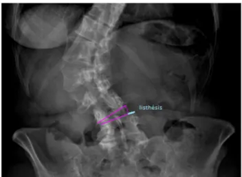
Fig. 1. Measurement method for rotatory subluxation (RS).
2.2. Radiographic analysis
Radiographic measurements were made by an experienced observer. 2D measurement on Surgimap software (Nemaris Inc., New York, USA) consisted in lateral listhesis alone: distance (in mm), on the convex side parallel to the cranial plate of the underlying vertebra, between the lateral edge of the underlying vertebra and the lateral edge of the overlying vertebra lowered per-pendicularly to the plate of the underlying vertebra (Fig. 1). RS was defined as axial rotation associated with > 5 mm lateral listhesis in the coronal plane[24,25]. The patient cohort was thus divided into two groups: with (RS) and without≥ 1 lateral listhesis exceeding 5 mm (non-RS).
3D spinal reconstruction used SterEOS®software, version 1.2.1 (EOS Imaging, Paris, France) (Fig. 2). To correct any pelvic rotation during acquisition, all parameters were measured with the patient-specific landmark defined by the Scoliosis Research Society (SRS) as the vertical plane through the acetabular centers[26]. Sagittal alignment assessment comprised global parameters (sagittal ver-tical axis [SVA], T1 spinopelvic inclination [T1SPi]) (Fig. 3), spinal parameters (T1T12 thoracic kyphosis, L1S1 lumbar lordosis [LL]) and pelvic parameters (pelvic incidence [PI], pelvic tilt [PT] and
Fig. 3. Global sagittal parameters. (T1SPi: spinal inclination; SVA: sagittal vertical
axis).
sacral slope [SS])[27]. The 3 parameters measured for SRS Schwab ASD classification were PT, SVA and the difference between PI and LL (PI-LL)[28]. In the coronal plane, lumbar Cobb angle (Cobb) and the C7 plumb-line with respect to the center of the sacrum (C7PL) were measured[29,30]. Vertebral and intervertebral rotations were measured in the axial, sagittal and coronal planes; intervertebral rotation was defined as superior vertebral rotation with respect to the underlying vertebra. Transverse plane parameters comprised apical axial vertebral rotation (apex AVR), axial intervertebral rota-tion at the limits of the curve (sup AIR, inf AIR) and maximal intervertebral rotation (AIR max). The lumbar curve torsion index (TI) was calculated as the sum of the axial intervertebral rotations in the curve[20](Fig. 4).
2.3. Statistical analysis
Statistical analysis used Stata software, version 13.0 (Stata-corp, College Station, Texas). Normal distribution was checked
Table 1
Comparison of demographic parameters and curvature types between patients with and without rotatory subluxation.
RS (n = 79) Non-RS (n = 51) P Mean SD Mean SD Age (years) 63.4 18.3 48.4 22.5 0.001 BMI (kg/m2) 25.9 6.0 24.1 5.5 0.096 Gender (% female) 85% – 80% – 0.921 Lumbar 75% 45% 0.003 Thoraco-lumbar 13% 45% Major double 12% 10%
RS: rotatory subluxation; SD: standard deviation; BMI: body-mass index.
on Shapiro Wilk test. Descriptive analysis was performed on the demographic and radiology data. Inter-group comparison used Chi2or Student tests as appropriate for normally distributed
vari-ables, and Kruskal-Wallis test for non-parametric variables. Finally, descriptive analysis was performed for the clinical variables, and correlations with radiologic parameters were calculated. The sig-nificance threshold was set at 0.05.
3. Results
3.1. Demographic analysis
One hundred and thirty patients with 3D EOS® imaging were included. Eighty-three percent were female; mean age was 57.6± 18.3 years; mean BMI was 25.2 ± 5.9 kg/m2.
Eighty-three patients (64%) had lumbar scoliosis, 33 (25%) thoraco-lumbar scoliosis, and 14 (11%) major double scoliosis. Lum-bar scoliosis was significantly more frequent in the RS group (75% vs. 45%, P = 0.003). Seventy-nine patients (61%) had > 5 mm lateral listhesis in the coronal plane and 51 (39%) were free of RS. Age in the RS group was significantly greater; there were no inter-group differences for BMI or gender (Table 1).
3.2. Radiographic analysis
Mean Cobb angle was 33.2± 15.6◦ and mean apex AVR
18.3± 14.3◦. Cobb angle was significantly greater in the RS group
(37.4± 16.7◦vs. 26.6± 10.8◦; P = 0.0001). There was no significant
difference in coronal C7PL. In 35 of the 79 RS patients (44%) RS
Table 2
Comparison of radiographic parameters between patients with and without rotatory subluxation. RS (n = 79) Non-RS (n = 51) P Mean SD Mean SD SVA (mm) 42.7 62.6 12.9 43.9 0.003 PI-LL (◦) 13.6 22.1 −1.4 18.4 < 0.001 PT (◦) 23.5 10.9 14.9 11.4 < 0.001 T1SPi (◦) −2.3 7.1 −3.6 4.6 0.22 T1T12 (◦) 38.0 21.3 41.4 19.9 0.68 L1S1 (◦) 38.9 19.6 51.4 17.4 < 0.001 PI (◦) 52.6 12.0 50.5 16.5 0.42 Apex AVR (◦) 22.9 15.9 11.3 7.3 < 0.001 TI (◦) 41.1 29.7 19.3 12.2 < 0.001 AIR max (◦) 19.5 11.9 9.8 5.2 < 0.001 Sup AIR (◦) 7.1 6.0 3.9 3.6 0.001 Inf AIR (◦) 5.6 5.4 3.7 3.8 0.07
RS: rotatory subluxation; SD: standard deviation; SVA: sagittal vertical axis, PI-LL: pelvic incidence minus lumbar lordosis; PT: pelvic tilt; T1Spi T1: spinopelvic inclination; T1T12: thoracic kyphosis between T1 and T12; L1S1: lumbar lordosis between L1 and S1; PI: pelvic incidence; apex AVR: apical axial vertebra rotation; TI: torsion index; AIR max: maximum axial intervertebral rotation; sup AIR: axial intervertebral rotation in the superior transitional level of the curve; inf AIR: axial intervertebral rotation in the inferior transitional level of the curve.
involved 1 level, in 28 (36%) 2 levels, in 15 (19%) 3 levels, and in 1 patient 4 levels. RS level was predominantly L3L4 (33%).
RS patients showed significantly greater sagittal malalignment in terms of SVA, PI-LL and PI. Transverse deformity was more severe in RS, with significantly greater apex AVR, TI, AIR max and AIR sup (Table 2).
Transverse plane analysis found significantly greater AIR in case of RS at the same level (except for L4L5). AIR range in non-RS patients was 0.1–28.3◦. In sub-analysis of patients with≥ 5◦AIR,
38 (29%) were free of lateral listhesis, as were 13 (10%) for≥ 10◦
AIR (Table 3). 3.3. Clinical analysis
ODI, available for 56 patients, showed moderate disability, without inter-group difference. Radicular and low back pains were more frequent in the RS group (Table 4).
RS number correlated with ODI (r = 0.362, P < 0.05) and radic-ulalgia (r = 0.380, P < 0.05). There were no significant correlations between transverse plane parameters and ODI.
4. Discussion
The present results for ASD assessed on EOS®found that patients with ASD and RS showed greater coronal curvature and sagittal and transverse deformity than patients with ASD without RS.
4.1. Assessment of rotary subluxation
RS was more frequent in lumbar scoliosis, notably of L3L4, in agreement with Freedman et al.[31]. RS was observed in the most severe transverse plane deformities (greater TI, apex AVR and AIR max). In almost a third (29%) of patients with > 5◦AIR, there was no lateral listhesis, and the range of AIR values was wide. Rotation is thus detected ahead of subluxation in the degenerative evolution toward RS.
Several authors focused on assessment of axial rotation on 2D plain radiographs. In 1948, Cobb developed a measurement method based on spinous projection; later, Nash and Moe and also Perdri-olle used pedicle projection[12,13,29]. However, these methods show > 5◦ measurement error [12,13,29]. Moreover, beyond 10◦ rotation, the discrepancies between 2D and 3D measurement become statistically and clinically significant[15,32,33].
4.2. 3D analysis of ASD
The recent development of 3D imaging has facilitated axial rota-tion analysis, which is now more widely recognized and studied. However, on MRI and CT it requires supine positioning, and involves a higher radiation dose. The EOS®system, which allows upright positioning, shows measurement error of± 1.6◦for coronal,± 2.0◦
for sagittal and± 3.8◦for axial rotation[18,20,34–36]. Several
stud-ies of adolescent idiopathic scoliosis demonstrated the prognostic importance of transverse plane analysis[35–37]. To the best of our knowledge, however, only two studies focused on ASD, only one of which analyzed axial rotation using the EOS®system[20,21]. Table 3
AIR according to RS.
Spinal level n Axial intervertebral rotation (AIR) (◦) P
Mean SD Min Max Median
L1L2 RS 32 13.1 10.4 0.3 29.2 9.8 < 0.001 Non-RS 98 5.1 4.6 0.1 19.6 4.1 L2L3 RS 31 12.0 9.8 0.8 44.9 12.4 < 0.001 Non-RS 99 6.2 6.4 0.1 24.6 4.1 L3L4 RS 43 11.5 11.3 0.1 35.6 6.6 0.045 Non-RS 87 7.4 8.9 0.3 28.3 4.2 L4L5 RS 96 5.5 7.1 0.3 28.2 4.4 0.11 Non-RS 34 7.3 7.1 0.1 20.1 3.5
RS: rotatory subluxation; SD: standard deviation; Min: minimum; Max: maximum.
Table 4
Comparison of clinical symptoms between groups with and without RS.
n RS Non-RS P n Mean SD n Mean SD ODI 56 37 35.9 21.5 19 24.9 22.3 0.06 VAS 119 71 5.0 2.5 48 4.4 2.7 0.26 Radiculalgia 119 71 46 (65%) 48 19 (40%) 0.004 LBP 119 71 64 (90%) 48 35 (73%) 0.019
4.3. Relation between radiologic and clinical data
In the present series, radicular and low back pain were signif-icantly more frequent in case of RS. Low back pain is a common symptom in degenerative spinal pathology, and especially in case of RS in ASD. Trammel reported an 80% rate of low back pain in patients with RS[38]. Marty-Poumarat reported similar findings, with 84% low back pain and 43% radiculalgia in ASD patients with RS. Ploumis reported more severe ODI in case of RS[25]. However, no correlation has been demonstrated between clinical symptoms and radiologic data[39].
Many cofactors certainly need to be taken into account in clini-cal analysis, but in the present study RS number showed moderate correlation with ODI (r = 0.362, P < 0.05) and radiculalgia (r = 0.380, P < 0.05). RS may increase underlying foraminal stenosis, which, when associated with radicular stretching, may exacerbate radic-ulalgia. The relation between transverse plane parameters and clinical symptoms has, to the best of our knowledge, never pre-viously been studied. Rotation-induced shear stress to the disk and paravertebral structures as a whole partly accounts for symptoma-tology.
4.4. Study limitations
The present study involved certain limitations. Firstly, detailed radiographic analysis of anatomic structures such as the zygapophysial joints and foramina was difficult in cases of severe deformity associated with osteoarthritis and osteoporosis, as is fre-quent in ASD. Secondly, the study design was retrospective, and only a limited number of clinical scores were available; this could be improved by a prospective study with systematic clinical scor-ing. Even so, the present series was larger than in the main previous studies on the subject.
5. Conclusion
The present study reports the first 3D description of ASD and RS in a significant cohort. 3D data were associated to 2D measurement of lateral listhesis, enabling analysis of the relations between 2D and 3D radiologic parameters and clinical symptoms.
Patients with RS showed more severe deformity in the sagittal plane. RS measurement seemed to be an objective criterion of rota-tory destabilization in ASD, showing acceptable clinical correlation. Moreover, presence of AIR in patients in whom lateral listhesis is not yet radiologically detectable is a determining finding in our understanding of the evolution of RS. These results show that 3D assessment is necessary for complete analysis of the deformity. Future studies are needed to analyze the evolution of ASD on 3D data, as has been done for adolescent scoliosis.
Disclosure of interest
The authors declare that they have no conflicts of interest con-cerning this article.
Acknowledgements
Master’s grant from the French Orthopedic and Traumatologic Surgery Society (SoFCOT), without which this research would not have been possible.
References
[1] Deyo RA, Mirza SK, Martin BI. Back pain prevalence and visit rates: estimates from U.S. national surveys, 2002. Spine 2006;31:2724–7,
http://dx.doi.org/10.1097/01.brs.0000244618.06877.cd.
[2]Schwab F, Dubey A, Gamez L, et al. Adult scoliosis: prevalence, SF-36, and nutritional parameters in an elderly volunteer population. Spine 2005;30: 1082–5.
[3] Liu W, Chen X, Jia L, Song D. The clinical features and surgical treatment of degenerative lumbar scoliosis: a review of 112 patients. Orthop Surg 2009;1:176–83,http://dx.doi.org/10.1111/j.1757-7861.2009.00030.x. [4]Jackson RP, Simmons EH, Stripinis D. Coronal and sagittal plane spinal
defor-mities correlating with back pain and pulmonary function in adult idiopathic scoliosis. Spine 1989;14:1391–7.
[5] Schwab F, Farcy J, Bridwell K, et al. A clinical impact clas-sification of scoliosis in the adult. Spine 2006;31:2109–14,
http://dx.doi.org/10.1097/01.brs.0000231725.38943.ab.
[6]Glassman SD, Berven S, Bridwell K, et al. Correlation of radiographic parameters and clinical symptoms in adult scoliosis. Spine 2005;30:682–8.
[7]Glassman SD, Bridwell K, Dimar JR, et al. The impact of positive sagittal balance in adult spinal deformity. Spine 2005;30:2024–9.
[8] Lafage V, Schwab F, Patel A, et al. Pelvic tilt and truncal inclination: two key radiographic parameters in the setting of adults with spinal deformity. Spine 2009;34:E599–606,http://dx.doi.org/10.1097/BRS.0b013e3181aad219. [9]Schwab FJ, Smith V, Biserni M, et al. Adult scoliosis: a quantitative radiographic
and clinical analysis. Spine 2002;27:387–92.
[10] Glassman SD, Hamill CL, Bridwell KH, et al. The impact of periopera-tive complications on clinical outcome in adult deformity surgery. Spine 2007;32:2764–70,http://dx.doi.org/10.1097/BRS.0b013e31815a7644. [11]Dubousset J. L’homme debout : le rachis et son plan horizontal, scolioses. La
scoliose est une« maladie » du plan horizontal : le secret pour comprendre les 3 dimensions [The standing man: scoliosis and horizontal plane defor-mity. The best way to understand 3D in orthopedics]. e-Mem Acad Natl Chir 2012;11:66–70.
[12]Nash Jr CL, Moe JH. A study of vertebral rotation. J Bone Joint Surg Am 1969;51:223–9.
[13]Perdriolle R, Vidal J. A study of scoliotic curve. The importance of extension and vertebral rotation (author’s transl). Rev Chir Orthop Reparatrice Appar Mot 1981;67:25–34.
[14]Stokes IA, Bigalow LC, Moreland MS. Measurement of axial rotation of vertebrae in scoliosis. Spine 1986;11:213–8.
[15]Weiss HR. Measurement of vertebral rotation: Perdriolle versus Raimondi. Eur Spine J 1995;4:34–8.
[16] Humbert L, De Guise J, Aubert B, et al. 3D reconstruction of the spine from biplanar X-rays using parametric models based on transver-sal and longitudinal inferences. Med Eng Phys 2009;31:681–7,
http://dx.doi.org/10.1016/j.medengphy.2009.01.003.
[17] Ilharreborde B, Steffen JS, Nectoux E, et al. Angle measurement reproducibil-ity using EOS three-dimensional reconstructions in adolescent idiopathic scoliosis treated by posterior instrumentation. Spine 2011;36:E1306–13,
http://dx.doi.org/10.1097/BRS.0b013e3182293548.
[18] Gille O, Champain N, Benchikh-El-Fegoun A, et al. Reliability of 3D recon-struction of the spine of mild scoliotic patients. Spine 2007;32:568–73,
http://dx.doi.org/10.1097/01.brs.0000256866.25747.b3.
[19] Deschênes S, Charron G, Beaudoin G, et al. Diagnostic imaging of spinal deformi-ties: reducing patients radiation dose with a new slot-scanning X-ray imager. Spine 2010;35:989–94,http://dx.doi.org/10.1097/BRS.0b013e3181bdcaa4. [20] Steib J, Dumas R, Mitton D, Skalli W. Surgical correction of
scolio-sis by in situ contouring: a detorsion analyscolio-sis. Spine 2004;29:193–9,
http://dx.doi.org/10.1097/01.BRS.0000107233.99835.A4.
[21] Steffen J-S, Obeid I, Aurouer N, et al. 3D postural balance with regard to gravity line: an evaluation in the transversal plane on 93 patients and 23 asymptomatic volunteers. Eur Spine J 2010;19:760–7,
http://dx.doi.org/10.1007/s00586-009-1249-5.
[22] Schwab FJ, Blondel B, Bess S, et al. Radiographical spinopelvic parameters and disability in the setting of adult spinal defor-mity: a prospective multicenter analysis. Spine 2013;38:E803–12,
http://dx.doi.org/10.1097/BRS.0b013e318292b7b9.
[23]Faro FD, Marks MC, Pawelek J, Newton PO. Evaluation of a functional posi-tion for lateral radiograph acquisiposi-tion in adolescent idiopathic scoliosis. Spine 2004;29:2284–9.
[24] Ploumis A, Transfeldt EE, Gilbert TJJ, et al. Degenerative lumbar scoliosis: radiographic correlation of lateral rotatory olis-thesis with neural canal dimensions. Spine 2006;31:2353–8,
http://dx.doi.org/10.1097/01.brs.0000240206.00747.cb.
[25] Ploumis A, Liu H, Mehbod A, et al. A correlation of radiographic and func-tional measurements in adult degenerative scoliosis. Spine 2009;34:1581–4,
http://dx.doi.org/10.1097/BRS.0b013e31819c94cc.
[26] Sangole A, Aubin C-E, Labelle H, et al. The central hip vertical axis: a reference axis for the Scoliosis Research Society three-dimensional classification of idiopathic scoliosis. Spine 2010;35:E530–4,
http://dx.doi.org/10.1097/BRS.0b013e3181da38b8.
[27]Protopsaltis T, Schwab F, Bronsard N, Smith JS, Klineberg E, Mundis G, et al. TheT1 pelvic angle, a novel radiographic measure of global sagittal deformity, accounts for both spinal inclination and pelvic tilt and correlates with health-related quality of life. J Bone Joint Surg Am 2014;96:1631–40.
[28] Terran J, Schwab F, Shaffrey CI, et al. The SRS-Schwab adult spinal deformity classification: assessment and clinical correlations based on a prospective
operative and nonoperative cohort. Neurosurgery 2013;73:559–68,
http://dx.doi.org/10.1227/NEU.0000000000000012.
[29]Cobb JR. Progress in orthopedic surgery for 1945; conditions involving the spine and thorax, exclusive of those in the lower part of the back. Arch Surg 1947;55:76–87.
[30] Weinstein SL, Dolan L, Spratt KF, et al. Health and function of patients with untreated idiopathic scoliosis. JAMA 2003;289:559–67,
http://dx.doi.org/10.1001/jama.289.5.559.
[31] Freedman B, Horton WC, Rhee JM, et al. Reliability analysis for manual radio-graphic measures of rotatory subluxation or lateral listhesis in adult scoliosis. Spine 2009;34:603–8,http://dx.doi.org/10.1097/BRS.0b013e31819a841e. [32] Lam GC, Hill DL, Le LH, et al. Vertebral rotation measurement: a summary
and comparison of common radiographic and CT methods. Scoliosis 2008;3:16,
http://dx.doi.org/10.1186/1748-7161-3-16.
[33]Skalli W, Lavaste F, Descrimes JL. Quantification of three-dimensional vertebral rotations in scoliosis: what are the true values? Spine 1995;20:546–53.
[34]Kalifa G, Charpak Y, Maccia C, et al. Evaluation of a new low-dose digital x-ray device: first dosimetric and clinical results in children. Pediatr Radiol 1998;28:557–61.
[35] Courvoisier A, Drevelle X, Dubousset J, Skalli W. Transverse plane 3D analysis of mild scoliosis. Eur Spine J 2013;22:2427–32,
http://dx.doi.org/10.1007/s00586-013-2862-x.
[36] Ilharreborde B, Dubousset J, Le Huec JC. Use of EOS imaging for the assessment of scoliosis deformities: application to postoperative 3D quan-titative analysis of the trunk. Eur Spine J 2014;23(Suppl 4):S397–405,
http://dx.doi.org/10.1007/s00586-014-3334-7.
[37] Nault M-L, Mac-Thiong J-M, Roy-Beaudry M, et al. Three-dimensional spinal morphology can differentiate between progressive and non-progressive patients with adolescent idiopathic scoliosis at the initial presentation. Spine 2014;39:E601–6,http://dx.doi.org/10.1097/BRS.0000000000000284. [38]Trammell TR, Schroeder RD, Reed DB. Rotatory olisthesis in idiopathic scoliosis.
Spine 1988;13:1378–82.
[39] Marty-Poumarat C, Scattin L, Marpeau M, et al. Natural his-tory of progressive adult scoliosis. Spine 2007;32:1227–34,