HAL Id: tel-01778529
https://tel.archives-ouvertes.fr/tel-01778529
Submitted on 25 Apr 2018HAL is a multi-disciplinary open access archive for the deposit and dissemination of sci-entific research documents, whether they are pub-lished or not. The documents may come from teaching and research institutions in France or abroad, or from public or private research centers.
L’archive ouverte pluridisciplinaire HAL, est destinée au dépôt et à la diffusion de documents scientifiques de niveau recherche, publiés ou non, émanant des établissements d’enseignement et de recherche français ou étrangers, des laboratoires publics ou privés.
helicases and polymerases
Samar Hodeib
To cite this version:
Samar Hodeib. Real-time unfolding of DNA G-quadruplexes by helicases and polymerases. Biological Physics [physics.bio-ph]. Université Paris sciences et lettres, 2017. English. �NNT : 2017PSLEE027�. �tel-01778529�
COMPOSITION DU JURY : Mme CRIBIER Sophie
UPMC, présidente du jury
Mme SAINTOME Carole
UPMC, Rapporteur
M. MERGNY Jean-Louis
IECB, Rapporteur
Mme ALBERTI Patrizia
MNHN, Membre du jury
M. NICOLAS Alain
Institut Curie, Membre du jury
M. BOULE Jean-Baptiste
MNHN, invité
M. CROQUETTE Vincent
ENS, Directeur de thèse
M. TANNER Kyle
IBPC, Codirecteur de thèse
Soutenue par Samar HODEIB
le 28 juin 2017
h
THÈSE DE DOCTORAT
de l’Université de recherche Paris Sciences et Lettres
PSL Research University
Préparée à L’école normale supérieure
Dirigée par M. Vincent CROQUETTE
M. Kyle TANNER
h
Real-time unfolding of DNA G-quadruplexes by helicases and
polymerases
Résolution des G-quadruplexes d’ADN en temps réel par les
hélicases et les polymérases
Ecole doctorale n°564
PHYSIQUE EN ÎLE-DE-FRANCE
i
Acknowledgments
First and foremost, I would like to express my sincere gratitude to my principal advisor
professor Vincent Croquette for the valuable opportunity to perform my ph.D study in his
group. I am sincerely thankful to him for his continuous guidance, patience, availability,
positive attitude and motivation during the last years. It is a pleasure to work with a
person who has a huge scientific knowledge, a lot of inspiring ideas and precious “human
qualities”. And it is an honor for me to be one of his ph.D students.
Next, I would like to thank my second advisor professor Kyle Tanner and his wife
Josette Banroques for their support, advices and kindness.
I would like to thank all my committee members: Mme Carole Saintomé, M. Jean-Louis
Mergny, Mme Patrizia Alberti, M. Alain Nicolas, Mme Sophie Cribier et M. Jean-Baptiste
Boulé.
I would like to acknowledge our collaborators: Michelle Spiering, Kevin Raney,
Shubeena Chib and Eric Johansson.
I would like to thank professor David Bensimon, the second leader of the group, for his
kindness and for the opportunity he provided me to join his team. Besides, I would like to
thank the permanent members of the group. A special thanks to Jean-François Allemand for
the musical atmosphere in the lab, and for all the Christmas lunches during the last four
years, and to Nicolas Desprat for the valuable advices for my presentations.
ii
I would also like to thank the current and former members of the team for their support
and for this lovely atmosphere in the lab Saurabh, Fatima, François-Xavier, Thibault,
Elena, Elise, Marie-Cécilia, Romain, Gaël, Hugo, Caroline and Bertrand.
I am also grateful for the members of Depixus especially to Jimmy Ouellet who taught
me how to make the DNA constructions. A special thanks to Laurène and Gordon for their
kindness.
I would like to thank Annie Ribaudeau, Nora Sadaoui, Fabienne Renia, Benoît Paulet
and Marie Gefflot.
I would also like to acknowledge Thierry Bizebard who provided me with the necessary
training during my graduate studies, and for his encouragement and kindness.
I would like to extend my gratitude to all my wonderful friends Elsa, Marilyne, Layal,
Hala, Elise, Hadia, Mathilde, Racha, Serine, Kaoula, Fida et Rim.
My deepest thanks go to my family for your sacrifices and for standing beside of me
through my tough times. For my loving parents and brother who supported me, for my
loving, encouraging, and patient husband Mahmoud who didn’t let me give up, and for my
precious daughter Yara who also had to be very patient, thank you.
iii
Table of contents
Acknowledgments ... i
Table of contents iii List of figures viii List of key abreviations xi Introduction 1 CHAPTER I: The DNA, Structure and mechanics ... 5
I.1 DNA History ... 5
I.2 DNA structure ... 6
I.2.1 DNA components ... 6
I.2.2 Primary structure or sequence ... 8
I.2.3 DNA secondary structure ... 8
I.2.3.1 B-DNA ... 9
I.2.3.2 Melting temperature Tm ... 11
I.2.3.3 Base pair Mismatch ... 11
I.2.3.4 Non-canonical secondary structures ... 11
a. A-DNA ... 11 b. Z-DNA ... 11 c. Cruciforms ... 11 d. Holliday junctions ... 12 e. Triple helices ... 13 f. H-DNA ... 13
iv
g. G quadruplexes ... 13
I.2.4 DNA tertiary structure ... 15
I.2.4.1 Topological parameters ... 15
a. Contour length: ... 15
b. Persistence length ... 15
c. Linking number LK ... 15
d. Twist ... 16
e. Writhe ... 16
I.2.4.2 DNA storage in chromosomes ... 16
I.3 DNA mechanics... 18
I.3.1 dsDNA extension model ... 18
I.3.1.1 Free jointed chain model ... 18
I.3.1.2 Worm like chain model ... 19
I.3.2 ssDNA elasticity ... 20
I.3.3 Unzipping a DNA double helix ... 20
I.3.4 DNA supercoiling ... 21
CHAPTER II: Overview of the DNA replication ... 23
II.1 Proteins and Enzymes ... 23
II.2 Proteins structure ... 24
II.2.1 The primary structure... 24
II.2.2 The secondary structure ... 24
II.2.3 The tertiary structure ... 24
II.2.4 The quaternary structure ... 25
II.3 Gene expression ... 26
II.4 DNA replication ... 27
II.4.1 Helicase ... 27
II.4.2 Polymerase ... 28
II.4.3 Telomere replication ... 29
CHAPTER III: DNA micromanipulation using Magnetic tweezers ... 31
III.1 Introduction ... 31
III.1.1 Atomic Force Microscopy AFM ... 31
III.1.2 Optical microfiber ... 32
v
III.1.4 Magnetic tweezers setup ... 32
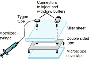
III.1.4.1 DNA molecules preparation ... 33
III.1.4.2 Fluid cell assembly ... 34
III.1.4.3 Bead tracking and molecule length ... 35
III.1.4.4 Force calibration ... 35
III.2 Enzymatic activity study using Magnetic tweezers ... 39
CHAPTER IV: Thesis project ... 45
IV.1 G-quadruplexes ... 45
IV.1.1 C-MYC G4 ... 46
IV.1.2 Human telomeric G4 ... 47
IV.2 This work ... 48
IV.3 G4 ligands ... 48
IV.3.1 Phen-DC3 ligand ... 49
IV.4 G4s kinetics and stability dependence on cation type ... 50
IV.5 G4 and helicases ... 51
IV.5.1 Pif1 helicase and G4s ... 51
IV.5.2 RecQ helicase and G4s ... 52
IV.6 G4 and Polymerases ... 52
IV.7 G4 and proteins ... 53
IV.7.1 RPA and G4s ... 53
IV.7.2 Sub1 and PC4 proteins ... 53
CHAPTER V: Results and discussion ... 55
V.1 Folding a G4 in a double-stranded DNA ... 55
V.1.1 Simple unzipping and re-zipping of DNA hairpins ... 55
V.1.2 Unzipping and re-zipping the DNA hairpin in presence of an oligonucleotide complementary to a segment on the DNA hairpin ... 56
V.1.3 Folding the c-MYC (14, 23) G4 embedded in the DNA hairpin in a K+ buffer ... 58
V.2 C-MYC (14, 23) G4 kinetics ... 62
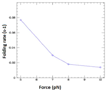
V.2.1 C-MYC (14, 23) G4 folding rate ... 62
V.2.2 C-MYC (14, 23) G4 Stability ... 63
V.2.3 Unfolding the c-MYC (14, 23) G4 by applying an external force ... 64
V.2.4 C-MYC (14, 23) G4 encircling ... 66
vi
V.2.5.1 Effect of Na+ on the c-MYC (14, 23) G4 folding ... 66
V.2.5.2 Effect of Na+ on the c-MYC (14, 23) G4 stability ... 68
V.2.6 Discussion of the c-MYC (14, 23) G4 Kinetics and stability ... 69
V.3 Folding other G4 sequences in the hairpin ... 73
V.4 G4 stability ... 75
V.4.1 Stability of different G4s sequences with and without phen-DC3 in K+buffer ... 75
V.4.2 Increasing the G quadruplex stability ... 77
V.4.3 Promoting the G-quadruplex folding ... 78
V.5 Does the c-MYC (14, 23) G quadruplex form a roadblock on the enzymes’ path? ... 80
V.5.1 Bacteriophage T4 replisome ... 81
V.5.1.1 gp41 helicase ... 82
V.5.1.2 gp43 polymerase ... 86
V.5.1.3 gp41 and gp43 coupled ... 88
V.5.2 Pif1 helicase ... 91
V.5.2.1 The helicase is added on the hairpins that closing is blocked by the lagging strand G4 ... 93
V.5.2.2 Pif1 heliacase is added to the hairpins having an encircled G4 that is located on the lagging strand in order to visualize its collision with the G4 ... 95
V.5.2.3 G4 on the leading strand ... 98
V.5.2.4 Does the Pif1 helicase have an affinity for the G4 structure? ... 99
V.5.2.5 Does Pif1 resolve the c-MYC (14, 23) at a high concentration of K+? ... 100
V.5.2.6 Does Pif1 resolve the c-MYC (14, 23) in the presence of Phen-DC3 ligand? ... 100
V.5.2.7 Does Sub1 protein enhance the G4 stability against unwinding by Pif1? ... 100
V.5.3 RecQ helicase ... 101
V.5.3.1 Does RecQ unfold the c-MYC(14,23) G4? ... 101
V.5.3.2 Folding of G4s due to hairpin opening by helicases ... 102
V.5.4 Does RPA or SSB unfold the G4? ... 103
V.5.4.1 G4 folding in presence of single stranded proteins ... 103
V.5.4.2 Unfolding the G4 by the single-stranded proteins ... 103
V.6 Unfolding the G4 structure by polymerases ... 105
V.6.1 T7 Polymerase ... 106
V.6.2 Manta polymerase ... 106
V.6.3 Pol ε polymerase ... 107
vii
CHAPTER VI: T4 replisome study using DNA rolling circle ... 117
VI.1 Rolling circle construction ... 117
VI.2 Rolling circle replication ... 118
VI.2.1 gp43 exo- polymerase ... 120
VI.2.2 Manta polymerase ... 120
VI.3 T4 coupled leading strand and lagging strand polymerases ... 121
VI.3.1 GP43 polymerase ... 122
VI.3.2 Light replisome (gp41 helicase and gp43 polymerase) ... 124
Résumé du travail de thèse en français……...………129
Annexe 1 ………...151 Annexe 2: ………...177 Annexe 3 ………...179 Annexe 4 ………...181 Annexe 5 ………...183 References ………...185
viii
List of figures
Fig. I.1: Nucleic Acid building blocks (nucleotides). ... 7
Fig. I.2: Glycosic bonds and sugar conformation. . ... 9
Fig. I.3: DNA double helix. . ... 10
Fig. I.4: DNA major and minor groove.. ... 11
Fig. I.5: DNA secondary structures. . ... 12
Fig. I.6: DNA triple helix. . ... 13
Fig. I.7: G-quadruplexes. ... 14
Fig. I.8: DNA storage in chromosome. ... 16
Fig. I.9: A chromosome at different magnifications. ... 17
Fig. I.10: G-quadruplex structures at the 3’ overhang of telomeres.. ... 18
Fig. I.11: FJC and WLC models.. ... 20
Fig. I.12: Unzipping a double stranded DNA helix. . ... 21
Fig. I.13: DNA supercoiling. ... 22
Fig. II.1: Schematic representation of proteins.. ... 23
Fig. II.2: Protein structure. . ... 25
Fig. II.3: Protein tertiary structure stabilizing interactions. . ... 26
Fig. II.4: DNA replication or DNA synthesis in the process of copying a double-stranded DNA molecule. . ... 28
Fig. III.1: The magnetic trap setup. ... 33
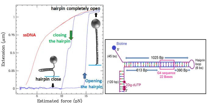
Fig. III.2: The DNA hairpin used in the current work. ... 34
Fig. III.3: Fluid chamber assembly.. ... 34
Fig. III.4: Calibration image. ... 35
Fig. III.5 : The forces acting on the magnetic bead in the magnetic tweezers setup. ... 36
Fig. III.6 : – Power spectrum density. ... 38
Fig. III.7: Schematics of the unzipping assay. ... 41
ix
Fig. IV.2: c-Myc(14,23) G4 structure... 47
Fig. IV.3: Ligand binding to G4. ... 49
Fig. IV.4 : Phen-DC3 structure ... 49
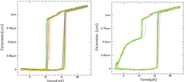
Fig. V.1: Force-Extension curves. ... 55
Fig. V.2: Unzipping and rezipping of the DNA hairpin in presence of an oligo. ... 57
Fig. V.3: A serie of opening – closing cycles of the hairpin having a G4 sequence on one strand before the folding of the G4 structure.. ... 58
Fig. V.4: The method used to fold the G4 in the hairpin. ... Erreur ! Signet non défini. Fig. V.5: Folding the G4 structure in the DNA hairpin using an oligonucleotide complementary to the hairpin loop.. ... 60
Fig. V.6: The hairpin extension curves before and after the G4 folding. ... 61
Fig. V.7: Folding the G4 in the DNA hairpin using a loop oligo... 62
Fig. V.8: C-MYC (14, 23) G4 folding rates. . ... 63
Fig. V.9: C-MYC (14, 23) G4 stability. . ... 64
Fig. V.10: G4 unwinding by applying an external force.. ... 65
Fig. V.11: The histogram representing the number of G4 resolved at different forces.. ... 65
Fig. V.12: C-MYC (14, 23) G4 encircling rates. ... 66
Fig. V.13: Force - extension curves before and after the G4 folding cycles in 60 mM Na+. ... 67
Fig. V.14: Unfolding a G4 structure by exchanging K+ by Na+.. ... 68
Fig. V.15: C-MYC (14, 23) folding and unfolding rates.. ... 72
Fig. V.16: hairpin having 2 consecutive telomeric G4s.. ... 74
Fig. V.17: Human telomeric G4 stability.. ... 76
Fig. V.18: G4C2 lifetimes in 150 mM K+ and in presence of 1 nM Phen-DC3.. ... 76
Fig. V.19 : G4 stabilizing agents. ... 77
Fig. V.20: G4 chaperone assay.. ... 78
Fig. V.21 : G4 folding at high force in the presence of Sub1 protein. ... 79
Fig. V.22: gp41 helicase (60 nM) unwinding a hairpin that does not have a G4 structure. . ... 81
Fig. V.23: gp41 helicase encountering a G4.. ... 82
Fig. V.24: gp41 helicase encountering a G4 that is blocking the hairpin closure. . ... 83
Fig. V.25: gp41 helicase opening a DNA hairpin having an embedded G4 structure... 84
Fig. V.26: gp41 helicase opening a folded hairpin containing a G4 structure on its lagging strand that is encircled in the duplex.. ... 85
Fig. V.27: The gp43 polymerase encountering a G4 structure.. ... 86
x
Fig. V.29: T4 light replisome encountering a G4. The T4 light replisome formed by the helicase and
the polymerase is tested on a hairpin having the G4 on the leading strand. ... 88
Fig. V.31: T4 replisome encountering a G4.. ... 89
Fig. V.31 : The T4 replisome replicating the G4 where the helicase and the polymerase are coupled. 90 Fig. V.32: Pif1 helicase unwinding a hairpin that does not have a G4. ... 92
Fig. V.33: Pif1 unwinding rate.. ... 93
Fig. V.34: Pif1 added to hairpins that are blocked by the lagging G4 structure. ... 93
Fig. V.35: Pif1 resolving the G4... 94
Fig. V.36: Pif1 helicase jumps the G4. ... 95
Fig. V.37: Pif1 unwinding a hairpin that has an embedded G4 structure. . ... 95
Fig. V.38: Pif1 resolving a G4 embedded in a hairpin. . ... 96
Fig. V.39: Zoom on the Pif1 pause on the G4. ... 97
Fig. V.40: Pif1 pause duration on the c-MYC (14,23) G4. ... 98
Fig. V.41 : Pif1 added to hairpins having a G4 on their leading strand.. ... 99
Fig. V.42: G4 unwinding by Pif1 in the presence of Sub1.. ... 100
Fig. V.43: RecQ delta opening the hairpin and leading to the G4 folding. ... 102
Fig. V.44: RPA unzipping DNA lex in the absence of ATP. ... 104
Fig. V.45 : Pif1 resolving a G4 embedded in a hairpin in the presence of RPA. ... 105
Fig. V.46: T7 polymerase replicating c-MYC (14, 23) G4. ... 106
Fig. V.47 : The Pol ε exo- replicating a molecule that does not have a G4 structure. ... 107
Fig. V.48: Polymerase Epsilon WT replicating a G4. . ... 108
Fig. V.49: The Polymerase Epsilon replicating a G4 in a primer extension mode. ... 109
Fig. VI.1: Construction of a rolling circle molecule. in of the DNA molecule. ... 117
Fig. VI.2: Replication of a rolling circle DNA molecule by a polymerase.. ... 119
Fig. VI.3: gp43 exo- polymerase replicating the rolling circle DNA molecule. ... 120
Fig. VI.4: Manta polymerase replicating the rolling circle DNA molecule.. ... 121
Fig. VI.5 : A model of the T4 bacteriophage DNA replisome[148].. ... 122
Fig. VI.6 : Rolling circle replication with the coupled lagging and leading strands polymerases. a) ... 124
Fig. VI.7 : The rolling circle is replicated without coupling between the lagging and leading strands polymerases. ... 124
Fig. VI.8 : Rolling circle replication showing a coupling between the lagging and lesding strands polymerases.. ... 125
xi
List of key abreviations
ATP: Adenosine triphosphate
Bp: Base pair
BSA: Bovine Serum Albumin
dNTP: deoxynucleoside triphosphates
dsDNA: Double-stranded DNA
DTT: Dithiothreitol
EDTA: Ethylenediamine tetracetic acid
G4 DNA: G-quadruplex DNA
k
off: Unfolding rate
k
on: Folding rate
RPA: Replication protein A
SF: Superfamily
SSB: Single stranded DNA-binding protein
ssDNA: Single-stranded DNA
1
Introduction
In an effort to understand what living organisms are made of, how parents transmit hereditable traits to offspring, and how do organisms age and fall sick, scientists have discovered an impressive microscopic world that is encapsulated in every micron-sized cell of our body. In the cell, the craft workers that intervene in the various processes dealing with the DNA masterpiece are the proteins and enzymes. Each one of them has a specific expertise and a function that is complementary to the others. The DNA that contains all the organism’s genetic information is compacted in entities called chromosomes and stored in the nucleus of every cell of the organism.
Cell multiplication is a process that ensures the growth and development of a living organism, where a cell divides into two daughter cells having the same genetic patrimony. The genetic code conservation among the cells after so many divisions is due to the complementarity between the two strands of the DNA double helix where adenine binds to a thymine, and a guanine binds to a cytosine. So before a cell division occurs, the DNA must be replicated in order to generate from a double-stranded DNA helix two similar double-stranded DNA helix. The replication is a process achieved by the replisome that is an assembly of different proteins and enzymes. It consists mainly of a helicase that unzips the double helix and polymerases that replicate the two DNA strands among other proteins.
Moreover, when the organism signals the need to express a protein, the transcription machinery first intervenes to copy the recipe of the protein that is encoded in segments of DNA called genes. Then, it synthesizes the protein following the given instructions.
Furthermore, proteins intervene in the aging process that was shown to be linked to the erosion of the ends of chromosomes called telomeres. The overexpression of some proteins is linked to diseases. For instance, the telomerase is overexpressed in 80% of cancers. It elongates and replenishes the telomeres of malignant cells, the thing that continuously rejuvenates them and makes them immortal.
Most of the time, the DNA forms a double-stranded helix in vivo. Some biological processes, such as the replication and the transcription mentioned above, require the opening of the helix in order to access to the genetic code embedded in its vicinity. But when a guanine rich sequence is single-stranded, it can form a kind of DNA knot called G-quadruplex (G4). The G4 consists of three or more parallel quartets of four guanines each. The intra-quartet guanines are linked by hydrogen bonds and the intra-quartets are coordinated by cations. These DNA knots are considered as roadblocks on the path of molecular motors. Their
2
occurrence can interrupt the replication and thus lead to genetic instabilities. If a G4 is present upstream of a gene, it can suppress the transcription of the gene and provoke diseases. The presence of putative G4 sequences in various biologically relevant regulatory regions in the human genome such as oncogenes and the telomeres, makes them interesting pharmacological targets for disease treatments. For instance, the folding of G4 in a proto-oncogene can silence gene expression, while the stabilization of those roadblocks on telomeres can inhibit the telomerase in cancerous cells. Until today, nearly 900 G4 stabilizing compounds have been developed, and some of them are used in medicine.
The G-quadruplexes have been extensively studied by different biochemical and biophysical methods, by both bulk and single molecule assays. Thermodynamics of some biologically relevant G4s as well as their folding and unfolding kinetics has been studied by different techniques. Furthermore, many papers have reported their stabilization or also their unfolding by some proteins and helicases. However in most of these studies authors do not have a direct insight on the folded G4 structure and thus the G4 might have been unfolded before the enzyme encountered it.
In this study we present a new method to study G-quadruplexes using magnetic tweezers. The designed DNA molecule mimics a G4 in a double-stranded region of the DNA such as those embedded in the promoters of the genes. The G4 folding gives a stable signal that ensures the G4 still folded during the entire assay. This method allows measuring the kinetics as well as the stability of a G4 structure using various buffer conditions. It also allows the detection of a G4 chaperone activity of some proteins. Moreover, it permits for the first time to get a real-time insight into the interaction between a molecular motor and a G4 roadblock encountered on its template, to visualize if it constitutes a real barricade and how long the enzyme will require to overcome such structures.
The manuscript is structured as follows:
The first part introduces the different players: DNA, G-quadruplexes, and proteins that are reported in two different chapters; the third chapter describes the single molecule magnetic tweezers technique used in this work.
In the second part, the thesis project is presented in the fourth chapter. It consists of studying G4 structures embedded in double-stranded DNA molecules and visualizing in real-time the effect of proteins and enzymes on the folding, the stability and the unfolding of such DNA structures. In the fifth chapter we explain, comment and discuss the results that we have obtained in our study. Finally, we present a general conclusion about the G4 study.
A sixth chapter presents another research topic about the lagging strand replication. We present the methods and the results that we have obtained. However, we did not get good statistics, which has not allowed leading the project to the end.
Part I
5
CHAPTER I: The DNA, Structure and
mechanics
I.1 DNA History
How do children inherit their parents’ traits? The mystery of inheritance has persisted over centuries before being finally solved by the discovery of deoxyribonucleic acid, famously known as “DNA”. This genetic material encodes the biological information for living organisms and permits its transfer from generation to generation. DNA studies have revolutionized medicine, it revealed mutations on the DNA that cause genetic diseases, and permitted to customize genetic drugs and therapies. For untreatable diseases, DNA analysis permitted an earlier diagnosis that could help avoiding some symptoms or reducing pain. Another important DNA application is crime scene investigations, where DNA analysis had become irrefutable evidence to identify the suspect as well as the victim and thus to resolve criminal cases. The DNA profiling or genetic fingerprinting is also used for parenting tests.
In the following paragraph, we present an overview about the most remarkable stages in the DNA discovery.
In 1865, the botanist Gregor Mendel, who was pursuing experiments on pea plants, concluded that the traits found in the offspring are determined as discrete factors inherited from the parents. Those factors will be later known by “genes”, DNA segments that encode proteins that build the organism’s tissues and organs among other functions. The genes carry the code that determines the traits of an organism. A gene could be involved in the determination of multiple traits, and a polygenic trait, such as eye color and skin color, could be affected by many genes. Mendel concluded that a feature is determined by two genes alternates, each inherited from a parent. A gene alternate could be dominant or recessive. The dominant is expressed in the new generation even if there is only one copy of it (i.e from only one parent). Contrarily, the recessive one, in order to be expressed, must be transmitted by both parents. The nature of genes, their localizations in the living organisms, and the way they
6
pass to the next generation was still unknown. In 1869, while the chemist Friedrich Miescher was trying to extract the proteins components in the nuclei of human white blood cells, he discovered a substance with a higher phosphorous content, that he called the nuclein. In 1919, the biochemist Phoebus Levene discovered that the nuclein is a deoxyribonucleic acid[1]. He found the components of DNA: a phosphate group, a deoxyribose sugar, and a nitrogenous base with basic qualities, all together form a DNA building block called nucleotide. Four types of nucleotides were discovered: Adenine (A), Thymine (T), Cytosine (C) and Guanine (G) that have different nitrogenous bases. Levene stated that the DNA is a polynucleotide chain that contains the same amount of each nucleotide. This latter finding will be later proven to be false. In 1944 Oswald Avery demonstrated that genes are composed of DNA[2]. But at the time, it had been thought that the DNA was the chromosome component but it was difficult to accept that it could hold biological information. In 1950 Erwin Chargaff found that the DNA of different species had the same properties but different nucleotides order[3], [4]. He also concluded that the amount of Thymine equals the amount of Adenine, while the amount of Cytosine equals the amount of Guanine.
In 1950, James Watson and Francis Crick, had been working on a 2D model of the DNA, and finally suggested that the DNA should have a 3D structure with the backbone located at its center and the bases pointing outward. Linus Pauling proposed another structure, that of a triple helix[5]. In 1951 Rosalind Franklin obtained the famous X-ray diffraction pattern of DNA, called the “Photograph 51”, that revealed the double helix structure of DNA[6]. It enabled her to know the helix diameter, the distance by turn, the distance between two consecutive nucleotides, and that the bases are located in the center of the DNA molecule. This finding paved the way for one of the most important discoveries of the twentieth century, the 3D structure of DNA in a right-handed double helix published by Watson and Crick in 1953. In fact, when Watson saw the photograph 51, which was not published at that time, he directly understood that it resulted from a helical structure. He pursued working with Crick on their model, improved it by putting the bases inside the molecule, and concluded without any DNA manipulation, that Adenine pairs with Thymine while Guanine pairs with Cytosine[7], [8]. The discovery of the correct DNA structure shed the light on different biological mechanisms in the cell, as well as the inheritance chemical basis.
While great progress has been made during the last decades, much more research is required to answer the remaining questions surrounding the DNA.
I.2 DNA structure
I.2.1 DNA components
A DNA strand is a long polymer chain composed of nucleotides. Each nucleotide itself consists of a nitrogenous base, a deoxyribose sugar (forming together a nucleoside) and a phosphate group negatively charged. The phosphate group attached to the 5’ sugar carbon of a nucleotide is linked to the hydroxyl group attached to the 3’ sugar carbon of the next
7
nucleotide by phosphodiester bonds (fig.I.3.b). Thus the nucleotides of a single strand of DNA are joined by the sugar phosphate backbone. There are four different nitrogenous bases and hence four different nucleotides. Purine bases are Adenine (A) and Guanine (G), they have two carbon cycles: a six membered and a five membered ring containing nitrogen.
Pyrimidine bases, that have only one carbon cycle, are Thymine (T) and Cytosine (C) (fig.I.1).
Similarly, the ribonucleic acid RNA is composed of nucleotides that, instead of having deoxyribose sugar, include a ribose sugar. The RNA nucleobases are the same as DNA bases, the only difference is that RNA does not have a thymine but instead it has a uracil (U).
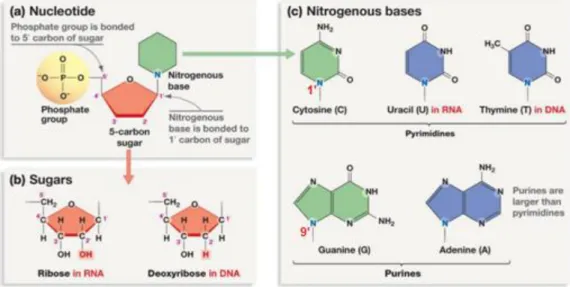
Fig. I.1: Nucleic Acid building blocks (nucleotides). a) A nucleotide is the association of a
nitrogenous base, a carbon sugar and a phosphate group. b) DNA nucleotides contain a deoxyribose sugar while RNA nucleotides contain a ribose sugar. The hydrogen on the 2’C of a deoxyribose is replaced by a hydroxyl group on the ribose. c) Four bases are possible for DNA: Purine bases (Adenine (A) and guanine (G)) and pyrimidine bases (Cytosine (C) and Thymine (T)). The first theree bases are also found in RNA, but instead of Thymine, RNA has a uracil (U). (www.sites.google.com)
In a nucleotide (fig.I.1):
- The ester bond is the linkage between the triphosphate group oxygen and the 5' carbon of the sugar.
- The glycosidic Bond is the linkage between the 9' nitrogen of purine bases or 1' nitrogen of pyrimidine bases and the 1' carbon of the sugar. The rotation of the base around the glycosidic bond gives rise to an anti or a syn conformation. The nucleotide has an anti-conformation if the base is projecting away from the sugar.
8
I.2.2 Primary structure or sequence
The primary structure of the DNA, in other words the DNA sequence, is defined by the order of its nucleotides from 5’ to 3’ end; the same direction of DNA synthesis. The DNA sequence is usually written as the sequence of bases it contains. A change in the DNA sequence within a gene, even in one nucleotide could have a large effect on the organism. For example, different eyes colors come from different DNA sequences called allele i.e the possible sequence within a gene or the gene variant. In fact, the DNA permits building the organism tissues and organs as well as ensuring its biological functions by translating the genetic code contained in the genes into functional proteins. Along the DNA chain constituting the gene, every three nucleotides will finally contribute by one amino acid in the protein chain. Thus, a mutant base will lead to a different amino acid and thus a different protein that does not fulfill the required function and could cause diseases like cancer. For instance, the sickle cell anemia disease is caused by a genetic mutation where an adenine is replaced by a thymine. However, some amino acids could be encoded by different base sequences, thus a mutation that does not change the resulting amino acid leads to the synthesis of the normal protein and is called a silent mutation. Moreover, a mutation that occurs between genes, in other words in the noncoding region is also neutral. Finally, in some cases, the mutation could be beneficial and lead to the improvement of a biological function.
I.2.3 DNA secondary structure
In the cell, the DNA is double stranded most of the time. This is due to the binding between two antiparallel DNA strands by means of nitrogenous bases. This base pairing is not random, and only takes place between complementary bases i.e. an A binds to a T by means of two hydrogen bonds, and a G binds to a C by means of three hydrogen bonds. Although the DNA strands are negatively charged due to the phosphate groups, cations in physiologic buffer shield the electrostatic repulsion between them and allow their hybridization.
Due to the complementarity between the bases, the two strands hold the same information and allow its faithful transmission to the next generation. However, some non-Watson-Crick base pairing could occur. Those are called base pairing mismatches (see below).
The DNA secondary structure or the DNA folding pattern arises from the specific base pairing as well as from the base stacking inside of the DNA duplex. In fact, the DNA forms a 3D double helix where the two nucleotides chains are twisted around each other. The sugar phosphate backbone is on the outside of the helix and the bases point inward. Actually, the hydrophobic nature of the bases, as well as their flat form, makes them stack within the double stranded DNA. This structure is made possible due to the hydrophilic nature of the sugar phosphate backbone and its flexibility. Moreover the bases are tilted like the steps of spiral staircase preventinginsertion of water molecules.
Each base of the base pair is linked to the sugar of its phosphodiester backbone via a glycosidic bond. The two glycosidic bonds of the same base pair are not symmetric with respect to the short base pair axis, but the angle between them is around 120° from a side and
9
around 240° from the other side. This leads to the existence of two different sized grooves for the double helix, called major and minor grooves. The grooves twist around the DNA helix on opposite sides.
The sugar ring of nucleic acids is not planar, but some atoms are out of its plane due to steric reasons. Endo-pucker refers to the case where the C2’ or C3’ are out of the plane toward the O5’. Exo-pucker refers to a shift of C2’ or C3’ in the opposite direction. C2’ endo and C3’ exo are equivalent, likewise C2’ exo and C3’ endo are equivalent.
Fig. I.2: Glycosic bonds and sugar conformation. (a) Glycosidic bond anti- and syn-
conformations: The rotation of the base around the glycosidic bond determines its conformation. If the base is pointing toward to the sugar the glycosidic bond is in the syn-conformation. If it is pointing away from the sugar, the glycosidic bond is in the anti-conformation. (b) Sugar A- and B- conformation: The sugar of a nucleotide is not planar, the C2’ and C3’ are shifted in opposite directions, thus the one that is shifted toward the O5’ is said endo-puckered. A-conformation refers to a C3’ endo (also called C2’ exo) and B-conformation to C2’ endo (also called C3’ exo).
I.2.3.1 B-DNA
The predominant DNA structure found in nature is the canonical B-DNA form (fig.I.3.a) that is the preferred conformation for the DNA double helix at high humidity and low salinity. It consists of a right handed double helix of 2 nm in diameter (while the electrostatic diameter is 5nm), a distance of 0.34 nm between adjacent nucleotides and a rise of 3.4 nm per helix turn involving 10-10.5 bases per turn. The B-DNA helix has a narrow minor groove 12 nm wide and a wider major groove of 22 nm. The sugar pucker is C3’ exo or C2’ endo, and the bases are nearly perpendicular to the helix axis. However, DNA was discovered to be highly polymorphic and can adopt other non-canonical conformations.
10
Fig. I.3: DNA double helix. a) A B-form DNA that is a right-handed double helix
formed by two anti-parallel strands. A DNA strand is a string of nucleotides that are joined via the sugar phosphate backbone. The bases are pointing to the interior of the double helix. The double helix has a diameter of 2 nm, a rise by Bp of 0.34 nm, and a major groove that is wider than the minor groove. b) An A is bound to a T via two hydrogen bonds while a G is bound to a C via three hydrogen bonds.
11
Fig. I.4: DNA major and minor groove. a) An A-T base pair bound by two hydrogen
bonds, and b) a G-C base pair bound by three hydrogen bonds. The glycosidic bonds linking the bases to the sugar phosphate cytoskeleton defines the major and the minor grooves.
I.2.3.2 Melting temperature Tm
The DNA melting temperature Tm is the temperature needed to denature the DNA double helix. It is the temperature required to dissociate half of DNA duplexes into single strands by breaking the hydrogen bonds. Hence, the more stable the double stranded DNA, the higher the Tm (molecules having a big content of G-C base pairs, long DNA chains, less mismatch base pairing, high cations concentration in the surrounding buffer). The average DNA Tm is
around 65°C.
I.2.3.3 Base pair Mismatch
During DNA replication, a strand is used as a template to synthesize the complementary strand. If a mistake is made and a wrong base is added, two types of mismatches could occur:
- Transition mismatch: When a purine pairs to the wrong pyrimidine (G-T and A-C baisepairs).
- Transversion mismatch: When a purine pairs to a purine or a pyrimidine to a pyrimidine.
I.2.3.4 Non-canonical secondary structures
a. A-DNA
A-DNA is also a widespread DNA conformation mostly adopted by DNA-RNA hybrids and purine DNA sequences. It consists of a right handed double helix that has a diameter of 2.3 nm, a distance of 0.26 nm between adjacent nucleotides, a rise of 2.8 nm per helix turn and 11 bases per turn. The minor groove of the A-DNA is wider than its major groove. The B-DNA is driven into an A-B-DNA form when proteins bind to it and shorten it, or under dehydrating conditions. The sugar pucker is C3’ endo, the bases are not perpendicular to the helix axis but inclined.
b. Z-DNA
The Z-DNA is a left-handed helix of 1.8 nm of diameter, an inter-base distance of 0.37 nm, and 12 bases per turn. This structure occurs in GC rich DNA stretches alternating pyrimidine-purine steps, and is promoted by DNA negative supercoiling.
c. Cruciforms
A DNA stretch can have direct repeated sequences; mirror repeated sequences, and inverted repeated sequences or palindromes. The first is a repeat of the sequence with the
12
same polarity, the second is a repeat with the opposite polarity (as if a mirror is placed between the two repeats) and the third is the complementary of the sequence (as if one has a mirror but this mirror returns for each base the image of its complementary base instead of his image). Inverted sequences are prone to form cruciforms under negative supercoiling. In order to form these non-canonical structures the DNA duplex, unzipping the DNA duplex is required. Then the sequence and its palindrom will bind to each other extruding a loop of unpaired bases, and this leads to a stem-loop structure.
d. Holliday junctions
It is a four way DNA junction that takes places as an intermediate structure during homologous genetic recombination. The homologous recombination happens when two homologous chromosomes each undergo a single strand break, and each incised strand invades the other DNA duplex leading to a four bridged Holliday junction. It was proposed by Robin Holliday in 1964. At high cation concentration permitting the phosphate repulsion to be overcome, the junction adopts a stacked conformation. While at small cation concentration, it adopts an open conformation, by moving every branch away from the others. At the end of the homologous recombination, the junction resolving enzymes separate the resultant duplexes as much as possible.
Fig. I.5: DNA secondary structures. a) A-DNA is shorter and wider than B-DNA. Both
are right handed double helices. Z-DNA is a left-handed double helix that is slightly longer and narrower than B-DNA. b) Palindromic sequences form cruciform structures under negative supercoiling: once unzipped, a linear DNA having inverted repeats can form a cruciform structure when palindromic sequences bind to each other.
13
e. Triple helices
A Triple helix can form during recombination when a strand of a DNA duplex interacts with another DNA duplex via Hoogsteen (or reversed Hoogsteen) bonds at the major groove of the Watson-Crick duplex. A polypyrimidine invading strand binds to the polypurine strand of the canonical DNA duplex via Hoogsteen bonds in a parallel orientation and leads to C+:G-C and T:A-T triplets (where C+ represents a protonated cytosine on the N3 position). Similarly, a polypurine invading strand binds also to the polypurine strand of the duplex but this time via reverse Hoogsteen bonds and in an anti-parallel orientation and leads to G:G-C, A:A-T, and T:A-T triplets.
f. H-DNA
H-DNA is an intarmolecular triple helix structure adopted in a negatively supercoiled B-form DNA by polypurine-polypyrimidine regions having a mirror repeat symmetry. If the strand that is going to bind to the canonical duplex via Hoogsteen binds is a polypyrimidne (leading to T-A:T or C-G:C+ triplets) the triple helix requires an acidic pH to form. Now if the invading strand is a polypurine a triple helix can form at neutral pH but requires divalent cations for stability (that will lead to T-A: or C-G:G base triplets).
Fig. I.6: DNA triple helix. a) Base triplets of a DNA triple helix: On the left panel, base
triplets that occur when the invading strand is composed of purines only. On the right panel, when the third strand is composed of pyrimidine only. b) H-DNA or intramolecular triple helix.
g. G quadruplexes
In 1910, Bang reported that higher order structures could be formed within G rich DNA sequences[9]. Fifty years later, in 1962, Gellert found that Guanine rich DNA sequences can form G-quadruplex structures[10], hereafter called G4. G4 is a non-canonical four stranded
14
DNA structure that arises from guanine quartets stacking (fig.I.7). A guanine quartet is a square planar arrangement formed by four guanine nucleotides where the neighboring guanines are linked by two hydrogen bonds. The minimal requirement to make an intramolecular G4 structure is four tracts of three or more Guanine (G≥3Nx G≥3Nx G≥3Nx G≥3),
however some intramolecular G-quadruplexes are formed by repetitions of two guanines. The G4 structure is stabilized by a monovalent or a divalent cation such as k+, Na+, Sr2+. In fact, depending on their size, these cations occupy the intra-quartet, or inter-quartets cavities, neutralizing therefore the electrostatic repulsion between negatively charged guanines oxygens[11]. The bases chains between the guanine tracts are extruded from the tetrads and are called loops. Shorter loops and longer guanine tracts lead to more stable G4s[12]. Intramolecular G4s are those that engage Guanines of the same DNA strand while intermolecular G4s engage guanines of different strands and could be bimolecular, trimolecular or tetramolecular.
A G quadruplex is parallel if all of its G strands are parallel. On the contrary an antiparallel G4 has two parallel strands that are antiparallel to the two others. Besides, if one strand is antiparallel to the three others, the G4 is called hybrid. The big majority of parallel G4s have guanines in the anti-conformation while antiparallel and hybrid G4s have a mixture of syn- and anti-conformations of guanines. Moreover, the loops joining the G runs could be lateral, diagonal or double chain reversal. For all those reasons, the G4 is considered a DNA structure of high polymorphism.
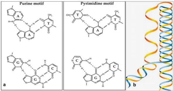
Fig. I.7: G-quadruplexes. a) Tetrads can stack to form G-quadruplexes. G
quadruplexes can fold into a variety of topology, which differ mainly by the relative orientation and number of strands (1 to 4), the number of tetrads (at least 2), and the geometry of the loops. b) Guanine tetrads contain four guanines linked by eight hydrogen
15
bonds (donor and acceptor groups in blue and green, respectively). Guanine O6 selectively coordinate a metal cation (red). [13]
I.2.4 DNA tertiary structure
The DNA tertiary structure refers to the three dimensional arrangement in space of the DNA double helix and its confinement in chromosomes that is influenced by geometrical and steric constraints.
I.2.4.1 Topological parameters
a. Contour length:
The contour length of a polymer is its total length Lc=N.l.
Where N is the number of monomers composing the polymer, and l is the length of a single monomer. The contour length of DNA is thus the number of nucleotides times the length of a single nucleotide.
b. Persistence length
A polymer persistence length is the length scale beyond which the elastic cost of bending is smaller than thermal energy. The stiffer the polymer, the bigger the persistence length. The DNA persistence length is about 50nm[14] and is much higher than PVC or polyethylene polymers. The DNA is compared to a random coil in the absence of a stretching force, and its end to end length is 𝑅 = √2𝐿𝑙𝑝, where lp is its persistence length. The DNA tertiary structure
refers to the topology adopted by DNA at length scales larger than the persistence length L>>>lp.
c. Linking number LK
It is the number of times one strand is wrapped around its complementary one in a circular double stranded DNA. This number cannot be changed in a double stranded circular DNA because it does not have free ends. Lk could be also considered as the number of times a
DNA strand has to be cut (breaking of a covalent bond) and turned around its complementary in order to entirely separate the two strands. For a given circular DNA of N base pairs, the more the number of base pairs per turn (n) the less the LK. A relaxed DNA has LK=LK0=N/n,
for B-form DNA n=10.5 base pairs. LK is an integer and it is the sum of two other
16
d. Twist
It is the number of helical turns of one strand about the other. It is determined by the number of times a strand wraps completely around the other. Therefore, for a planar circular double stranded DNA, Tw=LK.
e. Writhe
It is the number of times the DNA double helix crosses around itself. A right handed writhe is negative while a left-handed writhe is positive. When the DNA is overwound the helix wrap around itself in a left-handed fashion, writhe is then positive and the linking number is higher than that of relaxed DNA. The DNA is said positively supercoiled. If the DNA is underwound, the double helix wraps around itself in a right handed fashion leading to a negative writhe and then a linking number smaller than that of relaxed DNA.
I.2.4.2 DNA storage in chromosomes
Living organisms are sorted into two groups: Eukaryotes (human, animals and plants) and Prokaryotes (archaea and bacteria). In Eukaryotes, the DNA is stored in every cell nucleus, which is surrounded by the cytoplasm and enveloped by the cell membrane. Thus, every cell contains the whole code that characterizes the organism except red blood cells and blood platelets that lack the nucleus. In order to fit into the cell nucleus of 6um of diameter, the DNA, long about 2m, is condensed into compact chromosomes. In a human cell, there are twenty three pairs of chromosomes.
Fig. I.8: DNA storage in chromosome. From PMG biology October 2015
In Eukaryotes, the negatively charged double helix DNA is first wrapped around positively charged proteins called histones. Along the DNA molecule, every eight histone subunits form one nucleosome, around which 146 DNA base pairs are wrapped. The resulting complex DNA-protein that is three times shorter than the linear DNA is named chromatin and still accessible to enzymes and proteins. In this form, DNA strings link the beads-like nucleosomes, and hence the structure is called “beads on a string” and has a diameter of 10
nm. The DNA becomes more strongly compacted when every six nucleosomes are coiled together, and stack upon each other’s to form a solenoid of 30nm of diameter. In this form,
17
the DNA is 50 times shorter than the linear form. The chromatin form does not only make possible fitting DNA in a tiny space but also control gene expression. For instance, when the cells are not dividing, some chromatin regions becomes tightly packed and thus could not be transcribed, those are called heterochromatin. Heterochromatin accumulates near the nucleus envelop. Contrarily, euchromatin are the loosely packed regions of chromatin that are not stainable but dispersed in the nucleus and are prone to transcription. At this stage of the cell cycle, called interphase, the DNA is replicated to two sister chromatids in order to prepare to the mitosis, or the cell division stage where a cell divides into two daughter cells that will inherit the same genome. The chromatid sisters are joined by cohesion proteins to form an X shape chromosome and are bound together at its centrosome. The chromosomes become more condensed during the first phase of the mitosis called prophase, and during the metaphase they become 10 000 fold shorter than the linear form. The chromosomes at this stage align along the cell equator in order to let the sister chromatids detachment and migration to the opposed cell poles during the anaphase. At the end, during telophase, every cell is divided into two new cells that contain chromosomes of one chromatid each.
Fig. I.9: A chromosome at different magnifications. 1) DNA double strand. 2)
Double-strand DNA wrapped around histone proteins (nucleosomes). 3) Chromatine with a centromere during the interphase. 4) Following synthesis, two chromatids are joined by a centromere; each chromatid consists of highly coiled nucleosomes. 5) Two chromatids of a chromosome are tightly coiled in preparation for cell division. (From © 2012 T. F. Fletcher vanat.cvm.umn.edu)
In Prokaryotes, the cells are nucleus free and the DNA is located in the cytoplasm in a region called nucleoid that is not membrane bound. The majority of prokaryotes have only one circular chromosome and their DNA is even more compact than the DNA of Eukaryotes.
Eukaryotic telomeres refer to the terminal segments of chromosomes that seal the genes and protect them from erosion. They consists of a double stranded DNA regions and a 3’ single stranded DNA overhang, both are gene free and have a species-specific tandem repeats of G-rich sequence. Human telomeres contain repeats of TTAGGG (2-50 kilobase pairs) and a tail of 100–250 bases[15]. These single-stranded rich ends are believed to form G-quadruplex structures (fig.I.10) [16].
18
Fig. I.10: G-quadruplex structures at the 3’ overhang of telomeres. The repeated G
rich sequence on the single stranded telomeric overhang forms successive G4 structures.
I.3 DNA mechanics
I.3.1 dsDNA extension model
I.3.1.1 Free jointed chain model
In this model, the DNA is assimilated to a polymer chain of length L and having N rigid segments (fig.I.11.A). All the links have the same length b and an independent random direction 𝒓⃗ 𝒊. There is any self-avoiding condition and thus the chain can cross itself. The end
to end length of the chain 〈𝑹⃗⃗ 𝟎〉 is equal to 0 because the direction a link can adopt is random and thus two opposite directions are possible with equal chance:
〈𝑅⃗ 0〉 = 〈𝑟⃗⃗⃗⃗ − 𝑟𝑁 ⃗⃗⃗ 〉 = 〈∑ 𝑏𝑟1 ⃗⃗ 𝑖 𝑁 𝑖=1 〉 = 𝑏 〈∑ 𝑟⃗⃗ 𝑖 𝑁 𝑖=1 〉 = 0 The root mean square end-to-end distance is:
〈(𝑅⃗ 0)2〉 1 2 ⁄ = 〈(𝑟 𝑁 ⃗⃗⃗⃗ − 𝑟⃗⃗⃗ )1 2〉 1 2 ⁄ = 𝑏 〈(∑ 𝑟 𝑖 ⃗⃗ 𝑁 𝑖=1 ) 2 〉1⁄2 = 𝑏 〈∑ 𝑟⃗⃗ 𝑖 𝑁 𝑖=𝑗 . 𝑟⃗⃗ ⃗⃗ + ∑ 2𝑟𝑗 ⃗⃗ 𝑖 𝑁 𝑖,𝑗=1 𝑖≠𝑗 . 𝑟⃗⃗ ⃗⃗ 〉𝑗 1 2 ⁄ = 𝑏√𝑁
Although the FJC model is interesting since it explains the principle of entropic elasticity, it does not describe properly the elasticity of a DNA molecule.
19
I.3.1.2 Worm like chain model
The FJC model can be used for double stranded DNA at low forces. In fact, at intermediate and high forces the FJC model cannot describe the DNA behavior, because this latter is not a jointed chain but a double helix and its bending energy should be taken into account. The worm-like chain model (WLC) assimilates the DNA to a flexible rod that has a persistence length 𝜉 (fig.I.11.B). In order to align the polymer segments in the range of the persistence length, one should apply a force of 𝐾𝐵𝑇
𝜉 . If the DNA is subjected to a stretching,
then the energy of the system is: E= Ebending + Estreching 𝐸 𝐾𝐵𝑇= 1 2𝜉 ∫ ( 𝜕𝑟 𝜕𝑠) 2 𝐿 0 𝑑𝑠 − 𝐹 𝐾𝐵𝑇∫ cos(𝜃(𝑠))𝑑𝑠 𝐿 0
Where ξ is the persistence length, 𝒕 (𝒔) is the vector tangent to the chain at the curvilinear coordinate s, F is the stretching force modulus along the stretching direction z, and θ is the angle between 𝒕 (𝒔) and the z direction.
The force F required to induce an end to end extension of 〈𝐳〉 can be approximated to[17]:
𝐹 = 𝐾𝐵𝑇 𝜉 ( 〈𝑧〉 𝐿 − 1 4+ 1 4 (1 −〈𝑧〉⁄ )𝐿 2 + ∑ 𝑎𝑖 7 𝑖=2 (〈𝑧〉 𝐿 ) 𝑖 ) At low forces 𝐹 < 𝐾𝐵𝑇
𝜉 , the double stranded DNA acts like an entropic spring, its
extension is proportional to the force:
𝐹 =3𝐾𝐵𝑇 2𝜉 (
〈𝑧〉 𝐿 )
At forces up to 10 pN, the dsDNA extension curve can be modelled by the WLC model. Above 10 pN, the dsDNA is stretched and its end-to-end length exceeds the contour length. At 65 pN the DNA is overstretched and undergoes a phase transition from B-form to S-form. Moreover, above 300 pN the double stranded DNA is unzipped and gives single strands DNA.
20
Fig. I.11: FJC and WLC models. A) The FJC model assimilates the DNA to a chain of
rigid segments having a length b and linked to each other. θ is the angle between two consecutive segments. B) The WLC model assimilates the DNA to a smoothly bending flexible rod that has a total length L and a persistence length ξ. Θ is the angle between the pulling force axis and the tangent vector to the curvilinear position s.
I.3.2 ssDNA elasticity
Due to its high flexibility, the ssDNA is more contractile than dsDNA and needs a higher force to align it (around 5pN instead of 0.1pN F=KBT/ξ). Up to 5 pN, the single stranded
DNA is shorter than dsDNA, above 5 pN, however, the ssDNA length exceeds that of dsDNA. ssDNA can be modelized by the FJC model or the elastic FJC model.
I.3.3 Unzipping a DNA double helix
The force required to open a DNA double helix is around 15 pN[18]–[20]. The exact value, however, depends on the double helix G-C and A-T base pairs content. A G-C rich segment requires a higher force value.
21
Fig. I.12: Unzipping a double stranded DNA helix. On the top, the graph shows the
force required to open 100 bases of dsDNA depending on the GC content.
The fig. I.12 represents the curve of the Force versus the extension when opening a DNA hairpin. The hairpin opens around 15 pN and an abrupt extension of 10 nm is noticed.
I.3.4 DNA supercoiling
If the DNA molecule is tethered with multiple bounds at both extremities, twisting the DNA molecule becomes possible. If we increase the torsional constrain, the molecule extension remains constant until we reach a threshold above which plectonemes appear leading to the shortening of the molecule.
22
Fig. I.13: DNA supercoiling. a) A dsDNA pulled at a force F and twisted by rotating
23
CHAPTER II:
Overview of the DNA
replication
II.1 Proteins and Enzymes
-
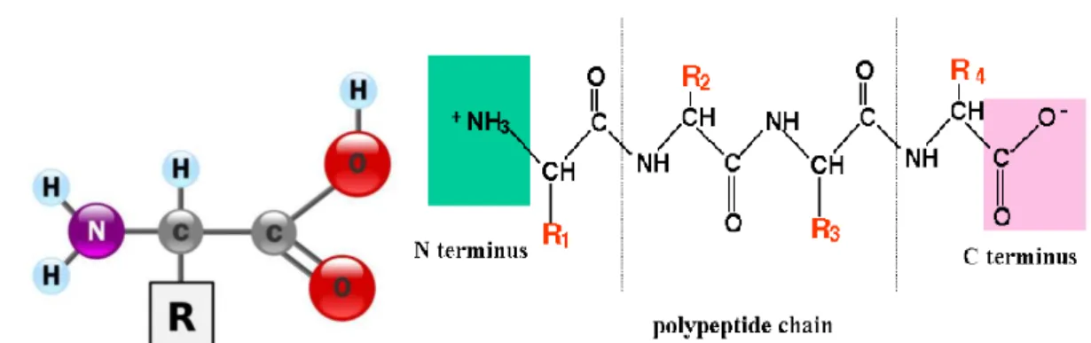
Fig. II.1: Schematic representation of proteins. a) Amino acid: It is formed by a
carboxyl group and a central atom to which is linked a lateral chain. The lateral chain determine the type of the amino acid. b) Polypeptide chain: It is a formed by the association of amino acids via peptidic bonds and is read from N-terminus to C-terminus domain. A protein is formed by one or several polypeptidic chain.
Proteins and enzymes are biological macromolecules that fulfill most functions in the organism. Enzymes are proteins that lower the activation energy for a reaction through a process called catalysis. Proteins consist of long chains of amino acids linked together by covalent peptide bonds (fig.II.1). If this polymeric chain contains up to fifty amino acids it is called a peptide, if it contains more, it is referred to by polypetide or protein. A protein could be formed by one or more polypeptides. All of the twenty amino acids that serve to build proteins have an amino group, a carboxyl group and a central carbon atom to which is linked a lateral chain (R-group). This lateral chain is specific for every amino acid that could be hydrophilic, hydrophobic or negatively or positively charged. Proteins are defined by their
24
sequence, structure and function, and are therefore classified in different categories. Among proteins categories, one finds: Structural proteins that build the cells and the tissues, transport proteins that carry molecules and ions, gene regulatory proteins that trigger or silence gene expression and enzymes that catalyze reactions.
II.2 Proteins structure
II.2.1 The primary structure
The protein primary structure or the sequence of a protein is defined by the order of amino acids forming the molecule from the N-terminus to the C-terminus. The primary structure of a protein determines the secondary structure; therefore a change in the first generally leads to a change in the secondary structure and may affect the protein function.
II.2.2 The secondary structure
The secondary structure of a protein is its shape, more precisely the shape of every petide chain within the protein, which is also called domain). In fact, every peptide chain folds in a three dimension structure owing to the hydrogen bonding between the backbone atoms (and not the side chains atoms). At small distances, this interaction gives the polypeptide chain a shape of α-helix (a rode shape), β-pleated sheet, or unstable random coil. The α-helix is a right handed helix that arises from the stable hydrogen bonds between the oxygen of C=O of an amino acid (i) and the hydrogen of the NH group of the amino acid (i-4) in the helix. There is 3.6 amino acids in every turn of the α-helix and the amino acids side chains point to the exterior. β-sheet arises from the hydrogen bonds between strands. These bonds take place between the oxygen of the carbonyl group of an amino acid and the hydrogen of the amino group of another amino acid. The beta sheet is called parallel (anti-parallel) when the directions of the strands forming it are the same (opposite). The pleated form of the β-sheet is conferred by the coplanar hydrogen bonds.
II.2.3 The tertiary structure
The tertiary structure of a protein is the overall 3-D structure that results from the folding of all the protein domains and their positioning with respect to each other, which is fashioned by the amino acids R-groups (side chains) interactions. In fact, many stabilizing interactions (Hydrogen bonds, salt bridges, ionic interaction, stacking) lead to this structure in order to achieve the lowest energy state. The hydrophobic R-chains are buried within the protein core while the hydrophilic ones are exposed to the aqueous medium. The tertiary structure of a protein is also highly stabilized by covalent bonds formed between oxidized cysteine amino acids, or disulfide bridges, that connect amino acids even if they are distant in terms of sequence.
25
II.2.4 The quaternary structure
The quaternary structure of a protein is the way subunits assemble to make a functionalized protein complex (fig.II.2). Depending on the protein, the subunits can be similar and form a homodimer, or different and form a heterodimer. This structure is also stabilized by the same interactions cited above between amino acids side chains (fig.II.3).
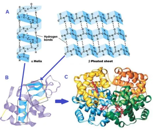
Fig. II.2: Protein structure. A) Protein secondary structure: α-helix and β-sheet
formed by hydrogen bonds between backbone atoms. B) Tertiary structure: The overall shape of the protein that is achieved by weak interactions between side chains and covalent bonds between oxidized cysteine amino acids. C) Quaternary structure: This is the fourth level of the protein structure to form a protein complex by assembling protein subunits.
26
Fig. II.3: Protein tertiary structure stabilizing interactions. Interactions between
amino acids side chains: Ionic bonds, hydrogen bonds, van der Waals interactions, and covalent disulfide bridges.
II.3 Gene expression
Gene expression refers to protein synthesis using the specific gene code in order to respond to the organism demand and provide a biological function. This is a two steps process: Transcription and translation. The transcription takes place in the nucleus and consists to copy the gene or the protein recipe encoded in the DNA. The transcript is an RNA called messenger mRNA. The mRNA quits the nucleus afterwards, and delivers the message to the DNA synthesis machinery, a nucleoprotein called ribosome located in the cytoplasm. The ribosome translates the code and synthesizes a polypeptide chain by adding one by one the amino acids according to the order provided by the messenger.
27
II.4 DNA replication
II.4.1 Helicase
Helicases constitute one of the largest class of enzymes [21], [22] , and are implicated in almost all cellular mechanisms that engage DNA and RNA such as DNA replication, transcription, translation,combination, DNA repair, and ribosome biogenesis [23]–[26] . A helicase is an enzyme that unzips the DNA double helix using ATP hydrolysis. In fact, the helicase opens an eye shaped region (also called replication fork fig. II.4) of two single strands in the nucleic acid duplex to initiate replication and allow the loading of different proteins in order to achieve the replication. The helicase unzips the double stranded DNA permitting the progression of the replisome that is formed by the helicase, the polymerases, the clamps and the clamps loaders (see next section).
The human genome codes for 64 RNA helicases and 31 DNA helicases [27]. Accordingly, defects in their function or deregulated expression lead to severe diseases [28], [29]. Helicases are classified into six super-families (SF) based on their sequences, structures, and mechanistic features [30], [31]. SF 3 to 6 include hexameric helicases that contain six RecA-like domains that form a ring shape complex, while SF 1 and 2 include non-ring-shaped helicases that contains two RecA-like domains [30]. The helicase core domains are referred to by RecA-like domain due to their resemblance to the ATP-binding core of the recombination protein RecA.
A Helicase of type alpha translocates on ssDNA while a helicase of type beta translocates on dsDNA. Another feature to distinguish helicases is the translocation polarity: A helicase A has a 3’-5’ directionality while a helicase B moves in the opposite direction. Conserved sequence motifs between helicases are those involved in ATP binding, ATP hydrolysis and translocation along nucleic acids. Seven of these conserved motifs had been identified by bioinformatics and led to the helicase discovery [32].
Helicases may be active or passive, in both cases they are molecular motors using ATP to translocate along ssDNA. The active or passive behavior characterizes the way they displace the opposite strand: a passive helicase cannot melt the dsDNA but wait for thermal fluctuations to open the dsDNA and then advances preventing the fork reclosing. Active helicases are able to translocate and melt dsDNA at the same rate. In fact an active helicase holds a special structure which enhances the melting fluctuations of dsDNA. The unwinding rate of an active helicase is nearly equal to translocation rate. Contrarily, the unwinding rate of a passive helicase is depends on the force applied to dsDNA and is lower than its translocational rate.