HAL Id: tel-01513121
https://tel.archives-ouvertes.fr/tel-01513121
Submitted on 24 Apr 2017HAL is a multi-disciplinary open access archive for the deposit and dissemination of sci-entific research documents, whether they are pub-lished or not. The documents may come from teaching and research institutions in France or abroad, or from public or private research centers.
L’archive ouverte pluridisciplinaire HAL, est destinée au dépôt et à la diffusion de documents scientifiques de niveau recherche, publiés ou non, émanant des établissements d’enseignement et de recherche français ou étrangers, des laboratoires publics ou privés.
Fabrizio Simonetti
To cite this version:
Fabrizio Simonetti. Study of the mechanisms of sexual differentiation in the fission yeast S. pombe. Molecular biology. Université Paris-Saclay, 2017. English. �NNT : 2017SACLS081�. �tel-01513121�
NNT: 2017SACLS081
T
HÈSE DE DOCTORAT
DE
L
’U
NIVERSITÉ
P
ARIS
-S
ACLAY
PREPARÉE
À
L
’U
NIVERSITÉ
P
ARIS
-S
UD
É
COLED
OCTORALE N°
577
Structure et dynamique des systèmes vivants
Spécialité de doctorat : Sciences de la vie et de la santé
Par
M. Fabrizio Simonetti
Study of the mechanisms of sexual differentiation
in the fission yeast S. pombe
Thèse présentée et soutenue à Paris, le 7 Avril 2017
Composition du Jury :
M. Daniel Gautheret M. Damien Hermand Mme Valérie Borde M. Benoît Palancade M. Mathieu Rougemaille
Professeur (Université Paris Sud, Orsay) Senior research associate (NARC, Namur) Directeur de recherche (Institut Curie, Paris) Chercheur (Institut Jacques Monod, Paris) Chercheur (Institut Jacques Monod, Paris)
Président Rapporteur Rapporteur Examinateur Directeur de thèse
To my mother, my father, my brother
and all my family
SUMMARY
In the fission yeast S. pombe, several meiotic genes are constitutively expressed during
the mitotic cell cycle. In order to avoid untimely entry into meiosis, cells have adopted a
degradation system that selectively eliminates the corresponding mRNAs. The YTH family
RNA-binding protein Mmi1 recognizes specific sequence motifs within these transcripts
(UNAAAC), and delivers them to the nuclear exosome for degradation. Upon entry into
meiosis, Mmi1 is sequestered in a ribonucleoprotein complex made of the meiotic protein Mei2
and the long non-coding RNA meiRNA, thereby allowing meiotic mRNAs to be exported and
translated. During my PhD studies, I focused my work on the role of Mmi1 in the degradation
of meiotic transcripts during vegetative growth. Consistent with recent studies, we showed that
Mmi1 stably interacts with the mRNA deadenylation complex Ccr4-Not. This interaction is
functionally relevant because Ccr4-Not is required for the degradation of meiotic mRNAs.
Surprisingly, however, the deadenylation activity of the complex is not involved. Rather, our
genetic and biochemical analyses indicate that the E3 ubiquitin ligase subunit Mot2
ubiquitinates a pool of the Mmi1 inhibitor, Mei2, to promote its degradation by the proteasome.
This regulatory mechanism ensures the maintenance of Mmi1 in a functional state, leading to
the persistent repression of meiotic mRNAs in mitotic cells. Thus, Mmi1 has a dual role: in
nuclear mRNA surveillance, by targeting meiotic transcripts for degradation by the exosome,
and in protein degradation, by recruiting Ccr4-Not to its own inhibitor Mei2. These results have
also revealed a novel role for the ubiquitin ligase activity of the Ccr4-Not subunit Mot2 in the
control of sexual differentiation in fission yeast.
Further experiments indicate that the YTH RNA-binding domain of Mmi1, but not the
non-coding RNA meiRNA, is required for the degradation of Mei2. Intriguingly, our results
strongly suggests that the YTH domain acts as a bifunctional module, binding not only to
meiotic RNAs but also to proteins. We discuss these results within the context of the current
literature and we propose a model for the control of sexual differentiation by the Mmi1-Mei2
RÉSUMÉ
Chez la levure fissipare S. pombe, certains gènes méiotiques sont exprimés de façon
constitutive pendant la croissance végétative. Cependant, pour empêcher le déclenchement
prématuré de la méiose, la cellule a mis en place un système de dégradation sélective des ARN
messagers correspondant. La protéine de liaison à l’ARN Mmi1, de la famille YTH, reconnaît des répétitions de motifs spécifiques (UNAAAC) au sein des transcrits et dirige ces derniers
vers la dégradation par l’exosome nucléaire. Lors de l’entrée en méiose, Mmi1 est séquestré par un complexe ribonucléoprotéique comprenant la protéine de méiose Mei2 et l’ARN noncodant meiRNA, ce qui permet aux ARNm méiotiques d’être exportés et traduits.
Au cours de ma thèse, je me suis intéressé au rôle de Mmi1 dans la dégradation des
transcrits méiotiques pendant la croissance végétative. En accord avec des études récentes, nos
travaux montrent que Mmi1 interagit étroitement avec le complexe Ccr4-Not de déadenylation
des ARNm. Cette interaction est fonctionnelle car Ccr4-Not est requis pour la dégradation des
ARNs méiotiques. De façon surprenante, cependant, l’activité de déadénylation n’est pas requise. Nos analyses génétiques et biochimiques suggèrent que la sous-unité E3 ubiquitin
ligase Mot2 de Ccr4-Not ubiquitine un pool de l’inhibiteur de Mmi1, la protéine Mei2, pour
faciliter sa dégradation par le protéasome. Cette voie de régulation permet de maintenir la
fonction de Mmi1 et donc la répression des ARNm méiotiques dans les cellules mitotiques.
Ainsi, Mmi1 a une double fonction: cibler les ARNm méiotiques vers la dégradation par
l’exosome nucléaire, et recruter Ccr4-Not pour ubiquitiner et dégrader son propre inhibiteur Mei2. Ces résultats mettent également en avant un nouveau rôle pour la sous-unité E3 ligase
du complexe Ccr4-Not dans le contrôle de la différenciation sexuelle.
Des expériences supplémentaires indiquent que le domaine YTH de liaison à l’ARN de Mmi1, mais pas l’ARN noncodant meiRNA, est requis pour la dégradation de Mei2. De façon
importante, nos données révèlent aussi que le domaine YTH de Mmi1 a un rôle clé dans
l’interaction avec Mei2. Ceci suggère fortement que le domaine YTH agit comme un module bifonctionnel, permettant la liaison non seulement aux ARNs méiotiques mais aussi aux
protéines comme Mei2. Nous discutons ces résultats dans le contexte de la littérature actuelle
et proposons un nouveau modèle du contrôle de la différenciation sexuelle par le système
CONTENTS
SUMMARY ... I
RÉSUMÉ ... III
CONTENTS ... V
INTRODUCTION ... 1
1
General principles of sexual differentiation in yeast ... 2
2
The meiotic program: from external signals to transcriptional
regulation ... 6
2.1 Signaling cascades ... 6
2.1.1 The cAMP pathway ... 7
2.1.2 The TORC1 pathway ... 7
2.1.3 The mating pheromone-responsive pathway ... 8
2.1.4 The stress-responsive pathway ... 8
2.2 The transcription factor Ste11 ... 10
2.2.1 General properties ... 10
2.2.2 Regulatory mechanisms of ste11+ expression ... 11
2.2.2.1 Transcriptional control ... 11
2.2.2.2 Post-transcriptional control ... 12
3
Regulatory mechanisms of sexual differentiation ... 16
3.1 The Pat1-Mei2 system: a central regulator of meiosis ... 16
3.1.1 The Pat1 kinase inhibits entry into meiosis ... 16
3.1.2 The RNA-binding protein Mei2: a key meiosis inducer ... 19
3.1.2.1 General properties ... 19
3.1.2.2 Evolutionary conservation ... 19
3.1.2.3 A key Mei2 cofactor: the long non-coding RNA meiRNA ... 20
3.1.2.4 Localization ... 21
3.1.2.5 Functions ... 22
3.1.2.6 Regulation of Mei2 expression and activity ... 23
3.2 The selective elimination of meiotic transcripts during vegetative growth ... 25
3.2.1 cis-regulatory sequences control meiotic mRNA levels ... 25
3.2.2 Factors involved in the selective elimination of meiotic mRNAs ... 27
3.2.2.1 The factors that recognize meiotic transcripts ... 27
3.2.2.1.1 The YTH-family RNA-binding protein Mmi1 ... 27
3.2.2.1.2 3’-end processing and polyadenylation factors ... 35
3.2.2.1.3 The MTREC complex ... 36
3.2.2.1.4 Erh1, enhancer of rudimentary homologue ... 38
3.2.2.2 The factors that contribute to meiotic mRNA elimination ... 39
3.2.2.2.1 The nuclear exosome ... 39
3.2.2.2.2 The Ccr4-Not complex ... 45
3.2.2.2.3 The RNA interference machinery ... 58
RESULTS ... 61
1
Summary and contribution ... 62
2
Article ... 64
3
Supplemental Results... 102
3.1 meiRNA is not required for Mmi1-dependent Mei2 degradation ... 102
3.2 The YTH domain of Mmi1 is required for degradation of Mei2 ... 104
3.3 The YTH domain of Mmi1 is required for interaction with Mei2 ... 105
3.4 The N-terminal region of Mmi1 mediates the interactions with the MTREC and Ccr4-Not complexes ... 108
3.5 N-terminal residues of Mmi1 are important for efficient meiotic mRNAs suppression ... 110
DISCUSSION ... 113
1
Mechanisms of sexual differentiation by the Mmi1-Mei2 system ... 114
1.1 YTH proteins and the Ccr4-Not complex: RNA deadenylation and/or protein ubiquitination? ... 114
1.2 Molecular basis for the inactivation of Mmi1 by Mei2 ... 115
1.3 Distinct Mmi1-containing complexes for specific functions?... 118
1.4 A general model for the regulation of meiotic mRNAs suppression ... 121
2
Beyond meiotic mRNA suppression: additional roles for the
RNA-binding protein Mei2 and the E3 ubiquitin ligase Mot2? ... 125
2.1 Towards a comprehensive view of Mei2 RNA targets ... 125
2.2 The E3 ligase Mot2 is required for the repression of Ste11 target genes... 126
BIBLIOGRAPHY ... 129
1 General principles of sexual differentiation in yeast
In eukaryotes, sexual differentiation is a process that relies on the conversion of external
signals into a stable phenotype, through temporary changes in the expression of genes. The
events occurring during this process are critical for the generation of diversity and for the
production of a normal offspring. In nearly all eukaryotes, meiosis has a central role in sexual
reproduction and is a highly-conserved process, from fungi to plants and animals. Meiosis
consists in the specialized division that halves the genetic material, reducing the diploid
genome of the progenitor cell to the haploid state and producing genetically different daughter
cells. The haploid products, also called gametes, are oocytes and sperm in animals, pollen in
plants, and spores in yeasts.
Over the last decades, great advancements in the study of meiosis came from a small
number of model organisms, including the fission yeast Schizosaccaromyces pombe. The
employment of fission yeast as a model system allowed a better understanding of the
mechanisms that govern sexual differentiation. S. pombe is an ideal organism to study the
meiotic process for several reasons. For instance, almost all yeast cells in a population, when
starved for nutrients, enter meiosis in a synchronous manner. This allows temporal analyses
using cytological, biochemical and molecular assays. Yeast meiotic mutants are easy to isolate,
and it is possible to rapidly clone and disrupt the corresponding genes.
The use of fission yeast as tool allowed elucidating to a great extent how cells enter and
proceed through meiosis.
In fission yeast the initiation of meiosis relies on a decrease of available nutrients in the
environment. This event induces yeast haploid cells of opposite mating types to mate and
conjugate, forming a zygote that enters the meiotic cell cycle. One round of DNA replication
spores can eventually re-enter the mitotic cell cycle in the presence of nutrients (Fig. 1). By
analogy, yeast haploid cells behave similarly to gametes in higher organisms, such as oocytes,
sperm or pollen.
Figure 1. Scheme of the life cycle of the fission yeast S. pombe.
Upon nutritional starvation, haploid cells arrest mitotic growth and initiate the mating process. Cells of the M and P mating types conjugate to form a zygote. The diploid zygote then undergoes the meiotic process, generating four ascospores. If nutrients are supplied in the medium the spore can germinate giving rise to a haploid cell. Figure adapted from Otsubo and Yamamoto [1].
Particularly, in S. pombe, the cell mating type (h+ or h-) is determined by the sequence at the mat1 locus, which can be either P (mat1-P for h+ cells) or M (mat1-M for h- cells) [2]. h+ cells synthesize the mating pheromone P-factor as well as the M-factor receptor, whereas h -cells produce the M-factor and the P-factor receptor. P and M mating pheromones are small
peptides that bind to their cognate receptor anchored in the plasma membrane of cells with
opposite mating type, thereby promoting mating and conjugation [3, 4]. Upon mating, cells
arrest transiently in G1 and undergo one round of DNA replication known as pre-meiotic S
phase, in which the DNA content of each cell is doubled. The nuclei of the two progenitor cells
then fuse, in a process called karyogamy, and undergo meiotic prophase, which features a
continuous movement of the nucleus, mediated by the spindle body (SPB), favors chromosome
pairing, which is essential for the following event of chromosome segregation.
Cells then perform two consecutive nuclear divisions, which are known as meiosis I
and meiosis II. During meiosis I, called reductional division, replicated homologous
chromosomes align at the equator of the cell and undergo high-frequency meiotic
recombination. Then, the first nuclear division occurs and homologous chromosomes
segregate, producing two daughter cells with halved genetic material [5-7]. Meiosis II instead
is described as an equational division; it resembles the normal mitotic division where sister
chromatids are separated. This division produces four genetically different cells, each carrying
a haploid content of DNA, which are then packed in order to form mature spores (Fig. 2).
Figure 2. The meiotic process in fission yeast.
Upon nutritional starvation, h+ and h- haploid cells arrest in G1 and secrete the respective mating pheromone. Upon exchanging the pheromones, cells elongate towards each other and fuse (conjugation process), followed by the fusion of the haploid nuclei. The diploid nucleus elongates and assumes a horsetail shape, then moves from one end of the cell to the other (meiotic prophase). After the horsetail nucleus ceases to move, it becomes round again and proceeds through the first and second meiotic divisions to form four haploid nuclei. Figure adapted from Asakawa [8].
The meiotic process is characterized by a complex sequence of events that irreversibly
alter cell morphology and growth capacities in response to specific developmental and/or
environmental cues. In order to prevent the initiation of sexual differentiation in conditions
suitable for vegetative growth, mitotic cells have evolved several regulatory mechanisms that
remain largely uncharacterized. Indeed, the molecular details underpinning the decision of a
cell to switch from mitosis to meiosis are far from being fully understood.
During my PhD studies, I focused my work on the molecular mechanisms involved in
the transition from mitosis to meiosis in fission yeast. In the next sections, I will describe the
several layers of the regulation of meiosis in S. pombe, ranging from gene transcription to
protein modification. First, I will give an overview on how environmental cues trigger and
establish the meiotic gene expression program, highlighting the main molecular determinants
at play. In a second part, I will introduce the factors involved in the control of meiosis initiation
and their associated function. Given the results obtained from my work, particular attention
2 The meiotic program: from external signals to
transcriptional regulation
Specific environmental and/or developmental cues trigger the entry into meiosis,
including nutrient starvation and the synthesis of mating pheromones. These external signals
are integrated by the cells, which modify and adapt their gene expression profiles to drive
sexual differentiation. The expression of hundreds of meiosis-specific genes is indeed induced
in a coordinated manner, thanks to several transcription factors that are themselves activated
sequentially. This temporal regulation is essential for the correct execution of the different
steps of the meiotic program.
In this section, I briefly summarize the current knowledge concerning the mechanisms
that link the sensing of environmental signals to the activation of a complex transcriptional
cascade, which ultimately leads to cell differentiation in fission yeast.
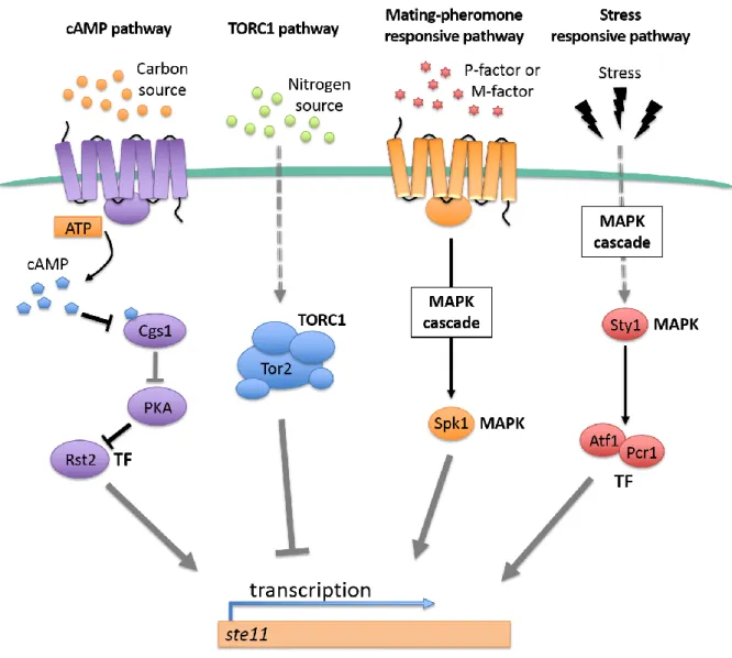
2.1 Signaling cascades
Fission yeast cells respond to environmental stimuli (e.g. nutrients, stresses, mating
pheromones) and activate specific signaling pathways to initiate the meiotic program. Four
transduction cascades, involved in the control of sexual differentiation, have been characterized
in fission yeast. Their activation relies on different input signals, including carbon and nitrogen
sources, mating pheromones and stress stimuli. These signaling pathways, which I describe
succinctly below, all converge to the activation of a master regulator of meiosis, the
2.1.1 The cAMP pathway
In an environment rich in carbon, which is favorable to vegetative growth, intracellular
ATP is converted into cAMP, a key messenger molecule that represses sexual differentiation
[9]. High concentrations of cAMP results in the inhibition of the Cgs1 protein and thereby
relieve the downregulation of the protein kinase PKA, which in turn phosphorylates and
inactivates the transcription factor Rst2 (Fig. 3) [10, 11]. As a consequence, expression of the
ste11+ gene is not induced, thereby maintaining cells in the mitotic cell cycle.
Instead, upon depletion of the carbon source in the environment, intracellular cAMP
levels rapidly decrease and prevent the activation of PKA. This allows Rst2 to escape inhibition
and to induce expression of ste11+, which promotes sexual differentiation (Fig. 3) [12].
2.1.2 The TORC1 pathway
Nitrogen starvation is a major determinant of sexual differentiation in fission yeast. The
signaling pathway that responds to nitrogen availability involves a TOR (Target Of
Rapamycin) family protein kinase (Fig. 3). Conserved in all eukaryotes, TOR kinases are key
regulators of cell growth that modulate gene transcription, protein synthesis and degradation
to adapt to environmental changes (reviewed in Loewith and Hall [13]).
S. pombe encodes two TOR homologues, tor1 and tor2, that incorporate in distinct
complexes, TORC2 and TORC1, respectively (reviewed in Loewith and Hall [13]).
Remarkably, mutations of tor2+, but not tor1+, mimic nitrogen starvation by initiating sexual
development regardless of the nutritional conditions [14, 15]. This is accompanied by an
increased expression of ste11+ and its target genes, indicating that Tor2 normally functions to
repress sexual differentiation [15, 16]. Interestingly, Tor2 forms a complex with Ste11,
although the biological relevance of this association remains unclear. Whether Tor2 directly
transcriptional regulators requires further investigation. More recently, Tor2 was shown to
phosphorylate the RNA-binding protein Mei2, another key meiosis inducer, to stimulate its
degradation by the proteasome and therefore prevent untimely entry into meiosis ([17]; see
next section). Thus, Tor2 is a central effector that functions to maintain robust vegetative
growth and inhibit sexual differentiation. Depletion of nitrogen causes the inactivation of Tor2,
thereby allowing initiation of the meiotic program.
2.1.3 The mating pheromone-responsive pathway
Upon meiotic conditions, fission yeast cells of either P or M mating type, produce and
secrete the mating pheromones (P-factor and M-factor) that will bind to their cognate receptor
on the cell membrane [3, 4]. This event allows cells of the opposing mating type to conjugate
and it triggers a MAPK (Mitogen Activating Protein Kinase) signaling cascade that leads to
the phosphorylation and activation of Ste11 by the Spk1 kinase (Fig. 3) [18-20]. Importantly,
Ste11 itself is required for the induction of mating-type specific genes, including the mating
pheromones and their receptors [21]. This reinforces the commitment of cells in sexual
differentiation and ensures that the process is irreversible.
2.1.4 The stress-responsive pathway
Other stimuli, such as heat shock and oxidative stress, activate a fourth signaling
pathway that controls ste11+ expression [22, 23]. The final effector of this pathway is the
MAPK Sty1, which accumulates in the nucleus and phosphorylates several targets, including
the CREB family transcription factors Atf1 and Pcr1 [24]. These latter are both required for
ste11+ expression (Fig. 3) [23, 25, 26], although it is unclear whether they directly control its
that phosphorylates the Ser2 within the Carboxy Terminal Domain (CTD) of the Rpb1 subunit
of RNAPII, a prerequisite for the correct expression of ste11+ [27, 28].
Figure 3. Scheme of the signaling pathways which control meiosis onset in S. pombe.
In fission yeast, four pathways mediate the transduction of external signals to the activation of ste11+ expression. (MAPK: MAP kinase; TF: Transcription factor). Figure adapted from Yamamoto [29].
2.2 The transcription factor Ste11
2.2.1 General properties
ste11+, which belongs to a group of genes whose mutations provoke meiotic deficiency
and complete sterility, encodes a major transcription factor required for sexual differentiation
[21, 30]. Supporting this notion, ectopic induction of ste11+ triggers uncontrolled mating and
sporulation, irrespective of nutritional conditions [21].
Ste11 is 486 amino acids in length and contains a HMG (high mobility group) domain
at its N-terminus that facilitates protein-DNA interactions [31, 32]. It is responsible for
inducing the expression of genes required for mating and meiosis, which feature one or several
10-base motifs in their 5’-upstream region (TTCTTTGTTY), called the TR boxes. The
selective binding of Ste11 to this cis-regulatory motif is essential for its proper function [21,
33]. Intriguingly, the ste11+ gene itself harbors a TR box [34], which likely serves to reinforce
its own expression and cell fate decision.
Ste11 controls the expression of approximately 80 genes, the majority of which is
induced in cells of both mating types [33]. These include the transcription factor Rep1,
involved in pre-meiotic S phase [35], Dhc1, a dynein protein necessary for nuclear movement
during meiotic prophase [36] and Tht1, required for karyogamy [37]. Other targets encode
positive (Mei2, meiRNA) and negative (Pat1) regulators of sexual differentiation, suggesting
the existence of complex feedback mechanisms [33] (see next section). Ste11 also promotes
the expression of mating type specific genes through its association with either Mat1-Pc or
Mat1-Mc, which encode cell type specific transcription factors that assist Ste11 in the binding
2.2.2 Regulatory mechanisms of ste11+ expression
2.2.2.1 Transcriptional control
Given the fundamental importance of Ste11 in sexual development, its expression must
be tightly regulated to avoid ectopic induction of meiotic genes during vegetative growth and,
conversely, to induce the meiotic program upon nutritional starvation. Interestingly, the ste11+
gene displays an unusually long 5’ UTR of more than 2 kb that comprises several binding sites for various transcription regulators. One of these is the zinc-finger protein Rst2, which
recognizes a stress response (STRE) element (i.e. 5’-CCCCTC-3’ motif) within the promoter
of ste11+ [34]. Rst2 relays the nutritional status to the expression of ste11+ via the cAMP
pathway [34, 39]. Ste11 itself promotes the transcription of its own gene by binding to a TR
box in its promoter [34]. The GATA protein Gaf1 instead represses ste11+ expression by
binding a canonical GATA motif 5’-CTATCT-3’ in the promoter, although the precise mechanism at play remains unknown [40].
Interestingly, the conserved coactivator SAGA (Spt-Ada-Gcn5-acetyltransferase)
complex also regulates the expression of ste11+ through the opposing roles of its Gcn5 and
Spt8 subunits [41]. It was suggested that the complex, which is recruited by Rst2, fine-tunes
the levels of Ste11 in response to cellular signals, although the mechanistic basis of this
regulation is still unclear.
As previously mentioned, phosphorylation on Ser2 (S2P) of RNAPII CTD responds to
cellular signaling and it is important for proper ste11+ expression [27, 28]. Generally, the
phosphorylation pattern of the RNAPII CTD has a key role in recruiting different factors
involved in transcriptional regulation [42]. Accordingly, it has been recently shown that the
presence of S2P, which depends on the activity of the Lsk1 kinase, activates ste11+ expression
histone deacetylases (HDACs) that negatively regulate transcription of ste11+ [43]. Moreover,
it has been shown that the transcriptional activation of ste11+ also depends on the
deubiquitynase Ubp8, which regulates the ubiquitination status of histone H2B to limit the
recruitment of Set1 to the ste11+ promoter [44]. Thus, phosphorylation of S2 on the RNAPII
CTD and removal of ubiquitin from H2B act in parallel to impair the Set1-dependent
recruitment of HDACs and in turn promote transcriptional induction of the ste11+ gene.
2.2.2.2 Post-transcriptional control
Beside its transcriptional regulation, ste11+ is also regulated at the protein level. Upon
nutritional starvation, the mating-pheromone pathway activates a signaling cascade and the
MAPK Spk1 binds and phosphorylates Ste11 on Thr305 and Thr317, triggering its activation [18, 19]. In mitotic cells, the Pat1 protein kinase represses the sexual differentiation program.
Indeed, Pat1 phosphorylates directly Ste11 on Thr173 and Ser218, inhibiting it in two ways: (1)
allowing binding of the 14-3-3 protein Rad24 to Ste11, hampering its nuclear accumulation
and therefore its role in transcription activation [45], and (2) promoting the ubiquitination and
subsequent degradation of the Ste11 protein [46].
Moreover, the Tor pathway, also affects Ste11 at the protein level. Indeed, Tor2 is able
to associate with Ste11 and the latter accumulates in the nucleus when the Tor2-containing
TORC1 complex is inactivated [14, 15]. It has been speculated that Tor2 might directly
phosphorylate Ste11, affecting its nucleocytoplasmic shuttling, although direct evidence is
2.3 The meiotic transcriptional program
Following the activation of Ste11, the meiotic program is initiated and leads to profound
modifications in gene expression profiles. Hundreds of genes are indeed activated in successive
transcriptional waves that correlate with the different phases of meiosis: starvation or
pheromone-induced genes, pre-meiotic S-phase and recombination (early meiosis), nuclear
divisions (middle meiosis) and spore formation (late meiosis) [47, 48]. Each group of genes is
associated with specific promoter motifs and contains defined transcription factors responsible
for the stepwise activation of the meiotic program (Fig. 4) [47, 48]. Below are summarized the
main regulators of meiotic transcription waves:
- Ste11 regulates the genes involved in the response to nutritional changes (e.g. Rep1)
(see above). It activates the pheromone communication system and the entry into
meiosis. Ste11 also promotes its own expression, therefore reinforcing the
commitment of the cell in sexual differentiation.
- The zinc finger transcription factor Rep1 induces genes required for early meiosis,
including those involved in premeiotic DNA synthesis and recombination [35, 49,
50]. However, about half of early genes do not depend on Rep1 for proper induction
[48], suggesting that additional transcription factors may also contribute to their
activation.
- The forkhead transcription factor Mei4 induces the transcription of middle genes
and is essential to complete meiosis I [51]. Mei4 binds to the FLEX motif present
in the promoter of its target genes via its forkhead domain and stimulates their
expression. Interestingly, the mei4+ gene also contains a FLEX sequence in its
- The bZIP transcription factors Atf21 and Atf31 mediate the transcription of more
than half of the late meiosis and sporulation genes in a sequence-dependent manner
[23, 47, 48, 53] and are essential to complete the meiotic program. Tough, Atf21
and Atf31 do not regulate all late genes and additional factors also contribute to
their expression, including the zinc finger protein Rsv2 [48].
The succession of the transcriptional waves ensures that the main biological events
occurring during meiosis are tightly coordinated over time. To reinforce the progression
through the meiotic cell cycle [48], transcription factors not only activate the expression of
transcription regulators involved in the next wave but also switch off the genes from the
previous wave. For example, Mei4 induces middle genes and represses some Rep1-dependent
early genes. Likewise, Rep1 negatively impacts expression of genes involved in response to
nutrient starvation and pheromone signaling (Fig. 4). However, the mechanisms at play are
likely indirect and additional work is needed to better understand the intricate coordination of
Figure 4. Transcriptional regulatory network regulating meiosis and sporulation.
The colors correspond to different phases of sexual differentiation. Arrows indicate induction and cross bars indicate repression. Mei4 stimulates its own production through a positive feedback loop, but no specific factor was shown to directly control its transcription. Rather, expression of mei4+ seems to be primarily regulated at the post-transcriptional level [54] (see next section). Figure adapted from Mata [48].
3 Regulatory mechanisms of sexual differentiation
Given the profound changes in gene expression, the meiotic process could be extremely
deleterious for the cell if executed in the inappropriate cell cycle context. Cells have therefore
evolved regulatory mechanisms that go beyond the transcriptional control of meiotic genes, to
avoid untimely entry into meiosis. These regulatory pathways act at the post-transcriptional
and post-translational levels to ensure a precise and proper switch from the mitotic to the
meiotic cell cycle. Here I will describe the molecular pathways implicated in the
mitosis-meiosis decision in fission yeast.
3.1 The Pat1-Mei2 system: a central regulator of meiosis
3.1.1 The Pat1 kinase inhibits entry into meiosis
Three decades ago, the pat1+ gene was isolated from a mutant undergoing meiosis even
in the presence of a nitrogen source and in a haploid state [55, 56]. These observations led to
propose that pat1 is a factor preventing sexual differentiation in nutrient-rich conditions.
The essential pat1+ gene encodes for a protein kinase for which two substrates have
been identified so far. The first one is Ste11 (see previous section), a key HMG domain
transcription factor involved in the expression of genes needed for mating and conjugation [35,
57, 58]. The second one is the RNA-binding protein Mei2, a major meiosis inducer [46, 59]
(see below).
In nutrient-rich conditions, Pat1 phosphorylates Ste11 at two sites (Thr173 and Ser218)
[60], allowing its association with the 14-3-3 family protein Rad24 which in turn inhibits Ste11
activity by preventing its nuclear accumulation [45]. These phosphorylation events have also
been suggested to reinforce the ubiquitin-dependent degradation of Ste11, although direct
residues, Ser438 and Thr527, both in vitro and in vivo [61]. Similarly to Ste11, phosphorylated
forms of Mei2 are bound by Rad24, which inhibits its function [46]. Further analyses showed
that phosphorylation of Mei2 is a prerequisite for its rapid turnover by the ubiquitin and
proteasome-dependent pathway involving the E2 ubiquitin-conjugating enzyme Ubc2 and
possibly the E3 ubiquitin ligase Ubr1 [46]. Remarkably, expression of a non-phosphorylatable
form of Mei2 (i.e. the Mei2-SATA mutant protein where serine and threonine residues targeted
by Pat1 are substituted by alanines) triggers ectopic entry into meiosis, similarly to the
inactivation of Pat1 [29, 61]. Based on these observations, it has been proposed that
phosphorylation of Ste11 and Mei2 by Pat1 during the mitotic cell cycle is essential to ensure
robust vegetative growth and avoid untimely entry into meiosis.
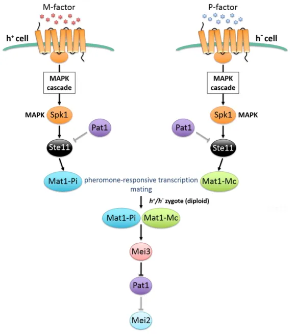
Upon nutritional starvation, the ste11+ gene is transcriptionally activated via the
downregulation of the cAMP-dependent kinase (PKA) [9, 21, 62]. In turn, Ste11 induces
several genes involved in mating and meiosis, including mating type genes, genes encoding
mating pheromones and their receptors, and mei2+ [21]. The binding of mating pheromones
to their receptors results in the activation of a MAPK cascade that promotes the induction of
mating genes required for the mating process and the formation of a zygote. In the zygote, h+ -specific Mat1-Pi and h--specific Mat1-Mc cooperate to induce the expression of the mei3+ gene, whose product inhibits Pat1 [60, 63]. The mei3+ gene indeed encodes a peptide that
functions as pseudosubstrate for Pat1, inhibiting its kinase activity [64]. Consistent with a
major role for Mei3 in the regulation of sexual differentiation, ectopic expression of mei3+ is
sufficient to trigger the switch from the mitotic to the meiotic cell cycle [65]. The association
of Mei3 results in the complete inactivation of Pat1, thereby allowing Ste11 and Mei2 to escape
Figure 5. The pheromone responsive pathway regulates the initiation of meiosis by inactivating the Pat1 kinase.
Binding of the P and M pheromones to their relative receptors triggers the activation of the MAPK cascade, which further activates ste11+. Ste11 then stimulates the transcription of pheromone-induced genes, which is a prerequisite for the initiation of mating and meiosis. The subsequent formation of a zygote provides the production of cell-type specific Mat1-Pi and Mat1-Mc within a single cell. Both these factors cooperate to stimulate transcription of mei3+. Mei3 inhibits the kinase Pat1, which cannot promote the inhibitory phosphorylation of Mei2. Figure adapted from Harigaya and Yamamoto [66].
3.1.2 The RNA-binding protein Mei2: a key meiosis inducer
3.1.2.1 General properties
The mei2+ gene was originally identified in a screen for fission yeast mutants that failed
to enter meiosis and/or to sporulate [67]. Genetic characterization then determined that mei2+
is required for efficient pre-meiotic DNA synthesis and the first meiotic division (meiosis I)
[30, 59, 68-70]. Transcription of mei2+ is induced by nutrient depletion and is directly
controlled by the transcription factor Ste11, which binds to its promoter region in a
sequence-dependent manner (e.g. TR boxes) [21, 70]. Later on, the mei2+ gene was found to encode an
RNA-binding protein harboring three RNA recognition motifs (RRM), two positioned in the
N-terminal half and one in the C-terminal half (Fig. 6) [59]. Importantly, the third RRM was
shown to be critical for entry into meiosis, highlighting a physiological role for Mei2
RNA-binding capacity in promoting sexual differentiation.
Figure 6. Schematic illustration of Mei2 protein.
Highlighted are the three RNA-recognition motifs (RRMs) and numbers indicate amino acid residues.
3.1.2.2 Evolutionary conservation
Analyses of amino acid sequences revealed the existence of Mei2-like proteins that
belong to an ancient family in eukaryotic organisms. The vast majority of the Mei2-like
proteins are found in plants - whereas none are present in mammals - and they are also key
regulators of differentiation and meiosis [71, 72].
Almost all these proteins are characterized by three identifiable RNA recognition motifs
identity in plants and fungi [71]. The strong conservation within the RRM3 suggests that all
Mei2-like proteins might use a common molecular mechanism. Supporting this notion,
expression of the Arabidopsis Mei2-like protein AML1 allows S. pombe cells that are defective
for meiosis to trigger sexual differentiation [73].
3.1.2.3 A key Mei2 cofactor: the long non-coding RNA meiRNA
The sme2+ gene (suppressor of mei2+) was isolated as a multicopy suppressor of a
thermosensitive mutant of mei2+ that does not undergo meiotic divisions [59]. Former analyses
showed that sme2+ encodes a non-coding RNA (named meiRNA) that is essential for meiosis
I [59, 74]. The RNA molecule is produced as two isoforms of different length, meiRNA-S, of
about 0.5 kb, and meiRNA-L, 1.0 kb longer, that are both polyadenylated [59, 74, 75]. meiRNA
directly interacts with Mei2 both in vivo and in vitro, preferentially via its 5’ region [59, 76],
and was shown to promote the transport of Mei2 from the cytoplasm to the nucleus, where the
latter exerts its function [74]. Because deletion of sme2+ prevents meiosis I but does not affect
pre-meiotic DNA synthesis, it was proposed that meiRNA acts as a key cofactor for Mei2 at a
specific stage in sexual differentiation [59].
The sme2+ gene also harbors multiple hexanucleotide motifs in its 3’ region that define
the so-called Determinant of Selective Removal (DSR). I will just mention here that these
sequences are critical for its degradation and function during vegetative growth (see next
section).
Recently, meiRNA was found to accumulate at its site of transcription, which is a
critical event to promote efficient pairing of homologous chromosomes during meiosis I [77].
It was proposed that meiRNA, retained at the sme2+ gene, favors the recognition between
homologous chromosomes, although this seems to occur independently of Mei2. Future work
3.1.2.4 Localization
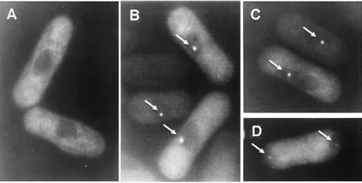
During vegetative growth, Mei2 is lowly expressed and resides essentially in the
cytoplasm (Fig. 7A). Upon nutritional starvation, Mei2 accumulates and a fraction of the
protein becomes apparent in the nucleus, forming a dot-like structure [61, 74]. The Mei2 dot,
which correlates with the ability to perform the first meiotic division, is observed in meiotic
prophase and persists until the end of the first division, segregating into the two daughter cells
(Fig. 7B-C-D) [74].
Further studies showed that meiRNA is a key constituent of the Mei2 dot, which
overlaps to the sme2+ locus on chromosome II [59, 74, 78]. It was proposed that meiRNA traps
Mei2 as a dot in the nucleus, thereby preventing its export to the cytoplasm [79]. Consistent
with this notion, sme2∆ cells are not able to form the dot, which prevents Mei2 nuclear
accumulation and function.
The exact nature and composition of the Mei2-meiRNA dot are far from being fully
understood but it is clear that it plays a key role in the initiation of the meiotic program.
Figure 7. Subcellular localization of the Mei2 protein during the mitotic and meiotic cell-cycle. Fluorescence of GFP-tagged Mei2 in fission yeast cells, which were grown vegetatively in rich medium (A) or depleted of nitrogen for 3,5 hs (B), 4 hs (C) and 6 hs (D). Specifically, cells in (A) are in mitotic growth, cells in (B) and (C) are undergoing meiotic prophase and cells in (D) are going through the first meiotic division. White arrows: Mei2 dot. Figure adapted from Yamashita [74].
3.1.2.5 Functions
Mei2 is crucial to initiate sexual differentiation in fission yeast and it has been
associated with several functions, although their mechanistic details are not fully understood
(Fig. 8).
First, Mei2 promotes pre-meiotic DNA synthesis [59]. Interestingly, the RNA-binding
capacity of Mei2, but not meiRNA, is required for this function. This led to the suggestion that
the binding of Mei2 to another RNA species may account for this phenotype. However, there
is currently no evidence for the existence of such a putative RNP complex.
Mei2 also contributes to telomere clustering, which is a prerequisite for the alignment
of homologous chromosomes during the horse-tail stage of meiotic prophase [18, 80].
However, and similarly to pre-meiotic DNA synthesis, whether this phenotype underlies a
direct function for Mei2 is still unclear but it is tempting to speculate that it is also linked to
the formation of an RNP complex with a specific RNA.
Finally, a key function for Mei2 in promoting meiosis I consists in sequestering, in
cooperation with meiRNA, a protein called Mmi1 (meiotic mRNA interception 1), which
targets meiotic transcripts produced during vegetative growth for degradation. This mechanism
is known as selective elimination of meiotic mRNAs, which is described in depth in the next
section. I will just mention here that upon nutritional starvation Mei2 and meiRNA trap Mmi1
in the dot-like structure, thereby inhibiting degradation of meiotic transcripts and favoring
Figure 8. Scheme of the known functions of Mei2.
To date, the role of Mei2 in both pre-meiotic DNA synthesis and telomere clustering is unclear; the most characterized function is the sequestration and inactivation of Mmi1 in an RNP complex containing Mei2 and the long non-coding RNA meiRNA.
3.1.2.6 Regulation of Mei2 expression and activity
As previously mentioned, the abundance and activity of Mei2 are regulated by the Pat1
kinase. Phosphorylated forms of Mei2 are indeed susceptible to ubiquitination and
proteasome-dependent degradation [46], and are also bound by the 14-3-3 protein Rad24, which prevents
the association of Mei2 with meiRNA [81]. These events prevent ectopic activation of Mei2
and therefore allow cells to sustain vegetative growth. Of note, the role of Pat1 in the inhibition
of the transcription factor Ste11 also contributes indirectly to lower mei2+ expression levels.
Recent work showed that Mei2 is also subjected to phosphorylation by the Tor2 kinase,
which, similarly to Pat1, accelerates its degradation through the ubiquitin-proteasome pathway
in mitotic cells [17]. Specifically, Tor2 phosphorylates nine residues within Mei2, which are
distinct form the ones targeted by Pat1 [17]. Therefore, two essential kinases independently
regulate the steady state levels and activity of Mei2.
The current model posits that, upon nutritional starvation, the expression of Tor2 is
accompanied by cell cycle arrest in G1 and mating, which in turn stimulates the transcription
of ste11+ and increases the levels of dephosphorylated Mei2 [28]. The subsequent inactivation
of Pat1 by Mei3 in zygotes contributes to the full activation of Mei2, leading to the initiation
of meiosis (Fig. 9).
Figure 9. Schematic illustration of the cascade of events leading to Mei2 activation and meiosis initiation.
A hypothetical model displaying the regulation of Mei2 in cells switching from mitotic growth to mating and meiosis. It is thought that, upon nitrogen starvation (-N), Mei2 is still phosphorylated by Pat1, but the inactivation of TORC1 reduces the Mei2 phosphorylation rate, allowing the mating process to occur. Whereas, it has been shown that inactivation of only Pat1 induces cells to enter meiosis without mating. It is likely that the stepwise inactivation of TORC1 and then Pat1 is crucial for the efficient progression of sexual differentiation. P: phosphorylation; Ub: ubiquitination. Figure adapted from Otsubo [17].
3.2 The selective elimination of meiotic transcripts during
vegetative growth
The selective elimination of meiotic mRNAs is a post-transcriptional regulatory
pathway that triggers degradation of cognate transcripts in a sequence-dependent manner.
Several factors recognize and remodel meiotic RNAs prior to target them for decay during the
mitotic cell cycle. In this section, I summarize the knowledge on the cis elements and the
trans-acting factors that play a key role in the regulation of this process that is essential for robust
vegetative growth and sexual differentiation.
3.2.1 cis-regulatory sequences control meiotic mRNA levels
Upon entry into meiosis, hundreds of genes are induced or up-regulated thanks to the
activity of specific transcription factors [47] (see above). However, in addition to
transcriptional regulation, another regulatory process distinguishes the mitotic and meiotic cell
cycles in fission yeast. Indeed, the expression of several meiotic transcripts is suppressed at the
post-transcriptional level during vegetative growth. This mechanism, known as the selective
elimination of meiotic mRNAs, was proposed to prevent untimely expression of the meiotic
program [54].
Originally, the Yamamoto lab made the surprising observation that some
meiosis-specific transcripts (e.g. mei4+, spo5+, ssm4+, mcp5+) do not accumulate in mitotic cells even
when artificially expressed from a constitutive promoter [54]. This suggested the existence of
cis-acting regulatory sequences that prevent the accumulation of these mRNAs in vegetative
cells. A screen for mutations that restore transcript levels allowed the identification of a region
Subsequent computational analysis showed that the DSR regions do not share extensive
homology between meiotic genes but contain several repeats of the hexanucleotide motif
U(U/C/G)AAAC (Fig. 10). Genetic experiments demonstrated that the UUAAAC and
UCAAAC sequences constitute the core motifs that exhibit functional DSR activity on their
own [75]. Insertion of multiple tandem repeats of UUAAAC at the 3’ end of a gfp+ reporter
gene expressed from the adh1+ promoter indeed precludes the accumulation of these
transcripts in mitotic cells [75].
Figure 10. Sequence and distribution of the hexanucleotide motif U(U/G/C)AAAC, in constitutively expressed meiotic genes.
Figure adapted from Chen [82].
Thus, a subset of meiotic transcripts that are constitutively expressed encode in cis the
elements that dictate their removal during the mitotic cell cycle, thereby preventing
inappropriate expression of meiosis-specific transcripts that might be deleterious for robust
3.2.2 Factors involved in the selective elimination of meiotic
mRNAs
Since the original discovery of the post-transcriptional regulation of meiosis-specific
transcripts, a number of factors have been shown to participate in this process. In this section,
I discuss the proteins involved in the recognition of meiotic transcripts as well as the effector
complexes that control and promote their elimination in mitotic cells.
3.2.2.1 The factors that recognize meiotic transcripts
3.2.2.1.1 The YTH-family RNA-binding protein Mmi1
Identification
A genetic screen was first designed to identify factors required for selective elimination
of DSR-containing meiotic transcripts [54]. Yeast wild type cells expressing a chimeric
transcript containing the ura4+ gene fused to the DSR region of mei4+ that prevents its own
accumulation, were mutagenized and selected for growth in the absence of uracile, i.e. in
conditions in which the reporter transcript is derepressed. Four of the isolated clones carried a
mutation in a gene encoding an RNA-binding protein of the YTH-family [83], named Mmi1
for meiotic mRNA interception factor 1 [54]. The fact that deletion of mmi1+ causes severe
growth and viability defects due to the ectopic expression of several meiotic genes highlights
the physiological relevance of the Mmi1/DSR system in mitotic cells.
Evolutionary conservation and structural properties
The YTH-family RNA-binding proteins are evolutionary conserved throughout the
eukaryotic kingdom. Originally identified by comparing protein sequences with the human
splicing factor YT521-B [83], the YTH domain (YT521-B Homology) defines a specific class
(Fig. 11). The fission yeast Schizosaccharomyces pombe and the budding yeast Saccharomyces
cerevisiae each encode one member of this family, Mmi1 and Mrb1 (also known as Pho92)
respectively, while mammals contain five YTH proteins, including the YTH Domain Family
(YTHDF) proteins 1, 2 and 3, and the YTH Domain Containing (YTHDC) proteins 1 and 2.
Figure 11. Schematic representation of the YTH-family RNA-binding proteins Mmi1 in S. pombe and YT521 in humans.
Numbers indicate amino acid residues. The black boxes correspond to the YTH domains in both proteins. Note that the human domain displays 24% identity and 45% similarity to Mmi1. Figure adapted from Harigaya [54] and Stowell [84].
The precise mode by which YTH proteins bind to RNA was discovered only very
recently. A study of the human YTHDF2 protein showed that specific RNA modifications, and
more precisely N6-methyladenosine (m6A) residues, confer selectivity for RNA binding both
in vitro and in vivo [85-87]. Crystal structures of the YTH domains of YTHDF2 and YTHDC1
bound to a methylated RNA revealed the existence of a conserved hydrophobic binding pocket
that can specifically accommodate m6A [87-89]. Thus, proteins of the YTH family directly “read” RNA methylation patterns, which in turn influence the fate of transcripts, thereby providing an additional layer in the control of gene expression. Landmark studies in metazoans
demonstrated that YTH proteins regulate many RNA-related processes, including splicing,
degradation, translation and transcriptional silencing [86, 90-92].
Similarly to their metazoan counterparts, the budding and fission yeast YTH proteins
(Fig. 12). Unexpectedly, however, structural and biochemical analyses demonstrated that m6A decreases the affinity of Mmi1 for RNA [92]. Contrary to Mrb1 and mammalian YTH proteins,
the arrangement of residues in the Mmi1 aromatic cage sterically hinders the recognition of
m6A (Fig. 12). Consistent with this notion, mutations of the residues delineating the cage do
not impact Mmi1 RNA-binding activity [92]. Another striking difference is the presence of
negatively charged residues surrounding the m6A-binding pocket, which creates a repulsive environment for RNA binding.
Figure 12. Comparison of the aromatic cages within YTH domains.
Above is the electrostatic potential of the surface, showing the positively charged residues (blue) in the m6A RNA-binding interfaces of YTHDC1, YTHDF2 and Mrb1 and the negatively charged residues (red) near the aromatic cage of Mmi1. Below is the enlarged view of the respective aromatic cages. Figure adapted from Wang [92].
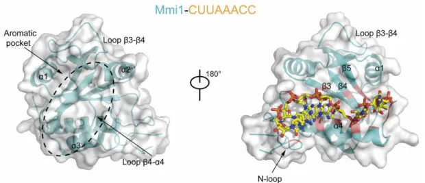
Mmi1 uses instead a long groove located at the opposite side of the m6A-binding pocket to associate with RNA, thereby defining a unique binding mode for YTH domains (Fig. 13)
enzymes homologous to METTL3 and METTL14 that catalyze methylation of adenosines in
metazoans (reviewed in Roundtree [94] and Zhao [95]). This suggests that m6A modification has been lost in S. pombe, although it encodes a putative RNA methyltransfease, ime4+, the
homologue of which is responsible for adenosine methylation in budding yeast.
Figure 13. Structural overview of the Mmi1 protein in complex with a DSR-containing CUUAAAC RNA.
The Mmi1 YTH domain is colored in cyan and the RNA-binding regions are in red. The structural elements and aromatic pocket implicated in RNA-binding are highlighted. Figure adapted from Wang [92].
Beyond their RNA-binding domains, YTH proteins do not share significant homology.
It has been proposed that the N-terminal regions are involved in protein-protein interactions.
Consistent with this notion, Mmi1 contains a serine rich low-complexity region at the
Targets
Former micro- and tiling-array analyses allowed the identification of a set of meiotic
genes whose expression is increased upon mutation of mmi1+ during vegetative growth [82,
96]. A common feature in these RNA targets is the presence of multiple DSR motifs (i.e.
repeats of the UNAAAC hexanucleotide sequence), generally clustered in the 3’ half of the genes. However, and because some Mmi1 targets include transcripts encoding meiotic
transcription factors (e.g. rep1+, mei4+), it has been difficult to discriminate between direct
and indirect effects on the expression of putative targets.
Recent genome-wide identification of direct Mmi1 RNA targets by CRAC (UV
Crosslinking and analysis of cDNA by high-throughput sequencing) revealed a large repertoire
of transcripts produced by all three RNA polymerases (PolI, II, III), including hundreds of
protein-coding and ncRNAs transcribed by PolII, many PolIII transcripts and a PolI-dependent
ribosomal RNA precursor [97]. Interestingly, many transcripts associated to Mmi1 display
fewer UNAAAC motifs, indicating flexibility in target recognition [97-99]. However, whether
this can impact Mmi1 function is not understood.
One peculiar target of Mmi1 is the lncRNA meiRNA, encoded by the sme2+ locus.
This non-coding RNA contains a DSR region with 13 copies of the core motif, especially in
the 3’ part of the transcript to which Mmi1 binds [75]. Its expression is increased in meiotic cells [59], like DSR-containing meiotic transcripts, and sme2∆ cells are not able to inhibit
Mmi1 via the Mei2 dot [54]. It has been proposed that meiRNA serves as a decoy to lure Mmi1,
favoring its inhibition upon meiotic conditions (see below).
Functions
As described above, Mmi1 recognizes, via its YTH domain, a cis-acting region within
meiotic transcripts (i.e. DSR motifs) [54, 75], which is essential to prevent their ectopic
I will simply mention here that one of the key functions of Mmi1 is the activation of
RNA degradation. Over the last few years, many studies have pointed a key role for the nuclear
exosome in the degradation of Mmi1 targets during vegetative growth (see below). The current
model posits that Mmi1 is co-transcriptionally loaded onto DSR-containing mRNAs and
recruits several factors that contribute to their rapid decay (Fig. 14).
Figure 14. Mmi1 promotes meiotic mRNA degradation by the nuclear exosome.
Mmi1 recognizes the DSR motif within meiotic mRNAs via its YTH domain (left) and then cooperates with other factors to promote RNA degradation by the exosome (right).
Previous work showed that Mmi1 also regulates the splicing of several
intron-containing meiotic mRNAs [82, 100]. Mutants of mmi1+ lead to the accumulation of spliced
rec8+ and crs1+ transcripts, suggesting that Mmi1 may be involved in intron retention. The
use of artificial constructs indicated that Mmi1 also affects intron splicing without impacting
transcript stability [82]. Recently, Kilchert and colleagues showed that mRNAs containing
introns with an Mmi1 binding site are rapidly targeted for degradation in conditions of
inefficient splicing [97]. Conversely, fast splicing events prevent the recruitment of Mmi1 and
degradation factors, thereby allowing accumulation of mRNAs [97].
Another function of Mmi1 lies in the control of transcription termination. It has been
shown that deletion of mmi1+ triggers the accumulation of RNAPII downstream of canonical
However, the mechanism by which Mmi1 may affect transcription termination is not fully
understood. One possibility is that Mmi1 co-transcriptionnally loads onto meiotic transcripts
and promotes the recruitment of additional factors that promote the dismantlment of elongation
complexes.
Localization and regulation
In mitotic cells, Mmi1 localizes to one or several scattered nuclear foci (Fig. 15A) [54,
76]. These nuclear bodies have been proposed to reflect RNA processing/decay centers,
whereby Mmi1 and several of its protein partners assemble onto newly synthetized meiotic
transcripts to mediate their elimination [76]. Microscopy experiments indicated that the dots
do not overlap with the transcription sites of canonical Mmi1 target genes, such as mei4+,
although a transient association of these bodies with meiotic gene loci cannot be formally
excluded [101]. One of the dots, however, was shown to precisely map to the sme2+ locus,
which encodes the DSR-containing lncRNA meiRNA that has been suggested to function as a
decoy to lure Mmi1 [76]. Removal of the Mmi1-binding sites within meiRNA (i.e. DSR motifs)
abolishes formation of the dot, suggesting that a pool of Mmi1 is associated with meiRNA at
the sme2+ locus in vegetative cells [76].
Upon nutritional starvation, the multiple Mmi1 foci converge into a single spot that
persists until meiotic prophase I before dispersing after metaphase I [54]. RNA-FISH and
immunofluorescence experiments revealed that this unique Mmi1 dot, observed upon meiosis
onset, overlaps with the Mei2 dot (Fig. 15B) [76]. Crucially, deletion of either mei2+ or sme2+
prevents the convergence of Mmi1 dots (Fig. 15C) and results in major sporulation defects,
highlighting the biological relevance of the Mei2 dot in sexual differentiation [54]. It has been
suggested that, upon meiosis, Mei2 anchors Mmi1 in a dot-like structure, to inhibit its function
However, and despite recent progresses, the exact composition and function of the
Mmi1-containing nuclear foci remain elusive.
Figure 15. The Mei2 dot sequesters Mmi1 allowing meiotic mRNAs to be translated
(A) Mitotically growing cells expressing GFP-tagged Mmi1 are shown; nuclei are counterstained with Hoechst 33342. (B) Cells during meiotic prophase, expressing CFP-Mmi1 and Mei2-YFP. (C) Meiotic cells, expressing GFP-Mmi1, show scattered foci when defective for mei2+ or sme2+. Figures in A-B-C adapted from Harigaya [54]. (D) Model showing the sequestration of Mmi1 by Mei2 and the non-coding meiRNA (left), which allows meiotic mRNAs to be free from degradation and therefore translated (right).
3.2.2.1.2 3’-end processing and polyadenylation factors
The Mmi1-dependent meiotic mRNA elimination process was initially found to require
components of the 3’-end processing and polyadenylation machinery. Yeast two-hybrid,
biochemical and genetic analyses uncovered several potential Mmi1 protein partners, including
Rna15, a subunit of the cleavage factor CF1A involved in mRNA processing, the canonical
mRNA poly(A) polymerase Pla1, the poly(A) binding protein Pab2 and the Rrp6 subunit of
the nuclear exosome [54, 102]. Functional analyses revealed that these factors cooperate with
Mmi1 to target DSR-containing meiotic mRNA for degradation [100, 102, 103].
Mechanistically, it was proposed that Mmi1 first binds to the transcript and, by virtue of its
affinity for Rna15 and Pla1, promotes its hyperadenylation. The resulting poly(A) tail is
subsequently bound by Pab2, which in turn recruits the exosome for rapid decay.
Recently, the 5’ to 3’ exoribonuclease Dhp1, the homologue of Rat1/XRN2, was also shown to prevent meiotic mRNAs expression in vegetative growth, possibly through a
mechanism that couples premature transcription termination to RNA decay [104]. However,
the requirement for the catalytic activity of Dhp1 is still unclear and it has been suggested that
the protein may serve as a scaffold for the recruitment of RNA elimination factors, including
Mmi1 and MTREC (see below) [104].
Interestingly, all of the above-mentioned factors form patchy subnuclear structures that
overlap with Mmi1 foci in mitotic cells. This further suggests that the polyadenylation control
3.2.2.1.3 The MTREC complex
A major effector in the selective elimination of meiotic mRNAs that is tightly
associated to Mmi1 is the multisubunit MTREC complex. Core components of this complex
include the zinc finger-containing protein Red1 (RNA elimination defective 1) and the RNA
helicase Mtl1 [101, 105].
Red1 was initially found as a factor localizing to nuclear foci in mitotic cells [106].
Functional analysis revealed that Red1 encodes a 712 amino acids protein harboring a
zinc-finger domain (often found in in DNA/RNA-binding protein) that is required for robust
vegetative growth and efficient mating/sporulation. Red1 was shown to associate with Mmi1
in mitotic cells and to colocalize with Pla1, Pab2 and the exosome in Mmi1 nuclear foci.
Expression profiling of red1∆ cells revealed that many meiotic genes are up-regulated in
vegetative cells which strongly overlap Mmi1 RNA targets. Importantly, point mutants in the
zinc finger of Red1 also display defects is meiotic mRNAs degradation, strongly suggesting
that RNA binding is important for function [106].
Subsequent affinity purifications provided for the first time a global picture of the
composition of MTREC. These include, together with Red1, the essential Mtr4-like RNA
helicase Mtl1, the Pro/Ser rich factor Iss10/Pir1, the zinc finger-containing protein Red5, the
RRM-containing protein Rmn1, and Ars2, the homologue of which is known to promote the
recruitment of the exosome and the cap-binding subunits Cbc1/Cbc2 in humans [101, 105, 107,
108]. Immunofluorescence experiments indicated that most MTREC subunits localize to
nuclear foci, overlapping those formed by Mmi1 [101, 105]. Deletion or mutation of the
MTREC subunits generally lead to a strong accumulation of meiotic transcripts, although some
components appear to have a weaker role [101, 105, 108]. It was suggested that submodules of
MTREC target distinct sets of RNA substrates to the nuclear exosome, including unspliced
subunits associate independently with the splicing factors Ctr1 and Nrl1, and the nuclear
exosome (e.g. Rrp6).
The precise mechanism by which MTREC promotes meiotic mRNA degradation
remains elusive. It has been proposed that MTREC may physically bridge RNA-bound Mmi1
to the nuclear exosome. Supporting this, Iss10, previously found in a genetic screen designed
to identify factors involved in the Mmi1/DSR system [109], is essential for the interaction
between Mmi1 and Red1. However, iss10∆ cells show only a moderate increase in meiotic
mRNAs steady state levels [101, 109], suggesting that the Mmi1-Red1 association is not
critical for meiotic mRNA degradation. It was proposed that MTREC also directly binds to
transcripts, which is supported by the presence of several RNA-binding motifs in its subunits.
A general model has emerged is which MTREC submodules target specific classes of
RNA substrates (e.g. DSR-containing meiotic mRNAs, unspliced pre-mRNA, CUTs, etc.) and
deliver them to a larger machinery that comprises MTREC itself, the poly(A) polymerase Pla1
and the nuclear exosome (Fig. 16) [108]. The reported physical interactions between Red1,
Mtl1, Pla1 and Rrp6 strongly support this idea. Polyadenylation by Pla1 is believed to provide
single-stranded A tails, thereby facilitating exonucleolytic attack by Rrp6. Whether the
catalytic activity of Mtl1 is required to unwind putative RNA secondary structures to further
assist the exosome remains to be established. This would be reminiscent of the function of the
Mtr4 subunit of the TRAMP complex in budding yeast, which facilitates exosome-dependent
degradation by remodeling RNA substrates. Interestingly, in human cells, there is only one
Mtr4-like protein that associates with two distinct complexes, one of which is similar to the
TRAMP complex of budding and fission yeast, and a second - known as CBCN complex -