The in vivo characterization of the DNA repair gene apn-1 in the model organism Caenorhabditis elegans
par Chadi Zakaria
Biologie Moléculaire Faculté de Médecine
Mémoire présenté à la Faculté de Médecine en vue de l’obtention du grade de M.Sc.
en Biologie Moléculaire
Aout 2009
© Chadi Zakaria, 2009
Ce mémoire intitulé :
The in vivo characterization of the DNA repair gene apn-1 in the model organism Caenorhabditis elegans
présenté par :
Chadi Zakaria
a été évalué par un jury composé des personnes suivantes : président-rapporteur : El Bachir Affar
directeur de recherche : Dindial Ramotar membre du jury : Darel Hunting
Résumé
Les sites apuriniques/apyrimidinique (AP) représentent une forme de dommage à l’ADN hautement mutagène et ce type de dommage peut survenir spontanément ou être induit par une variété d’agents. Afin de préserver la stabilité génomique, deux familles d’endonucléases de type AP, endo-IV et exo-III, sont nécessaires pour contrecarrer les effets mutagènes des sites AP. Malgré
l’identification de membres des deux familles dans plusieurs organismes
unicellulaire tels que E.coli et S. cerevisiae, aucun membre de la famille endo-IV n’a été identifié chez les organismes multicellulaires à l’exception de C. elegans et de C. briggsae. Nous avons donc décidé d’investiguer l’importance biologique de APN-1 chez C. elegans par l’utilisation d’une approche de knockdown du gène. Dans notre étude, nous avons montré que le knockdown du gène apn-1 chez C. elegans, en utilisant des ARN d’interférence (ARNi), cause une accumulation de mutations spontanées et induites par des drogues résultant en un délai de l’éclosion des œufs ainsi que par une diminution de la survie et de la longévité des vers adultes. De plus, nous avons montré que cette accumulation de mutations mène à un délai dans la progression du cycle cellulaire durant l’embryogénèse,
représentant possiblement une explication du délai dans l’éclosion des œufs. Nous avons montré qu’il y avait une augmentation du niveau de mutations dans la gorge des vers, sans toutefois pouvoir confirmer la distribution de APN-1 qui possède une étiquette GFP. Les animaux transgéniques APN-1-GFP n’exprimaient pas suffisamment de la protéine de fusion pour permettre une visualisation à l’aide d’un microscope à fluorescence, mais la protéine a été détectée par
immunobuvardage de type western. Les animaux transgéniques APN-1-GFP étaient instables et avaient des phénotypes concordants avec les défauts
génétiques. En conclusion, il semble que C. elegans aie évolué afin de retenir un niveau de base de APN-1 jouant ainsi un rôle versatile afin de maintenir l’intégrité génétique d’autant plus que cet organisme semble manquer plusieurs enzymes de la voie de réparation par excision de base.
Mots clés : C. elegans, Endo IV, réparation par excision de base, site AP,
endonucléase de type AP, stress oxidatif, agents causant des dommages à l’ADN, réparation de l’ADN.
Abstract
Apurinic/apyrimidinic (AP) sites are a form of highly mutagenic DNA damage that arise either spontaneously or by a variety of DNA damaging agents. To preserve genomic stability two AP endonuclease families, endo-IV and exo-III, evolved to counteract the mutagenic effect of AP sites. While members of both families were identified in multiple unicellular organisms, notably E. coli and S. cerevisiae, no members of the endo-IV family were identified in multicellular ones, with the exception of C. elegans and its close relatives, particularly C. briggsae. We set out to investigate the biological importance of APN-1 in C. elegans using gene knockdown approach. In our study, we showed that the knockdown of C. elegans apn-1 gene, using RNAi causes the accumulation of spontaneous and drug induced mutations, resulting in a delay in egg hatching, decreased survival and longevity. Furthermore, we have showed that the accumulated mutations lead to delays in cell cycle progression during early embryogenesis, thus providing a possible explanation for the observed delay in hatching. Although we showed increased mutations in the gut of the worm, we were unable to confirm APN-1 distribution tagged with GFP. The transgenic APN-1-GFP animal did not express enough of this fusion protein to be visualized by fluorescent microscopy, although it was detected by Western blot analysis. The transgenic animals over-expressing APN-1-GFP were unstable and showed phenotypes consistent with genetic defects. In conclusion, it would seem that C. elegans has evolved to retain a balanced level of APN-1, which plays a versatile role in maintaining genetic integrity, since this organism lacks a full complement of the enzymes in the base-excision repair pathway.
Key words: C. elegans, Endo IV, base excision repair, AP site, AP endonuclease, oxidative stress, DNA damaging agents, DNA repair
Table of contents
Resumé..……… i
Abstract……… iii
Table of Contents………. iv
List of Figures………... x
List of Abbreviations……… xii
Dedication……… xv
Acknowledgment……….. xvi
Chapter 1 – Introduction……… 1
1. General Review ……….. 1
2. DNA damage………... 2
2.1. Sources of DNA damage……… 2
2.2. Types of DNA damage………... 3
2.2.1.
Bulky lesions and inter/intra-strand crosslinks…… 32.2.2.
Modification of bases……….. 42.2.2.1. Deamination………. 4
2.2.2.2. Base oxidation……….. 5
2.2.2.3. Base alkylation………. 6
2.2.3.
Spontaneous base loss………. 62.2.4.
Single strand breaks……… 62.2.5.
DNA double strand breaks………. 72.2.6.
Result of unrepaired damage………. 83. DNA repair – Multiple pathways with one aim………. 8
3.1. Double strand break repair………... 9
3.3. Nucleotide excision repair (NER)……….. 10
3.4. Direct reversal……….. 11
3.5. Base excision repair pathway………. 12
4. AP endonucleases/3’-diesterases……… 13
4.1. AP endonuclease/3’-diesterase enzymes in E. coli………… 14
4.1.1.
Characteristics of E. coli exonuclease III…………... 154.1.2.
Characteristics of E. coli endonuclease IV…………. 164.1.3.
Importance of the AP endonuclease/3’-diesterase enzymes in E. coli– mutational analysis………. 174.2. AP endonuclease/3’-diesterase enzymes in S. cerevisiae……….. 18
4.2.1.
Characteristics of S. cerevisiae Apn1……….. 184.2.2.
Characteristics of S. cerevisiae Apn2……….. 204.2.3.
Importance of AP endonuclease/3’-diesterase enzymes in S. cerevisiae – mutational analysis……….. 214.3. AP endonuclease/3’-diesterase enzymes in humans………. 21
4.3.1.
General characteristics of human APE1……… 214.3.2.
General characteristics of human APE2……… 234.3.3.
The importance of the BER and the AP endonuclease enzymes in mammalian cells………… 245. Caenorhabditis elegan……….. 24
5.1. General information……… 24
5.2. Life cycle……….. 26
5.3. RNA interference in C. elegans……….. 27
5.4.1.
Known characteristics of C. elegansapn-1 and Ceexo-III……… 29
6. Research project and summary of results………. 30
Chapter 2 – Materials and methods………... 33
1.
C. elegans Strains and maintenance ofnematode cultures……… 33
2.
RNAi knockdown by feeding system…………...………... 34 2.1. L4440-apn-1 Plasmid preparation (prepared byDr. Andrea Shatilla)……… 35 2.2. Preparation of knockdown plates……….. 36
3.
Verification of the knockdown efficiencyusing RT-PCR……..……… 37 3.1. RNA extraction……… 37 3.2. Genomic DNA extraction……… 38 3.3. Reverse-transcriptase (RT)-PCR and
q-RT-PCR reactions………... 39
4.
Construction of the APN-1-GFP expressing plasmid,microinjection and generation of transgenic worms……… 40 4.1. Preparation of the pPD95.70-APN-1-GFP
plasmid (Prepared by Dr. Xiaoming Yang)……… 41 4.2. Generation of transgenic nematodes
(prepared by Dr. Jean-claude Labbe)………. 41
5.
Visualization of APN-1-GFP levels……….426.
Assessment of knockdown efficiency: Western6.1. Preparation of total protein extract………... 43
6.2. Western blot analysis……….. 43
7.
Detection of the AP endonuclease activity………. 447.1. Preparation of oligonucleotide substrates………. 44
7.2. Enzyme assays………. 45
8.
Assessment of the mutation frequency through the detection of β-galactosidase Activity………... 459.
Egg-hatching assay: Test for sensitivity to DNA damaging agents………. 479.1. Worm culture synchronization……….. 47
9.2. Drug plate preparation………... 47
9.3. Worm transfer and incubations……… 48
10.
Longevity assay……… 4811.
Cell cycle length assessment……… 49Chapter 3 – Results……….. 50
1. Transgenic APN-1-GFP shows increased levels of AP endonuclease activity ……….. 50
2. Overexpression of the long N-terminal domain of APN-1 is toxic……….. 53
3. Confirmation of knockdown by Q-RT PCR……….. 56
4. Confirmation of knockdown by Western Blot……….. 57
5. apn-1 knockdown increases the frequency of spontaneous frameshift mutations……….... 58
6. Knockdown of apn-1 causes the accumulation of unhatched eggs………. 61
7. RNAi-apn-1 knockdown delays the development of the parent N2 strain and sensitizes worms to
different DNA damaging agents………. 62 8. RNAi-apn-1 knockdown delays cell cycle progression…………. 64 9. Knockdown of apn-1 decreases lifespan in the
presence of agents that cause AP sites……… 66
Chapter 4 – Discussion………. 69
1. Genome-wide screen for the identification of genes involved in the maintenance of DNA
integrity failed to identify apn-1………... 70 2. APN-1 function seems essential
for C. elegans embryogenesis………. 71 3. Possible redundancy between APN-1 and EXO-3……… 72 4. APN-1 is essential for the repair of H2O2
induced damage ...………73 5. Overexpression of apn-1 is toxic………. 73 6. The C. elegans RNAi hypersensitive rrf-3 strain
behave similarly to the parental N2 strain……… 76 7. Future plans……….. 77
7.1.
Mutational spectrum resulting from apn-1knockdown……….. 77
7.2.
Study of the contribution of the two familiesof AP endonuclease/3’-diesterase in C. elegans ………….. 78
7.3.
In depth Cell cycle arrest studies following8. Conclusion……… 79
List of figures :
Figure 1. The APN-1-GFP transgenic line shows no detectable
GFP as compared to the parental N2 strain………..………...51
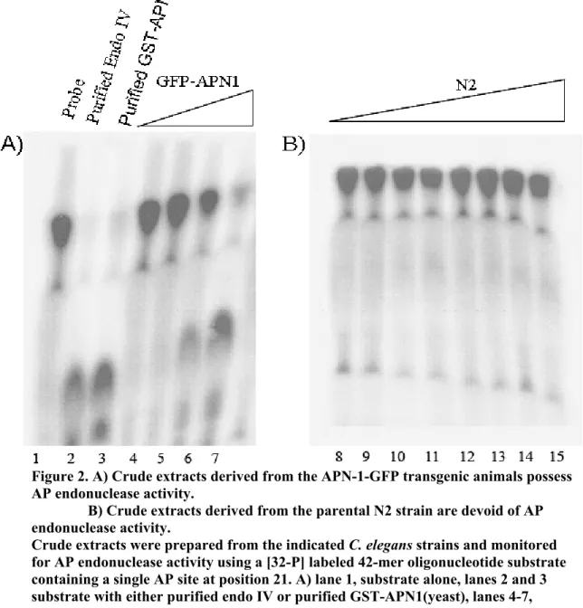
Figure 2. Crude extracts derived from the APN-1-GFP transgenic
animals possess AP endonuclease activity………52
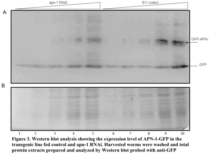
Figure 3. Western blot analysis showing the expression level of APN-1-GFP in the transgenic line fed control and
apn-1 RNAi..………...53 Figure 4. Alignment of the Endo IV family members of C. elegans,
C. briggsae and S. cerevisiae………....55 Figure 5. RNase free agarose gel revealing the quality of the
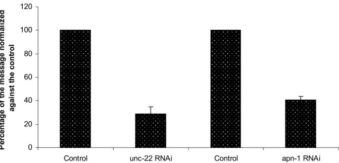
extracted RNA samples...……….56 Figure 6. Percentage of the remaining unc-22 and apn-1
mRNA following gene specific RNAi.……….57 Figure 7. apn-1-RNAi worms exhibit elevated mutations as
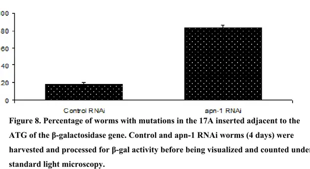
assessed by the β-galactosidase reporter gene……..……….59 Figure 8. Percentage of worms with mutations in the 17A inserted
adjacent to the ATG of the β-galactosidase gene…..………….…………..59 Figure 9. apn-1 RNAi worms exhibit elevated mutations
as assessed by the β-gal reporter.………..60 Figure 10. The X-Gal staining is mainly localized to the gut
and central nervous system of the worms….………...60 Figure 11. The knockdown of the apn-1 gene causes the
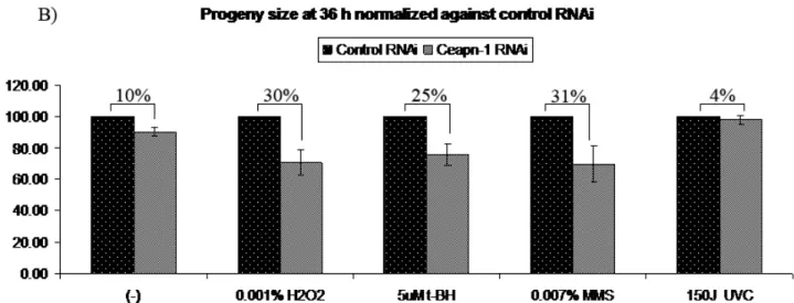
accumulation of unhatched eggs………….………..61 Figure 12. Spontaneous and drug-induced levels of mutations
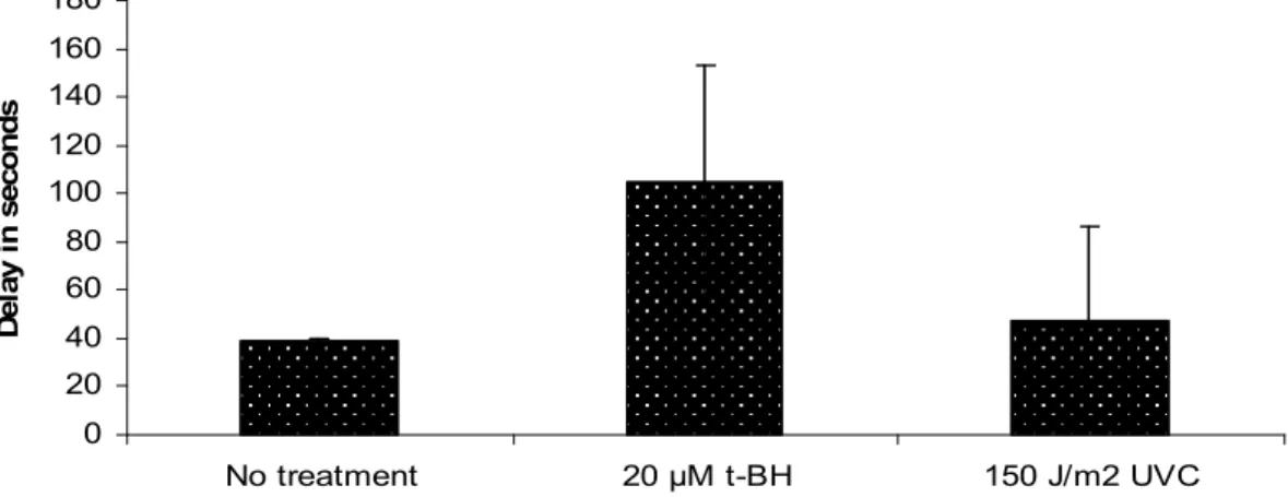
affect progeny size in apn-1 RNAi animals…….………...64 Figure 13. Cycle delay of embryonic P1 cells following apn-1 knockdown……...65
Figure 14. apn-1 RNAi worms display a shortened life-span when exposed to t-BH and MMS, but not to
List of abreviations
A Adenine
AID Activation-Induced cytidine Deaminase AP site apurinic-apyrimidinic site
APOBEC1 Apolipoprotein B mRNA Editing Catalytic subunit 1 Asn Asparagine
Asp Aspartic Acid
AT Ataxia Telangiectasia
ATP Adenosine-5'-triphosphate BER Base Excision Repair
BLM Bleomycin BRCA1 breast cancer 1, early onset C Cytosine C. elegans Caenorhabditis elegans
cDNA Complementary DNA
CIA Chloroform Isoamyl Alcohol
CIP Calf Intestinal Phosphatases
CS Cockayne Syndrome
Cys Cysteine
DEPC Diethyl pyrocarbonate
DNA Deoxyribonucleic acid
dRP Deoxyribosephosphate DSB Double Strand Break
dsDNA Double Stranded DNA dsRNA Double Stranded RNA E. coli Escherichia coli
EDTA ethylenediaminetetraacetic acid Endo IV Endonuclease IV
EtBr Ethidium Bromide
EXO III Exonuclease III
G Guanine GFP Green Fluorescent Protein
GG-NER Global Genomic nucleotide excision repair
Glu Glutamic Acid
H2O2 Hydrogen Peroxide
His Histidine
HR Homologous Recombination
kb Kilo bases
kDa Kilo Dalton
LB Luria Broth
LP Long Patch
MCS Multiple Cloning Site Mg2+ Magnesium
MGMT Methyl Guanine Methyl Transferase MMS Methyl Methane Sulfonate
mRNA Messenger RNA
N Nitrogen N3-meA N3-methyladenine N7-meG N7-methylguanine
NaOAc Sodium Acetate
NER Nucleotide Excision Repair
NGM Nematode Growth Media
NHEJ Non-Homologous End Joining NIR Nucleotide Incision Repair NLS Nuclear Localization Signal Ogg1 8-oxoguanine DNA glycosylase OH Hydroxyl
PCNA Proliferating Cell Nuclear Antigen
PMSF phenylmethanesulphonylfluoride
q-RT-PCR Quantitative Reverse transcription Polymerase Chain Reaction
RNA Ribonucleic Acid
RNAi RNA Interference
ROS Reactive Oxygen Species
rpm Revolutions Per Minute
RT-PCR Reverse Transcription Polymerase Chain Reaction S. cerevisiae Saccharomyces cerevisiae
SAM S-adenosylmethionine
SDS Sodium Dodecyl Sulfate
SP Short Patch
SSB Single Strand Break ssDNA Single Strand DNA SV40 Simian virus 40
T Thymine t-BH tert-butyl hydroperoxide TC-NER Transcription Coupled Repair
UV UltraViolet V(D)J Variable Diversity Joining
XP Xeroderma Pigmentosum
β-gal β-galactosidase
For mum and dad, who sacrificed their own life so I would have a good one. For those who never stopped offering me their unconditional love throughout the years, and their relentless support throughout the course of my studies and till
Acknowledgments
From the formative stages of this thesis, to the final draft, I owe an immense debt of gratitude to my supervisor, Dr. Dindial Ramotar. His sound advice and careful guidance were invaluable as I attempted to discover the scientific world which, if it wasn’t for him, wouldn’t have been a real pleasure over the past two years. I would also like to thank Dr Jean-Claude Labbé for his valuable help, guidance and major intellectual and experimental contributions to the project. He was always there when an emergent scientific advice was needed.
I cannot include an acknowledgment section without mentioning the positive and optimistic spirit of our lab, Dr Xiao-Ming Yang. It is not just for technical support I wanted to thank him, but for believing in me and making me believe in myself by saying: “Don’t worry, it works”, “you’re gonna be a big scientist!” in times when I was about to give up. I cannot forget to mention the kind and caring Rad Ramotar, our lab second technician, who gives without asking anything in return, as any mother would do to her children. We are lucky to have her.
Besides having a great scientific environment in Dr Ramotar’s lab, I consider myself very lucky to be surrounded by very kind, social and amazingly helpful student and post-doc friends: Dr Mustafa A., Dr Sonish A., Dr Karima A., Jeremy P., Nathalie J., Emily.A, Siham B. and my other colleagues in HMR research center. I would like to address a special gratitude to Dr Jim Daley and Dr Jasmine Lefebvre for reviewing this thesis and providing me with useful comments.
This acknowledgement would be incomplete without a mention of the support given me by my girlfriend, Emily Ayoub. She was my own "soul out of my soul," who kept my spirit up when the muses failed me. Without her lifting me up when this thesis seemed interminable, I doubt it would ever have been completed. Last but not least, I would like to thank my family especially my brother for being always there for me. I am also thankful for the constant encouragement and support of Uncle Michel and Aunt Norma Zakaria. They were always my role model in life as ambitious and dedicated people, making me aim for the highest of goals.
Chapter 1 - Introduction
1. General Review
A cell, the main constituent of any living organism, is constantly
subjected to insults from the surrounding environment as well as from byproducts of its own metabolism. These attacks can affect any macromolecule within the cell, from proteins and lipids to nucleic acids and may therefore hinder the cell’s capacity to grow normally and thrive [1-3]. While damage to lipids and proteins is restricted to the affected molecules and their function which can be replaced, damage to DNA represents a more widespread disastrous consequence as gene mutations may lead to the constant production of aberrant products with sometimes no or improper functions. As a result unrepaired DNA damage was found to cause apoptosis, cell cycle arrest, permanent mutational changes, and, in the worst scenario, a mutation that could lead to the formation of malignant
tumors; hence the importance of the presence of multiple DNA repair mechanisms to ensure genomic stability [4-7]. As such, heritable mutations in multiple DNA repair genes have already been associated with cancer-prone phenotypes such is the case of xeroderma pigmentosum (XP), cockayne syndrome (CS), ataxia telangiectasia (AT) breast cancer 1, early onset (BRCA1) patients among others [8-14].
Despite the relatively extensive knowledge that we possess regarding DNA repair, more detailed aspects of the pathways that govern this process and their inter-connectivity are yet to be discovered, thus the necessity of studying DNA repair and its relation to human syndromes notably cancer and other degenerative diseases.
2. DNA damage
In order to further understand the importance of DNA repair and its relation to human diseases, it would be essential to understand first the factors that contribute to the creation of the wide variety of DNA damages. For this purpose, the current section of the introduction is dedicated to discuss in detail the multiple sources as well as the types of DNA damages that can occur in the genome of a living cell.
2.1. Sources of DNA damage
The cell’s normal metabolic activity leads to the formation of reactive oxygen species (ROS) that are highly reactive due to the presence of unpaired valence shell electrons [15]. These small molecules that comprise oxygen ions, free radicals as well as peroxides originate primarily from cell respiration during electron transport reactions that occur in the mitochondria [15]. ROS molecules may also derive from a multitude of other endogenous or exogenous sources such as ultra violet (UV) light, ionizing radiation, gamma ray, metal-catalyzed
reactions, neutrophils and macrophages during inflammation and the atmosphere where they are present as pollutants [15].
ROS molecules present a beneficial role at low levels where they are implicated in cell signaling [15, 16]. It is only when a substantial increase in ROS production occurs, such in the conditions mentioned above, that significant damage takes place in the cell and jeopardizes its ability to normally grow and survive [15]. These conditions are known as oxidative stress and can generate as many as 10,000 to 20,000 oxidative DNA lesions/cell/day that include
apurinic/apyrimidinic (AP) sites and oxidized bases [17].
Other types of endogenously created DNA lesions are those created through programmed DNA breaks, such as V(D)J recombination, immunoglobulin
class switching and topoisomerase breakage of DNA covalent links for example [18, 19].
By contrast, multiple exogenous sources can account for DNA damage. Of these are multiple human-made chemicals such as methyl methane sulfonate (MMS), Radiations (UV, X-rays and gamma rays), viruses and other natural factors are also known to directly cause an important amount of DNA damage.
2.2. Types of DNA damage
The term DNA damage points to any type of aberrant changes to DNA integrity and these modifications can occur at any nucleotide or at any position of the DNA backbone. As previously discussed, the origin of these damages can be highly diverse. The variety of the factors causing DNA damage is the basis of the diversity in the type of the modifications observed. Base deamination,
methylation, and oxidation, AP sites, single strand breaks, double strand breaks and inter-strand and intra-strand crosslinks are all examples of damages that can occur in the genome of a given cell.
2.2.1.
Bulky lesions and inter/intra-strand crosslinks
Sunlight is composed of 3 main constituents: infrared, visible and ultra-violet light (UV). UV light is composed of 3 components: UVC (100 nm to 280 nm), UVB (280 nm to 315 nm) and UVA (315 nm to 400 nm). While UVC is largely blocked by the earth’s atmosphere, UVA and UVB have the ability to reach the surface of the earth. UVA photons generally increase ROS production that in turn causes damage to DNA and other macromolecules. On the other hand, UVB photons attack directly biological macromolecules such as the DNA, to cause bulky lesions and inter/intra-strand crosslinking [20]. The best known bulky
lesions created by the effect of UVB are cis-syn cyclobutane pyrimidine dimers and pyrimidine (6-4) –pyrimidone photoproduct, which connect adjacent
pyrimidine molecules present in the same or different strands of the DNA to create a distortion in the DNA structure [21].
Similarly, since DNA naturally interacts with proteins, UVB was also found to induce the formation of DNA-protein crosslinks [20].
2.2.2.
Modification of bases
In contrast to bulky DNA damages, single base modifications are also a well known type of aberrant change in the DNA. In fact, nitrogenous bases can be frequently modified by natural endogenous molecules as well as a variety of exogenous chemicals. Three main types of modifications are frequently observed: base deamination, base oxidation and base alkylation.
2.2.2.1. Deamination
Deamination is the process of eliminating of the amino group from a molecule. In DNA, the bases containing an amino group outside the cyclic structure of the base, such as cytosine, adenine and guanine, are susceptible to spontaneous deamination. The process of deamination leads to the formation of an alternate base that can cause, if unrepaired, multiple types of DNA damage such as transversion mutations and base mispairing. For example the deamination of cytosine leads to the formation of uracil, which normally is not present in DNA. If not removed, uracil has the ability to pair to adenine resulting in GC to AT
transversion mutation after DNA replication [22]. This mutation constitutes a significant source of spontaneous mutations in Escherichia coli (E. coli) [23]. Another example is T-G mispairs that arise from the spontaneous deamination of methylated cytosine to generate thymine and ammonia [24, 25].
Deamination is not only a spontaneously occurring event; multiple factors, such as reactive nitrogen species [26] as well as several proteins such as Activation-induced cytidine deaminase (AID) and Apolipoprotein B mRNA
editing catalytic subunit 1 (APOBEC1) dramatically enhance this process [27, 28].
2.2.2.2. Base oxidation
Base oxidation can originate from the incorporation of oxidized bases present in the nucleotide pool during replication, or it can directly occur at any base within the DNA sequence [29]. As mentioned previously, ROS, and more precisely hydroxyl radicals, are the main molecules accountable for oxidative damage that occurs in normal conditions but that is dramatically increased during oxidative stress [15]. Oxidation usually occurs on carbon 5 and 6 of pyrimidines and carbon 4 and 8 of purines [30]. Thymine glycol, 8-oxoguanine, 5-formyluracil, 5-hydroxyuracil and uracil glycol are all examples of oxidized bases [31]. These modifications, if not repaired, present a deleterious effect on a living cell as they may lead to replication fork arrest which can be lethal, many types of transversion mutations that are sometimes associated with cancer, as well as miss-incorporation of nucleotides opposite the oxidized bases [15, 29]. Oxidized bases can also be removed by DNA glycosylases to create AP sites, which are highly mutagenic as they lead to replication fork arrest and when bypassed, can lead to the
incorporation of an incorrect nucleotide [32]. It is important to mention that oxidation can also directly affect the sugar moiety. This generally leads to the formation of oxidized AP sites and single strand break formation with 3’-blocking groups [33].
2.2.2.3. Base alkylation
Alkylation is another type of modification that affects DNA bases. As for oxidation, alkylation can arise due to the action of endogenous molecules such as S-adenosylmethionine (SAM) as well as exogenous sources such as nitrate and other man-made drugs, e.g. MMS [34]. The N7-methylguanine (N7-meG) and the N3-methyladenine (N3-meA) are the most common endogenously formed alkylated bases [34]. These 2 alkylated bases are not directly mutagenic. However, the action of DNA glycosylases leads to the formation of AP sites, which in turn are mutagenic if not repaired [34].
2.2.3.
Spontaneous base loss
The N-glycosyl bond linking the base to the sugar phosphate backbone is relatively weak and therefore is susceptible to spontaneous hydrolysis that will lead to base loss and the formation of AP sites. Even though modified bases increase the fragility of the N-glycosyl bond and consequently increase base loss and AP site formation, spontaneous base loss still occurs in vivo at a
physiologically relevant rate [35]. Depurination in E. coli is estimated to occur at the rate of 0.5 purine / chromosome / generation [36]. In mammalian cells, it is estimated to occur at a frequency ranging from 2,000 to 10,000 purines / generation [36]. However, it has been noted that the rate of depyrimidination is lower than the rate of depurination [35, 36]. This is due to the double ring structure of purines that makes them more susceptible to hydrolysis than pyrimidine.
2.2.4.
Single strand breaks
Similarly to the other types of damages, the hydrolysis of the
can result from various exogenous as well as endogenous sources. On one hand, the action of free radicals, ionizing radiation and antitumor drugs such as
bleomycin can directly attack the DNA backbone to create SSBs with 5’-phosphate and 3’-phosphoglycolate or 3’-5’-phosphate termini [37-39].
On the other hand, endogenous breakage of the oxidized sugar and abnormal activity of cellular enzymes such as DNA topoisomerase 1, account for
endogenous SSB formation [40]. Moreover, modified bases, such as oxidized or alkylated bases, are usually removed by the action of multiple DNA glycosylases to create AP sites that can lead to the formation of SSBs through the activity of the AP endonucleases or AP lyases [40]. In this case, these SSBs acts as intermediates in the base excision repair pathway and thus are not a direct primary damage. Whereas AP endonucleases create a single strand break with a 3’-hydroxyl and a 5’-deoxyribose phosphate termini, AP lyases lead to the formation of 3’-α,β-unsaturated aldehyde and a 5’-phosphate termini through a β-elimination reaction [41]. All these 3’-blocking groups prevent the DNA polymerase from fulfilling its role and therefore needs to be processed to recreate the 3’-hydroxyl termini normally recognized by the polymerase for the subsequent repair steps.
2.2.5.
DNA double strand breaks
Another type of damage that occurs in the DNA can take the form of double-strand break (DSB) where both strands of the DNA phosphodiester
backbone are broken. Like many other types of damages, both endogenous as well as exogenous factors account for DSB formation. Replication, recombination, meiosis and endogenously produced ROS are all examples of endogenous sources while ionizing radiation, UV irradiation and multiple natural or man-made drugs are all examples of exogenous sources [42]. It is also important to note that DSBs can arise as a result of other damages localized closely in space on complementary strands of the DNA molecule. These can be SSB, AP sites or modified bases [43,
44]. Regardless of the cause of the DSB formation, these lesions are very dangerous as they may lead to chromosomal fusions and genome rearrangement [42].
2.2.6.
Result of unrepaired damageOne of the main processes that should be achieved with high fidelity in the cell is DNA replication. On some occasions, the bypass of a modified base or an AP site leads to the incorporation of an incorrect base [45, 46]. Usually, cells have evolved a proofreading and error-checking mechanism to ensure correct replication of DNA [47].
However, these replication errors sometimes escape the cell’s
surveillance systems and create mismatched bases (association of incompatible nucleotides). On other occasions, strand misalignments causes small insertions or deletions causing a shift in the coding sequence of the gene to create what are referred to as frame-shift mutations [48].
3.
DNA repair – Multiple pathways with one aim
The ultimate goal of any living cell in a multi-cellular organism is to ensure that it is fulfilling its role. To be able to do that, any cell has to maintain the stability of its genome to avoid the potential devastating effect of DNA damage. As briefly discussed previously, there are a multitude of types of DNA damage that arise as a result of a variety of endogenous as well as exogenous factors. The vast array of DNA damage requires the presence of highly complex mechanisms capable of specifically repairing each type of lesion. In fact, cells have evolved multiple mechanisms that can be grouped into 5 main pathways that are capable of repairing any type of change occurring in the genome as a result of an insult.
These mechanisms are direct reversal, double strand break repair, mismatch repair, nucleotide excision repair and the base excision repair.
After detection of the damage, multiple signaling pathways are triggered, leading to an appropriate cellular response [49]. The activation of cell cycle
checkpoints causes a cell cycle arrest allowing time for the cell to repair the damage [49, 50]. At this point, the choice of the DNA repair pathway largely depends on the type of the damage encountered. However, some types of DNA damages can be repaired through multiple repair pathways, which ensure alternatives for the cell to perform the appropriate corrections [51].
Although DNA repair is usually the best option for the cell, the extent of the damage can sometimes be large enough to force the cell into an irreversible state of dormancy (senescence) or programmed cell death (apoptosis) [4, 49, 52]. One should also keep in mind that in certain conditions, the cell is able to bypass some lesions that usually stall replication, through the use of alternate polymerases that are error-prone and may incorporate wrong nucleotides opposite to the site of damage. The bypass of lesions is termed as damage tolerance [45, 46].
3.1.
Double strand break repair
As previously described, DSB is a lesion in which two breaks on opposite DNA strands occurs. This type of damage is particularly hazardous as it can lead to a loss of information as well as genomic rearrangement which can be lethal [42]. Two distinct repair pathways exist to repair this kind of lesions: homologous recombination (HR) and non-homologous end joining (NHEJ) [42].
During HR, and after the occurrence of the DSB, a resection of the two strands of DNA occurs at the 5’-side of the break to generate 3’-overhangs. One of the two single stranded overhangs then invades the homologous chromosome. This allows the DNA polymerase to copy the information from the homologous
chromosome to fill the gap and thus preserve fidelity during repair. The contact point between the DNA strands of the 2 chromosomes form what are called
holiday junctions that should be resolved to recreate the normal dsDNA. The way this junction is cut determines whether a crossover occurs or not [53].
As opposed to HR, the NHEJ pathway is error-prone as it directly connects separated mismatched or damaged DNA ends without the use of the homologous chromosome [54]. It is important to note here that the choice between the two sub pathways depends on the cell cycle stage and is regulated at the resection step [53, 55].
3.2.
Mismatch repair
The DNA mismatch repair pathway is a conserved mechanism that allows the recognition and repair of mismatched bases as well as some insertions and deletions that occur during DNA replication or recombination [56, 57]. Since the above mentioned damages occur in the newly produced strand during DNA synthesis, the cell distinguishes first, in a process that is not yet very well
understood, between the newly synthesized strand and the template strand. After damage and strand recognition, an incision is created that can be up to thousands of base pairs away from the site of damage. The resulting nicked strand is then degraded, the created gap is filled with the correct nucleotides and the process finalized by ligation [57, 58].
3.3.
Nucleotide excision repair (NER)
Multiple forms of DNA damage lead to a distortion in the helical structure of the DNA such as thymine dimers and 6-4-photoproducts caused by multiple chemical drugs and UV radiation [20, 21]. These types of lesions are processed by the NER pathway through 4 main steps. The process starts with
lesion recognition that is followed by removal of the lesion along with a few nucleotides to create a small single stranded gap [59].
Within NER, one can distinguish two distinct sub-pathways that have been very well characterized: transcription coupled repair (TC-NER) and global genomic repair (GG-NER). TC-NER is activated when the RNA polymerase stalls as a result of a lesion. TC-NER is therefore associated with transcriptionally active genes while global genomic repair permits the repair of genes in regions of the genome that are not actively transcribed or the non-transcribed strands of transcribed regions[60]. These sub-pathways differ by the method of lesion recognition but share the same steps of damage processing [59]. NER is a particularly important DNA repair pathway as its inactivation is associated with multiple human diseases such as XP and CS [59].
3.4.
Direct reversal
Direct reversal is considered to be the simplest pathway to repair damaged DNA as it encompasses a single step, does not require a template and does not involve any phosphodiester breakage. Another factor of simplicity is the low number of enzymes identified so far that are directly able to reverse lesions. Thymine dimers, methylated guanine as well as certain methylation of cytosines and adenines are all known to be directly reverted by the cell to their original state. In this context, the UV-induced abnormal bonding between adjacent thymine to form thymine dimers can be reversed through a photoreactivation process that involves an enzyme called photolyase of which the activation depends on exposure to UV light [61]. It is important to note though that humans do not possess
photolyases and thus rely on other repair pathways, such as the NER pathway, to repair these types of lesions [61]. Likewise, methylated guanine can also be directly reversed to its original state by the enzyme methyl guanine methyl
transferase (MGMT) [62]. In the same way, methyladenine and methylcytosine were found to be reversed in E. coli through the action of AlkB [62, 63].
3.5.
Base excision repair pathway
The base excision repair (BER) pathway is a vital and a highly conserved mechanism that allows the processing of a variety of non helix-distorting DNA lesions. These comprise deaminated, oxidized or alkylated bases, AP sites and SSBs with 3’-blocked termini [64, 65]. The repair of these lesions occurs through a sequential process involving a set of highly conserved enzymes.
The first step of this pathway involves the recognition and processing of a modified base, which is generally done through the activity of one of several DNA glycosylases [64]. DNA glycosylases can be divided into two groups: mono-functional and bi-mono-functional glycosylases [66]. While the former is restricted to the removal of modified bases through hydrolysis of the N-glycosyl bond linking the base to the sugar, the latter is endowed with an additional AP lyase activity [66, 67]. This activity allows the direct processing of AP sites through a β-elimination reaction to generate single strand breaks with 3’-unsaturated aldehyde and 5’-phosphate termini [41]. Although these enzymes have certain substrate specificity, the recognition of multiple lesions by the same glycosylase, or the same lesion by multiple glycosylases still occurs, thus providing alternatives to repair damage [68]. However, despite the diversity of the substrates and their corresponding glycosylases, the resulting base removal induces the formation of an AP site. The second step of the pathway involves an AP lyase as described above, a delta lyase or an AP endonuclease, all with the ability to process AP sites. While delta lyases create an incision at AP sites and process 3’-aldehyde to create 3’-phosphate termini, AP endonuclease enzymes incise DNA backbone at the 5’-side of AP sites to generate 3’-hydroxyl and 5’-deoxyribosephosphate (dRP) termini [41]. AP endonuclease also possesses a 3’-diesterase activity allowing the
removal of 3’-blocking groups such as those left by AP lyase and delta lyases [41]. It is essential to create the hydroxyl group at the 3’-terminus of the DNA break as it serves as a primer for DNA polymerase for the subsequent steps of the pathway. At this point, the pathway can diverge into either short patch (SP) or long patch (LP) BER.
During SP repair, the 5’-dRP is removed by the action of a dRP lyase, resulting in the formation of a 5’-phosphate terminus. The removal of the 5’-dRP is followed by the incorporation of a single nucleotide by DNA polymerase, using the 3’-OH as a primer, to fill the created gap. Finally, the DNA ligase comes into play to reconnect the separated DNA fragments [69].
During LP repair, modified AP sites (e.g. oxidized) lead to the formation of modified dRPs which cannot be removed by the dRP lyase activity. As a result, multiple nucleotides are incorporated at the 3’-side of the damage leading to the displacement of the strand containing the 5’-dRP, thus creating a flap, which is then removed to create a phosphate terminus. Finally, the presence of 5’-phosphate and 3’-OH termini is essential for efficient ligation by DNA ligase that will reconnect the separated DNA fragments [69].
4. AP endonucleases/3’-diesterases
The frequent occurrence of AP sites necessitates efficient processing as they are highly mutagenic. The AP endonucleases and the AP lyases are two classes of enzymes with the ability to process AP sites. The incision made by AP lyase lead to the formation of blocked 3’-ends which need 3’-diesterase activity to be removed, whereas the ends created by AP endonuclease (i.e. 3’-hydroxyl and 5’-deoxyribose phosphates) can be directly used by dRP lyase and polymerase for the following steps of BER [41]. In addition, AP endonuclease enzymes possess a 3’-diesterase activity allowing them to process 3’-blocking groups left by AP lyase. Thus, AP endonucleases are considered to be far more important then AP lyases in processing AP sites [41]. This has also been supported by findings
showing that endonuclease deficient yeast strains are more sensitive to MMS, but not AP lyase deficient yeast strains [70, 71].
To date, two families of AP endonucleases / 3’-diesterases, termed Endo IV and Exo III after the ancestral bacterial enzymes, have been well characterized [41]. Multiple studies led to the identification of members of the Exo III family in many organisms both unicellular and multicellular (prokaryotes and eukaryotes). In contrast, members of the Endo IV family were only found in unicellular organisms with the exception of Caenorhabditis elegans (C. elegans). Most of these enzymes possess, in addition to the AP endonuclease and 3’-diesterase activities, 3’- to 5’- exonuclease and RNase H activities [72].
Despite the presence of a certain degree of redundancy between the two families, as shown in multiple studies of cross-species complementation, some differences regarding levels of expression and substrate specificity do exist [41]. However, the key feature used to differentiate between the two families is their magnesium dependency. While members of the Endo IV family are Mg2+ independent (EDTA resistant), it was shown that Exo III family members are magnesium dependent and therefore EDTA sensitive [41].
Since this project investigates the in vivo characteristics of the Endo IV family member in C. elegans, the current section of the introduction will address the present state of knowledge regarding AP endonucleases / 3’-diesterases in E. coli, Saccharomyces cerevisiae (S. cerevisiae), humans and C. elegans with special attention accorded to Endo IV family members.
4.1. AP endonuclease/3’-diesterase enzymes in E. coli
The first indication regarding the presence of a specific class of enzymes capable of processing AP sites came from studies in E. coli. These studies showed that an enzyme, first described as a 3’→ 5’ exonuclease with a 3’-phosphatase activity (named exonuclease III), also possess an endonuclease activity capable of generating single strand breaks through the hydrolysis of the phosphodiester bonds
of alkylated bases [73-76]. At this time, the second activity was thought to be generated from a different enzyme then exonuclease III, that was named endonuclease II [73, 74]. Later, it was found that the same mutation commonly disrupts exonuclease III and endonuclease II activities [77]. It wasn’t until 1976 that these enzymatic activities were attributed to the same enzyme. This discovery occured when it was found that the purification of exonuclease III always resulted in the co-purification of endonuclease II activity [78]. Moreover, denaturing polyacrylamide gel using the purified exonuclease III always showed a single band suggesting that one protein is responsible for both activities [78].
The hunt for the second AP endonuclease enzyme was initiated by the finding that exonuclease III mutants still possess a low AP endonuclease activity (~10%) [79, 80]. The endonuclease IV was isolated from crude extracts deriving from E. coli mutants lacking the exonuclease III enzyme.
4.1.1.
Characteristics of E. coli exonuclease III
Exonuclease III is the major AP endonuclease enzyme in E. coli as it accounts for 90% of the total AP endonuclease activity. It is encoded by the xth gene, and is predicted to produce a ~31 kDa protein [81]. It has been shown that exonuclease III requires magnesium for its activity and therefore is EDTA sensitive [41]. Moreover, exonuclease III was found to be heat sensitive as it degrades rapidly upon incubation of the purified protein at moderately high temperatures [79, 80]. Studies of this enzyme established that it possesses 4
different catalytic activities. It is a 3’ to 5’ exonuclease with an activity specific for dsDNA allowing it to degrade blunt ends, 5’-overhangs or nicks to produce
segments of ssDNA [72]. However, this activity was not observed on 3’-overhangs that are over 4 bases long similarly to what is observed on ssDNA [72].
Exonuclease III is also a phosphatase allowing the removal of 3’phosphate to generate a 3’-OH terminus and an RNase H that allows the degradation of RNA in DNA-RNA hybrids [72]. Finally, and most importantly, exonuclease III is an
apurinic/apyrimidinic endonuclease that has the ability to nick phosphodiester bond at AP sites as a step in BER [72].
4.1.2.
Characteristics of E. coli endonuclease IV
The endonuclease IV, product of the E. coli nfo gene, is a predicted 31 kDa protein that was discovered as a second and minor AP endonuclease enzyme in E. coli. Multiple studies have shown that this enzyme can incise the
phosphodiester bond at 5’ of AP sites and certain oxidized nucleotides (Nucleotide incision repair pathway NIR), but not other types of damages (e.g. bulky lesions), to create single strand breaks with 3’-OH terminus that is involved in subsequent steps of the BER pathway [79]. In addition, this enzyme was found to possess a 3’-diesterase activity allowing it to remove 3’-blocking groups at single strand breaks such as those resulting from the activity of AP lyase [82]. In recent studies, it was discovered that purified endonuclease IV also possess a 3’→5’ exonuclease activity with a preference for 3’-recessed ends of dsDNA [83]. By contrast to the AP endonuclease and 3’-diesterase activities, the 3’→5’ exonuclease activity was found to be inhibited in the presence of the metal chelator EDTA and reducing conditions [83].
As mentioned earlier, endonuclease IV is considered to be the minor AP endonuclease in E. coli and it was found to constitute 10% of the total AP endonuclease activity in bacterial cells [80]. However, it was demonstrated that this enzyme can be induced as much as 20-fold by superoxide anion generators such as paraquat [84]. As such, the level of endonuclease IV becomes comparable to that of exonuclease III [84] but its 3’-diesterase activity always remains lower than that of exonuclease III [85].
4.1.3.
Importance of the AP endonuclease/3’-diesterase
enzymes in E. coli– mutational analysis
The in vivo relevance of AP endonuclease/3’-diesterase enzymes in E. coli was investigated using mutants defective in either of the two families or both. These studies revealed an important degree of overlap between the two families [86]. Studies of the xth mutant, lacking the major AP endonuclease, was found to be very sensitive to MMS [77] whereas the nfo mutant, lacking the minor AP endonuclease, seems only slightly sensitive to the same drug [86]. These observations are consistent with the expression level of each family, allowing exonuclease III to highly complement the absence of endonuclease IV. The DNA damaging agent MMS mainly alkylates purines by methylating guanine at position N-7 and adenine at position N-3, among others [87, 88]. Another type of DNA damaging agent to which the xth mutant is sensitive to is hydrogen peroxide (H2O2). This oxidant causes base oxidation as well as single strand breaks with
3’-blocked termini which cannot be processed by DNA polymerases since they require a 3’-OH terminus to initiate DNA repair synthesis [89, 90]. By contrast, the nfo mutant shows no apparent sensitivity to H2O2 [86].
The fact that the double knockout xth nfo shows a higher sensitivity to the above-mentioned DNA damaging agents suggests that endonuclease IV acts in vivo as a backup enzyme in conditions of stress [86]. However, certain studies have demonstrated that the overlapping function of the two enzymes is not entirely accurate. In fact, it was found that some lesions repaired by endonuclease IV are not processed by exonuclease III. This was illustrated by the fact that the nfo mutant shows a very high sensitivity to the DNA damaging agent bleomycin (BLM) as well as tert-butyl hydroperoxide (t-BH) whereas the xth mutant seems only slightly affected by t-BH and not at all by BLM [86]. Furthermore, the double mutant xth nfo did not show any apparent increase in sensitivity to either of the two DNA damaging agents when compared to the single mutants [86]. BLM is an anti-tumor drug known to produce oxidized AP sites, single strand breaks with
3’-blocked ends as well as double strand breaks [91], while t-BH is an oxidant of which the resulting DNA damage is not very well understood.
Furthermore, endonuclease IV but not exonuclease III was found to directly incise at the 5’-side of various oxidized bases, independently of DNA glycosylases, to create 3’-hydroxyl and 5’-phosphate termini, in the NIR pathway [92].
4.2.
AP endonuclease/3’-diesterase enzymes in S.
cerevisiae
Shortly after the discovery of AP endonuclease enzymes in E. coli, evidence of the presence of their homologues came to light in the first eukaryotic organism, S. cerevisiae. These enzymes, Apn1 and Apn2, were found to have similar characteristics to their homologues in E. coli, endonuclease IV and exonuclease III, respectively. However, some differences are still noticeable.
4.2.1.
Characteristics of S. cerevisiae Apn1
The discovery of AP endonuclease enzymes in E. coli initiated efforts to find their homologues in higher organisms. The first hint of the presence of these enzymes in yeast cells came in 1978, when three AP endonuclease activities were partially purified by Paul R. Armel and Susan S. Wallace [93]. However, the actual major AP endonuclease enzyme in yeast was only purified and partially characterized in 1988 [94, 95].This 40.5 kDa monomer metalloenzyme was found to share significant amino-acid identity with its E .coli homologue, endonuclease IV, and was found to possess a similar zinc cluster in the active site [94, 96]. A couple of years later, the APN1 gene encoding the major yeast AP endonuclease enzyme was identified [97]. Disruption studies of this gene confirmed that its protein product accounts for 97% of the total cellular AP endonuclease as well as 3’-diesterase activities [97]. Further studies confirmed that, similar to its E. coli counterpart, the Apn1 protein possess a 3’→5’ exonuclease activity on duplex
DNA with preference for 3’-recessed DNA ends [98]. This activity was shown to permit the removal of 8-oxo-7,8-dihydrodeoxyguanosine (8oxoG) resulting from either the misincorporation of 8oxoGTP by DNA polymerase or the direct oxidation of guanines [98]. Although the processing of the 8oxoG lesions mainly occurs through the activity of 8-oxo-dGuo DNA glycosylase, OGG1 [99], the 3’→5’ exonuclease activity of Apn1 was found to constitute an alternative pathway for the repair of such lesions. Actually, the knockout of both ogg1 and apn1 was found to increase the rate of spontaneous mutations. These mutations are mainly due to G.C to T.A transversions which are normally observed in the Ogg1 single mutant [98]. Furthermore, Apn1 was shown to take part in the NIR pathway [100]. Similarly to E. coli endonuclease IV, Apn1 was not found to require
magnesium for its activity and thus is EDTA resistant [41].
The study of the structure of the Apn1 protein revealed the presence of a long C-terminal region rich in basic amino acids that is not present in its E. coli homologue endonuclease IV [97]. This extension contains two main
lysine/arginine clusters that serve as a nuclear localization signal (NLS) [101]. Removal of cluster one was not found to affect the enzyme’s stability and activity as this protein was still able to complement AP endonuclease deficient E. coli; instead it abolished its nuclear localization. Furthermore, the replacement of cluster 1 with the simian virus 40 (SV40) NLS was also associated with defective nuclear localization indicating the presence of highly specific transport machinery for the transport of Apn1. In fact, the C-terminal region was found to act as a bipartite NLS as neither cluster 1 nor cluster 2 alone can target the protein to the nucleus [101].
In addition to its role in the nuclear localization of the protein, the C-terminal region was also found to target the enzyme to the mitochondria through its interaction with Pir1, a previously identified cell wall protein. The absence of Pir1 was found to correlate with an accumulation of the protein in the nucleus and the cytoplasm and a parallel reduced level of Apn1 in the mitochondria [102].
Other structural studies pointed out two conserved amino acids (Glu 158 and Asp 192) the substitution of which disrupts the biological function of the protein, suggesting that these amino acids might constitute the actual active site [103].
4.2.2.
Characteristics of S. cerevisiae Apn2
The fact that apn1 knockout (Δ) yeast cells respond similarly to wild type yeast cells to certain DNA damaging agents such as BLM, raised the question of whether another enzyme was present to counter this type of lesions [104]. The effort of two independent groups led to the simultaneous identification of the Apn2 protein, the homologue of E. coli exonuclease III [88, 105]. By contrast to its homologue in E. coli, the Apn2 protein was found to possess AP endonuclease, 3’-diesterase and 3’→5’ exonuclease activities that do not require magnesium and that are maintained even after 24 hours of incubation with EDTA [70]. The transcription of the gene was found to be induced up to six-fold by treatment with MMS [105] and its 3’-diesterase and 3’→5’ exonuclease activities stimulated by proliferating cell nuclear antigen (PCNA) [106].
Structural studies revealed the presence of a large C-terminal extension that is not present in exonuclease III [105]. When deleted, the resulting truncated protein was found to retain its AP endonuclease activity in vitro but severely lack the ability to complement the AP endonuclease deficiency of the apn1 apn2 double mutant cells in vivo [70] suggesting that this portion of the protein might be
4.2.3.
Importance of AP endonuclease/3’-diesterase
enzymes in S. cerevisiae – mutational analysis
A mutational analysis revealed the major role of Apn1 in the BER pathway as compared to the lesser role of Apn2. In fact, the deletion of apn1 was found to increase 6- to12-fold the rate of spontaneous mutations in the cell, in contrast to apn2 where no increase was detected [104]. Furthermore, treatment of the apn1 mutant with DNA damaging agents such as MMS, H2O2 and t-BH, revealed an accumulation of DNA lesions that correlated with a hypersensitivity to the mentioned drugs [70, 104]. However, similar investigations using the apn2 mutant however, did not reveal any increased sensitivity [70, 105]. Interestingly, the apn1 apn2 double mutant was found to be even more sensitive than the apn1 single mutant to the same DNA damaging agents [70]. These results point to the fact that Apn1 provides the main AP endonuclease activity in the cell whereas Apn2 seems to be acting as a backup enzyme for Apn1. The study of the response of these mutants to BLM revealed that only the double mutant, but none of the single mutants, is sensitive to the drug, reflecting the inability of any of the two enzymes alone to process the damages created by the action of BLM [104, 105].4.3.
AP endonuclease/3’-diesterase enzymes in humans
Similarly to other organisms, human cells were found to contain AP endonuclease enzymes. However, the proteins found in human cells, APE1 and APE2, both belong to the Exo III family. To date, no homologues of the Endo IV family of the AP endonucleases/3’-diesterases have been identified in human cells.
4.3.1.
General characteristics of human APE1
The human AP endonuclease enzyme, APE1, was purified for the first time from HeLa cells [107] and later cloned and sequenced from cDNA expression
libraries obtained from human placenta and melanoma cells [108, 109]. The APE1 gene was found to encode a 318 amino acid protein with a molecular weight of 37 kDa [108, 109].
Functional analysis revealed that the enzyme, similarly to E. coli exonuclease III, possesses a strong AP endonuclease activity [110]. However, unlike the other Exo III family members, the 3’-diesterase activity of APE1 was found to be relatively weak [110-112]. Many studies of the 3’→5’ exonuclease activity of APE1 led to multiple publications with conflicting results. While some found that APE1 presented no exonuclease activity against blunt ended dsDNA [110, 113], others demonstrated that this exonuclease activity was structure specific and presented a preference for 3’-mismatched nucleotides [114-116]. The fact that this exonuclease activity processes only one to two nucleotides on duplex DNA suggests that this function might be providing a proofreading role to
compensate the absence of proofreading by the error-prone polymerase β [116-118]. Additionally, RNase H activity has been also associated with APE1 [113, 119].
While the APE1 enzyme has been found to possess a strong affinity for AP sites and thus requires no magnesium to bind the DNA substrate, Mg2+ was found to be essential for the enzymatic activities of the protein as well as for its dissociation form the incised AP sites [120].
Mutational analysis revealed the presence of a common catalytic site for the different DNA repair activities of the protein, as the same substitution was found to alter all of these activities [113, 121]. Specific amino acid substitutions revealed the function of multiple key amino acid residues in the protein. While Glu 95 was established to be implicated in metal binding [113, 121-123], Asn 212 and Asp 219 were found to be implicated in AP site recognition and binding [113, 124]. Moreover, Asp 210 was found to be responsible for phosphodiester bond cleavage and separation of APE1 from the DNA substrate [125]. The His 309 residue was found to likely be the catalytic residue of the protein as its mutation leads to a reduction of all of the activities of the enzyme [121]
In addition to its direct role in DNA repair, APE1 was found to
coordinate the whole BER pathway in human cells. In fact, it has been shown that APE1 interacts with multiple DNA glycosylases to stimulate their binding to DNA (i.e. adenine DNA glycosylase hMYH) [126] or to promote their dissociation from DNA (i.e. TDG, Ogg1…) [127-129]. Furthermore, studies have demonstrated the physical interaction of APE1 with DNA polymerase β, facilitating its access to AP sites and accelerating the removal of 5’-dRP resulting from the incised AP site [130]. APE1 was also found to interact with FEN1, PCNA and DNA ligase I, and to stimulate their activity [131, 132].
Very interestingly, APE1 was also found to be implicated in other processes that are not directly related to DNA repair. As such, it has been shown that APE1 is a redox factor and can activate and stimulate the binding of a number of transcription factors to DNA [133-135]. A mutational analysis identified Cys 65 as the redox active site of the protein [136].
While APE1 shares no sequence identity with Apn1 or endo IV, it was found to possess NIR activity, indicating that this enzyme has evolve with a broad substrate specificity and may compensate for the lack of endo IV-like enzyme in human cells [137].
4.3.2.
General characteristics of human APE2
The second AP endonuclease enzyme identified in humans, APE2, is a 518 amino acid protein with a calculated molecular weight of 57.3 kDa [138]. This enzyme was found to possess a weak AP endonuclease activity that is about seven fold higher than its 3’-diesterase activity [110]. Structural studies revealed the absence of the redox domain that is present in APE1 [138]. However, APE2 was found to possess a long C-terminal domain that was shown to contain a PCNA binding motif, similar to the one present in S. cerevisiae Apn2, suggesting a possible role in LB BER [139]. Multiple reports also suggested a possible role for the C-terminal domain of APE2 in localizing the protein to the nucleus [138].
Interestingly, in addition to its nuclear localization, APE2 was found to be
localized to the mitochondria through a potential mitochondrial localization signal in its N-terminal portion, suggesting a possible role in the maintenance of the mitochondrial DNA’s integrity [139].
4.3.3.
The importance of the BER and the AP
endonuclease enzymes in mammalian cells
As is the case in different biological processes, BER can be compromised at the level of its operating enzymes. In fact, it has been shown that mutations in multiple genes of this pathway, including AP endonucleases / 3’-diesterases, can be associated with many human diseases such as neurodegenerative disorders and cancer [140-143]. Additional reasons fueling our interest in further studying the families of AP endonucleases / 3’-diesterases came from multiple observations that AP endonuclease knockout mice were found to be embryonic lethal and the knockdown of in vitro human cancer cell lines were found to arrest cell
proliferation and activate apoptosis [144, 145].
5. Caenorhabditis elegans
5.1.
General information
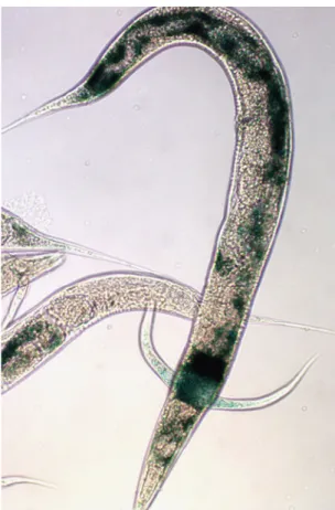
C. elegans is a free-living animal that is normally present in the soil and other bacteria-rich environments. It is a multi-cellular organism of the nematode phylum that has been very widely used in research since 1974 [146]. Multiple studies led to the portrayal of the complete cell lineage as well as the complete description of the anatomy of the whole animal [147]. The adult worm contains 1000 somatic cells, possesses a mouth, a pharynx, an intestine, a gonad, an excretory system, muscles an epithelial system and a cuticle made of collagen (wormatlas). It is also known to be the simplest animal with a central nervous
system [148]. In nature, one can find self-fertilizing hermaphrodites and males but no females [149]. The sexual organs differ between males and hermaphrodites as males have a single lobed gonade, a ductus deferens and a copulatory apparatus in the tail for mating while hermaphrodites, on the other hand, have two gonades, oviducts, a spermatheca, and a single uterus (wormatlas).
The reason behind the success of using C. elegans in research comes from the fact that it is easy to manipulate and can be easily grown for low cost in the laboratory on the bacterium E. coli [149]. It has a relatively short life span and can survive freezing for many years, which allows long-time storage of multiple strains. This organism is also transparent, allowing easy visualization of internal development and organs, as well as easy visualization of fluorescently tagged proteins and different types of staining [149]. Another strong point in the favor of the use of C. elegans in research is the relatively small genome in a small yet relatively complex metazoan. In fact, C. elegans was the first multi-cellular organism to have its genome sequenced and published in 1998 [150], with the last gap sequenced and published in 2002. The 100 million base pair genome contains an approximate 20, 000 genes that are represented on five pairs of autosomes and one pair of sexual chromosomes. The sexual orientation of the offspring is based on the XO sex-determination system, where XX signifies hermaphrodites and XO represents the rare males. Males are infrequently produced (0.1%) by spontaneous meiotic non disjunction in the hermaphrodites where one of the daughter cells inherits 2 X chromosomes while the other inherits none [151]. The production frequency of males increases by up to 50% upon introduction of males into a hermaphrodite population as male sperm preferentially fecund oocytes [149]. The complexity of this animal is exemplified by a diversity of intricate behavior including locomotion, foraging, feeding, defecation, egg laying, dauer larva formation, sensory response to touch, smell, taste, temperature, male mating, social behavior, learning and memory [152-156].
All the above mentioned characteristics render this organism a model of choice in multiple studies related to cellular processes such as cell fate
determination and differentiation as well as apoptosis. Genomics, aging and multiple studies concerning human nervous-based diseases such as Alzheimers and Parkinsons also used C. elegans as a model organism [157-160].
Similarly, this organism provides a big advantage to study DNA repair then yeast and bacterial cells do. This is mainly due to the complex behavior that this model possess, notably, egg hatching, speed of growth and individual survival, that can be studied upon a loss of a DNA repair protein and which cannot be assessed in other simpler organisms such as yeast and bacterial cells.
5.2.
Life cycle
The C. elegans life cycle comprises six stages of development, starting with embryonic development that occurs in part inside the hermaphrodite, while the second part takes place in the outside environment in the form of laid eggs [149]. Each egg gives rise to an L1 larva which then develops quickly through a series of four molts passing from L1 to adult [149]. Under starvation conditions and overcrowding, the L2 larva can enter an alternative 3rd stage called the dauer larva that is characterized by a thick cuticle, a sealed mouth and a very low metabolism, allowing it to survive for months under these conditions, whereas all the adult worms die off. The dauers resume development by entering the L4 stage upon re-introduction to a food source [149].
Since C. elegans is a cold-blooded animal, the temperature of the environment largely influences the speed of development and the time intervals between the molts. Incubation temperatures usually used in the laboratory are 16, 20 and 25˚C, where 16˚C allows the slowest growth rate and 25˚C, the fastest. In fact, it was found that growth at 25˚C is 2.1 times faster than it is at 16˚C and 1.3 times faster than it is at 20˚C [149]. Incubation of plates at lower temperatures, such as 6-8˚C, result in growth arrest and can be used to temporarily arrest