Combinatorial Design of a Nanobody that
Specifically Targets Structured RNAs
F. Cawez
1,†, E. Duray
1,†, Y. Hu
2, J. Vandenameele
3, E. Romão
2, C. Vincke
2,
M. Dumoulin
3, M. Galleni
1, S. Muyldermans
2and M. Vandevenne
11 - InBioS, Center for Protein Engineering, Biological Macromolecules and Biochemistry, Department of Life Sciences, University of Liege, B4000 Sart Tilman, Belgium
2 - Department of Cellular and Molecular Immunology, Vrije Universiteit Brussel, B-1050 Brussels, Belgium
3 - InBioS, Center for Protein Engineering, Protein Folding and Enzymology, Department of Life Sciences, University of Liege, B4000 Sart Tilman, Belgium
Correspondence toM. Galleni and M. Vandevenne:[email protected];[email protected] https://doi.org/10.1016/j.jmb.2018.03.032
Edited by Skerra Arne
Abstract
Recent advances in transcriptome sequencing and analysis have revealed the complexity of the human genome. The majority (≈ 98%) of cellular transcripts is not translated into proteins and represents a vast, unchartered world of functional non-coding RNAs. Most of them adopt a well-defined three-dimensional structure to achieve their biological functions. However, only very few RNA structures are currently available which reflects the challenges associated with RNA crystallization. Nevertheless, these structures would represent a critical step in understanding functions of non-coding RNAs and their molecular mechanisms in the cell. The overall goal of this study is to develop an innovative and versatile tool to facilitate the functional study and crystallization of structured RNAs (stRNAs). In this work, we have engineered an antibody fragment from camelid heavy-chain antibody (nanobody) able to specifically bind with low nanomolar affinity to stRNA, while no binding could be detected for single-stranded DNA/RNA, double-stranded DNA/RNA or a negatively charged protein. However, this nanobody recognizes different and non-related stRNAs, this observation suggests that it binds to an epitope shared by these stRNAs. Finally, our data also show that the binding of the nanobody does not alter the secondary structure content of the stRNA as well as its unfolding/refolding processes during heat treatment. This work constitutes a successful proof of concept demonstrating that nanobodies can be engineered to recognize RNA-related epitopes.
© 2018 Elsevier Ltd. All rights reserved.
Introduction
The recent discovery that most of the human transcriptome is not translated into proteins is probably one of the most important observations of this decade
[1]. Deciphering the function(s) of these non-protein-coding transcripts represents one of the major chal-lenges that molecular biology is facing nowadays. The biological relevance of these transcripts, named non-coding RNAs (ncRNAs), for the biology of the cell is becoming increasingly evident [2,3]. Numerous clas-ses of ncRNAs are highly expressed in adult and in developing tissues, and they often exhibit patterns of expression that are regulated precisely in time and space. These observations suggest that ncRNAs play
key roles in the cell fate and in the development of various tissues[4,5], and therefore, the deregulation of their expression is associated with multiple pathologies such as cancers, neurological disorders and type 1 and 2 diabetes[6,7]. Most ncRNAs adopt a complex three-dimensional (3D) structure to accomplish their biolog-ical functions [8,9]. Like proteins, RNAs present different organization levels to form their 3D structures
[10]. The first level of organization consists in the nucleotidic sequence (primary structure). In the presence of salt, RNAs fold on themselves via Watson–Crick base pairing to form segments of double-stranded helices (hairpins) interrupted by single-stranded regions (loops). These secondary structure elements constitute the second level of 0022-2836/© 2018 Elsevier Ltd. All rights reserved. J Mol Biol (2018) 430, 1652–1670
organization. Finally, the hairpins and unpaired regions are precisely organized in space to form the third organization level. This tertiary structure, formed in vivo and in vitro, is most of the time dependent both on the presence of divalent ions, such as Mg2 +, and the temperature. The biological functions of RNAs are mediated by their 3D structure[11–13].
Despite the increasing interest of the scientific community in these ncRNAs, only very few of them have been fully characterized to date. In this context, proteins engineered to specifically bind ncRNAs should allow their functions to be interrogated. Recently, this principle has been successfully applied in the case of engineered proteins that bind to single-stranded RNAs (ssRNAs). In particular, the most promising works so far were obtained with the Pumilio (PUF) proteins [14]. Engineered PUF proteins have been combined with various effector domains (GFP, PIN RNA endonucle-ase, RS-rich domains, GLD2 and CAF1 domains…) to monitor RNA localization, modulate splicing, generate site-specific RNA endonucleases and affect the translation and the stability of specific mRNAs in living cells [15,16]. These data suggest that fusing RNA-binding proteins (RBPs) to functional modules harbor-ing particular activities is a successful approach to investigate target RNAs in the cell. In contrast, the design of proteins able to bind structured RNA (stRNA) remains poorly explored and this is mostly due to the lack of knowledge regarding the 3D structures of RNAs. Indeed, currently, the number of available RNA 3D structures represents less than 1% of all the structures reported in the Protein Data Bank (PDB) which reflects the difficulties associated with RNA crystallization: (i) RNAs harbor negatively charged phosphate groups that create repulsive forces that are disfavorable for crystal contacts [17]; (ii) RNAs often present flexible regions that lead to a conformational heterogeneity and finally[18]; and (iii) phase solving techniques of RNA crystals can be complex and laborious[19].
This work is focused on a particular stRNA transcribed from a pseudogene of the murine brain cytoplasmic RNA 1 (BC1), that we named ϕBC1 (NCBI Gene ID: 12031) and was thought to be the “real” BC1 gene for a long time. Indeed, it is now well established that most pseudogenes are transcribed into functional RNAs and appear to play important regulatory roles[20]; this is why we were interested in the functional and structural analysis ofϕBC1. BC1, its cousin RNA, is a murine 154-nt-long stRNA expressed in neurons; its human functional analog (BC200) is deregulated in the brain of patients with Alzheimer's disease[21]. BC1 was shown to bind to translational regulators (eIF4A, eIF4B and PABP) and repress translation initiation[22]. BC1 has been well characterized: its secondary structure content has been experimentally determined [23] and has re-vealed that the 5′ region forms a long hairpin, the central poly A region is unpaired and the 3′ domain forms a second but smaller hairpin as shown inFig. 1.
Its molecular mechanisms are also well characterized; BC1 represses translation through its central polyA and 3′ domains by blocking the catalytic activity of eIF4A, an ATP-dependent RNA helicase, while stimulating its ATPase activity[22]. In contrast, barely any study was performed onϕBC1. This 167-nt-long stRNA mostly differs from BC1 in the central polyA and 3′ domains. Therefore, ϕBC1 lacks these two functional domains and consequently is predicted not to bind to translational regulators. This observation explains its attributed terminology as a pseudogene.
Engineering proteins able to bind to stRNAs (stRBPs) would have multiple applications. First, these stRBPs can be fused to different functional domains in order to investigate the biological function(s) of the targeted RNA in living cells or animals, similarly to what has been discussed above with PUF proteins[15,24]. Second, stRBPs can be engineered in order to inhibit the biological activity of catalytic RNAs, which are generally named ribo-zymes. Third, stRBPs can be used as crystallization aids to determine the 3D structure of the targeted RNAs. Indeed, a stRBP bound to RNA can increase the crystal contact surfaces and neutralize some of the negative charges that prevent efficient RNA crystal formation. Upon binding, the stRBP can also reduce the RNA flexibility. Moreover, stRBP struc-ture can be used to solve the phase of RNA crystal by molecular replacement [25]. Finally, stRBPs constitute potential new therapeutic agents given that many stRNAs were shown to be associated with important pathologies[26].
The variable domains of heavy-chain antibodies, which are devoid of light chains, constitute a protein scaffold of choice to engineer proteins that bind specifically to stRNA. These antibody fragments, named VHHs or nanobodies, consist in the N-terminal antigen-binding domain that is connected via the hinge region to the two constant domains (CH2 and CH3) in heavy-chain antibodies [27]. Nanobodies present several advantages compared to other anti-bodies or antibody fragments. First, they are much smaller (12–15 kDa) compared to conventional IgG (150 kDa) or antibody fragments (e.g., ≈50 kDa for Fabs) while recognizing the cognate antigen with high affinity and specificity. In contrast to the VH–VL pairing in Fabs, nanobodies are strictly monomeric and do not necessitate domain pairing to associate with antigen. In addition, nanobodies are more soluble and stable than other antigen-binding domains [28]. They are also poorly immunogenic in human and much easier to express and to purify from microbial hosts [28]. Furthermore, nanobodies present extended antigen-binding regions [especially complementary determin-ing region 3 (CDR3)] that can penetrate into the catalytic site of enzymes, and potentially inhibit the enzymatic activity [29–31]. Finally, nanobodies have been successfully used to trace their antigen within eukaryotic cells in real time and to relocate their target
or to modulate its activity in cells and even in complete organisms[32–34].
In the standard procedure to generate nanobodies, the first step consists of immunizing a camelid with the targeted molecule. We expected that stRNAs will not be immunogenic, and therefore, they will not elicit an immune response of the heavy chain-only antibodies. Therefore, we describe herein the design, the con-struction and the phage display selection of a synthetic nanobody gene library dedicated for nucleic acid binding. The screening of this library led to the selection of, at least, one nanobody, named cAbBC1rib3, that
binds toϕBC1 with nM affinity. We also investigated the thermal denaturation of the stRNA in the presence or the absence of the selected nanobody. Our data showed that the protein does not influence the thermal denaturation and renaturation process. Finally, we
demonstrated that although cAbBC1rib3 is specific for
stRNA, it recognizes different stRNAs, suggesting new strategies to improve the specificity of nanobodies.
Results
Design and construction of a synthetic library of nanobodies dedicated for RNA binding
We choose the nanobody, named cAb-BCII10, as the framework to create the synthetic library. It was initially selected from an immune library against BcII β-lactamase. This nanobody binds to the β-lactamase active site and inhibits its enzymatic activity[35]. cAb-BCII10 is stable, tolerant to mutations and has been Fig. 1. Nucleotide sequence alignment of BC1 (154-nt) and its pseudogene (ΦBC1) (167-nt) and schematic representation of their secondary structure content. The 5′ domain is shown in blue, the central polyA domain is shown in red and the 3′ domain is shown in green. Overall, these sequences share an identity of 72%. The represented secondary structure content of BC1 has been determined experimentally, whereas the secondary structure elements ofΦBC1 have been predicted using the RNAfold server (http://rna.tbi.univie.ac.at/cgi-bin/RNAWebSuite/RNAfold.cgi) from the Vienna RNA Web services website (http://rna.tbi.univie.ac.at).
successfully used several times as a universal scaffold to exchange its antigen specificities by complementarity determining region (CDR) grafting
[36,37]. In addition, its 3D structure has been solved and has revealed that its CDRs are well exposed on the protein surface for efficient binding. Finally, this nanobody is produced in Escherichia coli in high yields.
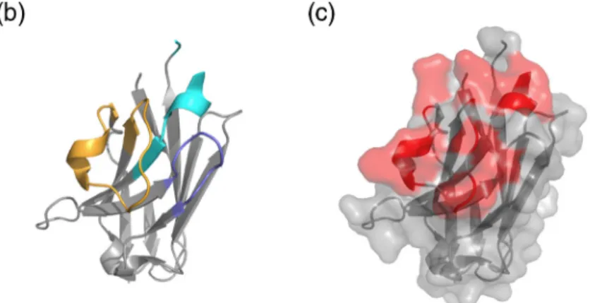
Based on different structural data available for nanobodies in complex with its antigen [36], 11 residues belonging to the antigen-binding regions (CDRs) were selected for randomization (3 residues within CDR1 and 8 residues within CDR3) (Fig. 2a). In the crystal structure of cAb-BCII10 (PDB ID: 3DWT), the side chains of these amino acids were all sticking out toward the solvent and are available for antigen interaction. Furthermore, this 3D structure also shows that these residues are clustered in the same area (Fig. 2c).
The gene library was synthetized using the “trinucle-otide mutagenesis” (TRIM) technology (GeneArt®, Combinatorial DNA library), in which the 11 mutated residues were randomized in a way that it incorporates preferentially 6 residues that are most commonly found at the interfaces of protein/RNA complexes available in the PDB[38–40]. In practice, each randomized position was mutated using a subset of trinucleotide blocks encoding: 10% for Arg, 10% for Lys, 10% for Ser, 10% for Asn, 10% for Gly, 10% for Tyr and 40% for all other residues, except Cys, in equal amounts. The theoret-ical diversity of this library is estimated to be≈1911. The non-amplified library effectively contained over 1011 independent molecules.
The library was amplified by PCR and ligated into the phagemid vector pHEN4[41]. The ligation product was then transformed into TG1 cells. The obtained library comprised ≈1010 individual transformants of which ≈80% harbored a pHEN4 vector with an insert compatible with the size of a cAb-BCII10 variant. To assess the quality of the library, the gene of 100 randomly chosen clones was sequenced in order to determine the percentage of amino acid incorporation for each randomized position. The amino acid distribu-tion in the randomized loops as obtained from DNA sequencing is presented in Fig. 3 and confirms an amino acid occurrence that correlates well with our design.
Selection of stRNA-binding nanobodies
The nanobodies of the dedicated RNA-binding nanobody library were displayed at the tip of M13 bacteriophages and fused to the coat protein PIII, and consecutive rounds of selection were performed against the target stRNA generated by in vitro transcription.
Importantly, since the function ofϕBC1 is unknown, we had no mean to assess the folding of this stRNA into its native conformation after in vitro transcription. This is why we decided to fuseϕBC1 to a catalytic stRNA, named ribglmS, as follows:5′ϕBC1–ribglmS3′ . This
172-nt-long ribozyme, is expressed by Bacillus anthracis and originates from the 5′ untranslated region of the glucosamine-6-phosphate (Glc6P) synthase messen-ger RNA[42]. In the presence of Glc6P, ribglmS
auto-cleaves itself from the mRNA from which it takes part.
Fig. 2. Representation of the amino acid sequence (a) and 3D structure of cAbBCII-10 (PDB ID: 3DWT) (b and c). The residues that belong to the CDRs are represented in cyan, blue and orange for CDR-1, -2 and -3, respectively, in the amino acid sequence (a) and on the 3D structure (b). The randomized resi-dues are highlighted in red in the amino acid sequence (A) as well as on nanobody structure surface shown in panel (c).
This catalytic activity has been well characterized[43]
and can be easily measured (see Materials and Methods). Therefore the ribglmSactivity was monitored
after each transcription as a quality control experiment. Although, the presence of the ribozyme activity does not reflect the conformation of theϕBC1 moiety of the fusion RNA, it indicates that the in vitro transcription conditions were compatible with the native folding an stRNA.
In total, we performed five rounds of selection against the fusion stRNA: 5′ϕBC1–ribglmS3′ , after checking the
presence of the ribozyme activity (data not shown). At the end of the selection process, 95 individual clones from rounds 3, 4 and 5 were cultured and recombinant proteins expressed in the periplasmic space of E. coli in fusion with a hemagglutinin (HA) tag at their C-terminal end. The periplasmic proteins were extracted and used in an ELISA to identify the variants that bind to the target RNA (data not shown). Only three of the tested clones (cAbBC1rib3, cAbBC1rib20, cAbBC1rib90) gave positive
signals in ELISA. The nanobodies cAbBC1rib3 and
cAbBC1rib90 were selected from the third round of
bio-panning, and cAbBC1rib20 was retrieved after the fifth
round of bio-panning. Bio-layer interferometry (BLI) measurements were performed to confirm the ELISA results. The data obtained show that significant binding against immobilized ϕBC1–ribglmS could only be
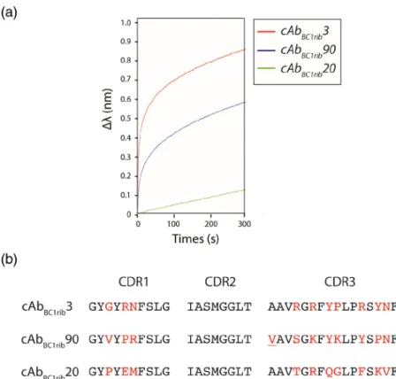
detected for cAbBC1rib3 and cAbBC1rib90 (Fig. 4a).
These two nanobodies were therefore sub-cloned into the pHEN6 expression vector for further charac-terization. Remarkably, the sequence of cAbBC1rib90
contains a single substitution in a non-randomized position of its CDR3 (A97V) (Fig. 4b). Given that the phage display screening led to the retrieval of this variant, we decided to keep this spontaneous mutation. After sub-cloning the nanobody genes in pHEN6, only cAbBC1rib3 could be expressed in significant
amount. We therefore focused our study on cAbBC1rib3.
When expressed from pHEN6, it harbors a C-terminal His6 tag to facilitate its purification by nickel ion
affinity chromatography. The protein sample was then
further purified to homogeneity by cationic exchange chromatography.
Characterization of the interaction between cAbBC1rib3 andϕBC1–ribglmS
Determination of the binding specificity of cAbBC1rib3
First, we analyzed the association of cAbBC1rib3
with different control molecules to assess its specificity for stRNAs. Besides the ϕBC1–ribglmS fusion, the
other target molecules include the following: ssDNA (ACTG)X6, ssRNA (ACUG)X6, dsDNA (ACTG)duplexX6,
dsRNA (ACUG)duplexX6 and bovine serum albumin
(BSA), a negatively charged protein. Apart from the strongϕBC1–ribglmSrecognition, a very weak binding
was observed for the double stranded RNA molecule (Fig. 5), whereas all the other tested molecules were not recognized at all. This result strongly suggests that cAbBC1rib3 is specific for stRNA.
Qualitative binding assays were then performed in order to identify the exact partner of theϕBC1–ribglmS
RNA fusion (i.e.,ϕBC1 or ribglmS) that is recognized by
the nanobody. The data presented inFig. 6a show that cAbBC1rib3 recognizes theϕBC1 part of the fused RNA
since hardly any binding could be detected for the isolated ribglmS. Moreover, to further investigate the
discrimination ability of cAbBC1rib3, BLI measurements
were performed against different stRNAs not related to ϕBC1 or ribglmSin terms of sequences and predicted
functions (seeMaterials and Methods). The stRNA ctrl 1 is 7SK, a human 331-nt-long small nuclear ncRNA that acts as a scaffold in the nuclear ribonucleoprotein complex. This complex was shown to play a key role in regulating the activity of the positive transcription elongation factor b (P-TEFb)[44]. The stRNA ctrl 2 is a bacterial 159-nt-long domain (the P4–P6 domain) of the group I intron ribozyme expressed Tetrahymena thermophyla[45]. Our data indicated that both control stRNAs tested are recognized by cAbBC1rib3, however,
the nanobody exhibits differential binding profiles to Fig. 3. Representation of the amino acids distribution at the ran-domized positions. The bars repre-sent the occurrence of a particular amino acid (in %) across all the randomized positions. The residues that were preferentially introduced during the mutagenesis are repre-sented in red and should theoretically be 10% (dashed line). The percent-age of amino acid occurrence was calculated from the sequences of 100 cherry picked clones from the initial library.
stRNAs, and it does not recognize ribglmS (Fig. 6).
These observations suggest that it probably recognizes a common epitope shared byϕBC1 and the 2 control stRNAs.
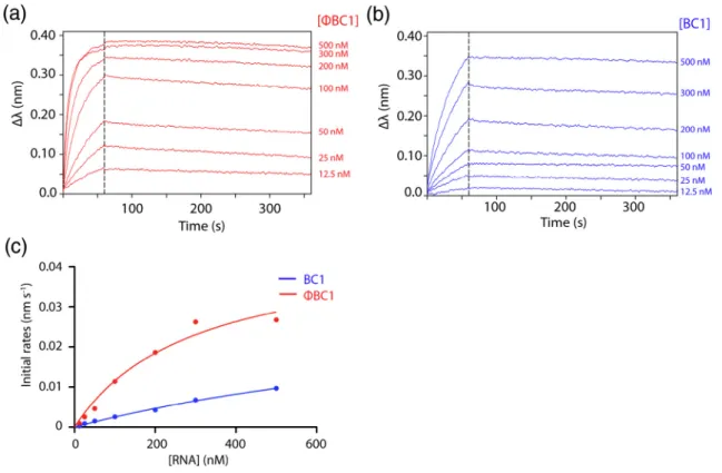
In order to further investigate the differential binding of cAbBC1rib3 among stRNA targets, qualitative binding
experiments were conducted on BC1 andϕBC1 and are compared in Fig. 7. As mentioned in the introduction, these two stRNAs mostly differ from each other in the central polyA and 3′ domains. Our results demonstrate that the observed association rates reach saturation at a lower concentration for ϕBC1 compared to BC1, whereas, as observed above, the dissociation rates are very slow and highly similar
for both stRNAs and can be considered as negligible. These data suggest that the binding of cAbBC1rib3 to
BC1 is weaker compared to that ofϕBC1. In order to further compare the binding efficiencies of cAbBC1rib3 to
these stRNAs, observed initial rates of association were plotted against the RNA concentration and fitted using a hyperbola equation (Fig. 7c). The fits predict at least a four‐fold higher binding efficiency for ϕBC1 compared to BC1. This result is important as it provides evidence that cAbBC1rib3 presents a differential
recog-nition between BC1 and ϕBC1 and across stRNAs, although the origin for this differential binding behavior remains unclear and will be discussed later (see Discussion). Notably, additional BLI control
experi-Fig. 5. Qualitative binding of cAbBC1rib3 to the fusion RNAϕBC1–
ribglmS and the different control
molecules measured by BLI. The experiment was performed using an“anti-His” sensor on which puri-fied cAbBC1rib3 was immobilized via
its C-terminal Hisx6 tag. The
con-centration of the different analytes used for these assays was 100 nM. The association and dissociation phases were both measured for 600 s.
Fig. 4. Binding measurements of the selected nanobodies with the fusion RNAϕBC1–ribglmSperformed
by BLI (a). The experiment was performed using an “anti-mouse” sensor on which an anti-HA antibody was immobilized. Then periplasmic extracts containing nanobodies were loaded onto the sensor via their C-terminal HA tag. The analyte (ϕBC1– ribglmS) concentration used for these
assays was 100 nM. The association was monitored for 300 s. CDR amino acid sequence alignment of
cAb-BC1rib3, cAbBC1rib90 and cAbBC1rib20
(b). The randomized residues are indicated in red; the A97V mutation present in the CDR3 for cAbBC1rib3 is
ments were performed in order to assess potential non-specific binding behaviors of both stRNAs. These data are presented in the Supplemental Data (Fig. S1). Briefly, a control nanobody was immobilized on BLI sensors and binding assays withϕBC1 and BC1 were conducted to assess potential differences in non-specific binding of these two stRNAs. The results indicate that only a very weak binding could be observed in the presence of an excess (1μM) of BC1 but not in the presence of 100 nM. In the case ofϕBC1, no binding could be detected in the presence of either 100 nM or 1μM. These data suggest that, at high concentrations, BC1 has higher tendency to exhibit non-specific binding compared toϕBC1.
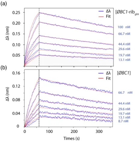
Determination of the binding affinity between cAbBC1rib3 andϕBC1–ribglmS
Quantitative BLI measurements were carried out to determine the binding affinity of cAbBC1rib3 for ϕBC1
alone or in the context of the RNA fusion. The binding assays (Fig. 8) show that cAbBC1rib3 binds toϕBC1–
ribglmS and to ϕBC1 with nM affinity (KD= 2.3 ±
0.8 × 10⁻⁹M and KD= 6.8 ± 2.8 × 10⁻⁹M,
respective-ly). The experimental data were adjusted using a 1:1 mathematical binding model. The equilibrium dissoci-ation constants and the kinetic parameters (kon and
koff) presented in Table 1 are very similar to those
measured for nanobodies directed against proteins selected from immune libraries[41].
As explained in the introduction section, it is well established that the 3D structure of folded RNAs is stabilized by the presence of bivalent ions such as Mg2+. Therefore, we have measured the binding affinities of cAbBC1rib3 for the fusion stRNA and the
isolated ϕBC1 in the presence of a chelating agent (10 mM EDTA) to assess whether the recognized epitope is a tertiary structure element or a secondary structure element. Circular dichroism (CD) spectra of theϕBC1–ribglmSrecorded in the presence of EDTA
Fig. 6. Qualitative binding measurements of cAbBC1rib3 to different stRNAs performed by BLI. (a) Binding curves of the
nanobody to the fusion RNAϕBC1–ribglmS(blue), the isolated ϕBC1 (red) and ribglmS(pink). (b) Binding curves of the
nanobody to different control stRNAs (orange and purple) andϕBC1–ribglmS(blue). These experiments were performed using
an “anti-His” sensor on which purified cAbBC1rib3 was immobilized via its C-terminal Hisx6tag. The analytes (stRNAs)
(data not shown) indicated that the stRNAs conserves its secondary structure content. The KD values
decrease 100-fold for bothϕBC1–ribglmS and ϕBC1
alone in the presence of EDTA (i.e., in the absence of Mg2+) (Fig. 9). The kinetic parameters (Table 1) indicate that, for both RNAs, this strong increase in the equilibrium binding affinity (decrease of the dissociation constant) in the presence of EDTA is mostly due to an increase of the kon(10- to 40-fold)
combined to with a 10- to 4-fold decrease of the koff.
Interestingly, qualitative binding assays conducted by BLI on the control stRNAs presented similar behaviors in the presence of EDTA, namely faster association and slower dissociation rates (data not shown). Additional control experiments were performed in the presence of EDTA in order to assess the non-specific binding of the stRNA on the BLI surface. These experiments are presented in the Supplemental Data (Fig. S2). Briefly, in the absence of Mg2+, no binding could be detected between ribglmS (a stRNA not
recognized by cAbBC1rib3) and BLI sensors on which
a control nanobody was immobilized, suggesting that the stRNA does not stick on the BLI surface when its 3D structure is abolished. In contrast, significant
signals could be detected between ribglmSand cAb-BC1rib3 loaded on BLI sensors in the absence of Mg
2 +
, whereas in the presence of Mg2+, no binding was observed (Fig. 6a). However, the signals measured in the absence of Mg2 + are not dose dependent and present atypical shapes, suggesting that these signals probably reflect non-specific binding of ribglmS on
cAbBC1rib3 only observed in the absence of Mg2 +.
All together, these data suggest that cAbBC1rib3
probably recognizes a secondary structure element present inϕBC1 and in the different control stRNAs tested. This observation will be further developed in the Discussion section.
Effects of the interaction of cAbBC1rib3 on the
properties ofϕBC1–ribglmS
In order to investigate the potential effects of the binding of the nanobody on the structure and stability of the stRNA, we recorded CD spectra at 25 °C and we monitored the changes of CD signal at 223 nm or 267 nm as a function of the temperature for the purified nanobody, the stRNA and for the nanobody: stRNA complex.
Fig. 7. Binding measurements of cAbBC1rib3 toΦBC1 (red) (a) and BC1 (blue) (b) performed by BLI. These experiments
were performed using an“anti-His” sensor on which cAbBC1rib3 was loaded and immobilized via its C-terminal Hisx6tag.
The stRNAs concentrations used for these assays were 12.5, 25, 50, 100, 200, 300 and 500 nM. The association and dissociation phases were measured for 60 and 300 s, respectively. The measured rates of association (initial rates) were plotted against the stRNA concentrations (c) and fitted to a hyperbola equation in order to compare the binding efficiencies of cAbBC1rib3 toΦBC1 and BC1.
Effects of the binding of the nanobody on the structure ofϕBC1–ribglmS
Figure 10 represents the CD spectra for ϕBC1– ribglmS and cAbBC1rib3 alone or in complex at a
1:1 molar ratio cAb:stRNA. The spectrum recorded forϕBC1–ribglmSindicates that the stRNA possesses
different secondary structure elements. The positive and intense signal observed around 267 nm is indicative for the presence of right-handed helices contained in hairpins, whereas the negative signal less intense around 210 nm is attributed to the presence of parallel duplexes[46]. The spectrum measured for the nanobody alone presents typical features of nanobo-dies, with a positive signal observed around 223 nm that indicates the presence of aromatic residues
immobilized within a particular chemical environment suggesting the presence of a well-defined 3D structure
[47]. The second positive signal recorded at 203 nm is due to peptide backbone, which, in this case, is an antiparallelβ-sheet[47]. It is interesting to see that the spectra of the complex are very similar to the one measured for the stRNA alone, suggesting that the binding of the nanobody does not alter the secondary structure content of the stRNA.
Effects of the binding on the thermal unfolding of ϕBC1–ribglmS
The thermal denaturation of cAbBC1rib3 alone,
ϕBC1–ribglmS alone or in complex with cAbBC1rib3
Fig. 8. Quantitative binding mea-surements of cAbBC1rib3 to the
fusion RNA ϕBC1–ribglmS (a) and
the isolated ϕBC1 (b) performed by BLI in the presence of 10 mM MgCl2. These experiments were
performed using an “anti-His” sen-sor on which purified cAbBC1rib3 was
loaded and immobilized via its C-terminal Hisx6 tag. The analyte
(stRNAs) concentration used for these assays are indicated on the graphs. The association and disso-ciation phases were monitored for 60 and 300 s, respectively. The experimental data were fitted using a 1:1 binding model.
Table 1. Equilibrium and kinetic constants values measured for the interaction between cAbBC1rib3 and ϕBC1 in the
context of the fusion RNA or isolated
EDTA kon(M−1s−1) koff(s−1) KD(M)
ϕBC1–ribglmS − 2.9 ± 2.5 × 10⁵ 1.7 ± 1.0 × 10⁻³ 2.3 ± 0.8 × 10⁻⁹
ϕBC1–ribglmS + 3.5 ± 1.2 × 10⁶ 1.1 ± 1.4 × 10⁻⁴ 2.5 ± 3.3 × 10⁻¹¹
ϕBC1 − 3.3 ± 1.7 × 10⁵ 2.1 ± 0.9 × 10⁻³ 6.8 ± 2.8 × 10⁻⁹
ϕBC1 + 1.2 ± 0.2 × 10⁷ 8.2 ± 4.8 × 10⁻³ 7.2 ± 4.8 × 10⁻¹¹
The association and dissociation kinetic constants (konand koff) and the deduced equilibrium dissociation constants (KD) were measured by BLI. The experimental data were fitted using a 1:1 model using at least six analyte (stRNA) concentrations. The KD values and associated errors correspond to averages and standard deviations calculated from at least three independent BLI experiments.
(1:1 molar ratio) was monitored by far UV CD at 223 nm for cAbBC1rib3 and at 267 nm for the stRNA
alone or in the complex. Note that at 267 nm, there is no signal from the nanobody, then the unfolding of the stRNA within the complex can be monitored specifical-ly. The results shown in Fig. 11a indicate that the denaturation and renaturation of cAbBC1rib3 are
coop-erative and characterized by an apparent two-state system. Noticeably, cAbBC1rib3 is thermostable since its
temperatures of mid-denaturation upon heating (Tm) is 68.4 ± 0.4 °C (Table 2). Most of the CD signal is recovered after the renaturation process, indicating that the thermal denaturation is highly reversible as regularly observed for nanobodies[45].
The denaturation/renaturation process of ϕBC1– ribglmSexhibits distinct features compared to that of
the nanobody. First, the denaturation is a lot less cooperative and occurs on an extended range of temperatures (from ≈ 25 to 90 °C). A plausible explanation is that the different secondary structure elements of the stRNA act as sub-domains that have different thermal stabilities and therefore explains this non-cooperative behavior. Another explanation would be that the RNA unfolding occurs similarly to a“zipper” in analogy to the complementary strands of RNA
duplexes and hairpins that separate from each other. Since the far UV CD measurements monitor only changes in the secondary structure of RNA and not its tertiary structure, it is not possible to determine if the denaturation of the two types of structures is cooperative. The temperature of mid denaturation of the secondary structure (Tm) obtained for the denaturation curve is 70.7 ±2.2 °C. Interestingly, the renaturation process occurs at much lower tempera-ture (Tm: 62.0 ± 4.0 °C) compared to the denaturation (Fig. 11b andTable 2). This discrepancy between the unfolding and refolding transitions, known as hyster-esis, has been described for nucleic acids[48]. Briefly, during the renaturation process, the probability of exact alignments of base sequences to reconstitute hairpins, duplexes and bulges present in the native stRNA conformation is very small. This means that non-native duplexes may form nucleation complexes that cannot be extended, resulting in large numbers of nonproduc-tive intermediate states. These nucleated regions form and disassociate as the system seeks the lowest free-energy (native) state. This phenomenon explains the observed hysteresis[49,50].
Notably, the binding of cAbBC1rib3 to the stRNA
does not affect its denaturation or renaturation Fig. 9. Quantitative binding mea-surements of cAbBC1rib3 to the
fusion RNA ϕBC1–ribglmS (a) and
the isolatedϕBC1 (b) performed by BLI in the presence of 10 mM EDTA. These experiments were performed using an “anti-His” sen-sor on which purified cAbBC1rib3
was immobilized via its C-terminal Hisx6 tag. The analyte (stRNAs)
concentrations used for these as-says are indicated on the graphs. The association and dissociation phases were monitored for 60 and 300 s, respectively. The experi-mental data were fitted using a 1:1 binding model.
processes (Fig. 11c and Table 2) since the mid-denaturation upon heating and cooling (Tm) for both processes is very similar to those measured for ϕBC1–ribglmSalone. Altogether, this result suggests
that cAbBC1rib3 does not modify the unfolding and
refolding behavior of the stRNA upon thermal denaturation.
Reversibility of the thermal denaturation monitored using far UV CD
The CD spectrum recorded after a complete denaturation/renaturation cycle superimposes the one recorded at the start of the experiment before
sample denaturation (Fig. 12), suggesting that the thermal denaturation is reversible.
Discussion
The overall goal of this study was to develop camelid antibody fragments or nanobodies able to bind with high affinity and specificity to a particular stRNA. Such nanobodies could be employed as innovative tools to investigate the structure and function of the non-protein coding part of genomes. Our strategy involved the construction of a synthetic library of nanobodies, in which 11 residues (3 from CDR1 and 8 from CDR3) Fig. 10. CD spectra recorded for the ϕBC1–ribglmS alone (a),
cAbB C 1 r i b3 alone (b) and the
cAbBC1rib3:ϕBC1–ribglmS complex
(c). The spectra measured for the nanobody and the stRNA alone were recorded with a 13-μM protein concentration and a 500-nM stRNA concentration, respectively. For the complex, the concentration of both the stRNA and the protein was 500 nM.
were randomized. This randomization introduced a bias in favor of 6 residues (Arg, Lys, Tyr, Ser, Gly, Asn) that are often found to interact with RNA in protein:RNA complexes. The first panning of this library by phage
display led to the selection of two individual variants (cAbBC1rib3 and cAbBC1rib90) that showed strong
binding to stRNA using the BLI technology. These data indicate that this library of camelid antibody fragments optimized for RNA interactions is a valid approach to select nanobodies able to associate with an RNA target and potentially other nucleic acid-derived molecules.
Importantly, up to date, only a couple of studies have reported the successful selection of conventional antibody fragments (Fabs) that bind either stRNAs
[51,52] or ssRNAs [53] in both cases also using a phage-display based selection approach. In these studies, the authors used two different synthetic Fab libraries harboring a“reduced genetic code.” In one of these libraries, named YSGR, the entire sequence of light-chain CDR-3 (CDR-L3), and heavy-chain CDR-1, -2 and -3 (CDR-H1, CDR-H2, CDR-H3) were mutated in Tyr, Ser, Gly or Arg, whereas in the second library, termed YSGRKX, the entire sequences of the 6 CDRs were mutated in Tyr, Ser, Gly, Arg, Lys, and (X) all the Fig. 11. Thermal denaturation curves (full circles) and renaturation curves (empty circles) measured for the cAbBC1rib3
(a) andϕBC1–ribglmSalone (b) or in complex with the nanobody using a 1:1 molar ratio (c). These curves were obtained
by monitoring the CD signal at 223 nm for the nanobody and 267 nm for the stRNA and the complex, while heating the samples from 25 to 92 °C (denaturation) and then cooling the samples from 92 to 25 °C, at a heating and cooling rate of 1 °C/min.
Table 2. Melting temperature values (Tm) calculated from the thermal denaturation and renaturation curves monitored by CD Tm denaturation (°C) Tm renaturation (°C) ϕBC1–ribglmSalone 70.7 ± 2.2 62.0 ± 4.0 ϕBC1–ribglmS:cAb (1:1) 70.3 ± 0.9 62.0 ± 4.2 The Tm values for denaturation and renaturation were measured for ϕBC1–ribglmSalone or in complex with the nanobody (1:1 molar ratio). The transition curves were all fitted using a sigmoidal dose response (variable slope) equation (seeMaterials and Methods). The presented Tm values and associated errors correspond to averages and standard deviations calculated from, at least, 3 independent experiments.
other residues except Cys, Ile and Met. Although these studies were successful and led to the co-crystallization of a Fab:stRNA complex, it is interesting to note that most of the selected Fabs from these libraries exhibited a poor binding affinity in the lowμM (≥ 1.5 μM) to high nM (≥160 nM) range compared to the nanobody-based binder (low nM) described in the present study. This is remarkable since the randomized surface in the Fab libraries was much larger than that in the nanobody library. This observation suggests that by mutating 11 residues only, across CDR1 and CDR3, it is possible to obtain tight binders against the target molecule instead of sampling a larger sequence space from libraries with more randomized residues.
This high binding affinity constitutes a strong advantage for co-crystallization experiments, given that nanobodies, like Fabs, are commonly used as chaperones in order to facilitate the crystallization of difficult protein targets (e.g., membrane proteins, etc…)
[25,54]. In addition, this strong binding is also interest-ing for functional studies in cultured cells (in vitro) or in animal models (in vivo).
Furthermore, in contrast to cAbBC1rib3, some of the
Fabs discussed above have a better differential binding specificity for particular stRNAs. A possible explanation for their enhanced discrimination among various stRNAs might originate from the use of competitor molecules, such as transfer RNAs (tRNAs) mixtures, during the phage display panning. The addition of increasing amounts of such compet-itors during the consecutive phage display selection rounds probably assisted in the selection of variants with higher specificity. The strategy of including competitor molecules during the selections could certainly be introduced in future pannings of our nanobody library to improve the binding specificity of the retrieved nanobodies.
Without any doubt, the binding specificity is a crucial parameter when designing antibody fragments for any sort of applications. Unlike proteins that are composed
of 20 possible subunits (amino acids) and therefore presenting a large chemical diversity, RNAs include only four different subunits (nucleotides). Consequent-ly, for molecules of similar size, the chemical diversity of RNA epitopes is much lower compared to protein epitopes. This RNA property probably explains why it is more challenging to obtain a high specificity for RNA antigens compared to protein antigens. Nevertheless, cAbBC1rib3 exhibits clear target specificity since no
association (or very weak) could be detected with ssRNA, ssDNA, dsRNA, dsDNA or BSA, demonstrat-ing that it is specific for stRNA. In addition, cAbBC1rib3
also discriminates among stRNAs since it does not recognize the isolated ribglmS.
Furthermore, our data also indicated that cAbBC1rib3
can also discriminate ϕBC1 from its cousin BC1, although these two stRNAs present a high degree of sequence identity in the 5′ domain (sequence identity of 92%) (Fig. 1). In contrast, the central polyA and the 3′ domains deviate significantly between BC1 andϕBC1, with sequence identities of only 44% and 54%, respectively. In order to explain the discrimination of cAbBC1rib3 forϕBC1 over BC1, we have analyzed their
secondary structure contents. Experimental determi-nation of the secondary structure content of BC1[23]
revealed that both the 5′ and 3′ regions form an hairpin, whereas the central polyA region is unpaired[22]. The secondary structure prediction performed on ϕBC1 using the RNAfold server (http://rna.tbi.univie.ac.at/ cgi-bin/RNAWebSuite/RNAfold.cgi) from the Vienna RNA Web services website (http://rna.tbi.univie.ac.at), indicates that the 5′ domain of ϕBC1 also forms a hairpin similar to that of BC1, whereas the central polyA and 3′ domains are predicted to be unpaired. Since our data showed that cAbBC1rib3 fails to
recognize ssRNA, such as the unpaired central polyA and 3′ domain of ϕBC1, it is therefore more likely that cAbBC1rib3 recognizes the 5′ hairpin of ϕBC1.
Notably, we hypothesized that the 5′ domain of BC1 contains subtle conformational or dynamic changes Fig. 12. CD spectra recorded forϕBC1–ribglmSalone (a) and the cAbBC1rib3:ϕBC1–ribglmScomplex at a 1:1 molar ratio
(b). These spectra were recorded before denaturation (full circles) and after renaturation (empty circles) with a 500-nM stRNA concentration. For the complex, the stRNA and protein concentrations used were 500 nM.
that might reshape the epitope surface, leading to lower binding efficiency compared toϕBC1, although at this stage, we consider the investigation of the bases causing this differential binding between BC1 and ϕBC1 to be outside the scope of this study. Neverthe-less, this differential binding between homologous stRNAs is important because it demonstrates the discriminating potential of nanobodies among various stRNAs.
In this work, we have also observed a strong increase in the binding affinity (100 ×) between cAbBC1rib3 and
ϕBC1 (isolated or in the fusion) in the absence of Mg2+
compared to the affinity measured in its presence. By chelating Mg2+ions, EDTA destabilizes the RNA tertiary structure, while the secondary structure content is maintained. Therefore, we postulate that cAbBC1rib3
recognizes one secondary structure element that is more accessible in the absence of Mg2+. This would explain the strong increase in affinity observed in the presence of EDTA.
At this stage, it remains difficult to describe the structural details of the interaction between cAbBC1rib3
and ϕBC1. We performed isothermal titration calo-rimetry experiments, microscale thermophoresis and gel filtration experiments in an attempt to explain the BLI data and determine other parameters such as the stoichiometry as well as the thermodynamic param-eters (ΔH, ΔS) of this interaction. Unfortunately, none of these methods led to conclusive and exploitable data. Nevertheless, the overall results generated in this study suggest that cAbBC1rib3 recognizes a
secondary structure element that is probably highly similar to the one included in the 5′ domain of ϕBC1 and common to the different stRNAs that were shown to interact with cAbBC1rib3.
The CD spectra of RNA alone and in the complex with the nanobody are virtually identical. This obser-vation suggests that the binding of the nanobody does not affect the RNA secondary structure content of the RNA partner, which is an important property in the context of nanobody:stRNA co-crystallization experi-ments. In addition, our CD studies have also demon-strated that cAbBC1rib3 is a thermostable protein which
is also advantageous for functional studies performed in cultured cells or animal models.
In conclusion, this study describes for the first time the successful design of a camelid antibody frag-ment able to recognize stRNA with high affinity retrieved from an innovative nanobody library dedicated for nucleic acid targets. Although the binding specificity of the obtained nanobody might be improved, this study represents a major step toward our long-term ambitious goal that aims to provide innovative tools for functional and structural RNA studies. Conversely, this lack of strict speci-ficity can be advantageous since cAbBC1rib3 could
potentially be used to assist the crystallization of several stRNA targets. This aspect will be the next focus of future investigations.
Materials and Methods
Nanobody library construction
The synthetic nanobody gene library harboring the 11 randomized residues in CDR1 and CDR3 regions was purchased from GeneArt with an estimated diversity of ≈1011 individual gene variants. The library was amplified using the following specific primers that included the Pst1 and Not1 restriction enzyme sites (underlined):
Fwd: 5′-GTTCAGCTGCAGGAAAGCGGTGGTG GTAGCGTTC-3′
Rev.: 5′-CGGGTAGCGGCCGCTCGAAACG-3′ These primers anneal to the framework 1 and framework 4 regions. The final and purified PCR product was digested with Not1 and Pst1, purified and then cloned into the phagemid vector pHEN4 using the protocol described by Hu et al. [55]. Briefly, the ligation of the nanobody gene library into the pHEN4 was performed at 16 °C for 16 h, and the ligation product was purified and then used to transform, by electroporation, TG1 electro-competent cells (Lucigen, Middleton, WI, USA). Immediately after the electro-transformation, the cells were allowed to recover with SOC medium, by incubation at 37 °C for 1 h. The transformed cells were then plated on LB/agar plates containing 2% glucose and 100μg/ml ampicillin. Serial dilutions of cell aliquot were plated on standard-sized Petri dishes to determine the total amount of transfor-mants, while the majority of the cells was plated on large Petri dishes. The plates were incubated overnight at 37 °C, and the colonies were scraped from the plates and stored at−80 °C in LB/20% glycerol at −80 °C for future use. The percentage of clones harboring a phagemid containing an insert corresponding to the size of a nanobody gene was assessed by colony PCR using the MP57 and GIII primers on 95 randomly chosen colonies.
MP57: 5′-TTA TGC TTC CGG CTC GTA TG-3′ GIII: 5′-CCA CAG ACA GCC CTC ATA G-3′ These primers anneal on both sides on the pHEN4 vector sequences abutting the inserted nanobody gene.
Display of the nanobody library on phage particles and selection of binders
The nanobodies within the library were expressed on phage particles after infection with M13 K07 helper phages. Enrichment of binders from this library was performed by five rounds of in vitro selection using a protocol adapted from Hu and coworkers[55]. Briefly,
the OD600value reached 0.6–0.9. Then, the cells were
infected with ~ 1012 M13 helper phage particles for 30 min at room temperature (RT). The infected cells were harvested by centrifugation at 1300g for 10 min and resuspended in 300 ml fresh 2xTY medium sup-plemented with 100 μg/ml ampicillin and 70 μg/ml kanamycin to secrete virions displaying nanobodies fused the coat protein pIII. After overnight growth, bacterial cells were removed by centrifugation and the phages particles secreted in the culture supernatants were precipitated using a polyethylene glycol/NaCl solution. The phage solution was re-suspended in a total volume of 1 ml sterile PBS and the phage concentration was calculated based on the OD260nm
value. For the selection rounds, 60μg of purified and in vitro transcribed ϕBC1–ribglmS was coated
over-night at 4 °C in a single well of a microtiter plate. Blocking of the stRNA-coated well and the non-coated control well (located at a distant position in the microtiter plate) was performed with different blocking solutions for the successive selection rounds. These blocking solution include the following: 4% (w/v) skimmed-milk proteins, protein free buffer (Thermo Scientific), 0.1% casein (w/v) or SEA BLOCK blocking buffer (Thermo Scientific). Changing of the blocking solution minimizes enrichment of phages that bind to components present in the blocking reagent. For each selection round, ~ 1011 phage particles were added per well and incubated for 1 h at RT. Increasing numbers of washings were performed with PBST (PBS containing 0.05% Tween-20) during the suc-cessive selection rounds. The elution of phages particles bound to the stRNA-coated well was performed with a triethylamine solution (100 mM TEA, pH 11.0) for 10 min at RT. The eluted phages were immediately neutralized with Tris–HCl (1.0 M, pH 7.4). Aliquots of the eluted phage solutions from each well (coated or not coated with stRNA) were serially diluted (10 ×) and used to infect exponentially growing TG1 cells. After 30-min incubation at RT, infected cells for each phage dilution were plated on selective medium to assess relative phage enrich-ment between the stRNA-coated and non-coated wells. The majority of the stRNA-eluted phages was also used to infect exponential growing E. coli TG1 cells in order to amplify and prepare phage particles for the next selection round. The panning was performed for five consecutive rounds of selection.
Screening of stRNA binders by ELISAs
Ninety-five individual colonies from rounds 3, 4 and 5 were randomly picked from the plates and grown in 100μl of 2xTY medium supplemented with 10% glycerol, 2% glucose and 100μg/ml ampicillin in a
mented with 0.1% glucose and 100μg/ml ampicillin in a 96-microwell plate (deep well). The leftover from the overnight pre-culture was frozen and stored at−20 °C for subsequent experiments. The 2-ml deep-well culture plates were incubated for 4–5 h at 37 °C with shaking before inducing the expression of the nano-bodies in fusion with the phage coat protein pIII (N-terminal end) and an HA tag (C-(N-terminal end) by the addition of 1 mM IPTG. After 4 h of incubation at 37 °C, the cultures were harvested by centrifugation, and the cell pellets were frozen at −20 °C. The nanobodies exported in the periplasmic space were extracted by freeze–thaw cycles and used in an ELISA to detected nanobody variants able to bind ϕBC1– ribglmScoated in a 96-microwell plate. Each soluble
extract was added to a well coated with 100μl ϕBC1– ribglmSat 70 μg/ml as well as a negative control well
(non-coated well). Nanobodies able to bind stRNA were detected with 1/2000 diluted mouse anti-HA IgG (primary antibody) and a 1/2000 goat anti-mouse IgG conjugated with alkaline phosphatase (secondary antibody) (ImTec Diagnostics NV, Antwerp, Belgium). After the addition of the substrate p-nitrophenyl phosphate (Sigma), the reaction was measured after 5, 15, 30 and 60 min at 410 nm. The colonies expressing nanobodies that exhibit a positive signal in the ELISA were cultured. The corresponding phagemid DNAs were extracted and purified, and the inserted nanobody genes were sequenced.
Expression of the nanobodies from the pHEN4 phagemid vector and periplasmic extract preparation
The gene of nanobodies corresponding to clones cAbBC1rib3, cAbBC1rib20 and cAbBC1rib90 cloned into the
phagemid vector pHEN4 were transformed into E. coli WK6 cells for expression of the nanobodies fused to an HA tag on their C-terminal end. Recombinant expres-sion of the nanobodies was performed in shake flasks using Terrific broth supplemented with 100μg/ml ampicillin until the OD600nm reaches ≈0.8. Then,
induction of protein expression was performed by adding IPTG (1 mM) and incubating for 16 h at 28 °C. After pelleting the cells, the periplasmic proteins were extracted by osmotic shock [56]. The periplasmic extracts were then dialyzed in Tris 50 mM (pH 7.5), NaCl 150 mM and 10 mM MgCl2 for the binding
assays performed using BLI.
Expression and purification of the nanobodies expressed from the pHEN6 plasmid
The nanobody genes corresponding to clones cAbBC1rib3 and cAbBC1rib90 were sub-cloned into
enzyme sites NcoI and BstEII. The plasmid constructs were transformed into E. coli WK6 cells for nanobody expression. Production of the recombinant nanobodies was performed in shake flasks using Terrific broth supplemented with 100μg/ml ampicillin until the OD600nm reaches ≈4. Then, induction of protein
expression was performed by the addition of 1 mM IPTG for 16 h at 28 °C. The cells were harvested by centrifugation and the pellets were resuspended in 50 mM Tris (pH 7.5), 150 mM NaCl and 10 mM imidazole. Total cell lysis was performed on an EmulsiFlex-C3 (Avestin). The soluble fraction of cell lysate was recovered and loaded on a Ni-nitrilotriacetic acid column (Qiagen), and after washing, the bound proteins were eluted with a 500 mM imidazole buffer. The purity of the protein sample was further increased using a cationic exchange chromatography. Briefly, the nanobody eluted from the first purification step was dialyzed overnight at 4 °C in a low salt buffer [50 mM Hepes (pH 7), 20 mM NaCl) and then loaded onto a 1 ml Mono-S (5/50 GL, GE Healthcare) column. The protein was eluted using a linear gradient performed with a 1-M NaCl buffer. The eluted fractions were dialyzed against 50 mM Hepes (pH 7) and 150 mM NaCl and then concentrated to≈15 μM before storage at −20 °C. The final protein concentration was determined from the UV absorption at 280 nm using the theoretical extinction coefficient of the nanobody calculated based on its amino acid sequence. The purity and integrity of the purified nanobody sample were checked using Coomassie-stained SDS-PAGE and mass spectrometry (ESI-Q-ToF) under reducing and non-reducing conditions in order to check the presence of the stabilizing disulfide bond between Cys 22 and Cys 96 (IMGT numbering scheme).
stRNA genetic constructs
The genes encoding the different stRNAs cloned downstream the T7 promoter sequence into the pUC57 plasmid in between the restriction sites BamH1 (5′ end of the genes) and EcoR1 (3′ end of the genes) were purchased from GeneCust (Luxembourg). Notably, for the RNA fusionϕBC1– ribglmS, we added a stretch of 10 adenines between
ϕBC1 and ribglmSto prevent any steric hindrances.
The stRNA gene sequences used in this study are detailed below: ϕBC1: GAAATTAATACGACTCACTATAG GGAAG G A G T T G G G G A T T T A G C T C A G T G G T A G A A T G C T T G C C T A G C A A G C G C A AGGCCCTGGGTTCAGTCCTCAGCTCTGT GAAAGGAAAAAAAAAGACAAAATAAATAATT TAGGAAGTATATGTTAATTGAGGGAAAGAA TATAAGTAAATCAAATTACATCATTATT. BC1: G A A A T T A A T A C G A C T C A C T A T A G G G G G G G T T G G G G A T T T A G C T C A G T G G T A G A G C G C T T G C C T A G C A A G C G C A A G G CCCTGGGTTCGGTCCTCAGCTCTGGAAA A A A A A A A A A A A A A A A A A A A A G A C A A A A T A A C A A A A A G A C C A A A A A A A A A C A A G G TAACTGGCACACACAACCTTT. RibglmS: G A A A T T A A T A C G A C T C A C T A T A G G G A A G C G C C A G A A C T G G C A C C A T T G CACTCCGGTGCCAGTTGACGAGGTGGGG TTTATCGAGATTTCGGCGGATGACTCCC GGTTGTTCATCACAACCGCAAGCTTTTACT T A A A T C A T T A A G G T G A C T T A G T G G A C A A A G G T G A A A G T G T G A T G A C G AGCAGGCCTGCGGAAT.
Fusion RNAϕBC1–ribglmS:
GAAATTAATACGACTCACTATAGGGAAG GAGTTGGGGATTTAGCTCAGTGGTAGAAT GCTTGCCTAGCAAGCGCAAGGCCCTGGGTT C A G T C C T C A G C T C T G T G A A A G G A A A A A A A A A G A C A A A A T A A A T A A TTTAGGAAGTATATGTTAATTGAGGGAAA GAATATAAGTAAATCAAATTACATCATTAT TAAAAAAAAAAAAAAAGGATCCAAGCGCCA G A A C T G G C A C C A T T G C A C T C C G G T G C C A G T T G A C G A G G T G G G G T T T A T C G A GATTTCGGCGGATGACTCCCGGTTGTTCAT C A C A A C C G C A A G C T T T T A C T T A A A T C A T TAAGGTGACTTAGTGGACAAAGGTGAAAGTG TGATGACGAGCAGGCCTGCGGAAT.
stRNA ctrl 1: 7SK: the human small nuclear RNA. GAAATTAATACGACTCACTATAGGGGGATGT G A G G G C G A T C T G G C T G C G A C A T C T G T CACCCCATTGATCGCCAGGGTTGATTCGGCT GATCTGGCTGGCTAGGCGGGTGTCCCCTT CCTCCCTCACCGCTCCATGTGCGTCCCTCCC GAAGCTGCGCGCTCGGTCGAAGAGGACGAC C A T C C C C G A T A G A G G A G G A C C G G T C T T C G G T C A A G G G T A T A C G A G T A G C T G C G C T C C C C T G C T A G A A C C T C C A A A CAAGCTCTCAAGGTCCATTTGTAGGAGAACG- TAGGGTAGTCAAGCTTCCAAGACTCCAGACAC A T TAGGGTAGTCAAGCTTCCAAGACTCCAGACAC TAGGGTAGTCAAGCTTCCAAGACTCCAGACAC A A A T G A G G TAGGGTAGTCAAGCTTCCAAGACTCCAGACAC G TAGGGTAGTCAAGCTTCCAAGACTCCAGACAC T G TAGGGTAGTCAAGCTTCCAAGACTCCAGACAC A T G T G G -CAGTCTGCCTTTCTTTT.
G A A A T T A A T A C G A C T C A C T A T A G G G GGAATTGCGGGAAAGGGGTCAACAGCCGTT CAGTACCAAGTCTCAGGGGAAACTTTGA G A T G G C C T T G C A A A G G G T A T G G T A A T A A G C T G A C G G A C A T G G T C C T A A C A C G CAGCCAAGTCCTAAGTCAACAGATCTTCT GTTGATATGGATGCAGTTCA.
stRNA sample preparation
All the in vitro transcribed stRNAs used in this study were produced by linearizing the correspond-ing pUC57-T7prom-stRNA plasmid, containing the
gene encoding the stRNA of interest downstream the T7 promoter sequence and harboring an EcoR1 restriction site at its 3′ end. Linearization of the plasmid with EcoR1 results in a 5′ overhang that was used to terminate the transcription reaction after the stRNA gene. Transcription was carried out using the in vitro transcription kit (RiboMAX™ Large Scale RNA production system-T7, Promega) following the manufacturer's instructions. The transcription prod-uct was purified and the unincorporated nucleotides removed using mini Quick Spin RNA columns (Roche). The final stRNA concentration was deter-mined from the UV absorption at 260 nm, using the theoretical extinction coefficient of the stRNA calcu-lated based on its nucleotide sequence. The transcripts were stored at−80 °C until needed.
To check the quality of the transcription product, ribozyme catalytic activity measurements were performed. Briefly, ϕBC1–ribglmS (3 μM) was
incu-bated at 37 °C for 30 min in the presence of 600μM GlcN6P, in 50 mM Hepes (pH 7.5), 150 mM NaCl and 10 mM MgCl2, in a final volume of 16 μl. The
reaction was stopped by adding dimethyl sulfoxide 80% (v/v). The sample was analyzed by electropho-resis on a 2% agarose gel supplemented with an intercalating agent (Midori Green DNA stain) to detect the RNA bands (data not shown). This experiment allows us to assess the homogeneity and integrity of the sample. In addition, although there is no way to assess the proper folding of the ϕBC1 moiety of the RNA fusion, the presence of a cleavage product indicates that the transcription was performed in conditions compatible with the native conformation of an stRNA.
Binding assays using BLI
BLI experiments were carried out at 30 °C in 96-well black polypropylene microplates (Greiner), with agitation set to 1000 rpm, on an Octet HTX instrument (FortéBio, Pall). All the protein and RNA solutions were diluted in the experimental buffer and supplemented with RNase inhibitor (0.4 U/μl)
extracts containing the nanobodies were carried out in 50 mM phosphate saline buffer (pH 7.5), 140 mM NaCl and 10 mM MgCl2 supplemented with 0.1%
BSA and 0.02% Tween-20. The binding experiments were performed by immobilizing a 1μg/ml anti-HA tag mouse antibody (Abcam ab18181) solution on anti-mouse IgG Fc capture biosensors (AMC, Pall) for 200 s. A baseline was then recorded for 60 s in the experimental buffer before loading the periplasmic extracts for 600 s to allow for immobilization of the nanobodies harboring a C-terminal HA-tag. A second baseline was then monitored for 120 s before recording the analyte (stRNA) association for 600 s with a 1-μM stRNA concentration. The dissociation was measured for 600 s in the experimental buffer.
The binding assays performed on the purified nanobodies were carried out in 50 mM Hepes (pH 7.5), 150 mM NaCl, 10 mM MgCl2 or EDTA,
supplemented with 0.1% BSA and 0.02% Tween-20 in order to minimize nonspecific interactions. All the binding experiments were carried out by immobilizing the purified cAbBC1rib3 harboring a C-terminal Hisx6tag
at a concentration of 1μg/ml on anti-His-coated biosensors (HIS1K, Pall) for 120 s. A baseline was then monitored for 300 s in the experimental buffer. For the qualitative binding assays, binding against the tested stRNAs was recorded for 600 s using an RNA concentration of 100 nM. The dissociation was re-corded in the experimental buffer for 600 s. For the quantitative binding assays, we recorded the binding for different stRNA concentrations ranging from 100 to 8.3 nM (1.5-fold dilution series) in the presence of 10 mM MgCl2or 10 mM EDTA. In all cases, binding
was measured for 60 s and dissociation in the experimental buffer for 300 s. Recorded data were corrected by subtracting the signals from a reference sensor immobilized with cAbBC1rib3 (baseline drift).
Data were analyzed using Octet software, version 8.0 (Pall) and fitted using a 1:1 interaction model and a global fit. Therefore, a single set of kinetic parameters was obtained for all tested concentrations by nonlinear least-squares fitting.
CD measurements
CD spectra were recorded with a Jasco J-810 spectropolarimeter equipped with a thermostatically cell holder from 210 to 360 nm in 50 mM Tris or Hepes buffer (pH 7.5), 150 mM NaCl and 10 mM MgCl2using
RNA and protein concentrations of ∼500 nM and 13μM, respectively. Spectra were acquired at 25 °C in a quartz cell with 0.1-cm path length at a scan rate of 50 nm/min, with a 1-nm bandwidth and a 2-s response. The presented spectra correspond to the average of three accumulations, from which the buffer spectrum acquired under identical conditions was subtracted.
The thermal denaturation transitions were monitored at 267 and 223 nm for the RNA and protein samples, respectively. Experimental conditions were kept as above, and the temperature was increased from 25 to 92 °C at a rate of 1 °C/min. Data were acquired every 0.5 °C, with a 4-s integration time and a 1-nm bandwidth. Notably, we tested different rates of temperature increase (0.2, 0.5 and 1 °C/min). All the tested rates gave the same denaturation curves and Tm*, indicating that the measurements performed at 1 °C/min were made at equilibrium. The transition curves were all fitted using a sigmoidal dose response (variable slope) equation: Y = Bottom + (Top–Bottom)/ (1 + 10^((LogEC50− X) * HillSlope)), where X is the temperature, Y is the CD signal intensity. Y starts at the bottom and goes to the top using a sigmoid shape. LogEC50 is the Tm, namely the X value when the response is halfway between the bottom and the top. HillSlope describes the steepness of the curve.
Acknowledgments
We thank the National Funds for Scientific Research of Belgium for funding most of the research and Dr. Ruth Kellner for providing assistance to design the graphical abstract. We also thank the Walloon Region (SPW, DGO6, Belgium) for supporting the ROBOTEINE project within the frame of the EQUIP2013 program, Convention 1318159. The salary of Julie Vandena-meele is currently supported by the AUTOBMP2 project (SPW, DGO6, convention 16100518).
Appendix A. Supplementary Data
Supplementary data to this article can be found online athttps://doi.org/10.1016/j.jmb.2018.03.032.
Received 12 January 2018; Received in revised form 8 March 2018; Accepted 27 March 2018 Available online 11 April 2018 Keywords: nanobody; non-coding RNAs; structured RNAs; RNA/protein interactions; antibody fragment engineering †F.C. and E.D. contributed equally to this work. Abbreviations used: stRNA, structured RNA; ncRNA, non-coding RNA; cAb, camelid antibody fragment; RBP, RNA-binding protein.
References
[1] I. Dunham, et al., An integrated encyclopedia of DNA elements in the human genome, Nature 489 (2012) 57–74.
[2] J.S. Mattick, I.V. Makunin, Non-coding RNA, Hum. Mol. Genet. 15 Spec No. 1 (2006) R17–29.
[3] T.R. Cech, J.A. Steitz, The noncoding RNA revolution-trashing old rules to forge new ones, Cell 157 (2014) 77–94.
[4] P.J. Batista, H.Y. Chang, Long noncoding RNAs: cellular address codes in development and disease, Cell 152 (2013) 1298–1307.
[5] V.J. Peschansky, C. Wahlestedt, Non-coding RNAs as direct and indirect modulators of epigenetic regulation, Epigenetics 9 (2014) 3–12.
[6] M. Esteller, Non-coding RNAs in human disease, Nat. Rev. Genet. 12 (2011) 861–874.
[7] R.A. de Almeida, M.G. Fraczek, S. Parker, D. Delneri, R.T. O'Keefe, Non-coding RNAs and disease: the classical ncRNAs make a comeback, Biochem. Soc. Trans. 44 (2016) 1073–1078.
[8] I.V. Novikova, S.P. Hennelly, K.Y. Sanbonmatsu, Tackling structures of long noncoding RNAs, Int. J. Mol. Sci. 14 (2013) 23672–23684.
[9] D. Bhartiya, V. Scaria, Genomic variations in non-coding RNAs: Structure, function and regulation, Genomics 107 (2016) 59–68.
[10]E. Westhof, P. Auffinger, RNA tertiary structure, in: R.A. Meyers (Ed.), Encyclopedia of Analytical Chemistry, John Wiley & Sons Ltd., Chichester 2000, pp. 5222–5232.
[11]V.K. Misra, D.E. Draper, On the role of magnesium ions in RNA stability, Biopolymers 48 (1998) 113–135.
[12]D.E. Draper, A guide to ions and RNA structure, RNA 10 (2004) 335–343.
[13]D. Grilley, A.M. Soto, D.E. Draper, Mg2 +-RNA interaction free energies and their relationship to the folding of RNA tertiary structures, Proc. Natl. Acad. Sci. U. S. A. 103 (2006) 14003–14008.
[14]A. Filipovska, M.F. Razif, K.K. Nygard, O. Rackham, A universal code for RNA recognition by PUF proteins, Nat. Chem. Biol. 7 (7) (2011) 425.
[15]R. Choudhury, Y.S. Tsai, D. Dominguez, Y. Wang, Z. Wang, Engineering RNA endonucleases with customized sequence specificities, Nat. Commun. 3 (2012) 1147.
[16]A. Cooke, A. Prigge, L. Opperman, M. Wickens, Targeted translational regulation using the PUF protein family scaffold, Proc. Natl. Acad. Sci. U. S. A. 108 (2011) 15870–15875.
[17]J.A. Doudna, J.H. Cate, RNA structure: crystal clear? Curr. Opin. Struct. Biol. 7 (1997) 310–316.
[18]A.R. Ferre-D'Amare, K. Zhou, J.A. Doudna, A general module for RNA crystallization, J. Mol. Biol. 279 (1998) 621–631.
[19]A.Y. Keel, R.P. Rambo, R.T. Batey, J.S. Kieft, A general strategy to solve the phase problem in RNA crystallography, Structure 15 (2007) 761–772.
[20]R.C. Pink, K. Wicks, D.P. Caley, E.K. Punch, L. Jacobs, D.R. Carter, Pseudogenes: pseudo-functional or key regulators in health and disease? RNA 17 (2011) 792–798.
[21]E. Mus, P.R. Hof, H. Tiedge, Dendritic BC200 RNA in aging and in Alzheimer's disease, Proc. Natl. Acad. Sci. U. S. A. 104 (2007) 10679–10684.
[22]D. Lin, T.V. Pestova, C.U. Hellen, H. Tiedge, Translational control by a small RNA: dendritic BC1 RNA targets the eukaryotic initiation factor 4A helicase mechanism, Mol. Cell. Biol. 28 (2008) 3008–3019.
(2001) 722–730.
[24] D. Matelska, K. Steczkiewicz, K. Ginalski, Comprehensive classification of the PIN domain-like superfamily, Nucleic Acids Res. 45 (12) (2017) 6995–7020.
[25] S. Koide, Engineering of recombinant crystallization chaper-ones, Curr. Opin. Struct. Biol. 19 (2009) 449–457.
[26] T.A. Cooper, L. Wan, G. Dreyfuss, RNA and disease, Cell 136 (2009) 777–793.
[27] S. Muyldermans, Nanobodies: natural single-domain anti-bodies, Annu. Rev. Biochem. 82 (2013) 775–797.
[28] M.M. Harmsen, H.J. De Haard, Properties, production, and applications of camelid single-domain antibody fragments, Appl. Microbiol. Biotechnol. 77 (2007) 13–22.
[29] J. Wesolowski, V. Alzogaray, J. Reyelt, M. Unger, K. Juarez, M. Urrutia, A. Cauerhff, W. Danquah, B. Rissiek, F. Scheuplein, N. Schwarz, S. Adriouch, O. Boyer, M. Seman, A. Licea, D.V. Serreze, F.A. Goldbaum, F. Haag, F. Koch-Nolte, Single domain antibodies: promising experimental and therapeutic tools in infection and immunity, Med. Microbiol. Immunol. 198 (2009) 157–174.
[30] M. Unger, A.M. Eichhoff, L. Schumacher, M. Strysio, S. Menzel, C. Schwan, V. Alzogaray, V. Zylberman, M. Seman, J. Brandner, H. Rohde, K. Zhu, F. Haag, H.W. Mittrucker, F. Goldbaum, K. Aktories, F. Koch-Nolte, Selection of nanobodies that block the enzymatic and cytotoxic activities of the binary Clostridium difficile toxin CDT, Sci. Rep. 5 (2015) 7850.
[31] J.S. Sohier, C. Laurent, A. Chevigne, E. Pardon, V. Srinivasan, U. Wernery, P. Lassaux, J. Steyaert, M. Galleni, Allosteric inhibition of VIM metallo-beta-lactamases by a camelid nano-body, Biochem. J. 450 (2013) 477–486.
[32] R. Chakravarty, S. Goel, W. Cai, Nanobody: the“magic bullet” for molecular imaging? Theranostics 4 (2014) 386–398.
[33] O.Y. Dmitriev, S. Lutsenko, S. Muyldermans, Nanobodies as probes for protein dynamics in vitro and in cells, J. Biol. Chem. 291 (2016) 3767–3775.
[34] S. Harmansa, I. Alborelli, D. Bieli, E. Caussinus, M. Affolter, A nanobody-based toolset to investigate the role of protein localization and dispersal in Drosophila, eLife 6 (2017).
[35] K.E. Conrath, M. Lauwereys, M. Galleni, A. Matagne, J.M. Frere, J. Kinne, L. Wyns, S. Muyldermans, Beta-lactamase inhibitors derived from single-domain antibody fragments elicited in the camelidae, Antimicrob. Agents Chemother. 45 (2001) 2807–2812.
[36] C. Vincke, R. Loris, D. Saerens, S. Martinez-Rodriguez, S. Muyldermans, K. Conrath, General strategy to humanize a camelid single-domain antibody and identification of a universal humanized nanobody scaffold, J. Biol. Chem. 284 (2009) 3273–3284.
[37] D. Saerens, M. Pellis, R. Loris, E. Pardon, M. Dumoulin, A. Matagne, L. Wyns, S. Muyldermans, K. Conrath, Identifica-tion of a universal VHH framework to graft non-canonical antigen-binding loops of camel single-domain antibodies, J. Mol. Biol. 352 (2005) 597–607.
[38] M. Treger, E. Westhof, Statistical analysis of atomic contacts at RNA–protein interfaces, J. Mol. Recognit. 14 (2001) 199–214.
[39] E. Jeong, H. Kim, S.W. Lee, K. Han, Discovering the interaction propensities of amino acids and nucleotides from protein–RNA complexes, Mol. Cells 16 (2003) 161–167.
[40] D. Lejeune, N. Delsaux, B. Charloteaux, A. Thomas, R. Brasseur, Protein–nucleic acid recognition: statistical
analy-Muyldermans, Selection and identification of single domain antibody fragments from camel heavy-chain antibodies, FEBS Lett. 414 (1997) 521–526.
[42] P.Y. Watson, M.J. Fedor, The glmS riboswitch integrates signals from activating and inhibitory metabolites in vivo, Nat. Struct. Mol. Biol. 18 (2011) 359–363.
[43] D.J. Klein, A.R. Ferre-D'Amare, Structural basis of glmS ribozyme activation by glucosamine-6-phosphate, Science 313 (2006) 1752–1756.
[44] M. Abasi, Z. Bazi, S. Mohammadi-Yeganeh, M. Soleimani, V. Haghpanah, N. Zargami, H. Ghanbarian, 7SK small nuclear RNA transcription level down-regulates in human tumors and stem cells, Med. Oncol. 33 (2016) 128.
[45] E.A. Doherty, J.A. Doudna, The P4–P6 domain directs higher order folding of the tetrahymena ribozyme core, Biochemistry 36 (1997) 3159–3169.
[46] M. Szabat, T. Pedzinski, T. Czapik, E. Kierzek, R. Kierzek, Structural aspects of the antiparallel and parallel duplexes formed by DNA, 2′-O-methyl RNA and RNA oligonucleotides, PLoS One 10 (2015), e0143354.
[47] M. Dumoulin, K. Conrath, A. Van Meirhaeghe, F. Meersman, K. Heremans, L.G. Frenken, S. Muyldermans, L. Wyns, A. Matagne, Single-domain antibody fragments with high conformational stability, Protein Sci. 11 (2002) 500–515.
[48] B.T. Andrews, D.T. Capraro, J.I. Sulkowska, J.N. Onuchic, P.A. Jennings, Hysteresis as a marker for complex, overlapping landscapes in proteins, J. Phys. Chem. Lett. 4 (2013) 180–188.
[49] F. Michel, Hysteresis and partial irreversibility of denaturation of DNA as a means of investigating the topology of base distribution constraints: application to a yeast rho- (petite) mitochondrial DNA, J. Mol. Biol. 89 (1974) 305–326.
[50] A.P. Russell, D.S. Holleman, The thermal denaturation of DNA: average length and composition of denatured areas, Nucleic Acids Res. 1 (1974) 959–978.
[51] E.M. Sherman, S. Holmes, J.D. Ye, Specific RNA-binding antibodies with a four-amino-acid code, J. Mol. Biol. 426 (2014) 2145–2157.
[52] Y. Koldobskaya, E.M. Duguid, D.M. Shechner, N.B. Suslov, J. Ye, S.S. Sidhu, D.P. Bartel, S. Koide, A.A. Kossiakoff, J.A. Piccirilli, A portable RNA sequence whose recognition by a synthetic antibody facilitates structural determination, Nat. Struct. Mol. Biol. 18 (2011) 100–106.
[53] Y. Shao, H. Huang, D. Qin, N.S. Li, A. Koide, J.P. Staley, S. Koide, A.A. Kossiakoff, J.A. Piccirilli, Specific recognition of a single-stranded RNA sequence by a synthetic antibody fragment, J. Mol. Biol. 428 (2016) 4100–4114.
[54] C. Low, Y.H. Yau, E. Pardon, C. Jegerschold, L. Wahlin, E.M. Quistgaard, P. Moberg, S. Geifman-Shochat, J. Steyaert, P. Nordlund, Nanobody mediated crystallization of an archeal mechanosensitive channel, PLoS One 8 (2013), e77984.
[55] Y. Hu, E. Romao, D. Vertommen, C. Vincke, F. Morales-Yanez, C. Gutierrez, C. Liu, S. Muyldermans, Generation of Nanobodies against SlyD and development of tools to eliminate this bacterial contaminant from recombinant pro-teins, Protein Expr. Purif. 137 (2017) 64–76.
[56] A. Skerra, A. Pluckthun, Assembly of a functional immunoglob-ulin Fv fragment in Escherichia coli, Science 240 (1988) 1038–1041.