i
Université de Montréal
Surveillance non invasive de la réponse neuroimmunitaire fœtale à l’infection
Par
Lucien Daniel Durosier, M.D.
Département des Sciences biomédicales Faculté de Médecine
Mémoire présenté à la Faculté de Médecine en vue de
l’obtention du grade de Maître ès sciences (M.Sc.) en Sciences Biomédicales option générale
Décembre, 2014
ii
Université de Montréal
Faculté des études supérieures et postdoctorales
Ce mémoire intitulé :
Surveillance non invasive de la réponse neuroimmunitaire fœtale à l’infection
Présenté par
Lucien Daniel Durosier, MD
a été évalué par un jury composé des personnes suivantes :
Dr. François Audibert, MD, MSc, président-rapporteur Dr. Martin G.Frasch, MD, PhD, directeur de recherche
iii
Résumé
Introduction. In utero, l’infection des membranes maternelles et fœtales, la chorioamniotite,
passe souvent inaperçue et, en particulier lorsque associée à une acidémie, due à l’occlusion du cordon ombilical (OCO), comme il se produirait au cours du travail, peut entrainer des lésions cérébrales et avoir des répercussions neurologiques péri - et postnatales à long terme chez le fœtus. Il n'existe actuellement aucun moyen de détecter précocement ces conditions
pathologiques in utéro afin de prévenir ou de limiter ces atteintes.
Hypothèses. 1)l’électroencéphalogramme (EEG) fœtal obtenu du scalp fœtal pourrait servir
d’outil auxiliaire à la surveillance électronique fœtale du rythme cardiaque fœtal (RCF) pour la détection précoce d'acidémie fœtale et d'agression neurologique; 2) la fréquence d’échantillonnage de l’ECG fœtal (ECGf) a un impact important sur le monitoring continu de la Variabilité du Rythme Cardiaque (VRCf) dans la prédiction de l’acidémie fœtale ; 3) les patrons de la corrélation de la VRCf aux cytokines pro-inflammatoires refléteront les états de réponses spontanées versus inflammatoires de la Voie Cholinergique Anti-inflammatoire (VCA); 4) grâce au développement d’un modèle de prédictions mathématiques, la prédiction du pH et de l’excès de base (EB) à la naissance sera possible avec seulement une heure de monitoring d’ECGf.
Méthodes. Dans une série d’études fondamentales et cliniques, en utilisant respectivement le
mouton et une cohorte de femmes en travail comme modèle expérimental et clinique , nous avons modélisé 1) une situation d’hypoxie cérébrale résultant de séquences d’occlusion du cordon ombilical de sévérité croissante jusqu’à atteindre un pH critique limite de 7.00 comme méthode expérimentale analogue au travail humain pour tester les première et deuxième hypothèses 2) un inflammation fœtale modérée en administrant le LPS à une autre cohorte animale pour vérifier la troisième hypothèse et 3) un modèle mathématique de prédictions à
iv
partir de paramètres et mesures validés cliniquement qui permettraient de déterminer les facteurs de prédiction d’une détresse fœtale pour tester la dernière hypothèse.
Résultats. Les séries d’OCO répétitives se sont soldés par une acidose marquée (pH artériel
7.35±0.01 à 7.00±0.01), une diminution des amplitudes à l'électroencéphalogramme( EEG) synchronisé avec les décélérations du RCF induites par les OCO accompagnées d'une baisse pathologique de la pression artérielle (PA) et une augmentation marquée de VRCf avec hypoxie-acidémie aggravante à 1000 Hz, mais pas à 4 Hz, fréquence d’échantillonnage utilisée en clinique. L’administration du LPS entraîne une inflammation systémique chez le fœtus avec les IL-6 atteignant un pic 3 h après et des modifications de la VRCf retraçant précisément ce profil temporel des cytokines. En clinique, avec nos cohortes originale et de validation, un modèle statistique basée sur une matrice de 103 mesures de VRCf (R2 = 0,90, P < 0,001) permettent de prédire le pH mais pas l’EB, avec une heure d’enregistrement du RCF avant la poussée.
Conclusions. La diminution de l'amplitude à l'EEG suggère un mécanisme d'arrêt adaptatif
neuroprotecteur du cerveau et suggère que l'EEG fœtal puisse être un complément utile à la surveillance du RCF pendant le travail à haut risque chez la femme. La VRCf étant capable de détecter une hypoxie-acidémie aggravante tôt chez le fœtus à 1000Hz vs 4 Hz évoque qu’un mode d'acquisition d’ECG fœtal plus sensible pourrait constituer une solution. Des profils distinctifs de mesures de la VRCf, identifiés en corrélation avec les niveaux de l'inflammation, ouvre une nouvelle voie pour caractériser le profil inflammatoire de la réponse fœtale à l’infection. En clinique, un monitoring de chevet de prédiction du pH et EB à la naissance, à partir de mesures de VRCf permettrait des interprétations visuelles plus explicites pour des prises de décision plus exactes en obstétrique au cours du travail.
Mots – clefs: Fœtus, RCF, EEG, VRC, monitoring, acidose, asphyxie, hypoxie, inflammation,
vi
Abstract
Introduction. In utero, the infection of maternal and fetal membranes, chorioamnionitis, goes
frequently unnoticed and, especially when combined with acidemia due to occlusions of the umbilical cord as they occur during labour, can result in brain damage and long term neurological sequelae peri- and postnatally. Currently, there is no way to early detect these pathological conditions to prevent or limit lasting neurological deficits.
Hypotheses. (1) the fetal electroencephalogram (EEG), obtained from the scalp could serve as
a useful ancillary tool to the existing fetal heart rate (FHR) monitoring for early detection of fetal acidemia and neurological injury; (2) the sampling rate of fetal ECG has a significant impact on the continuous FHR monitoring in the prediction of fetal acidemia; 3) patterns of FHR variability will reflect fetal baseline and inflammatory states; (4) FHR variability analysis should permit prediction of pH and base excess (BE) at birth.
Methods. In a series of studies using the chronically instrumented unanesthetized fetal sheep
and clinical cohort, we modeled 1) worsening fetal acidemia with intermittent hypoxia resulting from umbilical cord occlusions (UCO) of increasing severity as experimental model of human labour to test the 1st and 2nd hypotheses; 2) moderate fetal inflammation by administering lipopolysaccharide (LPS) to test the 3rd hypothesis and 3) prediction of pH and BE status at birth using clinically validated FHR variability measures in a clinical cohort of laboring women to test the 4th hypothesis.
Results. Repetitive UCO resulted in marked acidosis (pH arterial 7.35±0.01 to 7.00±0.01),
decreased EEG amplitudes synchronized with UCO-induced FHR decelerations and pathological arterial blood pressure decreases; in addition, we detected a significant increase in FHR variability with worsening acidemia when sampled at 1000 but not at 4 Hz, the sampling rate used clinically. LPS administration resulted in systemic fetal inflammation with IL-6 peaking at 3 h and FHR variability changes tracking this temporal cytokine profile precisely. In the clinical cohort, a statistical model based on a matrix of 103 FHR variability measures predicted pH (R2 = 0.90, P < 0.001), but not BE, from one hour of FHR recording prior to pushing stage.
vii
Conclusions. The decrease in the EEG amplitude suggests an adaptive and neuroprotective
brain shut-down; fetal EEG may complement the FHR monitoring during labour to improve early detection of incipient acidemia. FHR variability changes can detect early developing hypoxic-acidemia when sampled at 1000 Hz, but not when sampled at 4 Hz suggesting that a more sensitive mode of fetal ECG acquisition will improve early acidemia detection. Distinctive subsets of FHR variability measures permit online monitoring of fetal inflammation from ECG opening a new approach to characterizing the fetal inflammatory profile. Clinical bedside prediction of pH and BE monitoring at birth using FHR variability monitoring will allow more accurate decision making in obstetrics during labour.
Keywords: Fetus, FHR, EEG, HRV, monitoring, acidosis, asphyxia, hypoxia, inflammation,
viii
Liste des Abréviations
α7 nAChR α7 nicotinic acetylcholine receptor
BF Basse Fréquence
BHE Barrière Hémato-encéphalique
CIMVA Continuous individualized multiorgan variability analysis
DRO Derivés réactifs de l’oxygène
EB Excès de base
ECG Electrocardiogramme
ECGf Electrocardiogramme foetal
ECoG Electrocorticogramme
EEG Electroencéphalogramme
ELISA Enzyme-linked immunosorbent assay
H/OCO Animaux hypoxiques soumis à des occlusions du cordon ombilical
fHRV Fetal heart rate variability
FRSQ Fond de recherche du Québec – Santé
H/UCO Chronically hypoxic animals subjected to umbilical cord occlusions
HF Haute fréquence
HMGB1 High Mobility Group Box 1
HPA axis Hypothalamic-pituitary adrenal axis
HR Heart Rate
HRV Heart rate variability
ICU Intensive care unit
IL-1β Interleukin 1β
IRSC Institut de recherche en santé du Canada
LPS Lipopolysaccharide
M1 agonist Muscarinic 1 receptor agonist
MBI Mathematical Biosciences Institute
ix
N/OCO Animaux normoxiques soumis à des occlusions du cordon ombilical
OCO Occlusion du cordon ombilical
ON Oxyde nitrique
PAS Pression artérielle sanguine
PASM Pression arterielle sanguine moyenne
pCO2 Pression partielle en dioxide de carbone
QTNPR Réseau de formation en recherche périnatale du Québec
RCIU Retard de croissance intra-utérin
RMSSD Root mean square of successive differences of R-R intervals of ECG
SDNN Standard deviation of NN intervals
SEF Spectral edge frequency
SNA Système nerveux autonome
TNF-α Tumor necrosis factor α
TNFR Tumor necrosis factor α receptor
VRCf Variabilité du rythme cardiaque foetal
x
Liste des figures
Figure 1. Exemple d’une réponse ECOG / EEG à des OCO répétitifs ……….... 14 Figure 2. Réseau de la voie cholinergique anti-inflammatoire (VCA) ………..17 Figure 3. Les signaux cholinergiques provenant de la stimulation du nerf vague………18 Figure 4. Le point de réglage de la réponse du système immunitaire……….19 Figure 5. Interactions entre les systèmes nerveux, immunitaires et cardiaques……….. 19
xi
Table des matières
Résumé ... iii
Abstract ... vi
Liste des Abréviations ... viii
Liste des figures ... x
1. INTRODUCTION ... 13
1.1 L’arrêt de l’activité électroencéphalographique prédit l’acidémie critique chez le fœtus ovin proche du terme ... 13
1.2. Les aspects méthodologiques du monitoring du rythme cardiaque fœtal : le rôle important de la fréquence d’échantillonnage ... 14
1.3. Corréler l’analyse multidimensionnelle de la variabilité du rythme cardiaque fœtal avec l’équilibre acido-basique à la naissance ... 16
1.4. La variabilité du rythme cardiaque fœtal comme indicateur potentiel de l’inflammation ... 17
2. PUBLICATIONS ... 22
2.1 Adaptive shut-down of EEG activity predicts critical acidemia in the near-term ovine fetus .. 23
2.2 Sampling rate of heart rate variability impacts the ability to detect acidemia in ovine fetuses near-term ... 56
2.3 Does heart rate variability provide a signature of fetal systemic inflammatory response in a fetal sheep model of lipopolysaccharide-induced sepsis? ... 63
2.4 Correlating multidimensional fetal heart rate variability analysis with acid-base balance at birth ... 100
3. DISCUSSION ... 112
3.1 L’arrêt adaptatif de l’activité EEG prédit l’acidémie critique chez le fœtus ovin proche du terme ... 112
3.1.1 L’EEG fœtal durant le travail peut permettre une détection précoce de l’acidémie ... 112
3.1.2 Les effets d’une hypoxie antérieure sur les réponses cardiovasculaires et à l’EEG suite aux occlusions du cordon ombilical ... 113
3.1.3 L’arrêt cérébral adaptif est un mécanisme proactive qui n’est pas influencé par une hypoxie antérieure ... 115
3.1.4 Significations et orientations futures ... 116
3.2 Le taux d’échantillonnage de la variabilité cardiaque influence la capacité de détecter l’acidémie chez les fœtus ovins proches du terme ... 116
3.2.1 L’impact du taux d’échantillonnage sur l’estimation correcte des propriétés de la VRCf . 117 3.2.2 Signification clinique ... 118
3.2.3 Les forces et faiblesses de l’étude ... 118
3.2.4 Perspectives ... 118
3.3 Corréler l’analyse multidimensionnelle de la variabilité du rythme cardiaque avec l’équilibre acide-base à la naissance ... 119
xii
3.3.1 Signification clinique ... 119
3.3.2 La VRCf comme code neural véhiculant l’information de l’axe cerveau-cœur détectant acidémie et inflammation ... 119
3.3.3 Les forces et faiblesses de l’étude ... 121
3.3.4 Conclusions et directions futures ... 122
3.4 Une signature de la réponse inflammatoire systémique fœtale dans le patron temporel des mesures de variabilité de la fréquence cardiaque : une étude prospective de septicémie induite par le lipopolysaccharide chez le modèle ovin ... 122
3.4.1 Surveillance de la voie cholinergique anti-inflammatoires avec la VRC ... 123
3.4.2. Le cerveau est-il sensible dans la détection de l'inflammation fœtale encodée dans la VRCf ? ... 125
3.4.3 Signification et perspectives ... 126
3.5 REMERCIEMENTS ... 127
3.6 PUBLICATIONS DE CETTE THESE ... 127
13
1. INTRODUCTION
1.1 L’arrêt de l’activité électroencéphalographique prédit l’acidémie critique chez le fœtus ovin proche du terme
Des études cliniques indiquent une augmentation du risque d'issus défavorable pour les nouveau-nés issue et des séquelles à long terme notamment paralysie cérébrale avec des valeurs de pH <7,00 du cordon ombilical. ((1-3) Cette thèse est corroborée par des études effectuées chez les fœtus ovins montrant qu'un état d'hypoxie préexistant modifie les réponses physiologiques cérébrales et cardiovasculaires, similaire à ce qui pourrait se produire lors d'une occlusion du cordon ombilical (OCO) en phase active du travail. (4-6) Cela a conduit au développement et à l'utilisation de la surveillance électronique du rythme cardiaque fœtal (RCF) comme la clef de voûte de l'évaluation de la santé du fœtus pendant le travail. (1-3) La présence de variabilité du en absence de décélérations du RCF permet de prédire de gaz sanguins et de pH normaux à la naissance. (1-3) Cependant, la visualisation du monitoring clinique du RCF lors de l’accouchement ne peut, à elle seule, déterminer la détection de l'acidémie à la naissance. Donc, la valeur prédictive positive avoisinant environ les 50 %, il y a nécessité d'améliorer les technologies existantes pour la détection des états d'hypoxie-acidémie chez le fœtus pendant le travail.(1-3)
Nous avons récemment étudiés les patrons de l’activité électrocorticale (ECoG) enregistrée à partir de la périphérie du cerveau et du RCF chez le fœtus ovin proche du terme suite aux insultes répétées d’occlusion du cordon ombilical comparable à ce qui est observé chez une femme en travail, afin de déterminer le temps et la corrélation des changements qui se produisent à l’ECoG avec une acidémie aggravante.(7) Il y a des changements cohérents à l’ECoG avec une suppression de l’amplitude et une augmentation de la fréquence pendant les phases de décélérations du RCF accompagnées d’une diminution pathologiques de la pression artérielle sanguine (PAS). Ces modifications constatées à l’ECoG suggèrent un “arrêt cérébral adaptif ” et survient environ 50 minutes avant qu’un degré sévère d’acidémie ne soit atteint a (i.e., pH artériel fœtal <7.00, Fig. 1). Dans la quête de mettre en application cette technologie de surveillance du bien-être fœtal en phase de travail chez l’humain, nous avons montré que l’électroencéphalogramme (EEG) fœtal, l’équivalent clinique de l’ECoG actuellement, peut être acquise en modifiant une électrode de scalp fœtal. (8); (9) En conséquence, cette application accessoire de surveillance durant le travail peut être rentable et facilement adjointe au monitoring électronique du RCF fœtal actuel
14
grandement utilisé en clinique et dans les hôpitaux. (3) Nous émettons l’hypothèse qu’à la suite
de la modification du scalp fœtal à des fins d’enregistrements électroencéphalographiques, cette méthode permettra la détection précoce d’une acidémie aggravante similaire à nos précédentes trouvailles à l’ECoG fœtale (7, 10) qu’une hypoxie soit présente ou pas. En
conséquence, dans cette étude après voir optimisé le signal d’acquisition de l’EEG en utilisant une électrode modifiée du scalp fœtal analogue à celles utilisées au cours du travail chez la femme, nous avons soumis des fœtus ovins proche du terme à des insultes répétées d’OCO et comparé les réponses à l’ECOG et EEG fœtal pendant une acidémie aggravante.
1.2. Les aspects méthodologiques du monitoring du rythme cardiaque fœtal : le rôle important de la fréquence d’échantillonnage
Il est urgent d’identifier les signes précoces d’une acidémie fœtale durant le travail, en raison de l’association d’une acidémie sévère avec des séquelles neurologiques à long terme et des procédures diagnostiques actuelles sous-optimales.(3, 11, 12) Les variations de l’activité vagale chez le fœtus peuvent être mesurées en monitorant la variabilité du rythme cardiaque fœtal (VRCf). (13, 14) Le RCF et la VRCf sont régulés par une interaction complexe des systèmes nerveux autonome et sympathique qui contrôlent le RCF de base ainsi que la VRCf à court et long terme avec leurs propriétés linéaires et non linéaires.(15) Ces propriétés de la VRCf sont affectées différemment par l’acidémie. (16-18)
Le RMSSD, qui se définit comme “ root mean square of the successive differences of R-R intervals “ à l’ECG.(19), est une mesure centrale qui reflète les modulations de la VRCf au niveau l’activité parasympathique (vagale), battement-par-battement sur l’échelle du temps et est plus précise comparée aux actuelles mesures de variations à court terme de la RCF utilisé dans le monitoring électronique du RCF (MER) (20) Cependant, cette précision dépend de la fréquence d’échantillonnage du signal ECG, signal à partir duquel dérivent les intervalles R-R ainsi que la VRCf subséquente. (19)
15
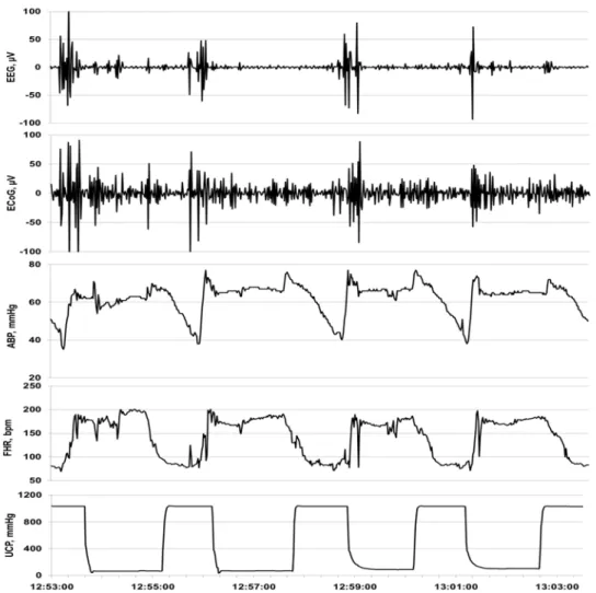
Fig. 1. Exemple d’une réponse ECOG / EEG à des OCO répétitifs. Vue de 10 min d’un patron d’un arrêt cérébral adaptatif visible à l’ECoG et à l’EEG en réponse aux changements de la pression artérielle sanguine (PAS) et du rythme cardiaque fœtal (RCF).
Nous avons montré que le RMSSD est une mesure de maturation de la branche vagale du système nerveux autonome et que cette mesure est réduite par l’atropine (un antagoniste cholinergique) chez le fœtus ovin proche du terme.(13, 16) Le RMSSD augmente également Durant une acidémie sévère (pH ~7.09) induite par 4 minutes d’occlusions du cordon ombilical (OCO) à intervalle de 30 min chez ce même modèle expérimental. (16) Donc le RMSSD est un marqueur potentiel de l’acidémie aggravante.
Dans cette étude, notre objectif est d’évaluer l’impact de taux d’échantillonnage de l’ECG fœtal afin de mesurer le bénéfice d’un monitoring continu de la VRCf dans la prédiction de l’acidémie fœtale. Nous avons testé la performance du RMSSD en tant que mesure de la VRCf et
son comportement en présence d’acidémie fœtale quand acquis à un taux d’échantillonnage de 4 Hz, actuellement utilisé dans les moniteurs de RCF, en comparaison au taux d’échantillonnage
16
expérimental de 1000 Hz. Basé sur des évidences démontrées à partir des études fondamentales chez l’animal et cliniques chez l’homme, nous proposons que le monitoring longitudinal de la
VRCf pendant le travail nous permettra d’améliorer le diagnostic précoce de l’acidémie fœtale , mais influencée par le taux d’échantillonnage de l’ECG.
1.3. Corréler l’analyse multidimensionnelle de la variabilité du rythme cardiaque fœtal avec l’équilibre acido-basique à la naissance
Les agressions cérébrales prénatales et périnatales restent une cause majeure de troubles neurologique et du développement à long terme.(21) Chez les enfants nés à terme, le contributeur le plus important d’une agression et de paralysie cérébrale est l’hypoxie ou l’asphyxie fœtale en intrapartum avec acidémie conséquente.(22) Bien que le monitoring fœtal électronique durant le travail ait considérablement réduit les décès (23) et les convulsions néonatales, il faillit de détecter avec précision, de manière anticipé, une hypoxie/asphyxie fœtale.(24, 25) Cette lacune a contribué à une épidémie de césariennes non nécessaire accompagnée de coûts élevés et de morbidité maternelle, sans pour autant diminuer le taux de paralysie cérébrale.(26) Selon la tendance, on pourrait dire qu’après 40 ans d’application cliniques extensives dans le monde entier, les limites du monitoring de RCF standard ne peuvent plus être surmontées.
Plusieurs raisons peuvent expliquer les raisons pour lesquelles le monitoring de RCF faillit dans la détection précoce d’une hypoxie associée à une acidémie. Premièrement, l’information provenant de l’analyse visuelle de la fréquence cardiaque (FC) et du mode de survenue des contractions utérines est limitée. Deuxièmement, la technologie actuelle de détection des évènements cardiaques fœtaux bioélectriques est limitée par un enregistrement insuffisant du vrai signal électrique. Les sondes trans -abdominales Doppler fonctionnent sur la moyenne des signaux biophysiques. Les électrodes du scalp, en plus d’être invasives, sont filtrées et échantillonnées à basse fréquence (BF), donc détectant un signal QRS avec le moins de bruit possible.(27) Troisièmement, alors que la VRCf est reconnue pour refléter les modulations par le système nerveux autonome, (13, 15) la perte d’information inhérente à ces méthodes minimise les données utilisables pour l’évaluation. L’échelle de temps de discrets évènements de la VRCf nécessite une résolution temporelle de détection de pics R au sein du complexe QRS en moins d’ 1 milliseconde.(15, 28, 29) (30)
17
Récemment, une approche multidimensionnelle de monitoring au chevet du patient a été développée afin de collecter les signaux cardiaques and respiratoires et en analyser la variabilité. La plateforme CIMVA (continuous individualized multiorgan variability analysis), développée par Seely et coll., est un logiciel basé sur la complexité de la science.(31) Cet outil a démontré son utilité dans les unités de soins intensifs pour adultes, en identifiant une septicémie, approximativement 60 heures, avant le diagnostic clinique (32) et pronostiquant choc et échec de l’extubation. (31) De même, Moorman et coll. ont montré que les moniteurs de VRC au niveau des unités de soins intensifs pédiatriques ont contribué à la détection d’une septicémie imminente par le personnel soignant.(33) Ces techniques ouvrent des avenues prometteuses avec une approche totalement différentes du monitoring fœtal, à condition que les signaux bioélectriques soient échantillonnés à une fréquence appropriée.
Dans cette étude, nous présentons une nouvelle méthode de monitoring en intrapartum monitoring basée sur l’électrocardiographie fœtale (ECGf) transabdominale acquise de manière non invasive à partir de la surface de l’abdomen maternelle, avec une fréquence d’échantillonnage suffisamment élevée pour détecter les modulations autonomiques du RCF. L’analyse repose de préférence sur la VRCf que les performances cardiaques en tant que tel.
Nous avons développé un modèle de prédiction mathématiques, utilisant une matrice multidimensionnelle de 101 mesures de la VRCf et CIMVA, la plateforme standardisée et cliniquement testée,(32) pour prédire le pH et l’excès de base (EB) à la naissance avec seulement une heure de monitoring d’ECGf.
1.4. La variabilité du rythme cardiaque fœtal comme indicateur potentiel de l’inflammation
La principale manifestation pathologique de l’inflammation de l’unité fœto- placentaire, la chorioamniotite, affecte 20% des grossesses à terme et plus de 60% grossesses prématurées; est le plus souvent de trouvaille fortuite. (34, 35) La chorioamniotite symptomatique et asymptomatique sont associées avec un risque d’environ 9-fois plus élevé de paralysie cérébrale. (36) Même une inflammation asymptomatique peut inhiber l’angiogenèse placentaire et ainsi moduler l’issue de la grossesse. (37) Ainsi, un nombre important de fœtus est exposé à des degrés variables d’inflammation qui peut influer sur le développement cérébral. Les méthodes actuelles de
18
diagnostic de détresse fœtale due à une condition infectieuse ou inflammatoire sont inadéquates. (38, 39) Il est urgent d’identifier les marqueurs précoces de risque foetal d’issues défavorables afin d’intervenir thérapeutiquement. (38, 39)
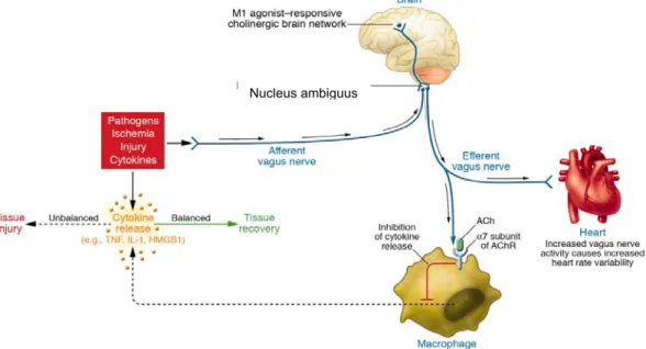
Via le nerf vague, la voie cholinergique anti inflammatoire (VCA) chez le fœtus entraine une rétroaction négative sur les niveaux systémiques des cytokines inflammatoires (Fig. 2-4). Ce contrôle homéocinétique rapide du milieu inflammatoire est reflété dans de subtiles modifications de la VRCf (Fig. 2).
Les variations de l’activité vagale chez le fœtus peuvent être mesurées de manière non invasive par le monitoring continue de la VRCf. (15, 40) Evidemment, de telles VRCf battements par battements sont plus précises que la moyenne des VRCf calculée dans le temps utilisée actuellement en clinique. Les études périnatales montrent que la VRCf monitorée battement par battement a le potentiel d’être une mesure non invasive, continue, sensible et spécifique de la réponse inflammatoire fœtale. (41-44)
Les mesures de la VRC peuvent dériver de plusieurs domaines analytiques du signal. Le RMSSD (Root Mean Square of Standard Deviation), une mesure à court-terme de la VRC, et d’autres mesures de complexité et de signaux liées à d’autres domaines, reflétant l’échelle de temps à court terme de la VRCf, peuvent servir d’indicateurs de cette modulation vagale complexe de l’activité inflammatoire (Fig. 2).
Fig. 2. Réseau de la voie cholinergique anti-inflammatoire (CAP), qui équilibre la production de cytokine. Les pathogènes aussi bien qu’une ischémie et autres formes d’agression activent la production de cytokines, laquelle restaure un état de santé normal. Cependant, si la réponse des cytokines est insuffisante ou excessive, alors ces mêmes
19
médiateurs causent des maladies. Les signaux efférents provenant du nerf vague inhibent la production de cytokines à travers des voies dépendant de la sous-unité α7 du récepteur d’acetylcholine (AChR) présent sur les macrophages et d’autres cellules. L’activité efférente du nerf vague augmente également la VRC instantanément (HRV). Les agonistes muscariniques M1 agissant sur le circuit cholinergique peut augmenter l’activité de la VAC et augmente aussi la VRC instantanément. Les signaux afférents transportés par le nerf vague peuvent active une réponse efférente qui inhibe la libération des cytokines, c’est le réflexe inflammatoire. (45)
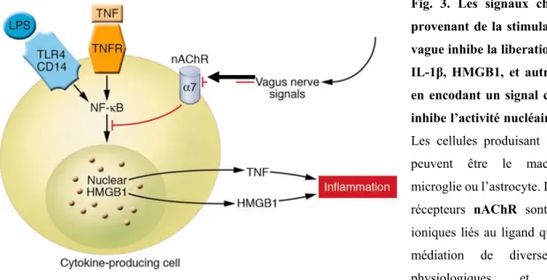
Fig. 3. Les signaux cholinergiques provenant de la stimulation du nerf vague inhibe la liberation de TNF-α, IL-1β, HMGB1, et autres cytokines en encodant un signal cellulaire qui inhibe l’activité nucléaire de NF-κB. Les cellules produisant les cytokines peuvent être le macrophage, la microglie ou l’astrocyte. La famille des récepteurs nAChR sont des canaux ioniques liés au ligand qui assurent la médiation de diverses fonctions physiologiques et ont été originellement identifiés au niveau du système nerveux. Ils consistent en différents sous-types formés par un assemblage spécifique de cinq sous-unités polypeptide incluant α1-10, β1-4, γ, δ, et ε. Les sous-unités sont divisées en 2 groupes : les récepteurs nicotiniques neuronaux (formés de α2–10 et β2–4) et des récepteurs nicotiniques musculaires (formés de α1, β1, γ, δ, et ε). Les sous-types neuronaux fonctionnelles de nAChR sont soit homomériques (formés de 5 sous-unités α identiques, telle que α7- ou α9 nAChR) ou hétéromérique (formé des combinaisons de sous-unités α et β, tels que α3β2 nAChR). Remarquablement, c’est le récepteur α7 nAChR qui est requis pour les effets de la VAC sur les cellules sur les cellules immunitaires innées cérébrales et périphériques et les. TNFR est l’abréviation pour le récepteur TNF. (45-48)
20
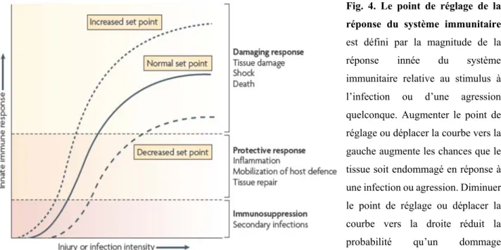
Fig. 4. Le point de réglage de la réponse du système immunitaire est défini par la magnitude de la réponse innée du système immunitaire relative au stimulus à l’infection ou d’une agression quelconque. Augmenter le point de réglage ou déplacer la courbe vers la gauche augmente les chances que le tissue soit endommagé en réponse à une infection ou agression. Diminuer le point de réglage ou déplacer la courbe vers la droite réduit la probabilité qu’un dommage tissulaire puisse arriver. La VCA est le circuit neural qui fournit une entrée compensatoire aigue afin d’ajuster la magnitude de la réponse immunitaire relative au point de réglage. (46)
Fig. 5. Interactions entre les systèmes nerveux, immunitaires et cardiaques. Les pathogènes ou cytokines envoient des impulsions au tronc cérébral via les nerfs afférents. Les signaux autonomiques afférents sont aussi déclenchés par les barorécepteurs en réponse aux changements de la pression sanguine. Les nerfs sympathiques et parasympathiques (vague) envoient alors des signaux efférents au niveau du nœud sino auriculaire (SA), menant, respectivement, à des accélérations et décélérations du rythme cardiaque (RC). Dans la septicémie, la VRC diminue avec quelques petites accélérations et décélérations, probablement reflétant une dérégulation des réponses autonomes. Les fœtus ou les nouveau-nés avec septicémie peuvent avoir une VRC diminuée et occasionnellement de larges décélérations. Le système nerveux autonome (SNA), en plus de réguler la VRC, joue également un rôle important dans la défense de l’hôte en envoyant des signaux adrénergiques et cholinergiques (via la voie cholinergique
anti-21
inflammatoire, VCA) à la périphérie modulant la libération des médiateurs inflammatoires tels que les cytokines. Cf. Fig. 2-4 ci-dessus. (44)
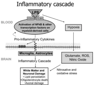
L’inflammation induite par le lipopolysaccharide (LPS) chez le fœtus ovin est un modèle bien établi de la réponse inflammatoire chez le fœtus humain à la septicémie (Fig. 5-6). Cependant, aucune étude n’a utilisé les mesures de VRCf pour décrire le processus inflammatoire. (cf. Fig. 5).
Fig. 6. Cascade inflammatoire en présence du LPS
Nous émettons l’hypothèse que les différents patrons de la corrélation de la VRCf aux cytokines pro-inflammatoires refléteront les états de réponses spontanées versus inflammatoires de la VAC.
22 2. PUBLICATIONS
23
2.1 Adaptive shut-down of EEG activity predicts critical acidemia in the near-term ovine fetus
1Martin G. FRASCH, MD, PhD, 1L. Daniel DUROSIER, MD, 1Mingju CAO, PhD, 2Brad MATUSHEWSKI, MSc, 3Lynn KEENLISIDE, 4Yoram LOUZOUN, PhD, 5Michael G. ROSS, MD, MPH, 2Bryan S. RICHARDSON, MD
1CHU Ste-Justine Research Center, Dept. of Obstetrics and Gynaecology, Université de Montréal, QC, Canada, 2Dept. of Obstetrics and Gynecology, Univ. Western Ontario, London, Ontario, Canada, 3Imaging Program, Lawson Health Research Institute, London, ON, 4Department of Mathematics, Bar-Ilan University, Ramat-Gan, Israel,
5Department of Obstetrics & Gynecology, LA BioMed at Harbor-UCLA Med. Ctr., Torrance, CA
Disclosure: BSR and MGF are inventors of related patent applications entitled “EEG Monitor of
Fetal Health” including U.S. Patent Application Serial No. 12/532,874 and CA 2681926 National Stage Entries of PCT/CA08/00580 filed March 28, 2008, with priority to US provisional patent application 60/908,587, filed March 28, 2007. No other disclosures have been made.
Source of Financial Support: Canada Research Chair Tier 1 in Fetal and Neonatal Health and
Development (BSR); CIHR and FRSQ (MGF); Women's Development Council, London Health Sciences Centre, London, ON, Canada (BSR, MGR, MGF).
Presentation Information: Presented in part at Society for Gynecologic Investigation Annual
Meetings 2012 and 2013.
Short Title: Fetal EEG – HR monitoring predicts fetal acidemia Please address reprint requests and correspondence to:
Martin G. Frasch
Département d'obstétrique-gynécologie Université de Montréal
CHU Sainte-Justine Centre de Recherche 3175, chemin de la Côte Ste-Catherine
24 Montréal, Québec H3T 1C5
Canada
Phone: +1-514-345-4931 x4048 Fax: +1-514-345-4648
25
ABSTRACT
In fetal sheep, the electrocorticogram (ECOG) recorded directly from the cortex during repetitive heart rate (FHR) decelerations predictably correlates with worsening hypoxic-acidemia. In human fetal monitoring during labour, the equivalent electroenkephalogram (EEG) can be recorded non-invasively from the sculp. We tested the hypothesis that fetal EEG – FHR monitoring allows for early detection of worsening hypoxic-acidemia similar to that shown for ECOG-FHR monitoring. Near term fetal sheep were chronically instrumented with arterial and venous catheters, ECG, ECOG and EEG electrodes and umbilical cord occluder, followed by four days of recovery. Prior to study, fetuses were identified as normoxic (n=9, N/UCO group) or chronically hypoxic (n=5, arterial O2Sat<55%, H/UCO group). One minutes lasting repetitive umbilical cord occlusions (UCO) of increasing strength were induced each 2.5 minutes until pH dropped to <7.00. Repetitive UCOs led to marked acidosis (arterial pH 7.35±0.01 to 7.00±0.01). In all groups, at pH of 7.20, 53 min prior to pH<7.00, both ECOG and EEG amplitudes decreased ~4 fold during each FHR deceleration in a synchronized manner. In the H/UCO group, fetal arterial blood pressure failed to show physiological increases during the repetitive UCOs, while in the N/UCO groups this was only observed once ECOG/EEG amplitude consistently decreased during the FHR decelerations. This suggests an adaptive brain shut-down mechanism that is triggered neurally regardless of preexisting hypoxia. Confirming our hypothesis, these findings provide proof of principle for fetal EEG as a useful adjunct to FHR monitoring during high-risk human labour for early detection of incipient fetal acidemia.
Word count: 247
26
INTRODUCTION
Human clinical studies indicate an increasing risk for neonatal adverse outcome and longer-term sequellae including cerebral palsy with umbilical cord pH values <7.00. ((Liston et al., 2002a, Liston et al., 2002b, Liston et al., 2007) This is supported by studies in the ovine fetus showing that pre-existing hypoxia alters cerebral and cardiovascular responses to labour-like umbilical cord occlusions (UCOs). (Gardner et al., 2002, Fletcher et al., 2006, Wassink et al., 2013) This has led to the use of electronic fetal heart rate (FHR) monitoring as the main stay for the assessment of fetal health during labour. (Liston et al., 2002a, Liston et al., 2002b, Liston et al., 2007) The absence of FHR decelerations along with presence of FHR variability are highly predictive for normal fetal blood gas/pH at birth. (Liston et al., 2002a, Liston et al., 2002b, Liston et al., 2007) However, clinical FHR monitoring has a low positive predictive value for concerning acidemia at birth (~50%) with need for improving existing technologies for the detection of fetal hypoxic-acidemia during labour.(Liston et al., 2002a, Liston et al., 2002b, Liston et al., 2007)
We recently studied patterns of electrocortical activity (ECOG) recorded from the cortex and FHR in the near term ovine fetus in response to repetitive UCOs insults as might be seen in human labour, to delineate the time-course and correlation of ECOG change with worsening acidemia.(Frasch et al., 2011) There were consistent changes in ECOG with amplitude suppression and frequency increase during FHR decelerations accompanied by pathological decreases in fetal arterial blood pressure (ABP). These changes in ECOG suggested an “adaptive brain shutdown” and occurred on average 50 minutes prior to attaining a severe degree of acidemia (i.e., fetal arterial pH<7.00).
As a first step toward implementing this technology in human labour surveillance of fetal well-being we have shown that fetal electroencephalogram (EEG), the clinically available equivalent of the ECOG, can be acquired with a modified FHR scalp electrode. (Frasch et al., 2010); (Frasch et al., 2012) Accordingly, this ancillary surveillance modality during labour could be added easily and cost-effectively to the current electronic FHR monitoring that is widely used. (Liston et al., 2007) We hypothesized that fetal EEG recorded from a modified FHR scalp electrode will allow for early detection of worsening acidemia similar to our previous findings for fetal ECOG (Prout et al., 2010, Frasch et al., 2011) and regardless of preexisting hypoxia. Consequently, in the present study after optimizing EEG signal acquisition using a modified FHR scalp electrode similar to that used during human labour, we subjected near term ovine fetuses to
27
repetitive UCO insults and compared the fetal ECOG and EEG responses during worsening acidemia.
28
MATERIALS AND METHODS Surgical preparation
Fourteen near-term ovine fetuses of 123±1 days gestational age (GA), (term=145 days) of mixed breed were surgically instrumented. The anesthetic and surgical procedures and postoperative care of the animals have been previously described (Kaneko et al., 2003, Frasch et al., 2009). Briefly, polyvinyl catheters were placed in the right and left brachiocephalic arteries and the right cephalic vein. Stainless steel electrodes were sewn onto the fetal chest to monitor the electrocardiogram (ECG). Stainless steel electrodes were additionally implanted biparietally on the dura for the recording of ECOG. A modified double spiral FHR electrode was placed midline just anterior to the ECOG electrodes to acquire EEG as successfully tested(Liston et al., 2007). An inflatable silicon rubber cuff (In Vivo Metric, Healdsburg, CA) for UCO induction was also placed around the proximal portion of the umbilical cord and secured to the abdominal skin. Once the fetus was returned to the uterus, a catheter was placed in the amniotic fluid cavity and another in the maternal femoral vein. Antibiotics were administered intravenously to the mother (0.2 g trimethoprim and 1.2 g sulfadoxine, Schering Canada Inc., Pointe-Claire, Canada) and the fetus and into the amniotic cavity (1 million IU penicillin G sodium, Pharmaceutical Partners of Canada, Richmond Hill, Canada). Amniotic fluid lost during surgery was replaced with warm saline. The uterus and abdominal wall incisions were sutured in layers and the catheters exteriorized through the maternal flank and secured to the back of the ewe in a plastic pouch.
Postoperatively, animals were allowed four days to recover prior to experimentation and daily antibiotic administration was continued. Arterial blood was sampled for evaluation of fetal condition and catheters were flushed with heparinized saline to maintain patency. Animals were 129±1 days GA on the first day of experimental study. Animal care followed the guidelines of the Canadian Council on Animal Care and was approved by the University of Western Ontario Council on Animal Care.
Experimental procedure
The animals were studied over a ~6 hour period in two groups. The first group comprised five fetuses that were found to be spontaneously hypoxic with arterial O2Sat<55% as measured on post-operative days 1 to 3 and at baseline prior to beginning the UCOs and formed an H/UCO group. This is in contrast to the second group of nine fetuses that were normoxic with O2Sat>55% before subjected to UCOs and formed a N/UCO group. After a 1-2 hour baseline control period,
29
both groups of animals underwent mild, moderate and severe series of repetitive UCOs by graduated inflation of the occluder cuff with a saline solution. During the first hour following the baseline period, mild variable FHR decelerations were performed with a partial UCO for 1 minute duration every 2.5 minutes, with the goal of decreasing fetal heart rate by ~30 bpm, corresponding to an ~50% reduction in umbilical blood flow. (Itskovitz et al., 1983, Richardson et al., 1989) During the second hour, moderate variable FHR decelerations were performed with increased partial UCO for 1 minute duration every 2.5 minutes with the goal of decreasing fetal heart rate by ~60 bpm, corresponding to an ~75% reduction in umbilical blood flow. (Itskovitz et al., 1983, Richardson et al., 1989) Animals then underwent severe variable FHR decelerations with complete UCO for 1 minute duration every 2.5 minutes until the targeted fetal arterial pH of less than 7.0 was detected or 2 hours of severe UCO had been carried out, at which point the repetitive UCOs were terminated. All animals were then allowed to recover for 48 hours following the last UCO. Fetal arterial blood samples were drawn at baseline, at the end of the first UCO of each series (mild, moderate, severe), and at 20 minute intervals (between UCOs) throughout each of the series, as well as at 1, 24 and 48 hours of recovery. For each UCO series blood gas sample and the 24 h recovery sample, 0.7 ml of fetal blood was withdrawn, while 4 ml of fetal blood was withdrawn at baseline, at pH nadir less than 7.00, and at 1 hour and 48 hours of recovery. The amounts of blood withdrawn were documented for each fetus and replaced with an equivalent volume of maternal blood at the end of day 1 of study.
All blood samples were analyzed for blood gas values, pH, glucose, and lactate with an ABL-725 blood gas analyzer (Radiometer Medical, Copenhagen, Denmark) with temperature corrected to 39.0°C. Plasma from the 4 ml blood samples was frozen and stored for cytokine analysis, and will be reported separately.
After the 48 hour recovery blood sample, the ewe and the fetus were killed by an overdose of barbiturate (30mg sodium pentobarbital IV, MTC Pharmaceuticals, Cambridge, Canada). A post mortem was carried out during which fetal sex and weight were determined and the location and function of the umbilical occluder were confirmed. The fetal brain was perfusion fixed and subsequently dissected and processed for later immunohistochemical study (data reported separately) as previously reported. (Keen et al., 2011)
30
A computerized data acquisition system was used to record fetal arterial and amniotic pressures, the ECG, ECOG and EEG electrical signals, as previously described (Richardson and Gagnon, 2008), which were monitored continuously throughout the baseline, UCO series, and first hour of the recovery period. Arterial and amniotic pressures were measured using Statham pressure transducers (P23 ID; Gould Inc., Oxnard, CA). Arterial blood pressure (ABP) was determined as the difference between instantaneous values of arterial and amniotic pressures. A PowerLab system was used for data acquisition and analysis (Chart 7 For Windows, ADInstruments Pty Ltd, Castle Hill, Australia).
Pressures, ECG, ECOG and EEG were recorded and digitized at 1000 Hz for further study. For ECG, a 60 Hz notch filter was applied, while for ECOG and EEG, a band pass 0.3 - 30 Hz filter was used. FHR was triggered and calculated online from arterial pressure systolic peaks.
Averaged values of FHR and ABP were calculated from artifact-free recordings of one hour of baseline, as well as between and during each consecutive variable FHR deceleration induced by the mild, moderate, and severe UCOs as previously reported.(Ross et al., 2013b) FHRnadir was measured as the minimal FHR during a UCO; ABPmax was measured as maximal ABP during a UCO; ABPmin was measured as the minimal ABP during a UCO; ΔFHR was calculated as FHR deceleration depth during UCO, i.e., the difference between mean FHR between UCO and FHRnadir; ΔABP was calculated as the difference between ABPmax and mean ABP between UCO; ΔABPUCO was calculated as the difference between ABPmax and ABPmin to capture the known biphasic change of ABP during each UCO.
The ECOG and EEG signals were sampled down to 100 Hz prior to the ECOG and EEG analysis. Subsequently, the voltage amplitude and 95% spectral edge frequency (SEF), this being the ECOG/EEG frequency below which 95% of ECOG/EEG spectral power is found, were calculated over 3 second intervals for the duration of the experimental monitoring. For each animal, mean values of the ECOG and EEG amplitudes and SEF were determined at baseline as well as during and between UCO for each of the deceleration series. To track the correlation between the ECOG/EEG and FHR, we determined the cross-correlation function (CCF) between the smoothed ECOG/EEG amplitudes (absolute value of the ECOG/EEG signals) and the smoothed FHR with a square smoothing kernel of 10 seconds with delays of -100 to 100 seconds (Matlab, Mathworks, Natick, MA, USA). CCF analysis tests the spectrographic similarity of ECOG and EEG recordings. The CCF were normalized to 1 and maxima of the normalized CCF were determined. The closer
31
these maxima (CCFM) are to 1, the higher is the correlation between both signals. We then compared the correlation between the different levels of occlusion (i.e., baseline, mild, moderate and severe UCO) as a function of the delay:
( )
(( ( ) ( ))( (( ) ( ) ) ( )) xy E x t E x y t E y C x y τ τ σ σ − − − =where
σ
( )x is its standard deviation, and E(x) is its expected value.To further validate the degree of synchronization between ECOG/EEG amplitudes and FHR, we compared the coherence in the 0.01-0.1 Hz band - representing the 10-100 seconds locking period - to all other frequency bands. 0.01-0.1 Hz represents the expected coherence time scale between these two signals (Low et al., 1995)_ENREF_13. The spectral coherence is the ratio between the squared Fourier transform of the cross-correlation function divided by the Fourier transform of the correlation function of each signal by itself. Since the correlated ECOG/EEG-FHR activities were consistently observed during the severe UCO series, the above comparisons were made accordingly between the severe UCO series versus all the preceding experimental stages (i.e., baseline, mild and moderate UCO).
Statistical analysis
Normal data distribution was tested using Kolmogorov-Smirnov test followed by parametric or non-parametric tests, as appropriate. Arterial pH and BD measurements in response to repetitive UCOs and associated variable decelerations were compared with the corresponding baseline values by one-way repeated-measures analysis of variance (ANOVA) with Student- Newman-Keuls post hoc analysis. One-way repeated measures ANOVA followed by Holm-Sidak (versus baseline) or Student-Newman-Keuls (pairwise) tests for multiple comparisons have been used to assess differences in ECOG/EEG and cardiovascular responses to UCO within N/UCO and H/UCO groups. Differences between ECOG and EEG within each group were assessed using t-test or signed rank test. Differences in ECOG/EEG and cardiovascular alterations during ECOG/EEG-FHR synchronized pattern were tested using t-test or signed rank test. Differences between the ECOG/EEG and cardiovascular variables of the N/UCO and H/UCO groups at each time point were assessed using rank-sum test. No adjustment for multiple comparison was undertaken at this point.(Rothman, 1990) A two-sided rank sum test was used to detect changes in CCF and spectral coherence between the ECOG/EEG amplitudes and FHR during the severe UCO series versus the preceding stages of the experiment (i.e., baseline, mild and moderate UCO).
32
All values are expressed as means ± SEM. Statistical significance was assumed for P <.05. Pearson or Spearman correlation analysis was performed as appropriate, and R values are presented where P < .05 (SPSS 19; IBM, Armonk, NY).
33
RESULTS
Responses of normoxic and hypoxic fetuses to umbilical cord occlusions
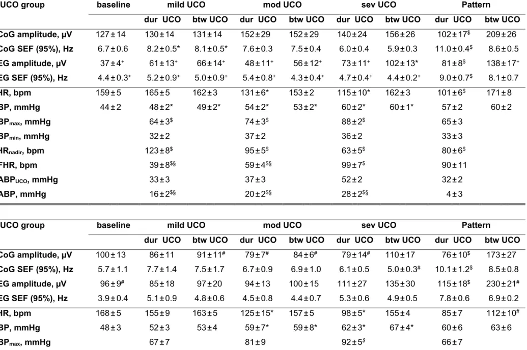
All results are summarized in Table 1 and Fig. 2. We present responses of the N/UCO group and highlight the H/UCO group responses where they were different from the N/UCO group.
In the N/UCO group, during the baseline period, fetal arterial pH (7.35±0.01) as well as FHR (159±5 bpm) and ABP (44±2 mmHg) were within the physiologic range. This was similar in H/UCO group (fetal arterial pH 7.34±0.01, Table 1). Arterial O2Sat measured 65±2% which was higher than in the H/UCO group where it measured 41±6% (p=0.02).
In the N/UCO group, baseline EEG amplitude measured 37±4 μV which was 3.5x lower than ECOG amplitude at 127±14 μV (p=0.01). Baseline EEG SEF was 4.4±0.3 Hz and 1.5x lower than baseline ECOG SEF at 6.7±0.6 Hz (p<0.01). In contrast, in the H/UCO group, ECOG and EEG amplitude and SEF were similar and EEG amplitude at 96±9 μV was ~2.6fold higher than in the N/UCO group (Table 1).
In both, N/UCO and H/UCO groups, repetitive UCO and associated variable FHR decelerations resulted in development of severe fetal acidosis (N/UCO: pH 7.35±0.01 to 7.00±0.03; BD -1.6±0.7 to 13.6±1.1 mEq/l; H/UCO: pH 7.34±0.01 to 7.01±0.02; BD -2.0±0.7 to 15.5±0.3 mEq/l) by the end of the severe UCO series (all p<0.01).
In the N/UCO group, ABP increased on average to 75±3 mmHg during each UCO versus 54±2 mmHg between each UCO (p<0.05). FHR deceleration depth averaged 66±6 bpm (decreasing to 94±6 during each UCO versus 159±3 bpm between each UCO, p<0.05). In contrast, in the H/UCO group, ABP remained unchanged at 59±5 mmHg during and between each UCO. Consequently, throughout the mild, moderate and severe UCO series, that is, well before the ECOG/EEG-FHR synchronized pattern onset, ABP showed pathologically low increases of 3±1 mmHg on average with each UCO-induced FHR deceleration. FHR deceleration responses in the H/UCO group were similar to those in the N/UCO group as intended.
In the N/UCO group, ECOG began to show cyclical behavior correlated with UCO-induced FHR decelerations starting at a pH of 7.22±0.03 (range 7.32 – 7.07), and 48±11 min (range 1h41min - 20 min) prior to the pH dropping <7.00: ECOG SEF began to consistently increase to 11.0±0.4 Hz from 8.6±0.5 Hz (p<0.01) and ECOG amplitude began to consistently decrease to 102±17 μV from 209±26 μV during versus between each UCO, respectively (p<0.001) (Fig. 1). In the H/UCO group, this dynamics was also observed at a similar pH of 7.23±0.01 (range 7.27 –
34
7.21), and 59±15 min (range 1h34min - 14 min) prior to the pH dropping <7.00 with similar values for ECOG SEF and amplitude (Table 1).
In both N/UCO and H/UCO groups, EEG behaved similarly to the cyclical ECOG behaviour correlated with UCO-induced FHR decelerations. In the N/UCO group, EEG began to show cyclical behaviour correlated with UCO-induced FHR decelerations at a pH of 7.22±0.03 (range 7.32 – 7.07), and 45±9 min (range 1h33min - 20 min) prior to the pH dropping <7.00, SEF began to consistently increase to 9.0±0.7 Hz from 8.1±0.7 Hz (p<0.05) and amplitude began to consistently decrease to 81±8 μV from 138±17 μV during versus between UCO and associated FHR decelerations (p<0.001) (Fig. 1). In the H/UCO group, this EEG dynamics was similar for the amplitude occurring at a pH of 7.20±0.03 (range 7.27 – 7.15), and 49±15 min (range 1h18min - 12 min) prior to the pH dropping <7.00; meanwhile EEG SEF did not change during compared to between UCOs averaging 7.4±0.4 Hz. Similar to the observation at the baseline, here we found that in the H/UCO group EEG amplitude between the UCOs was ~1.7fold higher than in the N/UCO group (Table 1). Consequently, while in the N/UCO group EEG measured ~66% of the ECOG amplitude between the UCOs, it was similar to ECOG amplitude at this time in the H/UCO group.
In the N/UCO group, 50±14 min (range 2h06min – 19 min) prior to pH<7.00, ABP began to consistently show pathologically low increases of 4±3 mmHg with each FHR deceleration compared to the dynamics during the mild, moderate and severe UCOs before the ECOG/EEG-FHR synchronized pattern onset (p<0.05, ΔABP in Table 1). The timing of the observed ECOG and EEG amplitude and SEF recurring pattern changes was highly correlated to the timing of the onset of the pathological ΔABP (both R=0.99, p<0.001) (Fig. 1). This cardiovascular behaviour was also observed in the H/UCO group at a similar time of 56±15 min (range 1h32min – 12 min) prior to pH<7.00. Notably, in the H/UCO group – in contrast to the N/UCO group – the pathologically low ABP increases of 3±3 mmHg with each FHR deceleration were similar in magnitude to the ΔABP dynamics during mild, moderate and severe UCO before the ECOG/EEG-FHR synchronized pattern onset (ΔABP in Table 1). That is, in the H/UCO group, the difference of this phenomenon to the ΔABP values before the pattern onset was not in absolute values but in the consistent lack of increase with each occlusion and FHR deceleration. Similar to the N/UCO group, the timing of the onset of the observed ECOG and EEG amplitude and SEF recurring pattern
35
changes was highly correlated to the timing of the onset of the pathological ΔABP pattern (both R=1, p<0.001) for individual animals.
Evidence of ECOG/EEG – FHR synchronization in N/UCO and H/UCO groups
To test for the assumption that ECOG/EEG amplitude and FHR show temporal synchronization early prior to the onset of severe acidemia (Frasch et al., 2011) in N/UCO and H/UCO groups, we computed in each group the correlation between the smoothed ECOG/EEG amplitudes and smoothed FHR with a 10 seconds moving average with a delay ranging from -100 seconds to 100 seconds (Figure 2). In both groups, only during the severe UCO series did a clear correlation with a phase lag appear between ECOG/EEG amplitude and FHR. Of note, the result was more pronounced for EEG-FHR cross-correlation function (CCF) than for ECOG-FHR CCF. If such synchronization exists, we expected the difference between the maximal and minimal correlation as a function of the phase to be maximal. We thus checked the distribution of this difference in all sheep fetuses of the N/UCO and H/UCO groups.
In the N/UCO group, the difference between the maximal and minimal correlation as a function of the phase was significantly larger during the severe UCO series compared to all previous time periods (baseline, mild and moderate UCO series), with an average difference of 0.16±0.08 (ECOG-FHR CCF) and 0.5±0.16 (EEG-FHR CCF) during the severe UCO series vs. 0.09±0.07 (ECOG-FHR CCF) and 0.31±0.14 (EEG-FHR CCF) for all previous time periods (p<0.01 and p<0.001, respectively, Figure 2A). To validate the synchronization (phase locking) between ECOG/EEG amplitude and FHR, we compared the coherence in the 0.01-0.1 Hz band - representing the 10-100 seconds locking period - to all other frequency bands. The average coherence in this band for ECOG-FHR and EEG-FHR was at 0.19±0.02 vs. 0.15±0.04 (p<0.01) and 0.19±0.04 vs. 0.15± 0.05 (p<0.05) indeed significantly higher during the severe UCO series than during all previous time periods.
In the H/UCO group, the difference between the maximal and minimal correlation as a function of the phase was significantly larger during the severe UCO series compared to all previous time periods (baseline, mild and moderate UCO series), with an average difference of 0.21±0.02 (ECOG-FHR CCF) and 0.25±0.02 (EEG-FHR CCF) during the severe UCO series vs. 0.15±0.01 (ECOG-FHR CCF) and 0.16±0.01 (EEG-FHR CCF) for all previous time periods (p<0.01 and p<0.001, respectively, Figure 2B). To validate the synchronization (phase locking) between ECOG/EEG amplitude and FHR, we compared the coherence in the 0.01-0.1 Hz band -
36
representing the 10-100 seconds locking period - to all other frequency bands. The average coherence in this band for ECOG-FHR and EEG-FHR was at 0.21±0.04 vs. 0.15±0.02 (p<0.01) and 0.25±0.05 vs. 0.16±0.02 (p<0.001) again significantly higher during the severe UCO series than during all previous time periods.
37
DISCUSSION
Fetal EEG during labour may permit early detection of acidemia
Our findings provide proof of principle for fetal EEG monitoring during high-risk human labour. EEG recorded from the scalp of near-term fetal sheep shows changes comparable to ECOG during normoxia (Frasch et al., 2010) and during worsening hypoxic-acidemia. (Frasch et al., 2012) Compared to the paradigm of fixed complete UCOs of increasing frequency we tested (Frasch et al., 2011), the frequency of UCOs does not impact on the pattern of the ECOG/EEG amplitude response, but it does impact on the ECOG/EEG frequency characteristics. The next step is to develop reliable online algorithms to detect such variable amplitude and frequency responses of fetal EEG. Overall, this confirms the notion that ECOG activity acquired from supradural electrodes and EEG activity acquired from scalp electrodes should similarly reflect the field potential neuronal activity, albeit with the ECOG amplitude larger than the corresponding EEG signals. Clinical studies with Cerebral Function monitors in newborns with suspected hypoxic-ischemic encephalopathy have demonstrated the feasibility of recording EEG activity from scalp electrodes and predictive value for longer term neurologic sequellae (Thordstein et al., 2004, de Vries and Hellstrom-Westas, 2005). However, this predictive ability relates to existent and evolving injury within the brain with variable degrees of necrotic/apoptotic cell death either primary or delayed (Williams et al., 1991) and the impact on EEG activity, rather than an adaptive suppression of synaptic activity as a protective mechanism. EEG activity as a measure of brain function has also been assessed in the human fetus during labour-related events after rupture of the membranes. Rosen et al. pioneered the human fetal EEG field in the 1970ies by placing two ‘suction-cup’ EEG electrodes transvaginally on the fetal scalp at some distance apart from each other and were able to acquire brain activity during uterine contractions, epidurals and drug administration. (Borgstedt et al., 1975, Chik et al., 1976, Sokol et al., 1977, Frasch et al., 2011, Prout et al., 2012, Ross et al., 2013a, Ross et al., 2013b) However, these studies were hampered by the lack of advanced computer-based technology for analyzing large data sets and the need for multiple scalp electrodes which made large scale clinical usage impractical. Recently, Thaler et al. used real-time power spectral analysis of fetal EEG during labour to facilitate signal processing and interpretation. (Thaler et al., 2000) However, while clearly demonstrating the presence of sleep state cycles in the human fetus, this study was limited to 14 healthy pregnancies with normal outcomes, and again used multiple scalp electrodes to acquire EEG which is not feasible for large
38
scale clinical use. As such, there has been continued need to develop a single transvaginal probe capable of acquiring EEG and FHR signals as an essential first step to ensure the clinical feasibility of monitoring both for the assessment of fetal health during labour.
Our findings overcome the limitations discussed above by providing 1) an EEG probe that can be used practically and by every obstetrician trained in placing the spiral FHR scalp probe during labour and 2) proving that use of such EEG probe would permit an early detection of worsening hypoxic-acidemia allowing for intervention. As an ancillary tool for intrapartum FHR monitoring, fetal EEG monitoring should provide additional decision making power to the delivering obstetrician whether to allow a labour to proceed or deliver acutely, thus minimizing the number of babies born with severe acidemia and increased risk for brain injury at one hand and decreasing the number of unnecessary Caesarian sections at the other hand.
Prospective clinical studies are urgently needed to validate this novel approach to electronic fetal monitoring during labour.
Effects of preceding hypoxia on EEG and cardiovascular responses to umbilical cord occlusions The chief finding is that ECOG amplitude of the H/UCO group fetuses was ~50% that of the N/UCO group for most of the UCO series. This was accompanied by a small but significant decrease in ECOG SEF between the occlusions during the severe UCO series although still remaining within the theta band range. This finding is in line with our previous ECOG findings in this and other laboratories. (Frasch et al., 2011, Wassink et al., 2013) In contrast, EEG amplitude was ~2fold higher in the H/UCO group than in the N/UCO group both at baseline and during the occlusions once the ECOG/EEG – FHR synchronized pattern was observed. This is likely due to artifacts in fetal sheep EEG. We anticipate that human fetal EEG will provide a cleaner signal while retaining the fundamental EEG-FHR pattern we report herein. This is supported by earlier studies that reported well delineated sleep states discernible from fetal EEG acquired during labour. (Thaler et al., 2000)
There was also a pronounced impact of preceding hypoxia on cardiovascular responses to the UCOs. An uncompromised fetus responds with an ABP increase during a UCO, a behaviour that is altered as the occlusions progress and the acidemia increases to the extent that ABP no longer increases during the occlusions. (Frasch et al., 2011) In the present study we observed that this relative change of ABP during each occlusion, ΔABP, was considerably lower throughout all H/UCO series and similar to values seen during the ECOG/EEG-FHR synchronized pattern in
39
N/UCO and H/UCO groups. Moreover, during the ECOG/EEG-FHR synchronized pattern, H/UCO group fetuses showed ~60% lower FHR deceleration depth than N/UCO group fetuses (cf. ΔFHR in Table 1) due to a ~35% lower mean FHR between UCOs. These findings suggest that the ECOG/EEG-FHR synchronized pattern onset, while correlated in time to the onset of the pathological ΔABP, is not solely secondary to the cardiovascular compromise of cerebral auto-regulation, but instead neurally mediated at an arterial pH around 7.20. Furthermore, chronic hypoxia preceding UCO of increasing severity had no impact on the average timing of ~53 minutes prior to pH drop to <7.00 when adaptive brain shut-down was observed. We discuss possible mechanisms linking systemic pH changes with adaptive brain shut-down elsewhere. (Xu et al., 2014)
Noteworthy, we observed hardly any effect of chronic hypoxia on EEG properties suggesting that EEG will readily track fetal brain electrical activity regardless of pre-existing hypoxia. During ECOG/EEG-FHR synchronized pattern, chronic hypoxia ablated the difference in EEG SEF during compared to between each UCO, seen in the N/UCO group. This suggests that efforts in development of robust automated algorithms for pattern detection should aim at using amplitude properties of EEG rather than its frequency properties. Chronic hypoxia likely resulted in ECOG suppression in the H/UCO group fetuses which thereby is the reason we saw no difference between ECOG and EEG amplitude values in the H/UCO group as we did in the N/UCO group fetuses. Growth restricted infants with chronic hypoxemia due to placental dysfunction are at a greater risk for concerning acidemia at birth and thereby subsequent adverse neurological outcomes due to superimposed acute hypoxemia during labour. (Kaneko et al., 2003, Liston et al., 2007, Frasch et al., 2009, Frasch et al., 2011) Our findings show that regardless of preceding hypoxia, the EEG-FHR monitoring in IUGR fetuses would allow detection of EEG-EEG-FHR synchronization pattern as an early warning for impending acidemia.
Fetal adaptive brain shut-down is a pro-active mechanism that is not impacted by preceding hypoxia
The above considerations lead us to propose that fetal adaptive brain shut-down revealed via ECOG/EEG-FHR synchronized behaviour is a pro-active and likely neuroprotective mechanism. First, there is a remarkable consistency of pH at ~7.20, when the pattern appears in N/UCO and H/UCO groups. This suggests an active mechanism triggering the adaptive brain shutdown. This is in line with literature suggesting an adenosine (A1) receptor mediated process of cerebral
40
metabolic shut-down in fetal brain. (Blood et al., 2003, Hunter et al., 2003, Pearce, 2006) In addition, using the same animal model with concurrent measurements of cerebral blood flow and metabolic rates we demonstrated that when cerebral oxygen delivery is severely compromised as during the complete UCOs, ECOG flattens reflecting decreasing synaptic activity as a neuroprotective mechanism. (Kaneko et al., 2003) Second, hypoxia effects are seen mostly in cardiovascular responses and ECOG amplitude (decrease in H/UCO group compared to N/UCO group), but not in the pH or average time when we observed the ECOG/EEG-FHR synchronized pattern. This is also consistent with literature in this animal model. (Keunen and Hasaart, 1999, Pulgar et al., 2007, Wassink et al., 2013)
Significance and future directions
The utility of joint EEG-FHR monitoring is based on consistent emergence of synchronized UCO-triggered EEG-FHR changes prior to reaching a severe degree of fetal acidemia at which brain injury might occur. These changes are due to the mechanism of adaptive brain shut-down triggered at pH ~7.20 and do not depend on preceding cardiovascular behavior due to chronic hypoxia. This makes it likely that the mechanism will be observed in a large population of fetuses with or without preceding hypoxia at labour onset. Fetal EEG monitoring during labour has the potential to serve as a valuable ancillary technique of electronic fetal monitoring. Together with FHR, EEG can provide an early, inexpensive and easily implementable and interpretable tool to accurately predict incipient fetal acidemia in fetuses.
Acknowledgements
The authors wish to thank Maria Sinacori, Carmen Movila, Ashley Keen, Jennifer Thompson, Karolina Piorkowska and Dora Siontas for technical assistance. We further gratefully acknowledge the contribution to the research presented thanks to a workshop organized by the Mathematical Biosciences Institute (MBI) at Ohio State University, Columbus, OH, and the Fields Institute at the University of Toronto.
41 References:
Blood AB, Hunter CJ, Power GG (2003) Adenosine mediates decreased cerebral metabolic rate and increased cerebral blood flow during acute moderate hypoxia in the near-term fetal sheep. J Physiol 553:935-945.
Borgstedt AD, Rosen MG, Chik L, Sokol RJ, Bachelder L, Leo P (1975) Fetal electroencephalography. Relationship to neonatal and one-year developmental neurological examinations in high-risk infants. American Journal of Diseases of Children (1960) 129:35-38.
Chik L, Sokol RJ, Rosen MG, Borgstedt AD (1976) Computer interpreted fetal electroencephalogram. I. Relative frequency of patterns. American Journal of Obstetrics and Gynecology 125:537-540.
de Vries LS, Hellstrom-Westas L (2005) Role of cerebral function monitoring in the newborn. Archives of disease in childhoodFetal and neonatal edition 90:F201-207.
Fletcher AJ, Gardner DS, Edwards CM, Fowden AL, Giussani DA (2006) Development of the ovine fetal cardiovascular defense to hypoxemia towards full term. Am J Physiol Heart Circ Physiol 291:H3023-3034.
Frasch M, Durosier L, Duchatellier C, Richardson B (2012) Fetal sheep
electrocorticogram and electroencephalogram changes accompanying variable fetal heart rate decelerations warn early of acidemia. Reprod Sci 19:F-090.
Frasch M, Keen A, Matushewski B, Richardson B (2010) Comparability of
electroenkephalogram (EEG) versus electrocorticogram (ECOG) in the ovine fetus near term. Reprod Sci 17:51A.
42
Frasch MG, Keen AE, Gagnon R, Ross MG, Richardson BS (2011) Monitoring Fetal Electrocortical Activity during Labour for Predicting Worsening Acidemia: A Prospective Study in the Ovine Fetus Near Term. PloS one 6:e22100.
Frasch MG, Mansano RZ, Gagnon R, Richardson BS, Ross MG (2009) Measures of acidosis with repetitive umbilical cord occlusions leading to fetal asphyxia in the near-term ovine fetus. American Journal of Obstetrics and Gynecology
200:200.e201-207.
Gardner DS, Fletcher AJ, Bloomfield MR, Fowden AL, Giussani DA (2002) Effects of prevailing hypoxaemia, acidaemia or hypoglycaemia upon the cardiovascular,
endocrine and metabolic responses to acute hypoxaemia in the ovine fetus. J Physiol 540:351-366.
Hunter CJ, Bennet L, Power GG, Roelfsema V, Blood AB, Quaedackers JS, George S, Guan J, Gunn AJ (2003) Key neuroprotective role for endogenous adenosine A1 receptor activation during asphyxia in the fetal sheep. Stroke 34:2240-2245.
Itskovitz J, LaGamma EF, Rudolph AM (1983) Heart rate and blood pressure
responses to umbilical cord compression in fetal lambs with special reference to the mechanism of variable deceleration. Am J Obstet Gynecol 147:451-457.
Kaneko M, White S, Homan J, Richardson B (2003) Cerebral blood flow and metabolism in relation to electrocortical activity with severe umbilical cord occlusion in the near-term ovine fetus. Am J Obstet Gynecol 188:961-972.
Keen AE, Frasch MG, Sheehan MA, Matushewski BJ, Richardson BS (2011) Electrocortical activity in the near-term ovine fetus: automated analysis using amplitude frequency components. Brain Res 1402:30-37.