Université de Montréal
The Nuclear Pore Complex and its Transporters:
From Virus-Host Interactors to
Subverting the Innate Antiviral Immunity
par Bridget Gagné
Programmes de biologie moléculaire Faculté de Médecine
Mémoire présenté à la Faculté de Médecine
en vue de l’obtention du grade de maîtrise en sciences (M.Sc.) en biologie moléculaire
option générale
Mai, 2015
Résumé
Les virus ont besoin d’interagir avec des facteurs cellulaires pour se répliquer et se propager dans les cellules d’hôtes. Une étude de l'interactome des protéines du virus d'hépatite C (VHC) par Germain et al. (2014) a permis d'élucider de nouvelles interactions virus-hôte. L'étude a également démontré que la majorité des facteurs de l'hôte n'avaient pas d'effet sur la réplication du virus. Ces travaux suggèrent que la majorité des protéines ont un rôle dans d'autres processus cellulaires tel que la réponse innée antivirale et ciblées pas le virus dans des mécanismes d'évasion immune.
Pour tester cette hypothèse, 132 interactant virus-hôtes ont été sélectionnés et évalués par silençage génique dans un criblage d'ARNi sur la production interferon-beta (IFNB1). Nous avons ainsi observé que les réductions de l'expression de 53 interactants virus-hôte modulent la réponse antivirale innée. Une étude dans les termes de gène d'ontologie (GO) démontre un enrichissement de ces protéines au transport nucléocytoplasmique et au complexe du pore nucléaire. De plus, les gènes associés avec ces termes (CSE1L, KPNB1, RAN, TNPO1 et XPO1) ont été caractérisé comme des interactant de la protéine NS3/4A par Germain et al. (2014) et comme des régulateurs positives de la réponse innée antivirale. Comme le VHC se réplique dans le cytoplasme, nous proposons que ces interactions à des protéines associées avec le noyau confèrent un avantage de réplication et bénéficient au virus en interférant avec des processus cellulaire tel que la réponse innée.
Cette réponse innée antivirale requiert la translocation nucléaire des facteurs transcriptionnelles IRF3 et NF-κB p65 pour la production des IFNs de type I. Un essai de microscopie a été développé afin d'évaluer l’effet du silençage de 60 gènes exprimant des protéines associés au complexe du pore nucléaire et au transport nucléocytoplasmique sur la translocation d’IRF3 et NF-κB p65 par un criblage ARNi lors d’une cinétique d'infection virale.
En conclusion, l’étude démontre qu’il y a plusieurs protéines qui sont impliqués dans le transport de ces facteurs transcriptionnelles pendant une infection virale et peut affecter la production IFNB1 à différents niveaux de la réponse d'immunité antivirale. L'étude aussi suggère que l'effet de ces facteurs de transport sur la réponse innée est peut être un mécanisme d'évasion par des virus comme VHC.
Mots-clés: Virus d’hépatite C, VHC, interactants virus-hôte, immunité innée antivirale, complexe du
pore nucléaire, transport nucléocytoplasmique, criblage ARNi, translocation nucléaire, IRF3, p65, microscopie, cinétique
Abstract
Viruses interact with cellular factors in order to successfully replicate and propagate in host cells. Germain et al. (2014) performed a proteomics analysis to elucidate viral-host interactors of hepatitis C virus (HCV). They found that the majority of host factors did not have an effect on viral replication, suggesting that these host proteins may be beneficial to the virus by affecting other cellular processes such as evading the innate antiviral immunity.
To test that hypothesis, 132 virus-host interactors were selected and silenced by RNAi for their effect on inteferon-beta (IFNB1) production as a readout of the innate antiviral response. 53 were found to modulate the response with enrichment in the gene ontology (GO) terms related to nucleocytoplasmic transport and the nuclear pore complex. An interesting point is that the genes associated with these terms (CSE1L, KPNB1, RAN, TNPO1, and XPO1) were previously elucidated as HCV NS3/4A interactors by Germain et al. (2014), as well as positive regulators of the innate antiviral response. Although it is surprising that a cytoplasmic-replicating virus like HCV would interact with proteins associated with the nucleus, we proposed that viruses interact with these proteins for their benefit to interfere with the innate immune response.
The innate antiviral response requires the nuclear translocation of IRF3 and NF-κB p65 for the production of type I interferons. As it is unclear which transporters or nucleoporins are involved, 60 genes associated with the nuclear pore complex and nucleocytoplasmic transport were studied for their effect on the nuclear translocation of IRF3 and NF-κB p65 via a microscopy-based RNAi screen during a 10-hour viral infection time course.
Overall, the study revealed that many of these proteins are involved in the trafficking of these transcription factors during a viral infection, and can affect the production of IFNB1 at different levels of the innate antiviral response. The study also suggests that the effect of these transport factors on the immune response may be an evasion mechanism for viruses such as HCV.
Keywords: Hepatitis C virus, HCV, virus-host interactors, innate antiviral immunity, nuclear pore
complex, NPC, nucleocytoplasmic transport, RNAi screen, nuclear translocation, IRF3, p65, microscopy, time course, kinetic
Table of Contents
Résumé ... i
Abstract ... ii
Table of Contents ... iii
List of Tables ... vi
List of Figures ... vii
List of Abbreviations & Acronyms... viii
Acknowledgements ... xii Chapter 1: Introduction ... 1 1.1 Hepatitis C Virus... 2 1.1.1 Epidemiology ... 2 1.1.2 Virology ... 3 1.1.2.1 Genome ... 3 1.1.2.2 Life Cycle... 4
1.1.3 Models for the Study of HCV ... 6
1.1.3.1 Replicon System ... 6
1.1.3.2 JFH-1 System ... 7
1.1.3.3 Mouse Models ... 8
1.1.4 HCV-Host Interactors ... 10
1.1.4.1 HCV and the Nuclear Pore Complex and its Transporters ... 12
1.2 Nuclear Pore Complex and Nucleocytoplasmic Transporters ... 13
1.2.1 Nucleocytoplasmic Transport ... 13
1.2.1.1 Protein Import ... 13
1.2.1.2 Protein Export ... 16
1.2.1.3 RAN Gradient ... 19
1.2.1.4 mRNA Export ... 20
1.2.2 Structure of the Nuclear Pore Complex ... 22
1.2.2.1 Cytoplasmic FG-Nups and Filaments ... 22
1.2.2.3 Outer-ring Nups ... 23
1.2.2.4 Linker Nups ... 24
1.2.2.5 Inner-ring Nups ... 24
1.2.2.6 Central FG-Nups ... 24
1.2.2.7 Nuclear FG-Nups and the Nuclear Basket ... 25
1.3 Viruses and the NPC & its Transporters ... 27
1.3.1 Nuclear-Replicating Viruses ... 27 1.3.1.1 DNA Viruses ... 27 1.3.1.2 RNA Viruses ... 28 1.3.2 Cytoplasmic-Replicating Viruses ... 29 1.3.2.1 DNA Viruses ... 29 1.3.2.2 RNA Viruses ... 30
1.4 Innate Antiviral Immune Response ... 31
1.4.1 Early Phase: Virus Recognition by PRRs ... 31
1.4.2 Signaling Cascade ... 32
1.4.2.1 IRFs ... 32
1.4.2.2 NF-κB ... 34
1.4.3 Late Phase: Interferon Activation of the JAK/STAT Pathway ... 34
1.4.3.1 Transcriptional Control by Type I Interferons ... 35
1.4.3.2 Regulation of IFNβ Expression ... 35
1.4.3.3 Type III Interferons ... 36
1.5 NPC and Transporters: Targets for Viral Immune Evasion... 37
1.5.1 Positive ssRNA Viruses ... 37
1.5.2 Negative ssRNA Viruses ... 38
1.5.3 Induced Nucleoporin Expression during an Innate Antiviral Response ... 38
1.6 Hypothesis & Objectives ... 39
Chapter 2: A Microscopy-based RNAi Screen Identifies Key Nucleocytoplasmic Transporters that Control IRF3 and NF-κB Nuclear Translocation and Innate Antiviral Responses Following Viral Infection. ... 40
3.1 Investigation of HCV-Host Interactors in the Modulation of the Innate Antiviral
Response ... 147
3.2 Validation of the Interactors on the Innate Antiviral Response ... 150
3.3 Epistatic Studies of the Interactors on the Innate Antiviral Response ... 151
3.4 Development of a Screen to Measure IRF3 and p65 Nuclear Translocation during Viral Infection ... 159
3.5 Microscopy Results: IRF3 and p65 Translocation... 162
3.6 Perspectives... 169
Conclusion ... 171
Bibliography ... i
Annex 1: Elucidating novel hepatitis C virus-host interactions using combined mass spectrometry and functional genomics approaches. ... xxix
List of Tables
List of Figures
Figure 1.1: HCV Genotype Prevalence by WHO GBD Region ... 2
Figure 1.2: HCV Genome ... 3
Figure 1.3: HCV Life Cycle... 5
Figure 1.4: Replicon System for the Study of HCV ... 6
Figure 1.5: JFH-1 System for the Study of HCV ... 7
Figure 1.6: Mouse Models for the Study of HCV ... 9
Figure 1.7: Protein Import ... 13
Figure 1.8: Protein Export ... 16
Figure 1.9: Kapα Recycling ... 17
Figure 1.10: Phylogeny of Karyopherin-β subfamilies ... 18
Figure 1.11: Recycling of RanGDP and the Renewal of RanGTP ... 19
Figure 1.12: Structure of the Nuclear Pore Complex ... 26
Figure 3.1: Silencing of Host Interactors in Modulation of SeV-mediated Innate Response of Different Human Cell Lines... 149
Figure 3.2: Epistatic study of XPO1 Silencing on the Innate Antiviral Response (1) .... 152
Figure 3.3.: Epistatic study of XPO1 Silencing on the Innate Antiviral Response (2) ... 152
Figure 3.4: Epistatic study of CSE1L Silencing on the Innate Antiviral Response (1) .. 154
Figure 3.5: Epistatic study of CSE1L Silencing on the Innate Antiviral Response (2) .. 154
Figure 3.6: Epistatic study of RAN Silencing on the Innate Antiviral Response (1) ... 155
Figure 3.7: Epistatic study of RAN Silencing on the Innate Antiviral Response (2) ... 156
Figure 3.8: Epistatic study of KPNB1 Silencing on the Innate Antiviral Response ... 157
List of Abbreviations & Acronyms
CARD Caspase-recruitment domainCD81 Cluster of differentiation 81 cGAMP cyclic-GMP-AMP
cGAS cGAMP synthase CLDN1 Claudin-1
CLR C-type lectin receptor cNLS Classical NLS
CSE1L Chromosome segregation 1-like DNA Deoxyribonucleic acid
dsRNA Double-stranded RNA EBV Epstein-Barr virus
EMCV Encephalomyocarditis virus ER Endoplasmic reticulum FG Phenylalanine-Glycine fLuc Firefly luciferase GDP Guanosine diphosphate GTP Guanosine triphosphate HBV Hepatitis B virus HCV Hepatitis C virus
HCMV Human cytomegalovirus
HIV-1 Human immunodeficiency virus type 1 HPV Human papillomavirus
HSV-1 Herpes simplex virus Type 1 IAV Influenza A virus
IFN Interferon IKKε IκB kinase-ε
ISG Interferon-stimulated gene ISRE IFN-stimulated response element JAK1 Janus kinase 1
JFH-1 Japanese patient with fulminant hepatitis Kapα Karyopherin-α
Kapβ Karyopherin-β kDa kiloDalton
KPNB1 Karyopherin (importin) beta 1
LD Lipid droplet
MAVS Mitochondrial antiviral signaling MDa MegaDalton
MDA5 Melanoma differentiation-associated gene 5 mRNA messenger RNA
NES Nuclear export signal NLR NOD-like receptor
NLS Nuclear localization signal
NOD Nucleotide oligomerization domain
NP Nucleoprotein
NPC Nuclear pore complex NS Non-structural
NTR Non-translated region NUTF2 Nuclear transport factor 2 Nup Nucleoporin
NXF1 Nuclear RNA export factor 1
NXT1 Nuclear transport factor 2-like export factor 1 OCLN Occludin
PAMP Pathogen-associated molecular pattern PI4K-IIIα Phosphatidylinositol 4-kinase III α PI4P Phosphatidylinositol-4-phosphate PIC Pre-integration complex
PPIase Prolyl-peptidyl isomerase PRD Positive regulatory domain PRR Pattern-recognition receptor
PRRSV Porcine reproductive and respiratory syndrome virus PY Proline-Tyrosine
RAN RAN, member RAS oncogene family RANGAP Ran GTPase-activating protein
RCC1 Regulator of chromosome condensation 1 RIG-I Retinoic acid-inducible gene 1
RLR RIG-I-like receptor RNA Ribonucleic acid RNAi RNA interference
SARS-CoV Severe acute respiratory syndrome coronavirus SCID Severe combined immunodeficiency
SeV Sendai virus
SR-BI Scavenger receptors class B type I ssRNA Single-stranded RNA
STAT Signal transducer and activator of transcription STING Stimulator of IFN genes
TBK1 TANK-binding kinase 1 TLR Toll-like receptor TNPO1 Transportin 1
TPR Translocated promoter region TYK2 Tyrosine kinase 2
uPA Urokinase-type plasminogen activator VACV Vaccinia virus
VLDL Very low density lipoprotein VSV Vesicular stomatitis virus VZV Varicella-Zoster virus
This is dedicated to everyone who believed in me,
supported me, and pushed me to my limits,
Acknowledgements
I would first like to thank Dr. Daniel Lamarre for giving me the opportunity to pursue my Master’s degree in an amazing laboratory, filled with innovative research projects and inspiring colleagues. He paved the foundation for the solid understanding I now have of my field of study, and a greater appreciation for the world of academic research. Most importantly, his guidance, support and patience were key in completing my memoir, and I cannot thank him enough for the time he spent in making me a better scientist.
I would next like to thank Martin Baril, the research associate and colleague I admired most as he taught me everything I needed to know to do good research in a lab from day one. I will remember his patience and support as a mentor and his enthusiasm as a researcher. He has left a deep impression in my scientific career, as I pushed myself to work hard and produce good results in the hope that someday I will make him proud.
To Nicolas Tremblay, my partner-in-crime and caring friend, I will definitely miss the good times we had in the lab, as well as your personal and professional advice which helped me grow and become a better person during my journey as a Master’s student. To Michael Meloche and Alex Park, my lab brothers who always know how to lighten up my day, and were always fun to work with in the lab. To Salwa Es-Saad, I will always remember your wisdom and kindness. To Bassim Mohammed, I am sure that you will cure HCV one day. To former lab members Laurent Chatel-Chaix and Marie-Anne Germain, also known as Team HCV, your focus, dedication, and commitment to doing good research is definitely not forgotten.
Last, but not least, I would not have gotten this far without the constant support of my family, friends and my special someone through early mornings, late nights and even weekends to further the knowledge of the scientific world by at least one step. I thank them all from the bottom of my heart.
This journey was an emotional rollercoaster for me, and I have learned more about myself and about science than I ever thought possible. Thank you all for this experience.
1.1 Hepatitis C Virus
1.1.1 Epidemiology
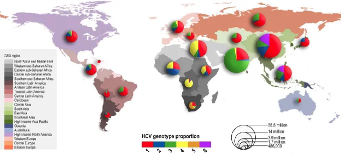
Hepatitis C virus (HCV) currently infects over 185 million individuals around the world [1]. The virus primarily infects hepatocytes, and only 20-30% of those individuals are able to clear the virus during the acute infection [2]. Persistence of the virus causes infected individuals to enter the chronic phase where their condition can lead to several different outcomes of liver disease such as cirrhosis and even hepatocellular carcinoma [2]. Therapies to counteract the virus have become more efficient in recent years, but there is still no cure for HCV globally due to treatment costs and the multiple genotypes of the virus that exist around the world (Figure 1.1) [1].
Figure 1.1: HCV Genotype Prevalence by WHO GBD Region
This figure visually represents the amount of individuals affected by HCV per Global Burden of Disease (GBD) region, as well as which genotypes are most common in each area. Overall, genotype 1 is found in all regions, while the occurrence of the other genotypes varies. (Figure from Messina JP, et al., Hepatology, 2015) [1]
1.1.2 Virology
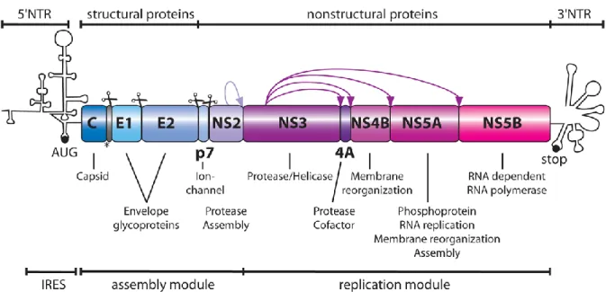
1.1.2.1 GenomeHCV, a member of the Flaviviridae family, is a positive-sense RNA virus, in which its single-stranded genome can be directly translated into a single polyprotein via the internal ribosome entry site (IRES) located on the 5' end of the virus coding sequence. This polyprotein is composed of 10 viral proteins, where the first cleavage by cellular proteases liberates core, E1, E2 and p7 proteins (Figure 1.2). The viral protein NS2 autocleaves itself from NS3, thus allowing the protease function of NS3 to then cleave the rest of the non-structural (NS) proteins: NS4A, NS4B, NS5A, and NS5B [2-4].
Figure 1.2: HCV Genome
This figure shows the HCV genome, as well as which regions encode which proteins. Scissors depict cleavage by cellular proteases, while the purple arrows depict cleavage by the viral proteins. The role of each viral protein in the HCV life cycle is also indicated. (Reprinted with permission from Lohmann V, et al., J Med Chem., 2014. Copyright 2014 American Chemical Society.) [4].
1.1.2.2 Life Cycle
1.1.2.2.1 Viral Entry and Viral RNA Release
Upon viral infection, HCV interacts with SR-BI, CD81, CLDN1 and OCLN on the surface of host cells via the envelope protein E2 and, through a pH-dependent mechanism, the virus is incorporated into the cell via clathrin-mediated endocytosis. Once the virus has entered the cell, its envelope fuses with an acidic endosomal compartment, releasing the viral RNA into the cytoplasm. (Figure 1.3) [3, 5].
1.1.2.2.2 HCV RNA Translation and Polyprotein Processing
The translation of HCV RNA is controlled by the IRES structure at the 5' non-translated region (NTR) of the genome, recruiting the factors necessary for cap-independent translation. The viral RNA is translated into a single polyprotein, which is then cleaved into 10 viral proteins by cellular and viral proteases. The mature proteins associate with ER membranes, as well as the region formed by NS4B called the membranous web, a compartment dedicated to HCV replication [3-5].
1.1.2.2.3 Viral Replication
HCV replication occurs within the membranous web, where the RNA polymerase NS5B will create negative RNA strands from the original positive RNA strand. The negative RNA strands will serve as template to further synthesize more positive-strand HCV RNA, which can then be translated into additional viral proteins or be packaged into newly-formed virions [3, 5].
1.1.2.2.4 Virion Assembly and Egress
Virion assembly is initiated via the interaction between the core protein and HCV RNA. The core protein induces lipid droplet (LD) formation and associates with these structures, which serve as the platform for virus assembly [3, 5]. NS5A binds to the 3' NTR of HCV RNA and transports it from the membranous web to the lipid droplets, where NS5A also plays a role in infectious particle formation [6, 7]. NS2 is also thought to play a role in viral assembly by interacting with multiple HCV proteins, such as E2, p7, NS3 and NS5A, to properly orchestrate their roles in the vicinity of LDs [8, 9]. The newly-assembled virions, composed of HCV RNA, core, E1, and E2, bud from the cells using a mechanism similar to the VLDL secretion machinery. However, it is thought that while the viral particles go through this secretory pathway, p7 protects them by neutralizing acidic compartments in the cell for successful virion release [3, 5, 10].
Figure 1.3: HCV Life Cycle
This figure outlines the major steps of the HCV life cycle from viral entry to viral particle budding from the cell. (Figure from Kim CW, et al., Clin. Mol. Hepatol., 2013) [3].
1.1.3 Models for the Study of HCV
1.1.3.1 Replicon SystemThe replicon system encodes the viral proteins NS3 to NS5B of the HCV genome for the study of HCV replication. The translation of the replicon is mediated by two IRES sequences on the 5’ end of the replicon: the first IRES is from HCV which drives the expression of the neomycin selection marker that is resistant against the cytotoxic compound G418, while the second IRES is from Encephalomyocarditis virus (EMCV) which drives the expression of the five viral proteins necessary for viral replication. The replicon also includes the firefly luciferase (fLuc) gene, whose expression is driven by the HCV IRES, to measure HCV replication by quantifying the luminescence produced. Having a quantifiable readout makes the replicon the model of choice for high-throughput RNAi and chemical screens to identify potential replication co-factors/restriction factors and stimulators/inhibitors, respectively. The replicon system known as Con1b signifies the replicon which contains the HCV 1b genotype. There are variations to the replicon system such as a monocistronic system, and an additional GFP within the NS3 to NS5B region to visually identify cells containing replication complexes in immunofluorescence microscopy. However, the replicon does not represent a complete viral infection [4].
Figure 1.4: Replicon System for the Study of HCV
This figure shows an example of a bicistronic replicon with two viral IRES: the first from HCV encoding the firefly luciferase promoter and selection marker, and the second from EMCV encoding NS3 to NS5B, the non-structural proteins of HCV important for viral replication. (Adapted with permission from Lohmann V, et al., J Med Chem., 2014. Copyright 2014 American Chemical Society.) [4].
5’
3’
HCV IRES
1.1.3.2 JFH-1 System
JFH-1 is a full-length virus that is able to replicate in cell lines, based on the HCV 2a genotype derived from a Japanese patient with fulminant hepatitis. To increase infectivity, multiple chimeras were created, one of which is JC1. JC1 is a chimeric HCV genome composed of J6 (encoding core to NS2 from another genotype 2a isolate), and JFH-1 (encoding NS3 to NS5B), which yields a viral titer 1000 times more efficient than the original JFH-1 genome [4]. Just like in the replicon, JC1 has also been modified to contain a luciferase gene, in this case renilla luciferase (RLuc), to measure the effects on the entire HCV genome, which can be used in high-throughput RNAi and chemical compound screens, as well as other constructs containing fluorescent proteins for visualization in microscopy [4].
There have been many mutations in the JFH-1 system in order to boost viral titers, but permissive cell lines such as Huh-7.5 can also increase viral replication due to a point mutation in RIG-I, a cytoplasmic RNA sensor, which disrupts the activation of IRF3 for the production of ISGs to induce an antiviral state in the cell [11, 12].
Figure 1.5: JFH-1 System for the Study of HCV
Figure 1.5.a shows the original JFH-1 genome. Figure 1.5.b shows one of the many chimeric versions of JC1 with the viral proteins from J6 and JFH-1 in dark grey and grey, respectively. The particular JC1 chimera shown in (b) is called J6/JFH-1/p7RLuc2a due to the inclusion of a RLuc promoter between viral proteins p7 and 2a. (Adapted with permission from Lohmann V, et al., J Med Chem., 2014. Copyright 2014 American Chemical Society.) [4].
a
1.1.3.3 Mouse Models
Mice have been used as a model to study HCV infection in vivo. The first method, viral adaptation, exposes the virus to mouse cells, which causes mutations in E1 and E2 in order to adapt to the murine version of entry factors CD81 and OCLN. This mouse-adapted virus was then tested on immortalized mouse liver cell lines, where the entire life cycle of the virus successfully occurs. However, these were in cell lines with an impaired innate antiviral immunity. Since this method involves the virus adapting to murine cell entry factors, studying viral entry in this model may be influenced by the mutations required to effectively enter mouse cells [13].
The second method uses mice transgenically-expressing HCV or human proteins in the liver. This type of study was used to understand the interactions between viral proteins and the effects on the host cell in an in vivo model. Expression of single viral proteins did not have an effect, but the expression of multiple proteins together caused hepatocellular carcinoma. The most well-known transgenic mice is the FL-N/35 mouse expressing the entire HCV polyprotein at close to physiological levels, where hepatic steatosis, increased liver fibrosis and increased risk of developing hepatocellular carcinoma was described. Another transgenic mice is the genetically-humanized mouse model, which involves the expression of human entry factors, such as CD81 and OCLN, delivered to the mouse via an adenoviral vector. The human wild-type virus is unable to replicate properly in murine liver cells unless the mouse is immune deficient, which then allows for a persistent infection. It is not yet known what kind of liver disease this mice develop due to this persistent infection [13].
The third method to study HCV in mice is the xenotransplantation model, which involves immunodeficient mice suffering from a liver injury. The immunodeficiency is required to prevent the rejection of the primary human hepatocyte transplantation, and the damaged liver is required for the human hepatocytes to have a growth advantage over the murine ones. This specific mouse model is the most used to study HCV, but this human-liver mouse chimera can only support the infection of HCV clones that are not based on JFH-1 [13]. Another mice model utilizing a human-liver chimera was the uPA-SCID mouse, which was homozygous for the uPA-transgene, the overexpression of which results in liver dysfunction.
the JFH-1 system or from viruses derived from patients of all genotypes [15], allowing for the study of the entire HCV life cycle, as well as a model to test antiviral therapies. One method tries to overcome the immunodeficiency aspect of these mice models by engrafting human hepatocytes with human CD34+ hematopoietic stem cells. However, even though one group was able to demonstrate a T-cell response to HCV infection, viral RNA could not be detected and the immune activity of CD34+ hematopoietic stem cells is suboptimal, thus making it too early to say whether this model will allow for the study of the adaptive immune response to HCV infection [13].
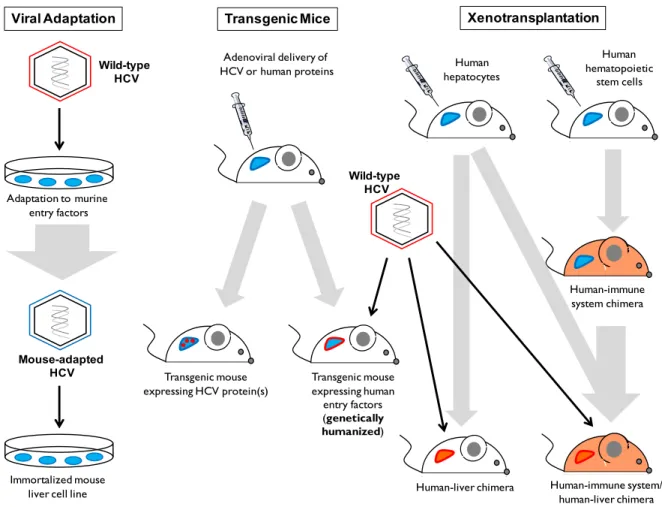
Figure 1.6: Mouse Models for the Study of HCV
This figure shows the three different mouse models: viral adaptation, transgenic mice and xenotransplantation. Viral adaptation involves the wild-type HCV to adapt to murine entry factors in order to successfully replicate in immortalize mouse cell lines. Transgenic mice involve the expression of HCV proteins. One method involves the transmission of human
Viral Adaptation Transgenic Mice Xenotransplantation
Wild-type HCV Mouse-adapted HCV Adaptation to murine entry factors Immortalized mouse liver cell line
Transgenic mouse expressing HCV protein(s) Transgenic mouse expressing human entry factors (genetically humanized) Adenoviral delivery of
HCV or human proteins hepatocytesHuman
Human hematopoietic stem cells Wild-type HCV Human-liver chimera Human-immune system chimera Human-immune system/ human-liver chimera
entry factors, delivered by an adenovirus, to support wild-type HCV infection. The third model involves the transplantation of human hepatocytes in immunodeficient mice with a damaged liver to favour the growth of human hepatocytes in the mouse liver. Mouse models using human immune cells and the human-liver chimera are still not well-characterized for the study of HCV infection and the adaptive immunity [13]. Blue indicates murine cells or liver, while red and orange indicates human liver or human immune system.
1.1.4 HCV-Host Interactors
HCV requires cellular factors in order to effectively replicate, assemble, and propagate to other host cells for its infection. Several RNAi studies have implicated a multitude of host proteins to be pro-viral factors or restriction factors for the different steps of the HCV life cycle [16, 17], but not many have addressed whether these host factors interact with viral proteins to do so. Other studies have identified HCV-host interactors, but have not yet elucidated the importance of all identified interactions to the HCV life cycle [18, 19].
Step of the Life Cycle HCV Genome Host Factor Effect of the Interaction
Cell Entry E1 & E2
CD81 CDLN1
OCLN SR-BI
The E1E2 heterodimer interacts with cell surface receptors and tight junction proteins for HCV
particle binding and entry into host cells [20]
IRES-mediated translation
5' NTR miR-122
miR-122 interacts with the 5' NTR of HCV to stimulate translation through the enhanced association of ribosomes with the viral RNA [21]
Replication 3' NTR miR-122 miR-122 interaction with the 3' NTR of HCV
reduces viral RNA expression [22]
Replication 5' NTR miR-122 miR-122 interacts with the 5' NTR of the HCV
genome to facilitate replication [23]
Replication NS3/4A YBX1 YBX1 interacts with NS3/4A and associates with
viral RNA for replication [24, 25]
Replication NS4B ATF6 ATF6, part of the unfolded protein response, is
Replication NS5A FKBP8 NS5A interacts with FKBP8 and Hsp90 to form a complex that is important for RNA replication [28]
Replication NS5A Hsp90
Replication NS5A PI4K-IIIα
NS5A interacts with PI4K-IIIα for proper membranous web formation by increasing levels of
PI4P in this region [29-31]
Replication NS5A Raf-1
NS5A interacts with Raf-1, causing Raf-1 to phosphorylate. Inhibition of Raf-1 reduced viral
replication [32]
Replication NS5A TIP47 NS5A interacts with TIP47 for efficient viral
replication [33]
Replication NS5A VAPA VAPA interacts with NS5A to positively regulate
viral RNA replication [34]
Replication NS5B Cyclophilin
A
Cyclophilin A is recruited to the replication complex by NS5B for its PPIase activity [35]
Replication NS5B HNRNPA1 HNRNPA1 interacts with NS5B, as well as the 5'
and 3' NTR of HCV RNA for viral replication [36] Viral Particle
Assembly
Core
NS3/4A YBX1
In a complex with DDX6, C1QBP, IGF2BP2 and LARP1, YBX1, a NS3/4A interactor, is re-localized to LDs by core to regulate the assembly
of viral particles [24, 25] Viral Particle
Assembly
HCV
RNA HNRNPK
HNRNPK interacts with HCV RNA and co-localizes with core protein and LDs for regulating
viral RNA incorporation into virions [37] Viral Particle
Assembly NS5A Rab18
NS5A interacts with Rab18 to associate with LDs [38]
Table 1.1: HCV-Host Interactors and their Effect on the HCV Life Cycle
The above table is a non-exhaustive list of characterized HCV-host interactors and their implication at various stages of the viral life cycle.
1.1.4.1 HCV and the Nuclear Pore Complex and its Transporters
Recent findings have implicated a relationship between the nuclear pore complex (NPC) and nucleocytoplasmic transporters with HCV proteins and viral replication. The first evidence of this is the existence of a nuclear localization signal and a XPO1-dependent nuclear export signal in the core protein, suggesting that the nucleocytoplasmic shuttling of core early in the HCV life cycle is important for viral replication [39, 40].
Neufeldt et al. demonstrated an increase in nucleoporins (Nups), components of the NPC, accumulating at infected areas in the cytoplasm during a HCV infection, which was supported by the overall increase in the mRNA levels of certain Nups during the infection. Several Nups, nucleocytoplasmic transporters and adaptors were identified to interact with HCV proteins, and elucidated to be important for viral infection. It was hypothesized that NPCs and nucleocytoplasmic transporters are beneficial to a cytoplasmic-replicating virus like HCV as it acts as a gate to different viral compartments, and protect the virus from being detected by cytoplasmic sensors, such as the pattern-recognition receptor RIG-I, of the innate antiviral immune response, and it would provide an explanation as to why several HCV proteins, such as core, NS2, NS3 and NS5A all encode nuclear localization signals [41]. This group then went on to demonstrate that the nuclear localization signals found in core and NS2 were found to be important for early stages of viral replication, while those found in NS3 and NS5A were important for later stages of viral replication such as viral particle assembly and egress. These signals may either serve to allow these viral proteins to enter NPC-gated viral compartments or to hijack nucleocytoplasmic transporters as a mode of facilitated transport within the cytoplasm [42].
Germain et al. (2014) identified several nucleocytoplasmic transporters, most of which were elucidated to interact with the NS3/4A protein of HCV such as KPNB1, RAN and TNPO1, which decreased viral replication when their individual gene expression was silenced. It was also suggested that NS3/4A can prevent the nuclear localization of STAT1 through its interaction with KPNB1, which is the main import carrier for STAT1, a novel viral immune evasion mechanism which supports the beneficial relationship between HCV and the NPC and its nucleocytoplasmic transporters [19].
1.2 Nuclear Pore Complex and Nucleocytoplasmic Transporters
1.2.1 Nucleocytoplasmic Transport
The nuclear pore complex (NPC) is the main gateway between the nucleus and the cytoplasm. Small molecules and proteins less than 60 kDa can passively diffuse through the pore, while larger proteins require the help of transporters for active transport to enter the nucleus. In order to enter or exit the nucleus, these larger proteins must contain a nuclear localization signal (NLS) or a nuclear export signal (NES), respectively [43, 44].
1.2.1.1 Protein Import
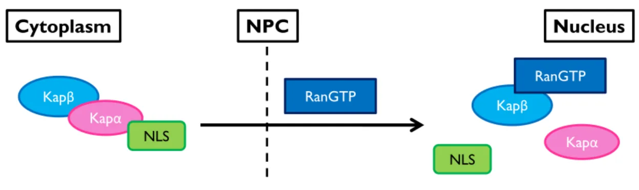
In order to be imported into the nucleus, a protein is bound by a Karyopherin-α (Kapα) adaptor protein via its NLS, and both of these proteins are then bound by a Karyopherin-β (Kapβ) carrier protein, which mediates the nuclear transport. Once this import complex enters the nucleus, Ran-GTP binds to the Kapβ to dissociate the complex and release both the NLS-containing protein and Kapα (Figure 1.7) [44].
Figure 1.7: Protein Import
The general principle of nuclear import of proteins is that a protein must contain a NLS, which will then be recognized by the Karyopherin-α (Kapα) adaptor protein. The Karyopherin-β (Kapβ) carrier protein forms a complex with the Kapα and its cargo to mediate the translocation of this import complex to the nucleus. The import complex is dissociated upon binding of RanGTP to the Kapβ import carrier.
Nucleus Cytoplasm Kapβ Kapα Kapα NLS NLS RanGTP Kapβ RanGTP NPC
1.2.1.1.1 Karyopherin-α Adaptors
There exists seven different Kapα adaptor proteins, split into 3 subfamilies (α1, α2, and α3) based on their sequence homology. The α1 subfamily is composed of KPNA2 (importin-α1) and KPNA7 (importin-α8); the α2 subfamily is composed of KPNA3 (importin-α4) and KPNA4 (importin-α3); and the α3 subfamily is composed of KPNA1 (importin-α5), KPNA5 (importin-α6), and KPNA6 (importin-α7). The subfamilies range from α1, α3 to α2 in terms of lowest to highest sequence homology within each subfamily [45].
In the literature, these Kapα adaptor proteins exist under a multitude of different names for each, such as in the case of KPNA1, previously known as NPI-1 due to its interaction with the nucleoprotein (NP) of the Influenza virus. The Kapα proteins are the most associated with nuclear import as over fifty cargoes have been identified over the years, whether it be for the nuclear translocation of transcription factors during an innate antiviral response or for entry into the nucleus by nuclear-replicating viruses like HIV-1 or Influenza virus [45].
1.2.1.1.2 Karyopherin-β Import Carriers
There are several Kapβ carriers involved in nuclear import (Figure 1.7) with the main one being KPNB1, also known as Importin-β. It is known to bind to classical NLSs (cNLSs) such as the monopartite-cNLS and the bipartite-cNLS, which contain one or two stretches of basic amino acids, respectively [46]. In the absence of Kapα, KPNB1 can mediate the nuclear transport of proteins containing an arginine-rich NLS [47].
Another Kapβ import carrier is transportin 1 (TNPO1), which is known to bind to a non-classical NLS known as the PY-NLS, where the proline-tyrosine (PY) amino acids are preceded by either a hydrophobic or basic stretch of amino acid residues for binding by TNPO1 [48]. TNPO1 and TNPO2 are thought to be redundant transporters, as they both can interact and mediate the nuclear import of HNRNPA1 and HUR [49]. However, TNPO2 has been implicated in mRNA export with NXF1 [50]. TNPO3 is known to import serine/arginine-rich (SR) proteins to the nucleus, hence its other name TRN-SR2 [51]. TNPO3 is also heavily
As shown in Figure 1.10, there are a variety of import carriers, which can specifically bind to certain cargoes. IPO4 has been noted to mediate FANCD2 nuclear import by interacting with C/EBPδ [54]. IPO7 is able to import supercoiled plasmid DNA (exogenous) and human mitochondrial DNA (endogenous) [55], as well as facilitate nuclear import of HIV-1 viral DNA [56]. It can also act as an adaptor in the KPNBHIV-1/IPO7 complex for Histone HHIV-1 nuclear import [57]. IPO8 is known for the import of the signal recognition particle 19 [58]. IPO8 also interacts with Argonaute 2 (Ago2) for cytoplasmic miRNA-guided gene silencing and the nuclear localization of Ago2 [59]. This IPO8-Ago2 is also important for the nuclear translocation of mature miRNAs [60]. IPO13, related to TNPO3 [61], is one of the few Kapβ carriers involved in both import and export, as it can import RBM8, but can also export translation initiation factor eIF1A [62, 63]. IPO13 can also interact with a sumoylated E2-conjugating enzyme, Ubc9, to suppress auto-sumoylation of this complex [64].
Import carriers can also have common cargo to transport to the nucleus, such as in the case of several ribosomal proteins which can interact with KPNB1, TNPO1, IPO5 and IPO7 for nuclear import [65]. However, some ribosomal proteins like RPL7 and RPL12 can only be imported by IPO5 and IPO11, respectively [66, 67].
Once the Kapβ import carriers have completed their function, they can automatically export back to the cytoplasm, independently of RanGTP, such as in the case of KPNB1 as part of its recycling mechanism to renew the formation of import complexes in the cytoplasm [68].
1.2.1.2 Protein Export
A protein containing a nuclear export signal (NES) can be exported to the cytoplasm when it is bound to a Kapβ export carrier. The Kapβ carrier, bound by RanGTP, can then mediate the translocation of the export complex to the cytoplasm, where RanGTP will be hydrolyzed to RanGDP, with the help of RANBP1 and RANGAP1 (their functions explained in section 1.2.1.3 RAN Gradient), for the dissociation of the complex (Figure 1.8) [44].
Figure 1.8: Protein Export
The general principle of protein export is that a protein must contain a NES and be bound by a Kapβ export carrier. Once the Kapβ carrier is bound to RanGTP, it can then mediate the translocation of the protein from the nucleus to the cytoplasm. Once in the cytoplasm, the export complex is dissociated upon hydrolyzation of RanGTP to RanGDP, causing the Kapβ carrier to release its cargo.
The main Kapβ export carrier is Exportin 1 (XPO1), also known as CRM1 (chromosome region maintenance protein), which specifically recognizes leucine-rich NES [44]. Many studies have used the XPO1 inhibitor, Leptomycin B, to determine whether the export of a protein is dependent on XPO1 function [69]. One co-factor that can interact with XPO1 for XPO1-mediated protein export is RAN binding protein 3 (RANBP3) [70, 71], which can act as a scaffold for efficient export complex assembly as RANBP3 can interact with the guanine nucleotide exchange factor RCC1, responsible for the exchange of RanGDP to RanGTP that is necessary to complete the export complex and provide the energy to drive
Nucleus Cytoplasm NPC RANBP1 RANGAP1 Kapβ RanGDP NES Kapβ RanGTP NES
Another export carrier is XPO6, which is known to specifically export profilin-actin complexes. These complexes are important for the proper cytoplasmic localization of NUTF2, a carrier which is responsible for recycling RanGDP back to the nucleus in order to restore RanGTP levels, via RCC1, for the formation of export complexes [73, 74].
XPO7 is known to export proteins in a XPO1-independent manner, as it recognizes short linear sequences and folded motifs, where it interacts with basic residues for export [75]. XPOT is involved in the export of tRNAs [76], and can interact with the nucleoporins RANBP2 and NUP153, located on the cytoplasmic and nuclear side of the nuclear pore complex, respectively, as part of its ability to shuttle when bound to RanGTP [77].
XPO4 is another Kapβ carrier known to function in both export and import as it can export eukaryotic translation initiation factor 5A and Smad 3 [78, 79], and can import the Sox family of transcription factors [80]. XPO5 is a Kapβ carrier that is also thought to be involved in both import and export of proteins, but has primarily been characterized in export as it can export dsRNA-binding proteins [81, 82], elongation factor 1A for translation [83], 60S subunit of the ribosome complex [84], and the export of pre-miRNA for regulating gene expression [85].
After the dissociation of multiple import complexes in the nucleus, the accumulated Kapα adaptors are recycled back to the cytoplasm by CSE1L, also known as XPO2 or CAS, to renew the formation of import complexes (Figure 1.9) [44].
Figure 1.9: Kapα Recycling
Kapα adaptors are recycled back to the cytoplasm by CSE1L. RanGTP is hydrolyzed to RanGDP to dissociate CSE1L and the Kapα adaptor.
CSE1L Nucleus Cytoplasm NPC CSE1L RanGTP Kapα Kapα RanGDP
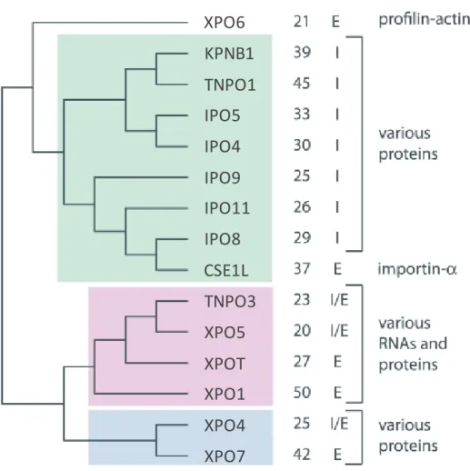
Figure 1.10: Phylogeny of Karyopherin-β subfamilies
The above figure shows the evolution of the Karyopherin-β (Kapβ) carrier proteins with each colour (white, green, pink and blue) representing a different subfamily. The percent identity (%ID) column lists the sequence similarity of each member in comparison to their subfamily. The I/E column identifies which carriers are involved in import (I) and export (E). Some Kapβ proteins are so similar in multiple aspects of their sequences, such as domains, length and composition, that they are considered as paralogous pairs: TNPO1-TNPO2, IPO5-RANBP6, IPO7-IPO8, TNPO3-IPO13, and XPO7-RANBP17 [61, 86, 87] (with the bolded protein of each pair corresponding to the figure above). (Figure adapted from O'Reilly AJ, et al., PLoS One, 2011) [86].
XPO6
KPNB1
TNPO1
IPO5
IPO4
IPO9
IPO11
IPO8
CSE1L
TNPO3
XPO5
XPOT
XPO1
XPO4
XPO7
1.2.1.3 RAN Gradient
The RAN gradient is what determines the directionality of transport through the nuclear pore complex, where the nucleus contains a high concentration of RanGTP for export complexes, and the cytoplasm contains a high concentration of RanGDP waiting to be recycled by the carrier, nuclear transport factor 2 (NUTF2). Once in the nucleus, the GDP bound to RAN is exchanged for a GTP by the guanine nucleotide exchange factor RCC1 to renew the formation of export complexes (Figure 1.11), and the dissociation of import complexes [44].
Figure 1.11: Recycling of RanGDP and the Renewal of RanGTP
To prevent the accumulation of RanGDP after the dissociation of export complexes, NUTF2 binds to RanGDP and transports it back to the nucleus, where RCC1 can exchange the GDP to a GTP to continue the formation of export complexes and the dissociation of import complexes in the nucleus.
The Ras-like nuclear protein (RAN) is the only member of the Ran family, which is part of the superfamily of GTP-binding proteins. This superfamily of proteins is known for their intrinsic GTPase activity, converting their GTP-bound active form to a GDP-bound inactive form [88, 89]. However, RAN is a weak GTPase and requires co-factors such as RAN binding protein 1 (RANBP1) and RAN GTPase-activating protein 1 (RANGAP1) to successfully hydrolyze RAN's GTP-bound form into a GDP-bound form for processes such as the dissociation of export complexes in the cytoplasm (Figure 1.8) [44, 90].
RCC1 NUTF2 RanGDP RanGTP Nucleus Cytoplasm NPC NUTF2
When import complexes are dissociated by the binding of RanGTP to Kapβ import carriers, these carriers can only be released from RanGTP when bound to RANBP1 such as in the case of CSE1L and TNPO1. KPNB1 requires the addition of a Kapα adaptor in order to be released from RanGTP. RANGAP1 prevents RanGTP from re-binding to these import carriers in the nucleus by hydrolyzing it to RanGDP [91].
RANBP1 and RANGAP1 are both considered as nucleocytoplasmic shuttling proteins, as they both function in the dissociation of export complexes in the cytoplasm, and the release of Kapβ from RanGTP in the nucleus [92, 93].
1.2.1.4 mRNA Export
Messenger RNAs (mRNAs) are other macromolecules which require an export pathway from the nucleus to the cytoplasm for the translation of mRNAs into mature proteins. The main carrier for mRNA export is nuclear RNA export factor 1 (NXF1), also known as TAP, specifically binds and exports RNA containing a constitutive transport element (CTE), originally found in the Mason Pfizer monkey virus [94]. The majority of mRNA is exported through the NXF1 pathway, where NXF1 forms a heterodimer with NUTF2-like export factor 1 (NXT1) to interact with the phenylalanine-glycine (FG) repeats of several NPC proteins for proper mRNA export [95-97], and this heterodimer also functions to promote the translation of unspliced mRNA [98]. NXT2, a homologue of NXT1, is localized at the nuclear rim and can interact with NXF1 for mRNA export as well [99]. NXF1 can also export mRNA through a TNPO2-mediated pathway, which has a higher export rate than the NXF1 binding to Nups for mRNA export [50].
The transcription-export (TREX) complex is important for mRNA export as the component ALY/REF can interact with NXF1 to expose its RNA-binding domain to increase the amount of mRNA that is bound, therefore increasing NXF1's RNA-binding affinity and increasing the amount of mRNA exported by NXF1. If components of the TREX complex are silenced, then the amount of mRNA bound is reduced, which will ultimately result in a mRNA export block [100, 101].
Other mRNA export factors are NXF2 and NXF3, where NXF2 can export cytoplasmic mRNAs by interacting with the cytoplasmic motor protein KIF17 [102]. NXF2 can also form a heterodimer with NXT1 in order to interact with Nups, similar to NXF1 [103]. NXF3 is another nuclear mRNA export protein related to NXF1 and NXF2, but lacks the domain to bind to Nups. However, it does contain an XPO1-dependent nuclear export signal, so it can still export mRNA via XPO1 function [104].
XPO1 can also mediate the export of certain mRNAs, but is not able to bind to mRNAs directly, such as in the case of the HIV-1 protein Rev which exports unspliced and partially spliced HIV-1 mRNA through its interaction with XPO1 [100]. NXT1 can also interact with XPO1 as a co-factor and is part of the terminal step of XPO1-mediated nuclear export for the release of XPO1 cargo to the cytoplasm, whether it be for mRNA or protein [105].
DDX19B, the DEAD-box helicase also known as DBP5, is another important mRNA export factor, and requires GLE1L, bound to inositol hexakisphosphate, to increase DDX19B's RNA binding and ATPase activity [106, 107], while NUP214 can interact with DDX19B to regulate those same activities [108]. GLE1L is localized to the NPC by the cytoplasmic FG-Nup NUPL2, and can also interact with NUP155 for mRNA export such as the export of HSP70 mRNA [109, 110].
1.2.2 Structure of the Nuclear Pore Complex
The nuclear pore complex (NPC) is a 125 MDa structure, and is embedded in the nuclear envelope (NE), a bilayer made up of the outer nuclear membrane and the inner nuclear membrane. Composed of 30 proteins known as nucleoporins (Nups), the NPC is made up of seven different parts: the cytoplasmic FG-Nups and filaments, the transmembrane-ring Nups, the outer-ring Nups, the linker Nups, the inner-ring Nups, the central FG-Nups, and the nuclear FG-Nups and the nuclear basket (Figure 1.12). These groups function in either the structural integrity of the NPC or in the regulation of macromolecular transport [43, 111-113].
1.2.2.1 Cytoplasmic FG-Nups and Filaments
The cytoplasmic phenylalanine-glycine (FG)-Nups and filaments are composed of NUPL2, RANBP2 (also known as NUP358), and NUP214 [112]. NUPL2 can bind to XPO1 to promote XPO1-dependent nuclear export, such as in the case of HIV-1 Rev export [114, 115]. RANBP2 acts a docking site, promoting nucleocytoplasmic transport for specific transporters and cargos [116], such as TNPO1 nuclear import [117], KPNB1 nuclear import of cNLS-containing cargo [118, 119], and XPO1 nuclear export [120]. RANBP2 can also interact with NXF1, in complex with NXT1, for mRNA export [95], as well as interact with mRNAs part of the alternative mRNA nuclear export (ALREX) pathway to promote their translation [121]. RANBP2 is also thought to play a role in presenting misfolded and ubiquitinated proteins for proteasomal degradation [122], as its cyclophilin-like domain acts as a modulator for the ubiquitin-proteasome system and may contribute to the compartmentalization of properly-folded protein for turnover [123].
RANBP2, part of the cytoplasmic filaments, is linked to the cytoplasmic side of the NPC by the NUP214/NUP88 complex [124], due to the association of NUP214 with the cytoplasmic ring [125]. NUP214 can also be found in the nucleus for its role in XPO1-mediated protein export [126], when in complex with NUP88, such as the export of the pre-ribosomal 60S subunit [127-129].
1.2.2.2 Transmembrane-ring Nups
The transmembrane-ring Nups, composed of NDC1, NUP210 and POM121, are all responsible for the anchoring of the NPC to the NE [130, 131]. NDC1 is important for NPC assembly as it can interact with Nups from different parts of the NPC such as NUP93 (linker Nup), NUP53 (central FG-Nup), and NUP205 (inner-ring Nup) [132, 133]. Out of the transmembrane-ring Nups, NDC1 is the one responsible for the targeting of ALADIN, a Nup thought to be associated with the outer-ring, to the NPC, where this interaction may be important for nuclear import selectivity [134, 135].
In comparison to POM121, NUP210 is thought to be redundant in the function of NE formation [136]. POM121 is thought to be the most important of the transmembrane-ring Nups for NPC assembly at the membrane [137, 138]. POM121 is important for recruiting RANBP2 (cytoplasmic FG-Nups) and NUP62 (central FG-Nups) to the NPC for assembly [130]. POM121 is also important for linking the NUP160 (outer-ring Nup) subcomplex and the NUP155 (inner-ring Nup) subcomplex together for nuclear pore formation and attachment to the NE [139]. POM121 is also thought to interact with KPNA/KPNB1 import complex and several nucleoporins in a NLS-dependent manner, which is important for NPC assembly at the NE and structural integrity of the NPC [140].
1.2.2.3 Outer-ring Nups
The outer-ring Nups are composed of ALADIN, NUP37, NUP43, NUP75, NUP96, NUP107, NUP133, NUP160, SEC13 and SEH1 [112]. ALADIN is targeted to the NPC by the transmembrane-ring Nup NDC1, where this interaction may be important for nuclear import selectivity [134, 135]. The NUP107-160 complex, composed of all the outer-ring Nups, with the exception of ALADIN, is important for nuclear import of NLS-containing cargos, as well as the organization of other complexes of the NPC such as the transmembrane-ring Nups [112, 141]. The NUP107 complex also plays a role in the diameter of the NPC, as it assembles into two reticulated rings, to accommodate the nucleocytoplasmic transport of large cargoes [142]. NUP107 itself is important for the proper assembly of multiple Nups to the NPC such as RANBP2, NUP214, NUP133, NUP153, and TPR [143, 144].
1.2.2.4 Linker Nups
The linker region of the NPC is composed of NUP88 and NUP93, which can be found on both sides of the NPC [112]. NUP88 can interact with lamin A, and NUP214 for XPO1-mediated export on the nuclear and cytoplasmic side of the NPC, respectively [128, 145]. NUP93 acts as a backbone to connect both the cytoplasmic and nuclear sides of the NPC, and in addition interacts with NUP205, NUP155 and NUP53 for proper NPC assembly, while recruiting NUP62 for nucleocytoplasmic transport-competent NPCs [146, 147]. The interaction between NUP93, NUP155 and NUP205 is known as the NUP93 subcomplex [148].
1.2.2.5 Inner-ring Nups
The inner ring of the NPC is composed of NUP155, NUP188 and NUP205 [112]. NUP155 interacts with NUP53 and NDC1 for NPC assembly [133, 149]. NUP188 is thought to regulate the movement of membrane proteins across the NPC [150], and can interact with FG-repeats and move through the NPC [151]. NUP155 and NUP205 interact with NUP93 and NUP53 for proper NPC assembly [146, 147].
1.2.2.6 Central FG-Nups
The central FG-Nups, composed of NUP53, NUP54, NUP58/NUP45, NUP62 and NUP98 [112]. NUP53 interacts and anchors the NUP93 subcomplex and interacts with NDC1 for proper NPC assembly at the NE [133, 148]. NUP98 is a mobile Nup, and is important for RNA export as it can interact with RAE1, a mRNA export factor [152-155]. During TNPO1 import, NUP98 competes for TNPO1 binding via its M9 motif, releasing the import cargo into the nucleus, and RanGTP bound to NUP98 dissociates it from TNPO1 [156]. NUP98, in complex with RANBP3, can act as a co-factor for XPO1-mediated export [157].
Within the central FG-Nups is the NUP62 complex made up of NUP62, NUP58/NUP45 and NUP54, which are all implicated in nuclear import as they all encode for FGs. NUP58/NUP45 complex is responsible for adjusting the diameter of the central channel [158], increasing in size to allow for nucleocytoplasmic transport to pass through the NPC [159]. NUP58 can complex with KPNB1 for SRP1α nuclear import, while NUP62 can interact
TNPO3-mediated nuclear import of serine/arginine-rich proteins [51]. NUP62 is also involved in export via interacting with NXT1 for NXF1-mediated mRNA export [97].
1.2.2.7 Nuclear FG-Nups and the Nuclear Basket
The nuclear FG-Nups and the nuclear basket are composed of NUP50, NUP153 and TPR [112]. Despite residing on the nuclear side of the NPC, NUP50 mediates XPO1-dependent export [164]. NUP50, depending on its isoform, can regulate the speed of NLS-cargo nuclear import, allowing CSE1L to properly dissociate the NLS-cargo from NUP50 for Kapα export [165, 166], such as in the case of KPNA1 and its cargo PB2 of influenza virus [167].
NUP153 interacts with NUP50 as a scaffold, supporting the NUP50/Kapα for efficient nuclear import [168]. NUP153 interacts with KPNA2 for nuclear export [169], and it is also responsible for the export of multiple classes of RNAs (snRNA, mRNA and 5S rRNA), as well as XPO1-dependent export for viral proteins such as HIV-1 Rev and its dependent RNA export, without affecting tRNA export and KPNB1 recycling to the cytoplasm [170]. Due to NUP153's involvement in export, it is thought to be mobile within the NPC, as it also plays a role in import through interacting with TNPO1 via NUP153's M9 shuttling domain [171]. NUP153 facilitates cNLS-mediated import by interacting with Kapα in the classical Kapα/Kapβ nuclear import pathway [172].
NUP153 interacts with TPR to localize it to the nuclear side of the NPC for the formation of the nuclear basket [173, 174]. TPR restricts the export of unspliced mRNAs via the NXF1-mediated mRNA export pathway [175, 176]. TPR is also thought to be involved in XPO1-dependent protein export [177].
Figure 1.12: Structure of the Nuclear Pore Complex
The nuclear pore complex (NPC) is composed of roughly 30 proteins known as nucleoporins (Nups), which are divided into 7 groups based on their location in the structure. (Figure adapted from Grossman E, et al., Annu Rev Biophys., 2012) [112].
RANBP2 NUP214 NUPL2 NUP210 NDC1 POM121 NUP153 NUP50 TPR NUP93 NUP88 NUP205 NUP188 NUP155 NUP98 NUP62 NUP54 NUP58/NUP45 NUP35 NUP133 NUP160 NUP96 NUP75 NUP107 SEH1 SEC13 NUP43 NUP37 ALADIN
1.3 Viruses and the NPC & its Transporters
Nucleoporins and nucleocytoplasmic transporters have been the target of a myriad of viruses for various reasons. Viruses that replicate in the nucleus usually target these proteins to enter and exit the nucleus for the life cycle and propagation of the virus.
1.3.1 Nuclear-Replicating Viruses
1.3.1.1 DNA VirusesThe Herpesviridae family is composed of several dsDNA viruses such as Herpes simplex virus type 1 (HSV-1), Varicella-Zoster virus (VZV), Epstein-Barr virus (EBV), and Human cytomegalovirus (HCMV), which are also known as Human herpesvirus 1 (HHV-1), HHV-3, HHV-4, and HHV-5, respectively. In the life cycle of these viruses, the capsid injects the viral genome into the nucleus, where replication and transcription occurs [178]. Multiple viral proteins then work in concert to take over cellular processes such as transcription for viral gene expression and nucleocytoplasmic transport of viral factors [179]. The HSV-1 proteins pUL25 and pUL36/pUL6, interact with the cytoplasmic FG-Nups NUP214 and NUPL2, respectively, for capsid import to the NPC, infection initiation, and viral DNA release into the nucleus [180]. ICP27 interacts with multiple export factors such as ALY/REF, NXF1 and XPO1 for efficient viral RNA export through the NXF1- and XPO1-mediated pathways [181-185]. ICP27 also interacts with NUP62 to block KPNB1- and TNPO1-dependent import to favour the export of viral mRNAs [186]. VZV interacts with ALY/REF and NXF1 via its IE4 protein for viral mRNA export through the NXF1-mediated RNA export pathway [187]. VZV can also interact with KPNB1 and XPO1 via its ORF9 protein for nucleocytoplasmic shuttling [188]. EBV can interact with NUP62 and NUP153 via its BGLF4 kinase, which phosphorylates these Nups to increase nuclear import of non-NLS-containing viral proteins and block the import of host proteins [189]. EBV protein SM is exported by XPO1 to the cytoplasm for its activity of increasing the expression of intronless genes, such as certain EBV genes involved in lytic replication [190]. The viral protein UL84 of HCMV can interact with KPNA1, KPNA2, KPNA3 and KPNA4, in complex with KPNB1 and RAN, for nuclear
import [191], while the viral protein UL79 interacts with TNPO1 for its import to the nucleus [192].
The Papillomaviridae family are also composed of dsDNA viruses of multiple types such as Human papillomavirus type 8 (HPV8), type 11 (HPV11) and type 16 (HPV16). HPV is internalized into cells via the L1 and L2 capsid proteins. The L2 minor capsid protein is responsible for the delivery of HPV viral DNA into the nucleus, while L1 remains at the endosome for degradation. The E2 protein binds to viral DNA and recruits the E1 helicase for viral DNA replication, while the E6 and E7 proteins are thought to promote cell proliferation [193, 194]. The L2 minor capsid protein uses IPO5 and TNPO1, as well as KPNB1 complexed with KPNA2, for viral DNA import in HPV11 and HPV16 [195, 196]. The E2 viral protein can also interact with KPNA1 and KPNA4 for nuclear import in HPV11 and HPV16 [197]. In HPV16, the E6 protein can enter the nucleus by interacting with KPNA2, in complex with KPNB1, and TNPO1 [198]. The E7 protein can mediate its nuclear transport by interacting with NUP62 in HPV8, HPV11 and HPV16, and NUP153 in HPV8 [199-201].
Hepatitis B virus (HBV) from the Hepadnaviridae family is a dsDNA virus, utilizing the reverse transcriptase enzyme for the synthesis of DNA from the viral mRNA [202]. The HBV core protein is exported by NXF1 for viral DNA synthesis, while the X protein is exported by and sequesters XPO1 as part of its role to induce liver carcinogenesis in chronically-infected hepatocytes [203, 204]. The HBV capsid interacts with NUP153 to regulate viral release into the nucleus [205].
1.3.1.2 RNA Viruses
Influenza A virus (IAV) is one of the few negative-sense RNA viruses to replicate in the nucleus. The polymerase basic protein 1 (PB1), part of the viral RNA polymerase complex, is imported to the nucleus by IPO5 for the accumulation of viral RNA required for efficient viral growth [206, 207]. PB1 also interacts with NUP54 for viral polymerase transcription and viral replication [208]. The viral protein PB2 can interact with KPNA1 and KPNA2 for nuclear import [209, 210], while the nucleoprotein (NP) interacts with KPNA2 for nuclear import and with KPNA4 for the regulation of viral replication, independent of its
NXT1 and RAE1, to block mRNA export [212]. The NP and NS2 viral proteins of IAV interact with XPO1 for the export of viral ribonucleoprotein complexes (vRNPs) to the cytoplasm, disfavouring the export of other XPO1 substrates [213-215]. The viral protein NP can also be exported directly to the cytoplasm by interacting with XPO1 for viral replication [216]. NXF1 is thought to play a role in the viral RNA export of most late proteins [217]. The phosphorylation of RANBP3, a XPO1 co-factor [70], is required at early and late stages of viral infection for vRNP export [218].
Human immunodeficiency virus type 1 (HIV-1) is retrovirus, which requires the use of the reverse transcriptase enzyme to convert its RNA genome into DNA for integration into the host genome. HIV-1 is part of a subgroup of retroviruses known as lentiviruses, as it can enter the nucleus through the NPC and infect non-dividing cells [219]. The pre-integration complex (PIC) of HIV-1 is able to enter the nucleus by using the viral protein integrase to interact with KPNA2, KPNA4 and TNPO3 [52, 53, 220, 221], as well as directly with the cytoplasmic FG-Nup RANBP2 [222]. Vpr also helps with PIC docking at the NPC by interacting with NUP54 and NUPL2 [223, 224]. HIV-1 also recruits other Nups and transporters such as NUP98 and IPO7 to facilitate with the nuclear import of its viral cDNA [56, 225]. The HIV-1 rev protein interacts with XPO1, NUP98 and NUP214 for the export of Rev and viral mRNA through the XPO1-dependent export pathway [226, 227], while suppressing the export of mRNAs by the NXF1 pathway [228].
1.3.2 Cytoplasmic-Replicating Viruses
Viruses that replicate in the cytoplasm do not require entry into the nucleus for its replication, but they can interact with nucleoporins and transporters to disrupt transport through the NPC to give the virus a growth advantage in the cell.
1.3.2.1 DNA Viruses
The Poxviridae family is the only viral family with a dsDNA genome to replicate in the cytoplasm. RNAi screens have elucidated multiple proteins from multiple functional groups which are important for cytoplasmic replication of Vaccinia virus (VACV), making the
requirement for NPC genes for replication to be unexpected. Out of the NPC and transporters elucidated to have an effect on VACV replication, NUP62 was shown to block viral morphogenesis when silenced [229].
1.3.2.2 RNA Viruses
The Picornaviridae family is composed of positive, single-stranded RNA viruses such as those from the Cardiovirus and the Enterovirus genus. The relationship between this viral family and the NPC, specifically, is that of nucleocytoplasmic transport disruption. Cardioviruses encode the Leader (L) protein, which has been shown to hyperphosphorylate multiple NPC proteins such as NUP62, NUP153 and NUP214 to inhibit protein import and export in Encephalomyocarditis virus (EMCV) [230], and NUP62 in Mengovirus, an EMCV subtype, to permeabilize the nuclear envelope and alter nucleocytoplasmic transport [231]. Enteroviruses encode the 2(A) protease, which can cleave NUP62, NUP98 and NUP153 in Poliovirus, and only NUP62 in Human rhinovirus, to disrupt nucleocytoplasmic trafficking like protein import and mRNA export [232-234].
The relationship between the Flaviviridae family and the NPC and its transporters, as seen with HCV in a previous section, is not well-understood. Other members such as Dengue virus have shown to interact with KPNB1 and XPO1 via its NS5 protein, suggesting that this nucleocytoplasmic shuttling is important for viral replication [235, 236]. Japanese encephalitis virus, another member, was shown to have increased replication when RANBP2 is silenced [237], which could suggest disrupting nucleocytoplasmic transport, such as protein import by KPNB1 [118], may provide a replication advantage in host cells.
Negative-sense RNA viruses, such as Human respiratory syncytial virus (RSV) from the Paramyxoviridae family and Vesicular stomatitis virus (VSV) from the Rhabdoviridae family, can interact with the NPC and its transporters via its Matrix (M) protein. In the case of RSV, the M protein interacts with KPNB1 for nuclear import and XPO1 for nuclear export later in the infection to localize at regions of virus assembly for viral production [238, 239]. On the other hand, the M protein of VSV can interact with NUP98 and RAE1 to block mRNA export to inhibit host protein transcription [240-242], and can interact with RAN to block the
1.4 Innate Antiviral Immune Response
1.4.1 Early Phase: Virus Recognition by PRRs
The innate immunity involves the recognition of pathogen-associated molecular patterns (PAMPs) by pattern-recognition receptors (PRRs). In humans, there currently exist five different PRR pathways: Toll-like receptors (TLRs), RIG-I-like receptors (RLRs), NOD-like receptors (NLRs), C-type lectin receptors (CLRs) and cGAS/STING. However, only the TLR, RLR and cGAS/STING pathway contain receptors recognizing viral genomes, while the NLR and CLR pathways have receptors recognize other macromolecules such as bacterial peptidoglycans and carbohydrates on the surface of pathogens, respectively [244-246].
The TLR pathway consists of extracellular receptors ranging from TLR1 to TLR11. TLR3, TLR7, and TLR9 recognize viral dsRNA, ssRNA and CpG-DNA, respectively in endosomal compartments. Once the receptor recognizes the virus, TLR3 signals through the TRIF adaptor protein, while TLR7 and TLR9 signal through MYD88 for the innate antiviral response [244-246].
The RLR pathway is composed of two cytoplasmic sensors RIG-I, which recognizes short dsRNA and 5’ triphosphate dsRNA, and MDA5, which recognizes long dsRNA. RIG-I recognizes a multitude of viral RNA such as HCV and influenza virus. Sendai virus (SeV) is often used to specifically activate the RLR pathway via RIG-I sensing. When RIG-I and MDA5 sense viral RNA, their CARD domain becomes exposed and interacts with the CARD domain of MAVS, which causes MAVS to translocate to the mitochondria where it mediates its downstream signaling of the immune response [244-247].
In the cGAS/STING pathway, intracellular viral DNA is recognized by cyclic-GMP-AMP (cGcyclic-GMP-AMP) synthase (cGAS), which will then synthesize cGcyclic-GMP-AMP. The newly-synthesized cGAMP binds to STING localized at the ER, where it then signals downstream for the innate antiviral immunity [248].