Eur. J. Biochem. 88, 297-305 (1978)
Solubilization and Isolation
of the Membrane-Bound DD-Carboxypeptidase
of Streptococcus faecalis
ATCC
9790
Properties of the Purified Enzyme
Jacques COYETTE, Jean-Marie GHUYSEN, and Roberta FONTANA
Service de Microbiologie, Faculte de Medecine, Institut de Botanique, Universite de Liege, and Istituto di Microbiologk, Universita di Sassari
(Received January 25, 1978)
Streptococcus faecalis ATCC 9790 possesses six membrane-bound, penicillin-binding proteins. That numbered 6 ( M , 43000) is the most abundant one and is the DD-carboxypeptidase studied
previously. The enzyme has been solubilized and purified to the stage where one single protein band can be detected by gel electrophoresis. The purification procedure does not alter the properties that the enzyme exhibits when it is membrane-bound. The DD-carboxypeptidase itself may be a killing target for penicillin in S. faecalis.
Streptococcus faecalis ATCC 9790 possesses a membrane-bound DD-carboxypeptidase (standard re- action catalysed : Acz-LLys-DAla-DAla
+
HzO+~-ala- nine+
Acz-LLys-DAla) [l - 31 which at alkaline pH is able to perform simple exchange reactions (standard reaction catalysed : Acz-~Lys-DAla-~Ala+
~ - [ ' ~ C ] a l a -nine + D-alanine
+
Acz-LLys-DAla-~['~C]alanine)[2]. The isolated membranes bind about 100 pmol of ['4C]benzylpenicillin/mg of protein and about 30 of these binding sites are on the DD-carboxypeptidase [3]. The enzyme (E) forms with 8-lactam antibiotics (I) inactive complexes EI* which, depending upon the antibiotics, have half-lives either equivalent or, more often, much longer than the generation time of the bacterium [3]. During breakdown of complex EI*,
the enzyme is reactivated and the p-lactam is released in a degraded form. Phenylacetylglycine is formed from benzylpenicillin, a reaction which requires frag- mentation of the antibiotic molecule [3]. With various p-lactams it was observed that the higher the ability of the antibiotic to form an inactive complex EI*, the higher was the ability of the same antibiotic to inhibit cell growth, suggesting that in S. faecafis the
DD-carboxypeptidase was an important target for penicillin [3]. Attempts were therefore undertaken to solubilize and purify the enzyme without altering the properties that it exhibits in its natural membranous environment.
Enzymes. OD-Carboxypeptidase (EC 3.4.12.6); penicillinase (EC 3.5.2.6); phospholipase C (EC 3.1.4.3).
MATERIALS AND METHODS
Membranes
Membranes, in 1 mM phosphate pH 7, 1 mM MgCl2 and at 20mg protein/ml, were prepared as described previously [2].
Measurement of Enzyme Activities
Enzyme preparation (either the isolated mem- branes or the solubilized fractions) and 3 mM AcZ- LLys-DAla-DAla were incubated in 20 p1 (final volume) of 50 mM carbonate buffer pH 10 for 30 min at 37 "C in the absence of ~-['~C]alanine (DD-carboxypeptidase activity) or in the presence of 10 mM ~-['~C]alanine (0.49 Ci/mol; exchange activity). The amounts of D-alanine liberated (enzymic procedure) or of Ac2- LLyS-DAh-D [14C]Ala formed (after separation by paper electrophoresis) were estimated as described previously [2,3]. Specific enzyme activity is expressed in pmol of Acz-LLys-DAla-DAla utilized x min-'
x (mg protein)-'. Unless otherwise stated, the experi- ments were carried out with the same batch of Ac2- LLys-DAla-DAla as that used previously [2,3]. This tripeptide was a gift from UCB, Brussels, Belgium. Another tripeptide preparation, a gift from Reanal, Budapest, Hungary, was used in one experiment (see text). Variations in the K , and V values were
observed depending upon the tripeptide preparation used.
298 Membrane-Bound Do-Carboxypeptidase of Streptococcus fueculis ATCC 9790
Protein Estimation
The technique of Lowry et al. [4] was used with bovine serum albumin as standard. However, all the reagents and solutions were supplemented with 1
%
(final concentration) of sodium dodecyl sulfate.0 rgan ic Phosphate Estimation
glycerol phosphate as standard.
The technique of Lowry et al. [5] was used with
Penicillinase and Phospholipase C
Penicillinase Riker (Neutrapen) was purchased from Serva and was used with a specific activity of 0.68 IUjpl; 1 IU catalyzes the hydrolysis of 1 pmol of benzylpenicillin/min. Phospholipase C, type I, was purchased from Sigma and was used with a specific activity of 10 U/mg protein; 1 U catalyzes the libera- tion of l pmol of water-soluble organic phosphate from lecithin per min (at 37 "C and pH 7.3).
Detergents
Genapol X-100 (isotridecanolpolyglycol ether,
ii = 10) was a gift from Farbwerke Hoechst A.G.,
Belgium. Nonidet P-40 (polyoxyethylene p-t-octyl phenol) and N-cetyl-N,N,N-trimethyl ammonium bro- mide were purchased from BDH Chemicals Ltd.
Radioactive Benzylpenicillin
['4C]Benzylpenicillin (with the radioactive label on the carbonyl group of the phenylacetyl side chain; either 52 or 54 Ci/mol) was purchased from the Radio- chemical Center. Amersham.
Affinity Chromatography
on Ampicillin-Bound CH-Sepharose 4B
CH-Sepharose 4B (4 g) was equilibrated against a 0.5 M NaCl solution for 15 min at 22 "C, washed successively with 800 ml of 0.5 M NaCl and 200 ml of water (pH 4.5) and collected by filtration on a sintered glass filter. Sepharose was resuspended in 30 ml of water containing 160 mg ampicillin (pH 4.8) and after dropwise addition of 1 ml of a water solution containing 575 mg of l-ethyl-3-(3-dimethylamino- propy1)-carbodiimide HCI, the suspension was slowly stirred for 90 min. During this treatment, the pH was maintained between 4.8 and 6 by adding 0.1 M HCl. The ampicillin-bound Sepharose was washed with
500 ml of 10 mM phosphate pH 8
+
0.2 M NaCland finally, with 500 ml of 10 mM phosphate pH 8
+
0.1%
Genapol X-100.Preparative Polyacrylamide Gel Electrophoresis in Genapol X-100
Proteins were separated by electrophoresis on cylindrical polyacrylamide gels (9 x 0.7 cm) for 4 h at 22 "C (4 mA per gel) according to [6] except that both the gels and the cathode buffer contained 0.1
%
Genapol X-100. The buffers used were 0.42 M Tris pH 9.18 at the anode and 40 mM Tris/40 mM boric acid (pH 8.64) at the cathode. The stacking gel was 3% polyacrylamide and the separation gel 5 % poly- acrylamide.Analytical Polyacrylamide Slab Gel Electrophoresis in Sodium Dodecyl Sulfate
The method used was essentially that described by Laemmli and Favre [7]. The slab gels were 20-cm long, 15-cm wide and 1-mm thick. The stacking and separation gels were 6 % and 10% polyacrylamide, respectively. The electrophoreses were carried out for 6 h at 4 "C and 20mA constant current (the voltage varied from 30 to 300V) in 50mM Tris/ 383 mM glycine buffer pH 8.3
+
0.1%
sodium dode- cyl sulfate. Bromophenol blue migrated 15 cm toward the anode. Proteins were stained with Coomassie brilliant blue (0.07%
in methanol/acetic acid/water, 5/1/4, v/v/v) for 2-3 h. Destaining was carried out overnight in methanol/acetic acid/water (5/7.5/87.5, v/v/v). The molecular weight of the proteins was estimated on the basis of the migration of the following standard proteins : Escherichia coli RNA polymerase,/3
andp'
subunits (mean M I 160000), bovine serumalbumin (dimer M , 136 000, monomer M I 68 000), catalase ( M , 60 000), leucine aminopeptidase ( M , 53000), ovalbumin ( M , 43000) and E. coli RNA poly-
merase, CI subunit ( M , 40000).
Detection of Penicillin-Binding Proteins
Samples smaller than 20 p1 were treated with ['4C]benzylpenicillin at a 20 pM, final concentration, for 15 min at 37 "C followed by non-radioactive benzylpenicillin (10 mM final concentration) and de- naturing buffer. The resulting solutions (at the most 50 pl) containing 1
%
sodium dodecyl sulfate, 10% glycerol, 60 mM Tris-HC1 pH 6.8, 5%
mercapto- ethanol and 0.001%
bromophenol blue, were boiled for 1 min.Samples larger than 20 pl were treated with ['"CI- benzylpenicillin and non-radioactive benzylpenicillin as above, and were precipitated with 4 vol. of acetone at 4 "C. The precipitates were collected by centrifuga- tion, dried in vacuo, redissolved in the denaturing
buffer containing the same reagents at the same final concentrations as above and the solutions were boiled for 1 rnin. Precipitation with acetone eliminated Gena-
J. Coyette, J.-M. Ghuysen, and R. Fontana 299
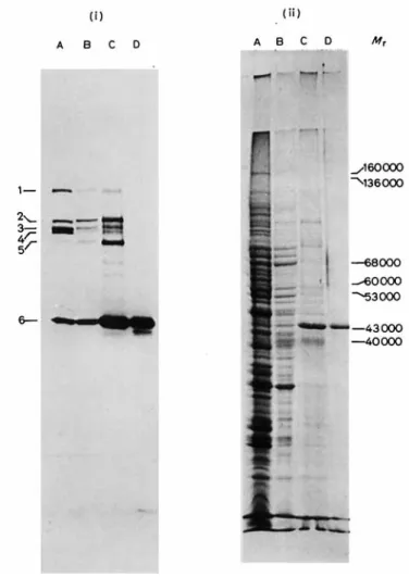
Fig. 1. Isolation of the on-carboxypepiidase andpenicillin-binding protein 6 by afJnity chromatography andpolyacrylamide gel electrophoresis in Genapol X-100. Analysis of the isolation procedure by dodecyl sulfate/polyacrylamide gel electrophoresis. (i) Penicillin-binding proteins as detected by fluorography (numbers on the left). (ii) Proteins as detected by staining with Coomassie brilliant blue R-250. Tracks: (A) isolated membranes; (B) after solubilization with Genapol X-100 (step 1); (C) after adsorption and elution from ampicillin-bound CH-Sepharose 4B (step 2); (D) final enzyme preparation after polyacrylamide gel electrophoresis in Genapol X-100 (step 3). The M , values are those of proteins of known molecular weight
pol X-100 from the samples. When present at too high concentrations, this detergent alters proteins migra- tion.
After polyacrylamide slab gel electrophoresis in the presence of sodium dodecyl sulfate, the [’“CI- benzylpenicillin binding proteins were detected by fluorography [8,9]. Kodak X-omat L films XL-1 were used (without preflash) and the time of exposure was of 15 days at - 70 “C.
RESULTS
Membrane- Bound Peniciflin- Binding Proteins
The isolated membranes of S. fuecalis contain six penicillin-binding proteins exhibiting M , values of
122000 (l), 97000 ( 2 ) , 92000 (3), 91 000 (4), 82000 ( 5 )
and 43000 (6) (Fig. 1, track A). Number 6 is the major one and is most likely the DD-carboxypeptidase since
this enzyme is known to represent one third of the total penicillin-binding sites of the membranes [3]. Solubilization and purification of the membrane- bound DD-carboxypeptidase were monitored on the basis of both enzymic assays and [‘“C]benzylpenicillin binding experiments.
Solubilization of the Membrane-Bound DD- Carboxypep tidase
Preliminary attempts to solubilize the enzyme with 1-2 M NaC1, 2-8 M urea with and without EDTA and 4- 6 M guanidinium HCl either failed or yielded poor yields. In these assays, 100-200 p1 membrane samples were centrifuged, the pellets re- suspended in 100- 200 p1 of the reagent solutions and the enzyme activity estimated in the dialysed super- natant fractions obtained after additional centrifuga- tion. Cetyltrimethylammonium bromide inactivated
300 Membrane-Bound Do-Carboxypeptidase of Streptococcus faecalis ATCC 9790
Table 1 Soluhilization of the membrane-hound Do-carboxypeptidare
Expt Phosphate buffer Membranes Genapol X-100 Yield of the extraction Specific activity
~~ ~
__
~~ ~
concn PH total protein enzyme activity
mM 1 0 (Hz0) 10 10 10 10" 10" 2 5 10 20 50 3 10 10 10 10 10 10 4 10 10 10 ~. ~- - ~ pmol min-' mg
'
~ mg protem/ml (v/v) % 10 1 5 11 0 043 6 10 1 23 93 0 065 7 10 1 34 90 0 043 8 10 1 39 90 0 040 9" 10 1 45 100 0 038 10" 10 1 64 100 0 027 8 10 1 29 87 0 050 8 10 1 30 87 0 047 8 10 1 33 93 0 043 10 1 38 91 0 038 8 8 10 0 1 13 38 0 052 8 10 0 2 17 69 0 060 8 10 0 5 21 87 0 062 8 10 1 23 91 0 058 8 10 2 28 98 0 052 0 042 8 10 5 36 100 8 5 1 38 100 0 038 8 10 1 36 97 0 040 8 20 1 32 79 0 037 -~ ~~~ ~ _ _ _ ~ _ . ~____ -~ ~- _ _ _ _ _ _ _ _ ~ ~~ ~~ ____ -~ ~~~ ~ a Carbonate buffer.the enzyme. Both Nonidet P-40 and Genapol X-100 solubilized it quantitatively. Genapol X-100 was selected because of its negligible absorbance in ultra- violet light.
The effects of pH (expt l), phosphate concentra- tions (expt 2), Genapol X-100 concentrations (expt 3) and membrane protein concentrations (expt 4) were studied separately (Table 1). In all cases, membrane suspensions were incubated under the conditions indicated for 15 min at 37 "C and the supernatant fractions obtained after centrifugation at 150 000 x g for 60 min at 4 "C, were analyzed. In terms of the amount of enzyme activity solubilized, an alkaline pH (8- 10) was most favorable, the phosphate buffer concentration (at pH 8) had little influence and a Genapol X-100 concentration at least equal to 0.1 "/, and a membrane concentration not exceeding 10 mg protein/ml should be used. The procedure finally selected was to treat a membrane suspension (10 mg protein/ml) in 10 mM phosphate pH 8 with 1
%
(final volume) Genapol X-100. As illustrated in Table 1 the yields were 90-97%
of enzymic activity solubilized and 25 - 40%
of total proteins solubilized. The super- natant fraction thus obtained had a 2.5 - 3.5-fold increased specific activity. Penicillin-binding and pro- tein analyses by polyacrylamide slab gel electrophore- sis in sodium dodecyl sulfate (Fig. 1, track B) confirm- ed the relatively high selectivity of the procedure as a means for solubilizing penicillin-binding protein 6 (as well as 2 and 5).Purification of the Solubilized Do-Carboxypeptidase Step 1. A membrane suspension containing 860 mg of protein in 86 ml of 10 mM phosphate pH 8 was treated with 1
%
Genapol X-100 (final concentration) for 30 min at 37 "C. After centrifugation at 4 "C and 150000 x g for 100 min, the supernatant fraction was supplemented with 4 g of ampicillin-bound CH- Sepharose 4B and the suspension was slowly shaken for 30 min at 37 "C (with the help of a Biichler rota-vapor). The ampicillin-bound Sepharose with the enzyme fixed on it was collected by filtration in a small chromatography column (2.3-cm diameter) and wash- ed with 10 mM phosphate pH 8, 1 M NaCl, 0.1% Genapol X-100 until the effluent had a negligible absorbance at 280 nm. About 250 ml of buffer were used.
Step 2. The enzyme
.
ampicillin.
Sepharose com- plex was resuspended in 15 ml of 10 mM phosphate pH 8 containing 0.8 M neutral hydroxylamine and 0.1%
Genapol X-100. After 5 min at 22 "C, the extract was collected by filtration (extract 1). The hydroxylamine treatment was repeated three times, each time by maintaining the suspension for 30 rnin at 22 "C (extracts 2-4). Washing with the above phosphate buffer+
0.1yo
Genapol X-I 00 (without hydroxylamine) until the effluent was virtually free of materials absorbing at 280 nm yielded the extract 5. Extracts 1 - 5 were dialysed separately for 30 h at 4 "C against the phosphate/Genapol X-100 buffer. They301
J. Coyette, J.-M. Ghuysen, and R. Fontana
Table 2. Soluhilization und purification Enzyme activity was measured at a 3
hydrolyzed
of the membrane-bound Do-curhoxypeptidase
mM Acn-LLys-DAla-DAla concentration and in 50 mM carbonate buffer pH 30, as pmol tripeptide
Purification Step Total protein Total enzyme activity Specific activity Yield
mg pmol/min wmol x min-' "/, -fold
x (mg protein)-' 9.6 0.01 1 1 1 24 1 8.1 0.034 84 3 2 14 3 0.22 31 20 3 0.8 1.17 1.45 12 130 Isolated membranes 860
exhibited a specific DD-carboxypeptidase activity which was 15 - 30-fold higher than that of the membranes, These extracts were pooled together and concentrated to 3.3 ml by ultrafiltration on Amicon UM 10 mem- branes. Fig. 1 (track C) shows the protein and peni- cillin-binding patterns of the preparation thus obtain- ed. Note that under the above conditions proteins 2 and 5 behaved exactly like protein 6. A 6th extract obtained after an additional hydroxylamine treatment for 15 h at 22 "C and dialysis as above gave rise to a preparation exhibiting the same specific enzyme activ- ity as the membranes; this extract was discarded.
Step 3. The enzyme preparation after step 2 (14mg)
was divided into 32 samples (100 pl each) and the samples (4 runs of 8 samples) were submitted to poly- acrylamide gel electrophoresis in Genapol X-100. After each run, the gels were cut into 2-mm slices and those slices originating from identical positions on the gels were mixed together and extracted, fives times successively, with 800 p1 of 10 mM phosphate pH 8
+
0.01%
GenapolX-100 for 16 h at 4°C. Theextracts were dialysed for 40 h at 4 "C against 10 mM phosphate pH 8
+
0.01%
Genapol X-I00 and con- centrated to 1 ml by ultrafiltration. Protein estimation, measurement of enzyme activity and protein and penicillin-binding analyses by polyacrylamide gel electrophoresis in sodium dodecyl sulfate showed that altogether the extracts originating from 4 or 5 contigu- ous slices of the gels contained all the enzyme activity but that two of them (corresponding to a migration of about 4.9-
5 cm from the top of the stacking gel toward the anode) (a) had most of the enzyme, (b) exhibited the same high specific enzyme activity and (c) gave rise by polyacrylamide gel electrophoresis in sodium dodecyl sulfate to one single band whose migration coincided with that of penicillin-binding protein 6 (Mr 43000). The extracts containing the purified enzyme and originating from the four different electrophoretic runs were pooled together and concen- trated to 3 ml by ultrafiltration, yielding the final enzyme preparation (Fig. 1, track D). Table 2 gives the total recoveries and enrichments in specific DD-carboxypeptidase activity after each step of the purif- ication procedure. Note that the ratio of DD-carboxy-
peptidase activity to exchange activity (not shown in Table 2) remained unchanged throughout the purifica- tion.
Interaction between Purified Enzyme and Peptide Donor Acz-LLys-DAla-DAla
From Lineweaver-Burk plots obtained with the purified enzyme, the K, value for the tripeptide (in
50 mM carbonate buffer pH 10) was 6 mM and V was 4.8 pmol of tripeptide hydrolyzed x min-' x (mg protein)-'. The corresponding values with the mem- branes were K, = 11 mM and V = 33 nmol x min-'
x (mg protein)-'. p-Chloromercuribenzoate (1 mM), EDTA (5 mM) and dithiothreitol, mercaptoethanol or iodoacetate (10 mM) had no effects on the hydro- lytic reaction and/or the exchange reaction (in 50 mM carbonate buffer pH 10).
Interaction between Purfied Enzyme and &Lactam Antibiotics
As observed with the membrane-bound enzyme, the solubilized enzyme (E) reacted with [)-lactams (I) to form enzyme . antibiotic complexes (EI") devoid of DD-carboxypeptidase-exchange activity and ex- hibiting rather high stability. Breakdown of complex EI* caused reactivation of the enzyme and the release of the antibiotic molecule as inactive metabolites. The general equation for the interaction is thus E
+
I -% EI*5
E+
antibiotic metabolites (kr= kformation and k b = kbreakdown).
Breakdown of Complex E P
Enzyme (20 pl of the final preparation containing 5.2 pg of protein) and 20 mM p-lactam (previously lyophilized in the test tube) were incubated together for 15 min at 37 "C. The excess of p-lactam was destroyed by treatment with 5 p1 penicillinase for
5 min at 22 "C. The solution containing the complex EI* was diluted to 200 pl with 5 mM phosphate pH 7.5
+
3 mM NaN3 and 0.01:d
Genapol X-100 and in-302 Membrane-Bound DD-Carboxypeptidase of Streptococcus faecalis ATCC 9790 Table 3. Effects of j-lactam antibiotics on the S. faecahs Do-carboxypeptidase
kb is the rate constant for the breakdown of the enzyme . antibiotic complex EI* at 37 "C, kr is the rate constant for formation of the enzyme
. antibiotic complex EI* at 37 "C
Antibiotic lo5 x kb for Half-life of kr for
_ _ _ _ _ ~
- --
_ _ _ _
membrane- purified membrane- purified membrane- purified enzyme bound enzyme enzyme bound enzyme enzyme bound enzyme
S - l min M-1 s - l Phenoxymethylpenicillin 9.6 8.6 130 135 560 1220 Benzylpenicillin 4.4 2.8 265 410 445 1045" Ampicillin 1.5 1.3 820 912 230 600 Carbenicillin 3.1 3.5 310 330 19 27 Oxacillin 23.5 15 50 15 4.5 4.6 Cloxacillin 6 8 205 145 0.8 0.8 Methicillin 6.3 7 185 165 1.85 3.6
cubated at 37 "C. Samples (20 pl) were removed after increasing times and the extent of enzyme reactiva- tion was determined. After correction as indicated elsewhere [3], plots of In [l - (E,/Eo)] versus time
(where Et = the concentration of active enzyme present at time t and Eo = the total, active
+
inhibit- ed, enzyme concentration) showed that reappearance of enzyme activity was a first-order reaction (kb in s-') at least when enzyme recovery did not exceed 60 - 80%
(depending upon the antibiotic used). The kb values thus obtained ranged from 1.3 x s-' (i.e. a half- life of 910 min) to 15x s-' (i.e. a half-life of 75 min) (Table 3). They were very similar to the kb values obtained previously with the membrane-bound enzyme.An enzyme sample, previously filtered on Sephadex G-75 in 10 mM phosphate pH 8
+
0.01%
Genapol X-100 (KD = 0) was used to form complex EI* with an excess of ['4C]benzylpenicillin. The enzyme . anti- biotic complex was reisolated by filtration on Sepha- dex G-75. Spontaneous breakdown gave rise to ['4C]- phenylacetylglycine as observed with the membrane- bound enzyme [3].Formation of Complex EI
*
Enzyme (20 pl of the final preparation containing 5.2 pg protein) and various B-lactam antibiotic concen- trations were incubated together at 37 "C (in 100-pl final volumes). The molar ratios of antibiotic to enzyme were at least equal to and most often higher than 10. After increasing times (0- 10 min), 2 0 4 samples were removed from the reaction mixtures, supplemented with 2 pl penicillinase to destroy the excess of the antibiotic and the residual activity was estimated. After correction of the data as described elsewhere
[ 3 ] , the apparent rate constants k, for formation of
complex EI* at each p-lactam concentration used
were estimated from the slopes of the straight lines obtained by plotting In (A,/Ao) versus time.
With benzylpenicillin, the secondary plots of k,
versus [I] were straight lines passing through the origin of the coordinates at low [I] values and showing devia- tion from linearity at high [I] values (Fig. 2A). This behaviour is compatible with a two-step process for the formation of complex EI* : E
+
I =&= EI EI* where EI is an intermediate enzyme . p-lactam com- plex characterized by a dissociation constant K (in M) and k3 the first-order rate constant for the transforma-tion of complex EI into complex EI*. Such a mecha- nism was found previously for the interaction between p-lactams and both exocellular DD-carboxypeptidases, transpeptidases R61 and R39 [ l l , 121. From the reci- procal plot l/ka versus l/[I] (Fig. 2B), a value of K
= 24 pM for the complex formed between benzyl- penicillin and the S . faecalis enzyme and a value of k3 = 2.5 x lo-* s-' were obtained (Table 3).
With the other /?-lactams and within the limits of the concentrations used, the secondary plots k,
versus [I] did not show deviation from linearity. The second-order rate constant
kf
for formation of com- plex EI* was estimated from the slopes of the lines(kf in M-' s-' is equivalent to
k3/f4.
The kf values thus obtained ranged from 0.8 to 1200 M-' sC1 (Table 3). They were either similar to or at least of the same order of magnitude as those obtained with the membrane-bound enzyme.With all the antibiotics tested, the half-lives of the complexes EI* formed with the DD-carboxypeptidase were longer than the generation time of S. faecalis.
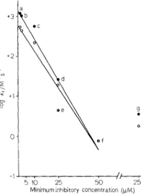
Hence, if one assumes that the inactivation of the DD-carboxypeptidase is lethal for the cell, the effec- tiveness of an antibiotic as a growth inhibitor should be related to its
kf
value. With six of the seven anti- biotics tested, the higher thekf
value, the lower was the minimum antibiotic concentration required toJ. Coyette. J.-M. Ghuyscn. and R. Fontana 303 I 600 200 0 2 4 6 1~[BenryIpenicillin] ( W M ~ ' )
Fig. 2. Forinrrfion of' c oi7iplr, i- El* hetwecw ben;yl~~riirc~illin uml tlrr
p w i f i e d S. faecalis u~-c.trcho.\-~~)~~rictlrsc.. (A) Plots of apparent rate constanl I(, for formation of complex E1* versus increasing concen- trations of benrylpenicillin. (B) Reciprocal plots 1 , k 4 versus 1 , [ben- zylpenicillin]
inhibit cell growth and within the limits of experi- mental error, the plot of log kf versus the minimum
inhibitory concentration gave rise to a straight line (Fig. 3). This relationship, however, did not apply to methicillin which exhibited an unexpectedly high minimum inhibitory concentration (see Discussion).
Corzcoin itun t Interact ion he t ween Enzyme,
Clo.uucilliii und Peptide Donor.
A c ~ - L L ~ s - D A ~ u - D A ~ u
Cloxacillin was selected for this study because of its low kf value (Table 3). Under these conditions,
high [l]/[E] molar ratios could be used. Enzyme (0.26 pg) and tripeptide (from Reanal, see Materials and Methods) at 4.2, 6, 7.8, 9.6 and 11.4 mM (final concentrations) in 20 pl (final volume) of 10 mM carbonate buffer pH 10 were incubated in the presence of 0, 1, 2, 4 and 6 mM final concentrations of cloxa- cillin at 37 'C for 30 min, after which time the amounts of D-alanine liberated were estimated. Graphically, the inhibition was competitive (Fig. 4). The validity of this conclusion was examined by computer analyses using the linear regression program devised previously [13]. The weight 11: of each experimental valuey = 1 / 2 3
-I -/ , 5 10 25 50 250
Minimum inhibitory concentratlon (KM)
Fig. 3. Rc~lutioriship hetwcwi //ie ~nirriim/ni utltihioiic. cotmnityitioi/
r('qiii~ctl to inhihil cell groic.tli and the .sec~0ncl-ur~k~~r c ( J I ~ . Y ~ u ~ / k fbr.
rhc f o r m ~ t i o i i o f comp1e.y EI* hemc.ren untihiotic mid /lie S. faecalis on-i~ui~/~o.\:1./,~~.(,idfirrsc~. (0) Membrane-bound enzyme; ( 0 ) purilied enzyme. (a) Phenoxymethylpeiiicillin, (b) beiizylpenicillin. (c) a m p - cillin. (d) carbenicillin, (e) oxacillin, (0 cloxacillin and (g) methicillin. The iuinimuin inhibitory concentrations are those published previously [ 3 ] . The lines were drawn according to a curve-fitting linear regression program. Coefficients of determination : r' = 0.95 with the membrane-bound enzyme and 1.' = 0.92 for the purified enzyme E" 'L 1.0 1
.
-
0.5was estimated both as 1/y and l / y 2 . The error function which was minimized was
Z
u.: ( y t - yih)2. With )I' = l/y, the residual variance values for a competitive model (&)c and a non-competitive model (s&&304 Membrane-Bound nu-Carboxypeptidase of Strcyfotocc U F fact a2rA ATCC 9790
were 0.00389 and 0.00404, respectively, and the Fisher-Snedecor variable ( F ) was 0.13. With M; = l/y2,
the (,s&&, (S&C and F values were 0.00106, 0.00109
and 0.39, respectively. The higher the F value, the higher is the probability that the reaction is non- competitive; in the present case F values of 2.98 and
4.32 indicate a non-competitive model with levels of confidence of 90 and 95
%,
respectively. Owing to the very low Fvalues obtained, there was no indication that a ternary complex enzyme . substrate . antibiotic may be formed under the experimental conditions used. The values of the parameters involved in the reaction (with confidence intervals t99 x li) were: K, = 29F
18 mM, P' = 14.4 6.8 pmol of tripeptide hy- drolyzed x min-' x (mg protein)-' and Ki (app) = 2.5-
+
0.6 mM. The half-life of complex EI* (145 min) is much longer than the incubation time used in the present experiments (30 min). Hence, the kinetics were not carried out at the steady state and the apparent Ki value thus obtained was necessarily different from the tii value as defined by the equation= 0.1 mM (on the basis of the data of Table 3).
Ki
= kbK/(kb+
k3), or if kb<
k3, Ki = kb/krDISCUSSION
Multiple membrane-bound penicillin-binding pro- teins have been shown to occur in a wide variety of bacteria [14,15]. Penicillin binding to one or more of them is thought to cause cessation of cell growth. The penicillin-binding proteins found in E.vcht.richiu
coli K12 [15], in SuluFiorzella t.yphinzurium [16], in other gram-negative bacteria [17], in Bucillus subtilis
[18,19] and in B. megaterium K M [20] have approxi- mately similar profiles. Those of low molecular weight
( M , = 40000-50000) are the major ones; they have
been identified as Du-carboxypeptidases. The uD-car- boxypeptidase 1 A, which presumably corresponds to penicillin-binding proteins 516 in E. coli and 5 in
S. typhimurium, and the m-carboxypeptidase 1 B, which presumably corresponds to proteins 4 in both
E. coli and S. typkiinurium, react with the pentapeptide
LAla-DG&(L)msA2pm(L)-DAla-DAla from which they catalyse the synthesis of a cross-linked peptide dimer [16,21- 241. However, the DD-carboxypepti- dases from B. subtilis and B. megaterium fail to catalyse this reaction. Curiously, no role in the physiological effects that the /3-lactams produce on E. coli and on bacilli could be attributed to any of these m-carboxy- peptidases. Apparently, the most important penicillin targets are among the penicillin-binding proteins of high molecular weight [15,25]. Thus in E. coli, pro- teins 1B (Mr 91000), 2 ( M , 66000) and 3 ( M r 60000)
play a role in cell elongation, rod shape determination and cell division, respectively. Similarly, protein 1 in
B. megaterium [20] and protein 2 in B. subtilis [I91 might be the targets for killing by penicillin. Quanti-
tatively all those proteins are minor components; their enzyme properties are unknown.
S. jirecalis possesses six penicillin-binding proteins
with a profile similar to those observed in gram- negative bacteria and in bacilli. Protein 6, which is the one with the lowest molecular weight ( M , 43000), is the most abundant one and is a DD-carboxypeptidase.
In vitro and at alkaline pH, the S. fuecalis DD-carboxy-
peptidase is able to catalyse exchange reactions where the C-terminal D-alanine residue of Acz-LLys-uAla- DAla is replaced by simple amino compounds such as D-alanine or glycyl-glycine, but is unable to catalyse more complex reactions which resemble the trans- peptidation reaction in vho [2]. The S.Jueca1i.r DD-
carboxypeptidase (penicillin-binding protein 6) has been solubilized froin the membranes and purified to the stage where it consists of one single protein band by gel electrophoresis. The procedure does not alter the properties found previously for the mem- brane-bound enzyme. In particular, the kf and l i b
values which characterize the interactions between the enzyme and p-lactanis are identical or at least of the same order of magnitude whether the assays are per- formed with the membrane-bound enzyme or with the purified enzyme. The lower kf values observed with the inembrane-bound enzyme may be due either to a more favourable environment for complex forma- tion to occur or to the fact that the inactivation of the membrane-bound enzyme is a phenomenon in which the other five binding proteins are also involved. Formation of complex EI* between benzylpenicillin and the purified enzyme is compatible with the two- step mechanism E
+
I & El & EI* proposed pre- viously for other model enzymes [ll, 121 and the inter- action between purified enzyme, Acz-LLys-DAla-DAla peptide donor and cloxacillin is competitive at least under the conditions of inhibitor and substrdte con- centrations used (for further discussion, see [26,27]).The relative effectiveness in vivo of six out of the
seven antibiotics tested as growth inhibitors for S.fae-
ccrlis (and as expressed by the minimum inhibitory concentration) is related to the ability of the same anti- biotics to inactivate the mi-carboxypeptidase in the form of complex EI* (whether the enzyme is mein- brane-bound or purified and as expressed by the kl
values for the enzyme-antibiotic interaction). The higher the k r value, the lower is the minimum inhibitory concentration. However, the relationship between the two is complex, a 50-fold decrease of the mini- mum inhibitory concentration being paralleled by a 1000-fold increased kf value and furthermore, it does not apply to methicillin for which the minimum in- hibitory concentration is about 8-fold higher than that expected from the corresponding kf value. A correct interpretation of these observations must await until work with the other five penicillin-binding proteins has been undertaken.
J. Coyette, .I.-M. Ghuysen, and R . Fontana 305
The work was supported in part by the Fond,s Nu/ionn/ rie Iu
Reclirrclic .Scientific/ue, Brussels. Belgium, and by the Natioiial Institutes of Health, Washington, D.C. (contract 1 R 0 1 A1 13364-
01 MRC). We thank Dr J. M. Frere for his interest and the computer analyses he made for us.
REFERENCES
1 . Coyette. J.. Perkins, H . R., Polacheck, I., Shockman, G. D. & Ghuysen. J. M. (1974) Eur. J . B i / J d l < ~ l ? . 44. 450 -468.
2. Coyetlc. J., Ghuysen, J. M. & Perkins, 13. R. (1977) Eur. J . Rioc,hciw. 75. 225 -228.
3. Coyette, J . . Ghuyscn, J . M., Binot. P., Adriaens. P., Mees- schaert, B. & Vanderhaeghe, H . (1977) E w . J . Bioiheni. 75. 231 -239.
4. Lowry. 0. H.. Rosebrough, N . J.. F;arr, A. L. & Randall, R. J. (1951) J . Bio/. C'hi.rii. lY3, 265-275.
5. Lowi-y. 0. H.. Roberts. N. R., Leiner, K. Y., Wu, M. L. & Parr, A. L. ( 1 954) J . Bioi. C'hern. 207, ! - 17.
6. Neville, D. M. (1971) J . B i d . C ' h ~ w . 246, 6328-6334.
7. Laeminli, 1J. K . & Favre. M. (1973) J . Mol. B i d 80. 575- 599. 8. Honncr. W. M. & Laskey. R. A. (1974) Ew. J . Biochcm. 46.
X3-88.
9. Laskep. R. A . & Mills, A. D. (1975) Eur. .I. Biociiem. 56. 335- 341.
10. Schilf. W. & Martin. H. H. (1977) Ah.vtruci.r of L~~~n7pos.ium U I I
Fuiiciions o / Microhid Meinhrarrc~s, Tubingen.
11. Frire. J. M., Ghuysen. J . M. & Iwatsubo, M. (1975) Eirr. .I. Rioc~/icn?. 57> 343 - 351.
12. Fuad. N., Fi-ere. J. M.. Ghuysen. J. M.. Duez, C. & Iwatsubo,
M . (1976) Bioi./rcni. J . /5S, 623 - 629.
13. SchilC, W.. Frere. Ph.. Frcre, J . M., Martin, H. H., Ghuysen, J. M.. Adriaens, P. & Mcesachacrt, B. (1978) Eur. .I. Biochtwi.
8.5, 125 - 330.
14. Blumbcrg, P. M . & Strominger, J . L. (1974) Bl/c'iCYi<J/. Rcv.
15. Sprall, B. G. (1977) h r . J . Bioclimi. 72, 341 - 352.
16. Shepherd, T., Chase, H. A. & Reynolds, P. E. (1977) Dir. ./. Biocltein. 78, 521 - 532.
17. Noguchl, H., Itoh, J., Matsubara, N., Mitsuhashi, S., Nikaido, T.. Tamaki, S. & Matsuhashi, M . (1977) Ah\irait,s of ilic 2nd
To/cjv Syinpo.siunz oti Microhid Drug Rcsictcincc~. Tokyo, Japan.
18. Blumberg, P. M. & Strominger. J. L. (1972) J . Bio/. C/wiii. 247.
19. Ruchanan. C . E. & Strominger, J . 1. (1976) Pro<. Nail A ( u d .
20. Chase, H. A,, Shepherd, S. T. & Reynolds, P. E. ( 1 977) /.'IIBS
22. Pollock, J. J., Nguyen-Disteche, M., Ghuyscn. J. M., Coyette, J., Linder, R., Salton. M. R. J., Kim. K . S.. Perkin\, H. R. & Reynolds, P. E. (1974) Eur. J . Biocllcw. 41. 439-446. 22. Nguyen-Distkche, M., Ghuysen, J. M., Pollock, J . J., Rcqnolds,
P. E., Perkins, H. R., Coyette, J . & Sallon, M . R. J. (1974) Eur. .I. Biochcn?. 41. 447-455.
23. Nguyen-Dislkchc, M., Pollock, J. J., Ghuyscn, J. M., Puig, J.. Reynolds, P. E., Perkins, H. R., Coyelte, I . & Salton. M . R . J. 24. Tamura, T., Imae, Y. & Strominger. J . L. (1976) J . Ilioi. C'/KWI. 25. Matsuhashi, M., Takagaki, Y., Maruyama, I. N., Tamaki, S.,
Nishiniura, Y., Suzuki. H., Ogino, U. & Hirota. Y. (1977) Proc. N u t i Acud. Sci. C.S.A. 74. 2976-2979.
26. Ghuysen, J . M . (1977) 7'lw Bat,fcviu/ Dn-('cii.ho\-yi"'l)fiifii.se-
7iorispt~pticla.ve En:j.mc, .Yystem. A N c ~ t , h?.sight h t o /hi,
.l//ot/c of' A c l h i of' Penic,i//in. E.R. Syuihh Lrc~jiit'c..t 011 ~ ' / U Y W
i.vrri, of'Mic,rohiu/ Proi/uc~t.r (Series ed.. Brohn.
w.
E.) 162 p. University of Tokyo Press.38,2Y 1 - 33 5. 8107-8113. Sci. l'.S.A. 73, 1816-1820. Lt'ti. 76, 199-203. (1974) Ew. J . Bioch<,/?I. 41, 457-463.. 251,414-423.
27. Frere. J. M . (1977) Bioc~hon7. Pllcirnluid. 26, 233-2108.
J . Coyettc and J.-M. Ghuysen. Service de Microbiologie, Eaculti. de MPclzcinc. Institut dc Botanique Univer5ite de LEgc a u Sart-Tilman, 8-4000 Liige, Belgium