Université de Montréal
Parenteral Nutrition as a Risk Factor for
Bronchopulmonary Dysplasia: its Role and Possible
Mechanisms in Infants Less than 29 Weeks Gestation
Par
Ibrahim Mohamed
Département de Sciences Biomédicales Faculté de Médecine
Thèse présentée à la Faculté des études supérieures et postdoctorales en vue de l’obtention du grade de Doctorat
en Sciences Biomédicales
Avril, 2018
Université de Montréal
Faculté des études supérieures et postdoctorales
Cette thèse intitulée:
Parenteral nutrition as a risk factor for bronchopulmonary dysplasia: its role and possible mechanisms in infants less than 29 weeks gestation
Présentée par Ibrahim Mohamed
a été évaluée par un jury composé des personnes suivantes :
Dr Sylvie Girard Président-rapporteur Dr Jean-Claude Lavoie Directeur de recherche Dr Diana Avrill-Bates Membre du jury Dr Ghilherme Sant’Anna Examinateur externe
Résumé
Une complication dévastatrice de la prématurité est la dysplasie bronchopulmonaire (DBP). Son étiologie est liée au stress oxydatif précoce. L’impact du stress oxydatif non-radical (stress redox) provoqué par les peroxydes contaminant la nutrition parentérale (NP) sur la DBP est peu étudié chez l’humain. Cependant, une réduction de l’incidence de DBP de 25-30% a été atteinte en appliquant une photoprotection complète de la NP (réduisant de moitié la contamination par les peroxydes). Une corrélation entre le potentiel redox sanguin (indicateur du stress redox) au jour 7 de vie et la sévérité de la DBP a aussi été rapportée.
Hypothèse et objectifs:
Hypothèse: chez les grands prématurés, les peroxydes contaminant la NP induisent un stress redox prolongé contribuant au développement de la DBP. Cet effet est causé par une détoxification insuffisante des peroxydes.
Objectifs:
- Vérifier l’impact de la NP sur le stress redox prolongé et la DBP relativement à d’autres oxydants (O2, infection, transfusion sanguine).
- Examiner différents mécanismes expliquant l’inefficacité de la détoxification des peroxydes. - Évaluer la mesure des peroxydes urinaires spécifiques comme biomarqueur précoce de DBP.
Patients et méthodes:
Cent-seize nourrissons < 29 semaines de gestation étaient suivis jusqu'à 36 semaines d’âge post-menstruel. Pour le premier objectif, la concentration sanguine de glutathion réduit (GSH) et disulfure (GSSG) a été mesuré à 36 semaines d’âge post-menstruel pour le calcul du
potentiel redox par l'équation de Nernst. Pour le second objectif, le glutathion plasmatique et les activités érythrocytaires de la glutathion peroxydase et réductase (GPx et GR) ont été mesurés à 7 jours de vie. Pour l’objectif 3, l’ascorbylperoxyde (AscOOH) urinaire était mesurés aux jours 3, 5, 7 de vie. Le test χ2, le test t-Student, l'ANOVA et la régression linéaire (r2 ajusté) ont été utilisés avec un P <0,05 comme seuil de signification, lorsqu’approprié.
Résultats:
Une NP > 14 jours a induit un potentiel redox plus oxydé (-193±5 versus -203±2 mV) et plus de DBP (89% versus 24%). FiO2 ≥ 25% au jour 7 était également associé un redox plus oxydé (-191±2 versus -198±2 mV) et plus de DBP (90% contre 45%). La régression logistique suggérait qu’une augmentation de 1% de FiO2 et une augmentation quotidienne de NP entrainent une augmentation du odds ratio pour la DBP de 1,57 (1,09 - 2,28) et 1,17 (1,03 - 1,33). Comparativement aux valeurs normales, la concentration plasmatique de glutathion était faible (1,02, 0,49-1,76 μmol/l: médiane, 25e -75e centile) alors que les activités GPx et GR étaient normales. L’AscOOH urinaire augmentait au fil du temps et était plus élevé chez les nourrissons qui ont développé une DBP.
Conclusion:
La durée de la NP et le supplément d'O2 ont des effets additifs sur le stress redox prolongé et un risque accru de DBP. Un faible taux de glutathion pourrait limiter la capacité à détoxifier ces peroxydes. Une diminution de la durée ou le développement d'une formulation plus sûre de NP et une thérapie de remplacement du glutathion peuvent être utilisés comme stratégies possibles pour diminuer le risque de DBP chez les grands prématurés.
Mots-clés : Oxygène, nutrition parentérale, glutathion sérique, potentiel redox du glutathion,
stress oxydatif, stress redox, ascorbylperoxyde, glutathion peroxydase, dysplasie bronchopulmonaire, nouveau-né prématuré.
Abstract
One of the most devastating prematurity complications is bronchopulmonary dysplasia (BPD). BPD etiology is multifactorial and one major factor is early oxidative stress due to high oxygen exposure. The relationship between non-radical oxidative stress (redox stress) caused by the peroxides contaminating the parenteral nutrition (PN) and BPD is less studied. Studies applying complete photoprotection of PN, which can decrease the peroxide contamination of PN by half, decreased BPD by 25-30%. A correlation between the redox potential on day 7 of life and BPD severity was demonstrated in preterm infants.
Hypothesis and objectives:
Hypothesis: Peroxides contaminating PN lead to prolonged redox stress and contribute to the development of BPD. This effect is due to deficient detoxification of peroxides.
Objectives:
- To study the post PN redox stress duration and its impact on BPD in relation to other oxidants (O2, infection, blood transfusion)
- To test if the deficient peroxides’ detoxification is related to glutathione deficiency - To evaluate first week urinary ascorbylperoxide concentration as a BPD biomarker
Patients and methods:
One hundred sixteen infants < 29 weeks of gestation were followed until 36 weeks postmenstrual age (PMA). Fifty-one families gave consent for urine and blood samples. The first objective was achieved by measuring total blood reduced (GSH) and disulfide (GSSG)
glutathione at 36 weeks PMA using capillary electrophoresis to calculate the redox potential using Nernst equation. For the second objective, total plasma glutathione, red blood cell glutathione reductase (GR) and peroxidase (GPx) activities were measured on day 7 of life. The third objective was achieved by measuring ascorbylperoxide (AscOOH) in the urine on days 3, 5 and 7 of life. Chi-square, t-student, ANOVA, linear regression and repeated measure ANOVA were used as appropriate; p< 0.05 was significant.
Results:
PN duration > 14 days was associated with more oxidized redox potential (-193±5 versus -203±2 mV) and more BPD (89% versus 24%). FiO2 ≥ 25% on day 7 of life was also associated with a more oxidized redox potential (-191±2 versus -198 ±2 mV) and more BPD (90% versus 45%). The effects of PN and FiO2 on redox potential and BPD were additive. In logistic regression model, each 1% increase in FiO2 and each day increase on PN resulted in an increase in the OR for BPD by1.57 (1.09 - 2.28) and 1.17 (1.03 - 1.33) respectively. Compared to normal values, total plasma glutathione concentration was low (1.02, 0.49-1.76 µmol/l: median, 25th – 75th percentiles) whereas GPx and GR activities were sufficient. Urinary AscOOH increased overtime (p=0.001) and was higher in infants who later developed BPD or death (p=0.037).
Conclusion:
Early O2 and peroxides contaminating PN have additive effects associated with prolonged oxidative stress and increased BPD. Extremely preterm infants have low glutathione level that limits their capacity to detoxify peroxides. Higher first week urinary AscOOH levels are associated with increased BPD or death. Judicious use of oxygen decreasing the duration or
developing a safer formulation of PN and GSH replacement therapy should be investigated as strategies to decrease BPD in extremely preterm infants.
Keywords: Oxygen, Parenteral nutrition, serum glutathione, redox potential of glutathione,
oxidative stress, redox stress, ascorbylperoxide, glutathione peroxidase, bronchopulmonary dysplasia and preterm infants.
Table of Contents
Résumé ... iii
Abstract ... vi
Table of Contents ... ix
List of Tables ... xiii
List of Figures ... xiv
List of Abbreviations ... xvi
Acknowledgements ... xix
Prologue ... 1
Get science out of the laboratory ... 1
My journey in the laboratory in preparation to this work ... 2
Living in many worlds! ... 6
Chapter 1: Introduction ... 7
Prematurity ... 8
1.1 1.1.1 Incidence, definition and classification... 8
1.1.2 Causes of preterm birth ... 8
1.1.3 The Costs of prematurity ... 9
1.1.4 Overtime evolution of survival and outcome of preterm infants ... 10
1.1.5 Short- and long-term complications of prematurity ... 10
Normal lung development and prematurity ... 11
1.2 1.2.1 Normal lung development... 12
1.2.2 Alveolarization phases and its regulation ... 16
1.2.3 Prematurity effects on lung development ... 21
Bronchopulmonary dysplasia (BPD) ... 24 1.3
1.3.1 The diagnostic criteria of BDP ... 25
1.3.2 Epidemiology of BPD ... 26
1.3.3 Short and long-term consequences of BPD ... 28
1.3.4 Economic impact of BPD ... 28
1.3.5 Pathogenesis of BPD... 29
Oxidant load and antioxidant defenses in preterm infants ... 33
1.4 1.4.1 Oxidants in the context of prematurity ... 35
1.4.2 Prematurity and antioxidant defenses ... 37
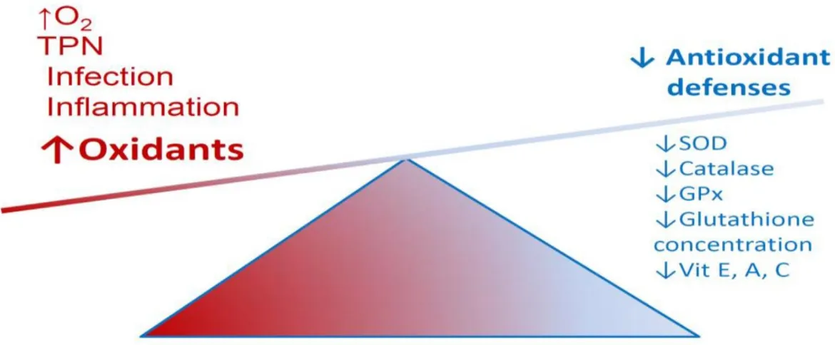
1.4.3 Oxidant-antioxidant imbalance: macromolecular injury and redox biology ... 49
Parenteral nutrition contamination with peroxides and its relation to BPD ... 61
1.5 1.5.1 Peroxides contaminating PN and its sources ... 62
1.5.2 In vitro effects of PN peroxide contamination ... 63
1.5.3 Effects of PN peroxide contamination in animal model ... 63
1.5.4 Effects of PN peroxide contamination in preterm infants ... 66
Thesis rational, hypothesis and specific objectives: ... 68
1.6 Specific objectives: ... 69
2 Chapter 2: Oxygen and parenteral nutrition - two main oxidants - for extremely preterm infants: ‘it all adds up’ ... 70
Complete reference, abstract and keywords ... 71
2.1 Introduction: ... 74 2.2 Methods: ... 75 2.3 2.3.1 Patients ... 75 2.3.2 Local practices ... 75 2.3.3 Measurements ... 76 2.3.4 Definitions... 77
2.3.5 Study groups and comparisons ... 77
2.3.6 Statistical analysis ... 77 Results: ... 78 2.4 Discussion ... 80 2.5 Conclusion: ... 84 2.6
References ... 85 2.7 Tables ... 92 2.8 Figures... 97 2.9 Supplementary data:... 100 2.10 3 Chapter 3: Ascorbylperoxide contaminating parenteral nutrition is associated with bronchopulmonary dysplasia or death in extremely preterm infants ... 101
Complete reference, abstract and keywords ... 102
3.1 Introduction ... 104 3.2 Methods... 105 3.3 3.3.1 Subjects: ... 105
3.3.2 Measurements and definitions: ... 105
3.3.3 Local practices ... 106 3.3.4 Data analysis ... 107 Results ... 107 3.4 Discussion ... 108 3.5 3.5.1 Prematurity, glutathione system and ascorbylperoxide detoxification: ... 109
3.5.2 Ascorbylperoxide and BPD or death: ... 110
3.5.3 Strengths and limitation: ... 111
Conclusion: ... 113 3.6 References ... 114 3.7 Tables ... 120 3.8 Figures... 124 3.9 4 Chapter 4: Appendices ... 127
5 Chapter 5: General discussion ... 135
Summary and novel contributions ... 136
5.1 Why choosing urinary AscOOH? ... 138
5.2 Limitations ... 139
5.3 5.3.1 The non-interventional /analytical /cohort approach ... 139
5.3.3 The limited number of samples... 140 Strengths ... 141 5.4
5.4.1 Our research questions ... 141 5.4.2 Methodology used in our research ... 142 Current insights and directions for future research ... 143 5.5
5.5.1 Putting the puzzle pieces together: ... 143 5.5.2 What is next?... 144 Conclusions ... 149 5.6
List of Tables
Baseline clinical characteristics of all eligible infants and blood sample consent Table I.
infants ……….. 92
The effect of O2 on days 7 of life and the duration of PN on GSH, GSSG and the Table II.
redox potential measured at 36 weeks PMA: ... 93 Severity of BPD in function of FiO2 measured on day 7 of life ... 94 Table III.
Effect of O2 and PN on the incidence of BPD: ... 95 Table IV.
Logistic regression of risk factors of BPD ... 96 Table V.
Components of Primene 10% received by the newborns. ... 120 Table VI.
Components of the multivitamin preparation received by the newborns ... 122 Table VII.
Baseline clinical characteristics of all eligible infants and consent available Table VIII.
infants. ……….
123
Basline clinical characteristics of non consent infants versus biological samples Table IX.
consent infants ... 129 Patients’ Characteristics and outcomes descriptive statistics: ... 130 Table X.
Percentiles of patients’ characteristics and outcomes: ... 131 Table XI.
The effect of FiO2 on day 7 of life and PN duration on total blood GSH, GSSG Table XII.
and redox potential at 36 weeks PMA: ... 133 Risk factors Logistic regression of BPD or death in chapter 2: ... 134 Table XIII.
List of Figures
Figure 1. Human lung development time scale. ... 12
Figure 2. The lung development stages. ... 13
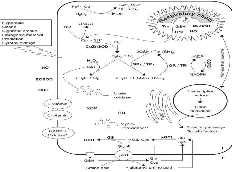
Figure 3. Significant intra- and extracellular sources of oxidants and antioxidants. ... 34
Figure 4. Summary of important antioxidants. ... 38
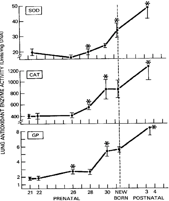
Figure 5. Developmental changes in antioxidant enzyme activity in lungs of fetal and newborn rabbits. ……….. 41
Figure 6. Glutathione metabolism. ... 46
Figure 7. Reduction of peroxides by GPx using GSH. ... 47
Figure 8. Imbalance between oxidants and antioxidants in preterm infants. ... 50
Figure 9. The two mechanisms of oxidative stress. ... 52
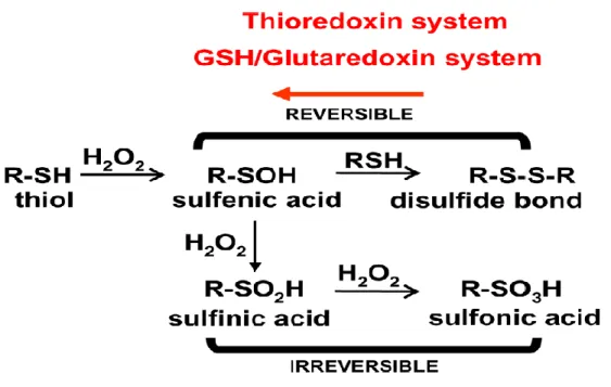
Figure 10. Different oxidation states of a sulfur atom upon H2O2-dependent oxidation. ... 54
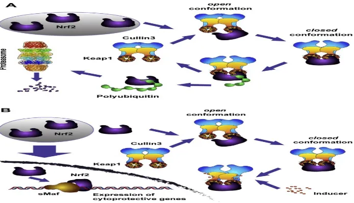
Figure 11. Inducers (oxidants and electrophiles) block the cycle of Keap1-mediated degradation of Nrf2 ... 55
Figure 12. Schematic classification of oxidative stress. ... 56
Figure 13. Cellular response to increasing exposure to reactive species (RS) ... 59
Figure 14. The relation between the redox potential on day 7 and 10 of life and BPD. ... 68
Figure 15. Participant flow. ... 97
Figure 16. Effect of FiO2 on day 7 of life and duration of PN on redox potential of glutathione at 36 weeks PMA. ... 98
Figure 17. Relation between severity of BPD and duration of PN. ... 99
Figure 18. Recruitment flow chart. ... 124
Figure 19. Evolution of urinary ascorbylperoxide (AscOOH) level over the first 7 days of PN. ……… 125
Figure 20. Evolution of urinary ascorbylperoxide (AscOOH) level over the first week of life. ……… 126
Figure 21. Combined participants’ flow for patients in chapters 2 and 3: ... 128
List of Abbreviations
ANOVA Analysis of varianceAscOOH 2,3-diketo-4-hydoxyperoxyl-5,6-dihydroxyhexanoic acid (Ascorbylperoxide) BPD Bronchopulmonary dysplasia
BW Birth weight
CIHR Canadian institutes of health research CLD Chronic lung disease
DTT Dithiothreitol
EDTA Ethylenediaminetetraacetic acid FGF Fibroblast growth factor
FGFR Fibroblast growth factor receptor FiO2 Fraction of inspired oxygen
g Gram
GA Gestational age
GPx Glutathione peroxidase (EC 1.11.1.9) GR Glutathione reductase (EC 1.8.1.7) GSH Reduced glutathione
GSSG Disulfide glutathione H2O2 Hydrogen peroxide
kg Kilogram
MAT Methionine adenosyl transferase (EC 2.5.1.6)
MMPs Matrix metalloproteinases
NF-κB Nuclear factor kappa-light-chain-enhancer of activated B cells NICU Neonatal intensive care unit
NIH National institutes of health (United States) Nrf2 Nuclear factor (erythroid-derived 2)-like 2
O2 Oxygen
PAS Pediatric academic societies PDGF-A Platelet-derived growth factor A PMA Post menstrual age
PMA Postmenstrual age PN Parenteral nutrition
RA Retinoic acid
RDS Respiratory distress syndrome SEM Standard error of the mean
SNAP Score for neonatal acute physiology SOD Superoxide dismutase (EC 1.15.1.1) TGF-β Transforming growth factor-Beta
TIMPs Tissue inhibitors of matrix metalloproteinases US United States of America
Acknowledgements
First and foremost, I offer my deepest gratitude my supervisor and mentor: Dr. Jean-Claude Lavoie for his guidance, supervision, inspiration, timely advice, constant encouragement and enthusiasm from the very early stages of this research. I also thank him for giving me extraordinary experiences, for creating an engaging working environment that accommodates my clinical duties, for always lending an ear and for his endless support. I have learned a great deal from his unique perspectives on research, his sharp insight on many issues and his personal integrity. My transformation from a standard clinician to a scientific clinician-researcher would have never happened without his mentorship.
I extend my thanks to Thérèse Rouleau for her deep questions, stimulating conversations, and her support with my laboratory work.
I thank as well my fellow lab mate Dr. Wesam Elremaly for the stimulating discussions and collaborations on different projects.
I would like to send many thanks my parents, Latifa and Sobhy, for their unflagging love. Even though I lost them early in my life, their love and unforgettable memories still surround me all the time. My sincere thanks go to my sisters and brothers: Sawsan, Nora, Mohamed and
Abdelrhman for being my best friends, for all their advices and their absolute confidence in me. Many thanks go to my beloved wife, Marwa and my kids (Basma, Salaheddin, Jenna and Abdelrahman) for their love and understanding during all these years. I love you.
A sincere thanks to my colleague Dr Michael Andrew-Assad for his meticulous thorough English editing of the thesis.
Finally, special thanks go to my examiner committee: Dr Sylvie Girard, Dr. Diana Avrill-Bates, Dr Ghilherme Sant’Anna for enthusiastically accepting to be my examiners and for reviewing my thesis.
Prologue
I was a 10 years old young boy when I had this sever abdominal pain and my father took me to the emergency room. A very calm and smiling physician took care of me. With the gradual relief of my pain I fell in love with this profession. Nothing in is this world equals the satisfaction of changing another person’s life. Babies are unique in that they are the most vulnerable patients a physician can care for. This is what attracted to the pediatrics and neonatology. I’m passionate about neonatal care and I enjoy watching every single sick little one gets better.
Get science out of the laboratory
Neonatology is a field in rapid evolution where interventions can have life-long impacts. For example, the surfactant treatment saved so many lives since 1980. At international conferences there are increasing amount of laboratory-based works presented year after year. However, minority of these make their way to clinical research testing and even less are integrated into clinical care. At CHU Sainte-Justine, the laboratory of Dr Jean-Claude Lavoie has produced state-of-the-art research for the last 30 years, with about 70 peer reviewed articles. Unfortunately, only a handful of his work has been translated into human clinical research. the fact that no clinical intervention resulted from this long-lasting amazing work perplexed me greatly. I felt as a clinical researcher that it our duty and mission to bridge the gap between laboratory and clinical research, to ensure that we create real change in preterm infants’ lives. With this dream and mission, I started my PhD studies in this laboratory
When I started this journey, I knew there would be many challenges as laboratory and clinical research are 2 unique fields. I knew there would be intellectual and cultural barriers. Basic science research starts with a hypothesis and designs specific experiments to validate or refute it, with the final aim of gaining new knowledge. Translational research starts with a health need and looks for scientific tools to fulfil that need.
I have savoured every moment of this journey so far. I have put all my efforts in understanding and analysing the works of Dr Lavoie and started actively participating in ongoing laboratory projects. The deep understanding gained helped me conceptualize my translational study and choose the appropriate biological markers that would apply to the clinical setting.
My journey in the laboratory in preparation to this work
I began by reading all the works produced form this lab in the last 30 years. Dr Lavoie’s interest in PN began with the discovery of peroxides’ contamination of PN (1). His team was first to describe that non-lipid peroxides form more than 80% of peroxides contaminating PN. (2) They were also first in describing the role of light in accelerating peroxide production reaction through riboflavin excitation (3, 4). Afterwards, they documented a reduction in infants’ urine peroxides following complete photo-protection of PN (5). This was followed by a randomised controlled trial of photo-protection which demonstrated a 30% reduction of bronchopulmonary dysplasia if the PN was completely photo-protected (6). This study showed many other benefits of photoprotection as well (7, 8). The challenge of integrating protection into daily neonatal practice is the technical difficulty of large-scale complete photo-protection as partial photo-photo-protection was found to be of no benefit (9). The team’s research then examined the effect of redox potential changes resulting from PN contamination with
peroxides on lung development (10-13). More recently the team discovered.an important biologically active peroxide that could be specifically measured and named it ascorbylperoxide (13-16). With all this background in mind I started actively participating in ongoing laboratory studies at that time with the team aiming to be able to extrapolate the clinically available biological markers. Being familiar with these biological markers, their limits and their significance was an important step before starting my translational research program in preterm infants.
In the following paragraphs, I will describe the laboratory studies that I participated in. This canvas will outline my own evolution and understanding of the subject matter and highlight the laboratory origin of my translational research program. The first laboratory study was entitled “Ascorbylperoxide from parenteral nutrition induces an increase of redox potential of glutathione and loss of alveoli in newborn guinea pig lungs” and it was published in the Redox Biology journal in May 2014 (17). In this study our neonatal guinea pig model was used. The group receiving an increasing dose of a peroxide contaminating the parenteral nutrition (PN), Ascorbyl peroxide (AscOOH), was compared to a group receiving the same dose of AscOOH with H2O2 in a concentration similar to existing PN solution (350 µM). AscOOH alone was associated with dose dependent activation of caspase-3 (the executive enzyme in apoptosis) and decreased alveolar index. The addition of H2O2 did not affect the alveolarization index while it decreased the activation of caspase-3. The dose dependent increase of redox potential with AscOOH reached a plateau in the presence of H2O2. It is important to note that only H2O2 stimulated Nrf2 (transcription factor that controls the expression of antioxidant proteins) and NF-κB (protein complex that controls DNA transcription of many proteins including inflammatory pathway). In this study we showed that some specifically measured PN
peroxides are involved in the induction of hypo-alveolarization (key feature of BPD) whilst also increasing in the redox potential of glutathione. We highlighted the importance of works aiming to develop safer PN compounding or administration strategies. This study was one of the foundations of our second clinical study presented in chapter 3 of this thesis. We suspected that Both H2O2 and AscOOH are peroxides that are most likely detoxified by the glutathione antioxidant system involving GSH and GPx. Our laboratory team had already demonstrated that a decrease in guinea pig plasmatic level of GSH was related to inhibition of methionine adenosyl-transferase (MAT) caused by the PN peroxides (18). following this study, the next logical direction was to document weather GSH replacement by adding GSSG to PN could resume the normal pulmonary development.
Consequently, the second laboratory study “Adding glutathione to parenteral nutrition prevents alveolar loss in newborn Guinea pig” was published in the journal of Free Radical Biology and Medicine in October 2015 (19). The gamma-glutamyl transpeptidase enzyme has similar affinity for both GSH and GSSG as a precursor for cysteine that is used for de novo production of GSH. In addition, GSSG is much more stable in PN than GSH. These two factors led us to use GSSG addition to PN as a strategy for GSH replacement therapy. In this study we first confirmed that AscOOH is a substrate to GPx in Michaelis-Menten kinetics. Six groups were compared sham, PN with and without photoprotection, 180 µM of AscOOH with and without 10 μM GSSG and PN exposed to light with 10 μM GSSG. The addition of GSSG to PN resulted in decreasing the redox potential and the level of activated caspase-. It also normalized the alveolarization index on histology sections. This work provided a confirmation of our suspected mechanism of PN peroxide contamination on redox potential and alveolar integrity. This paper on GSSG supplement addressed the effect of GSSG supplementation on
the lungs. Our other question was its effect on the liver and specifically on MAT activity as the liver is the most important organ involved in de novo GSH production and distribution. This question was addressed in our next laboratory work.
The third laboratory study “Impact of glutathione supplementation of parenteral nutrition on hepatic methionine adenosyl transferase activity” was published in Redox Biology journal in August 2016(20). In this study, we had 6 groups in 2 series of solutions. The first included sham, PN and PN with GSSG. The second included dextrose infusion, dextrose with H2O2 and dextrose with AscOOH. The MAT inhibition was more pronounced in the groups with PN as its activity was decreased by 45 ± 4 % compared to 23±7% decrease in the peroxides’ groups without amino acid and lipid supplement. The hepatic MAT activity correlated significantly with the redox potential, but the magnitude of the effect was different according to amino acid and lipid supplement (peroxides solutions versus PN). In addition, the use of dithiothreitol (DTT) reversed the effect of peroxides solution without methionine supplement but the inhibition of MAT persisted in the PN group. This indicated that the inhibition cannot solely be explained by a peroxide-oxidation of this thiol function and that other molecules must be involved. This study emphasized that with the current approach of PN compounding and administration, prevention of peroxide formation or GSH replacement to correct the redox potential is not sufficient in this model to restore the MAT activity.
With my initial reading and understanding of Dr Lavoie’s contributions and the added personal growth by participating in the previously described research studies I believed that translational research to test these results in human preterm infants is the logical next step before proposing the solutions created in Dr Lavoie’s laboratory to help prevent BPD.
As such, this thesis aims to test the hypothesis that there is an association between the PN contaminated with peroxide and the outcome of BPD through redox potential changes. In addition this thesis will test if the mechanism suggested by our guinea pig model, a decreased GSH limiting peroxides detoxification, is pertinent in human preterm infants.
Living in many worlds!
For the last few years, I have truly appreciated the diverse nature of being a clinician researcher with translational research interest. Specifically, while fully immersed in my basic science projects and the translational work under Dr Lavoie’s direction, I also designed and conducted separate clinical research projects These clinical projects were focused on osteopenia of prematurity, early neonatal hyperbilirubinemia and PN related liver disease including the role of ursodiol in its treatment (21-24). From the 8 articles I generated during my PhD period, I have chosen to present in this thesis two articles that were directly in line with the translational objective under the supervision of Dr Lavoie. I’m proud of this accomplishment and feel that this life changing experience will help propel me as a distinguished translational research scientist in the filed of oxidative stress in neonatal medicine.
Prematurity
1.1
1.1.1 Incidence, definition and classification
Each year, around 15 million babies are born preterm around the world (25). Prematurity is defined as live births with a gestational age (GA) less than 37 weeks. GA refers to the number of completed weeks after the onset of the last menstrual period. Prematurity is a worldwide major health problem ranging between 5% to 18% of all births across different countries with very serious health and economic consequences. In Canada, during the year 2013 there were 380,323 births, of which 29,716 (7.8%) were preterm births (26). While all infants less than 37 weeks gestational age are all considered preterm, all prematurity related complications are increasing with decreasing gestational age at birth. That is why this World Health Organization categorization of preterm infants based on their gestational age is important when comparing different outcomes in different preterm infants’ populations:
Extremely preterm infants < 28 weeks GA Very preterm infants ≥ 28 to < 32 weeks GA Moderate to late preterm ≥ 32 to < 37 GA
1.1.2 Causes of preterm birth
Preterm birth is an outcome that could be initiated by multiple mechanisms. The most known mechanisms include infections, inflammation, placental insufficiency or hemorrhage and uterine anomalies or over distension (27, 28) yet spontaneous preterm birth can occur without any identifiable mechanisms of these mentioned above. In addition to spontaneous
preterm births the category of medically indicated preterm delivery is well recognized and saw an increase in its percentage of all preterm births in the last few years (estimated at around one third of all preterm births) (28). This category includes maternally indicated deliveries as severe preeclampsia or fetus indicated deliveries as severe intrauterine growth restriction (29). The fact that we fail to predict and/or prevent preterm birth despite several years of research and numerous clinical studies reflects the complexity and variability of causes (30).
1.1.3 The Costs of prematurity
In a relatively recent and one of the most comprehensive evaluations of preterm birth costs, Institute of Medicine (US) Committee on Understanding Premature Birth and Assuring Healthy Outcomes estimated the societal cost of prematurity in the USA annually to be up to $26 billion (31). In addition to the maternal delivery and early neonatal intervention costs this evaluation included disability-specific lifetime medical, special education, and lost productivity costs for four specific developmental disabilities that are associated with preterm birth (cerebral palsy, mental retardation, vision impairment, and hearing loss). On individual level the cost was inversely correlated to gestational age with infants less than 28 weeks GA at $ 190,467 on the year of birth, infants between 28- and 31-weeks GA at $ 94,785, and between 32 and 36 weeks GA at 13,621 compared to $ 3.325 for the full term newborn (31). It should be noted that one of the limitations of this analysis is that it did not include lifetime costs of the caregiver due to lack of adequate data. These out-of-pocket costs incurred by the families of preterm infants include the cost of transportation, accommodations, and childcare for other siblings during hospitalizations as well as during outpatient visits (29, 32). The heavy
cost of prematurity on societal and on individual level makes it a priority to support the research aiming to prevent preterm birth or to prevent any of its major complications.
1.1.4 Overtime evolution of survival and outcome of preterm infants
Due to the better understanding of the pathophysiology, in addition to the technical and pharmaceutical advancements the rate of preterm infants’ survival has increased dramatically in the last few decades with a marked decrease in the mortality rate even for the smallest infants. While a preterm 1 kg infant chance of survival was about 5% in 1960 it reached 95% in 2000 (33). A large recent cohort including 355 806 infants born between 2000 and 2009 with birth weight of 501g to 1500 g, mortality rate decreased from 14.3% to 12.4% (34). This improvement of survival was accompanied by an improvement in outcomes. While outcome of infants between 1000 to 1500 g was poor in the 60s now most of them are doing well (35). However, with the increase of survival at lower gestational age the number of infants with disabilities has stayed approximately the same (33).
1.1.5 Short- and long-term complications of prematurity
Prematurity is now recognized as the leading cause of mortality among children under five years of age (25). In addition, preterm infants are at risk of having many perinatal complications including respiratory distress syndrome, nosocomial infections, necrotizing enterocolitis, intraventricular hemorrhage and retinopathy of prematurity (35, 36). This initial respiratory distress syndrome could precede the development of bronchopulmonary dysplasia (BPD), the most frequent and most severe complication in extremely preterm infants (37-39). It is important to note that the prematurity related complications are not limited to the perinatal period but could also extend far beyond this period. Many respiratory, metabolic and
neurocognitive complication are now recognized to be caused by preterm birth (37, 40-44). The origin of these multiple short- and long-term complications is related to the abrupt change in environment from the intrauterine to extrauterine while being in a period of active organogenesis and organ maturation. This change in environment negatively affects the normal development of fetal organs particularly the lung which is the primary subject of this thesis.
Normal lung development and prematurity
1.2
The main function of the respiratory system is to provide the organism with external oxygen and remove excess carbon dioxide from the blood and this function takes place in the lungs. However, the respiratory system includes many organs that are necessary to its function. It is composed of the upper and the lower respiratory tract. The upper respiratory tract filters, warms and moistens the air and it includes of the nasal cavities, sinuses, nasopharynx and the larynx above the vocal cords. The lower respiratory system is composed of the conducting system and the lung. The conducting system includes the larynx below the vocal cords, the trachea, the main bronchi, and the bronchioles that distribute the air throughout the lung. The lung itself is composed of respiratory bronchioles, alveolar ducts and the alveoli, the place of oxygen and carbon dioxide exchange. The pulmonary blood vessels are part of the lung. The pulmonary arteries bring the deoxygenated blood with excess carbon dioxide from the heart to the lung and the pulmonary veins return oxygenated blood with normal carbon dioxide to the heart. In this section, I will describe the normal development of the lung with special emphasis on lung alveolarization and the impact of antenatal conditions as well as preterm birth on this process.
1.2.1 Normal lung development
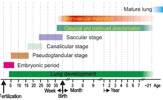
Five overlapping chronologic stages characterize the human lung development (Figure 1). They describe the structural and histological changes that occur during the lung morphogenesis and maturation (Figure 2). These stages include the embryonic, pseudoglandular, canalicular, saccular and alveolar stages extending throughout the gestation period into the postnatal period.
Figure 1. Human lung development time scale.
As the processes of lung development are starting centrally and spread peripherally, the stages of lung development are overlapping. From (45) under Creative Commons Attribution 4.0 International License (http://creativecommons.org/licenses/by/4.0/)
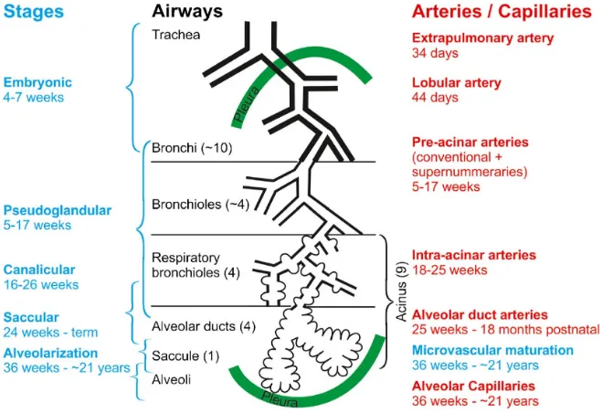
Figure 2. The lung development stages.
The stages of lung development (blue) are correlated to the development of the airways (black) and the arteries (red). From (45) under Creative Commons Attribution 4.0 International License (http://creativecommons.org/licenses/by/4.0/)
1.2.1.1 Embryonic stage (3-7 weeks GA)
During this phase lung bud arises from the ventral foregut endoderm. From the trachea primary, secondary, tertiary bronchi arise by branching morphogenesis. Pulmonary arteries develop from the aortic arches and pulmonary veins develop as outgrowth of the left atrium. Autonomic innervation extends to the trachea and bronchi during this period (45, 46).
1.2.1.2 Pseudoglandular stage (5-17 weeks GA)
Branching morphogenesis continues during this period and the tracheal tree formation is completed by 17 weeks of gestation. In the tracheal, cartilage and mucus glands develop and serous, ciliated, mucus, basal neuroendocrine cells differentiate during this period. In distal lung, respiratory bronchioles, acinar buds/tubules form in this time frame. The pulmonary vascular and lymphatic systems continue their development; pre-acinar blood vessels form the distal mesenchyme. Parallel to airway branching pulmonary arterial development continues. Pulmonary lymphatics arise from veins. Pulmonary lymphatics and veins extend into interlobular septa. The autonomic innervation also parallels airway branching all through this period (45, 46).
1.2.1.3 Canalicular stage (16-26 weeks GA)
While the mesenchyme thins, canalicular and acinar tubules lengthen, subdivide and widen. This period is characterized by the formation of the primitive future alveolar capillary network with the beginning of the formation of the blood air barrier. At the cellular level, type I and type II alveolar cells differentiate. Surfactant starts to be synthesized and stored in the lamellar bodies of type II cells (45, 46). This stage witnesses an increase of fetal lung fluid production and the initiation of the fetal breathing like movements.
1.2.1.4 Saccular stage (24-38 weeks GA)
Fluid filled saccules as acinar buds expand. Saccular distal airspaces continue to grow and branch. Alveolar septal walls arise as mesenchymal condensation. These septa contain well formed double capillary network. Alveolar septal crest formation is marked by elastin deposition at its sites. At the cellular level, type I alveolar cells flatten and elongate. Type II
cells gradually produce more surfactant. With these changes in lung structure, air breathing and gas exchange become feasible. However, premature infants born during the saccular stage of lung development are at high risk to complications related to biochemical immaturity of the lung (lung surfactant deficiency) leading to the respiratory distress syndrome or hyaline membrane disease. This acute lung injury during the saccular stage can alter the subsequent alveolar growth and differentiation leading to BPD (45, 46).
1.2.1.5 Alveolar stage (36 weeks GA to young adulthood)
This stage is characterized by a dramatic increase in the gas exchange surface area caused by the subdivision of the primitive saccular wall (primary septa) by new inter air-space walls (secondary alveolar septa) resulting in new alveoli. These secondary alveolar septa partition the transitory ducts and terminal saccules into true alveolar ducts and alveoli. The number of alveoli in human lung increases very rapidly during the first 2 years of life and recent human studies demonstrated an increase with slower rate throughout adolescence (45, 47). At the cellular level, there is marked interstitial fibroblast proliferation and differentiation with increased collagen, elastin and fibronectin deposition. Both type I and type II alveolar cells rapidly increase in number, but type II cells are the ones proliferating actively suggesting that type II cells are progenitor cells for type I cells. While type II alveolar cells represent two thirds of all alveolar epithelial cells in human adult lung, it only represents about 7% of total alveolar surface while the larger squamous type I cells account for the rest of the surface area. Surfactant production by type II alveolar cells significantly increases in this period. Although type I alveolar cells form a tight barrier that is impermeable to ions and fluid, they are easily injured by oxidants, infection and barotrauma (46). This stage is also characterized by the
maturation of the alveolar-capillary membrane. Throughout this stage the secondary alveolar septa lengthen and thin and its interstitial tissue is reduced. Another main characteristic is the remodelling of the capillary bed is the fusion of the two septal capillary networks, a capillary network on each side of a central core of connective tissue, into one. Pulmonary vascular resistance decreases because of this pulmonary vasculature and capillary bed remodelling. Injury to the lung early during this stage of development may result in abnormal lung remodelling with a reduction of number of alveoli. The most illustrative pathology of impaired alveolar multiplication in human is BPD, the most common chronic respiratory complication of prematurity. Impaired alveolarization (alveolar hypoplasia) and altered microvascular maturation are the main pathophysiologic features of BPD. In the next section, I will discuss the phases of alveolarization process and the mechanisms regulating it.
1.2.2 Alveolarization phases and its regulation
The time line of this process is relatively long; starting around 36 weeks of gestation and continuing till early adulthood (45). While definitive alveoli can be found in the lungs as early as 36 weeks of gestation, the average number of alveoli in a full-term baby is about 150 million (110 to 175 million). This very rapid increase in alveolar number (also called bulk alveolarization phase) continues during the first 6 months of life when 430 million (250 to 710 million) alveoli are already formed. The alveolarization rate then decreases progressively until about 8 years of age when lung growth becomes proportional to body growth. By adulthood the alveolar number reaches an average of 480 million (275 to 790 million) (47-49).
1.2.2.1 Vascular endothelial growth factor (VEGF)
The expression of VEGF and its receptor peaks in the developing lung during the bulk alveolarization (50). Several experiments in the developing rat involving VEGF receptor inhibitors demonstrated that normal angiogenesis is required for normal alveolarization (51, 52). In these experiments rarified peripheral vessels and decreased airspace-parenchyma ratio were noted. These observations were confirmed in extremely preterm baboon, with impaired VEGF found in animals developing BPD (53). Treatment with Recombinant human VEGF treatment enhanced alveolarization in hyperoxic lung injury model of neonatal rats (54, 55).
1.2.2.2 Elastogenesis
The generation of elastic fibers following the incorporation of elastin into microfibril bundles is called elastogenesis. Experimental approaches that disrupt this process elucidated the essential role of elastin in distal lung development. Fewer, dilated distal air sacs with attenuated septa were found upon inactivation of elastin gene in mice (56). Animals in this model die shortly after birth before reaching the alveolarization phase. Another model used Platelet-derived growth factor A (PDGF-A), chemoattractant of myofibroblasts, to demonstrate the role of elastin deposition in alveolarization indirectly (57, 58). Platelet-derived growth factor A- deficient mice surviving postnatally develop lung emphysema with failure of alveolar septation. In this model the absence of secondary septation results from a profound reduction of elastin deposition. As both fibroblast growth factor (FGF) and retinoic acid (RA) are key regulators of elastogenesis their role in lung alveolarization will be discussed in the next section.
1.2.2.3 Fibroblast growth factor (FGF)
FGF increases the expression of lysyl oxidase and tropoelastin, both are essential for elastogenesis, in myofibroblasts (59). FGF is also involved in the signaling for migration of alveolar myofibroblasts during postnatal alveolarization (60). FGF receptors (FGFR) 1 to 4 are present in the developing lung with increased expression of FGFR 3 and 4 during alveolar formation (61).
Homozygous disruption of FGFR 3 and 4 genes in murine model resulted in animals with normal lungs at birth but with completely blocked alveologenesis. These animals were not able to form secondary septa to produce new alveoli (62). This failure in secondary septation production was not noticed with either single mutant. Neonatal rats exposed to hyperoxia had reduced expression of both FGFR 3 and 4 (63).
1.2.2.4 Retinoic acid (RA)
RA enhances the gene expression of tropoelastin (64) and PDGF-A (65). In vitamin A mild deficiency neonatal rats, the number of alveoli was reduced as well as the total alveolar surface area (66). Treatment with retinoic acid resulted in 50% increase in alveolar number in neonatal rats (67). In rat hyperoxia model, RA treatment from day 3 to day 14 resulted in normalisation of the alveolarization with no significant difference from control group on day 42 of life, while hypo-alveolarization persisted in hyperoxia-exposed group that was not treated with RA (68). These observations were confirmed in premature infants as well. Preterm infants < 32 weeks GA who developed BPD were found to have significantly lower concentration of plasma vitamin A compared to the group without BPD (69). In a recent
Cochrane review of 9 studies, vitamin A supplement appeared to be beneficial in decreasing the risk of oxygen needs or death at one month of age in infants < 32 weeks GA (70).
1.2.2.5 Matrix metalloproteinases
One of the very important elements of normal lung development is extracellular matrix remodelling. It is important to realise that almost 40% of synthetized collagen will be degraded in few hours during bulk alveolarization in neonatal rat and this will allow the interstitium to become thinner and less cellular (71). Matrix metalloproteinases (MMPs) and namely MMP 2 and MMP 14 are actively involved in this remodelling and their expression and activity increase progressively during rat lung alveolarization (71, 72). Tissue inhibitors of MMPs (TIMPs) modulate the MMPs’ activity to create the required balance (73). The importance of MMPs in lung alveolarization was highlighted by the finding that low MMP 2 concentrations in tracheal aspirate of preterm infants was significantly and independently associated with the development of BPD (74).
1.2.2.6 Transforming growth factor-Beta (TGF-β)
TGF-β is a 25 KD protein implicated in cellular proliferation and differentiation (75). The TGF-β and its signaling were found to be essential for normal late lung development (76). The important role of TGF-β in lung alveolarization was documented as conditional overexpression of TGF-β resulted in a phenotype similar to histologic picture of BPD in neonatal mouse lung (77). In neonatal C57BL/6J mice hyperoxia model, exposed to 85% FiO2, β signaling was potentiated with increased susceptibility of alveolar type II cells to TGF-β induced apoptosis (78). Moreover, in the murine hyperoxia model, treatments with TGF- TGF-β-neutralizing antibodies improve pulmonary alveologenesis and vasculogenesis (79). In human
preterm infants < 30 weeks GA, abnormally high concentration of TGF-β was found in the endotracheal secretions of infants who later on developed sever BPD with need to home O2 therapy (80).
1.2.2.7 Hormonal regulators
Steroids have significant short and long-term effects on the lung development as demonstrated in cases of antenatal and post-natal administration for fetus and preterm infants. Antenatal administration of corticosteroids for women at risk of preterm birth is a standard of care that is supported by the most recent Cochrane review including 30 RCT with 7774 women and 8158 infants (81). In this meta-analysis antenatal steroids were shown to decrease neonatal mortality and RDS after preterm delivery. In sheep model, antenatal corticosteroids were associated with a decrease in the lung mesenchyme, an increase in the lung airspace and mRNA for surfactant proteins within 24hours of administration (82, 83). It should be noted that in the same model, decreased alveolar septation was noticed 7 days after steroids administration (82). When some treated fetuses were allowed to continue till term without further treatment, the neonates had decreased lung capacity compared to untreated fetuses. Thus, the beneficial effect of antenatal steroids seems to be limited to its short-term action (84). In post-natal life, the main effects of postnatal dexamethasone include the acceleration of alveolar wall thinning and the two capillary layers fusion (85-87). While these acute effects ameliorate the respiratory condition, this process prevents further septation and results in fewer and larger alveoli (85-87) which was found to be persistent in adult rats (86). It is also to be noted that postnatal dexamethasone does not affect endothelial cells replication, but it decreases the replication of fibroblasts and type II cells.
The thyroid hormone plays an important role in lung maturation and alveolarization (88, 89). Compared to control mice and euthyroid offspring of hypothyroid mother, postnatal hypothyroid mice offspring of hypothyroid mothers had decreased postnatal alveolarization due to decreased alveolar septation resulting in fewer large saclike alveoli (90). More recently, a model of iodine deficiency in female Sprague Dawley rats’ pups were compared to iodine sufficient mothers matched age pups. Structural wise, larger and irregularly shaped alveoli were documented in the iodine deficient pups (91). This resulted in reduce tidal volume, peak inspiratory and expiratory flow, and dynamic lung compliance in iodine deficient pups compared with iodine sufficient pups when double-chambered plethysmograph assessment was performed (91). In addition to these structural changes, significantly lower concentrations of surfactant protein B and C were observed in iodine deficient pups indicating significant delay of lung maturation (91).
It should be noted that all these mechanisms controlling lung development and maturation are programmed for the normal intrauterine environment. Preterm birth with the significant transition to extra-uterine life before the appropriate lung development and maturation by term gestation leads to significant consequences that will be addressed in the next section.
1.2.3 Prematurity effects on lung development
In utero the placental unit takes care of all gas exchange needed by the foetus. Programmed developmental changes in lung morphology and physiology occur in preparation to delivery near term so that this transition from a relatively hypoxic environment to ambient air environment causes minimal impact. Preterm birth disrupts this preparation and leads to birth with immature lung that is not ready for ambient air breathing. This lack of preparation
leads to specific pathologies in the acute phase ‘respiratory distress syndrome’ and over longer period of time ‘chronic lung disease of prematurity (CLD) or bronchopulmonary dysplasia (BPD)’. In this section we will discuss the normal lung preparation before term birth first and then we will address both the acute phase pulmonary pathological changes related to preterm birth.
1.2.3.1 Pulmonary preparation for term delivery
The goal of the morphological maturation that is described in the 1.2.1 section is to increase the surface area for gas exchange and to decrease to minimum the thickness of the alveolar-capillary membrane. This goal is met by secondary septation leading to the creation of alveoli and the fusion of the 2 capillary beds in the secondary septa into one single capillary bed, in concomitance with thinning of the matrix of alveolar septa (45, 92, 93). Infants born 24 weeks of gestation are born during the transition from canalicular to saccular stage of lung development which is much less effective in gas exchange compared to near term infants at 36 weeks of gestation who started the formation of lung alveoli (45). This explains the need for mechanical ventilation assistance and the need of O2 supplement in the extremely preterm infants group.
Another important factor that helps near term neonatal smooth transition is the adequate production of surfactant. Surfactant is essential in reducing surface tension at air-liquid interface in the airspaces (94, 95). While surfactant components can be detected within type II alveolar cells as early as 20 weeks GA (96), evidence of its presence in the amniotic fluid is not present until 26 weeks GA (96, 97). In addition, detection of surfactant proteins B and C occurs later in development around 30 weeks GA (during the saccular stage) (96). Many of the
preterm infants who are born before late third trimester are at high risk of developing hyaline membrane disease or respiratory distress syndrome due to surfactant deficiency that leads to alveolar collapse and increased work of breathing (96).
One other very important developmental preparation before near-term birth is the switch of secretory activities from chloride secreting membrane that produce the lung fluid necessary for normal lung growth to sodium absorbing membrane. Without this switch the presence of fluid in the potential airspace and the interstitium can impede gas exchange as diffusion is much faster in gas phase than in water (98, 99).
Another cornerstone aspect of developmental preparation for near term birth is the rapid upregulation of antioxidant defenses near the end of gestation. This part will be discussed in more details in section 1.4.
Preterm birth means simply that these essential developmentally programmed changes will not or will partially take place. This will result in difficult transition from the intrauterine to ambient air environment with variable degrees of respiratory failure that is known in the acute phase as respiratory distress syndrome (30).
1.2.3.2 Respiratory distress syndrome (RDS)
RDS is also known as hyaline membrane disease and is by far the most common cause of respiratory distress in preterm infants. Its name highlights the importance of surfactant deficiency in the pathogenesis of the disease (30, 100). Alveoli with insufficient surfactant tend to collapse. This alveolar collapse increases the necessary work of breathing to re-open the alveoli during inspiration leading to respiratory distress. This repetitive opening and collapse of alveoli leads to shear stress that damages the fragile lung architecture with leakage
of proteinaceous debris into alveoli forming the ‘hyaline membrane’ (30, 101). Clinically the preterm infants have tachypnea, chest wall retraction, grunting and in severe cases cyanosis. A ‘ground glass’ appearance on the chest X-ray represents the diffuse atelectasis and the ‘air bronchogram’ reflects the contrast between the airless parenchyma and the air-filled bronchi. In near term infants, surfactant replacement can lead to rapid improvement with independent spontaneous breathing with no marked long-term consequences. While the surfactant deficiency is a major contributor to the pathology of RDS, treatment with exogenous surfactant will not be enough to achieve independent spontaneous ventilation in the most extremely preterm infants due to the structural immaturity described in 1.2.3.1. In these infants needing prolonged neonatal respiratory support, the acute pulmonary pathology progress toward a chronic lung disease that is known as bronchopulmonary dysplasia (30).
Bronchopulmonary dysplasia (BPD)
1.3
Since the term of BPD was coined 50 years ago by Northway et al, it has become both the most common serious complication of prematurity and the most common form of chronic lung disease during infancy (102-105). Concomitant to the increasing rates of survival of extremely preterm infants, the incidence of BPD continues to be high especially in infants less than 28 weeks GA (106-108). Despite the enormous research and clinical advancement efforts for several years the impact on the incidence, severity and long-term outcomes of BPD is relatively minor (106). In this section I will discuss the diagnostic criteria, the epidemiology, the short- and long-term consequences and including the related costs as well as the pathogenesis of BPD.
1.3.1 The diagnostic criteria of BDP
One of the particularities of BPD is that it is defined by its treatment and not based its pathophysiology. This led to frequent changes in nomenclature and may be decreased our ability to understand and follow the progression of this important pathology (109, 110). In the original paper that described BPD, the disease was characterized by prolonged cyanosis, O2 requirements clinically and by radiologic changes resulting from a chronic lung disease that represents prolonged healing of RDS under the effect of O2 toxicity and mechanical ventilation (105). The definition of BPD diagnosis was largely debated and concluded in the U.S. National Institutes of Health (NIH) workshop held in 1979 that proposed the BPD definition of "continued O2 dependency during the first 28 days plus compatible clinical and radiographic changes" (111). With the increase of very preterm infant survival, this definition
became less relevant and in 1988 a large cohort study concluded that the need of O2 at 36 weeks postmenstrual age better predicted abnormal pulmonary findings at 2 y of age than the oxygen therapy requirement at 28 d of life (112). This definition was followed by a severity dependent definition that was proposed by a NIH workshop (113). In this definition the need of O2 for 28 days of life indicated mild BPD whereas if this need continues (> 21% FiO2 but < 30%) till 36 weeks postmenstrual age in infants < 32 weeks GA (or 56 days of life in infants > 32 weeks GA), this defines the moderate BPD and if FiO2 needs are ≥ 30% this is categorized as severe BPD (113). This definition was validated using data from 4688 infants in a retrospective study that concluded that this definition accurately predicts pulmonary outcomes including percent of patients needing treatment with pulmonary medications and rehospitalization for pulmonary causes by 18-22 months corrected age (114).
It is obvious that all the previous definitions have an inherent limitation, which is that the need for oxygen is determined by physicians or nursing staff rather than by a physiologic assessment with predetermined saturation targets. This led to the development of the physiologic definition of BPD that requires an oxygen room air challenge for all infants who need FiO2 < 30% and classify infants as having BPD if only they fail to maintain their Saturation > 90% for 30 minutes in room air following O2 weaning at 36 weeks postmenstrual age (115). A follow up validating study showed that many of the infants judged by the treating team as needing O2 at 36 weeks postmenstrual age did maintain their saturation > 90% during the room air challenge (116). In this study while 35% of infants were classified as having BPD with O2 needs at 36 weeks postmenstrual age, only 25% had BPD according to the physiological definition.
This wide variability in the definition of diagnostic criteria of BPD stimulated the recent NIH study group, Prematurity Respiratory Outcomes Program, to compare these definitions in a prospective multicenter study that concluded that the evolution in the management of preterm infants (like the wide use of high-flow nasal cannula) limits application of existing definitions and can potentially lead to misclassification. The study pointed out the need for a contemporary definition of BPD that correlates with respiratory morbidity in childhood (117).
1.3.2 Epidemiology of BPD
In the recent cohort of infants between 22- and 28-weeks GA that is including 8515 infants between 2003 and 2007, Stoll et al. reported the incidence of BPD depending on the definition used. While BPD severity definition resulted in an incidence of 68% of all infants
(27% mild, 23% moderate and 18% severe BPD), this incidence was down to 42% using the definition of O2 need at 36 weeks postmenstrual age and to 40% if the physiologic definition was used (103). The same previously mentioned group of Stoll et al. extended their time interval between 1993 and 2012 and included all infants between 22 and 28 weeks GA in the biggest cohort of 34 636 infants and reported an increase in the BPD incidence defined as O2 needs at 36 weeks postmenstrual age from 32% in 1993 to 45% in 2000, then a decrease to 40% in 2008 close to their previous report (103, 118).
Other studies reported different incidences of BPD depending on the population used. In a recent cohort of very low birth weight infants (infants less than 1500g), the Vermont Oxford group reported rates between 26.2% and 34.2% between 2000 and 2009 with significant decrease of BPD overtime (34). A comparative study between Canada and Japan neonatal research networks between 2006 and 2008, infants less than 1500 g, the incidence of BPD defined as the need of O2 at 36 weeks postmenstrual age was 12.3% and 14.6%respectively (119). It should be noted that the saturation target differed between institutions in these studies (34, 103, 119). In a very recent study (2011-2013) that compared the current BPD definitions and their limitations in 765 infants between ≥ 23 weeks GA and < 29 weeks GA, the incidence of BP was found to be 40.8, 58.6, and 32.0% of infants, respectively, with O2 needs at 36 weeks postmenstrual age, severity and physiologic definitions respectively (117).
This incidence makes BPD the most common serious complication of premature infants with 12 000 to 14 000 new cases diagnosed each year in USA alone (104, 120).
1.3.3 Short and long-term consequences of BPD
BPD carries heavy short- and long-term consequences. In the neonatal initial hospitalization BPD is associated with increased risk of impaired postnatal growth, higher mortality and prolonged hospitalization (121, 122). After discharge, infants with BPD are at higher risk of rehospitalisation -especially in the first 2 years of life- and to increased postnatal mortality (123-125).
Long-term consequences of BPD include pulmonary complications including asthma (126). Adults with BPD history are twice like to report wheezing and three times likely to use asthma medications (127). Studies using CT imaging in adults with history of BPD reported emphysema to be common in this population (128). Pulmonary hypertension is another major long-term complication of BPD (129).
In addition to long-term pulmonary complications many children with the history of BPD will suffer from cognitive impairment and neurodevelopmental deficit (130-132).
1.3.4 Economic impact of BPD
While there are relatively many studies reporting the economic impact of prematurity, fewer studies discussed the economic short and long-term impact of BPD. This could be related to the fact that it is difficult to dissect the cost of BPD from the total cost of co-existing prematurity complications. In the elegant work by Johnson et al. that included 425 surviving VLBW infants between 2005 and 2009, the estimated direct cost of the initial hospital stay for infants without BPD was 44,465 ± 23,300 US $ compared to 103,151 ± 43842 US $ for infants who have BPD (133). When trying to dissect direct costs related to BPD, it came to
31,565 US $ (133). It should be noted that these direct hospital costs do not account for physician fees or for family’s direct and indirect expenses. Recently, the article from Alvarez-Fuente group in Spain went a little bit further trying to estimate the economic impact of BPD in premature infants who do not have any other prematurity complications in the first two years of life. While these costs where between 45,050 € and 118,760 €, it increased for those who needed O2 treatment at home to 48,032 – 121,742 € compared to a cost of 910 € for term infants with no complication (134).
With all the above described heavy short- and long-term consequences and costs, BPD became a research priority with expected health, social and economic high impact of any intervention that can lead to a significant reduction in its incidence or severity.
1.3.5 Pathogenesis of BPD
About 50 years ago when BPD was first described, it was a result of the exposure of moderate to late preterm infants to high oxygen and invasive ventilation support. This ‘old BPD’ was characterized by severe airway epithelial lesions (hyperplasia and metaplasia), airway smooth muscle hypertrophy, extensive diffuse fibrosis, and decreased internal surface area and alveoli (135, 136) . With the advances in neonatal care, specifically: the increasing use of antenatal steroids, the administration of surfactant, the use of less invasive ventilation and improved neonatal nutrition, the number of surviving very and extremely preterm infants increased with a remarkable shift of the pathologic picture of the disease. Now, the new BPD is characterized by decreased, large and simplified alveoli, decreased and dysmorphic capillaries, and negligible fibrosis and airway epithelial lesions (135, 136) . It appears that these lesions of arrested alveolar and vascular development are mainly related to the fact that
most of these infants are born at the late canalicular and early saccular phase with exposure to certain factors that leads to arrested lung development.
While it is widely accepted that the pathogenesis of BPD is multifactorial, there are many controversies about the role of each specific pathogenic factor. In the following paragraphs I will critically summarize the most accepted contributing factors.
1.3.5.1 Oxidative stress and BPD
Historically oxidative stress related to long duration of high O2 exposure was considered the principal cause of BPD (105, 137). In addition, in the rodent animal models, the exposure to high O2 without mechanical ventilation is enough to produce structural lung changes similar to BPD (138). These historical cohorts and animal models depend on creating hyperoxia and increased oxidative stress. This situation is much less relevant to the current clinical practices where the minimal necessary supplement of O2 is provided under continuous saturation monitoring. It was not till late nineties when a large animal model was used in a situation that mimics clinical settings (extreme prematurity, antenatal steroids, postnatal surfactant, intravenous parenteral nutrition, gentle ventilation and minimal O2 supplements guided by oximetry). This study permitted to conclude that while mechanical ventilation and O2 toxicity despite being still important factors in the development of BPD, their role is likely not central in the new BPD, as observed in current practice (139). This conclusion became more accepted following the evidence provided by multiple large multicenter studies using two different oxygen saturation targets and comparing their effects on neonatal mortality and morbidities (140-143). The meta-analyses of these trials suggested no effect on the incidence of BPD (144, 145). Putting these data together suggest that while hyperoxia and oxidative