Université de Montréal
The expression of netrin-1 in the intact and injured adult mice retina
par
Pegah CHEHRAZI Département de Neuroscience
Faculté de la médecine
Thèse présentée
en vue de l’obtention du grade de Maîtrise en Neuroscience
April 2017
Résumé
La netrine-1 joue un rôle important en tant qu’un élément de guidance pour la croissance axonale au début du développement du système nerveux central. Des études récentes ont démontré une expression de la netrin-1 dans le cerveau antérieur adulte, où elle régule la fonction synaptique et la plasticité dans les neurones corticaux. Cependant, la contribution de la netrine-1 dans la rétine adulte reste encore inconnue. Le but de cette étude est donc de caractériser l'expression de la netrine-1 dans la rétine des souris adultes sauvages (rétine intacte) et malades (rétine blessée).
L'expression rétinéene de la netrine-1 et de son récepteur, supprimée dans le cancer colorectal (DCC), a été déterminée, à partir des immunobuvardages, chez des souris post-natales de jour 0 (P0), P14 et adultes. Le recours au double marquage de la netrine-1 avec un anticorps spécifique contre RBPMS, un marqueur sélectif pour les cellules ganglionnaires de la rétine (RGC), a permis l’identification de sa localisation sur les sections transversales de rétine. De plus, les niveaux de netrin-1 ont également été quantifiés à trois et sept jours après l'axotomie du nerf optique.
Nous avons démontré que la netrine-1 et DCC sont fortement exprimés dans la rétine à P0, toutefois l’expression de netrin-1 est relativement stable pendant le développent alors que l’expression de DCC est remarquablement réduite à l'âge adulte. De plus, ces expériences ont conclu une expression robuste de la netrin-1 dans le soma RGC adulte et, une expression des récepteurs DCC autour du corps cellulaire. Fait important, nous avons aussi pu démontrer que les niveaux d’expressions de la netrine-1 et DCC sont réduits à trois et sept jours suivant l'axotomie du nerf optique. Cependant, la surexpression de la protéine netrin-1 n'a pas eu un effet significatif sur la survie du RGC par rapport aux témoins injectés par un véhicule.
Les résultats obtenus suggèrent que : (i) la netrine-1 est abondamment exprimée dans la rétine néonatale et subit une diminution importante à l'âge adulte, (ii) la netrine-1 et son récepteur DCC sont présents dans les RGC et, (iii) l'expression de la netrine-1 diminue considérablement suite à une lésion axonale. Ensemble, ces résultats suggèrent un rôle pour la netrin-1 dans le système visuel chez les adultes.
Mots-clés : Rétine, Adulte, Netrine-1, DCC, Axotomie
Abstract
Netrin-1 plays a highly-conserved role as a guidance cue directing axonal growth during the early stages of central nervous system development. Recent data has shown that netrin-1 is continued to be expressed in the adult forebrain where it regulates synaptic function and plasticity in cortical neurons. However, the contribution of netrin-1 in the adult retina remains unknown. The purpose of this study was to characterize the expression of netrin-1 in the intact and injured adult mouse retina.
The retinal expression of netrin-1 and its receptor, deleted in colorectal cancer (DCC), was examined at postnatal day 0 (P0), P14, and adult mice using western blots. Double labeling of netrin-1 with an antibody against RBPMS, a selective marker for retinal ganglion cells (RGCs), was used to determine its location on retinal cross-sections. Netrin-1 levels were also quantified at 3 and 7 days after optic nerve axotomy.
We demonstrate that netrin-1 and DCC are abundantly and widely expressed in the retina at P0, but although the expression level of netrin-1 remains relatively stable during development, the expression level of DCC is markedly downregulated in adulthood. Adult RGC soma were shown to be endowed with robust netrin-1 expression. DCC receptors were also found to be expressed around the cell body. Importantly, we show that netrin-1 and DCC levels are further downregulated at 3 and 7 days after optic nerve axotomy. However, the over-expression of netrin-1 protein failed to exert any significant effect on RGC survival in comparison to vehicle-injected controls.
Our data support that: 1) netrin-1 is abundantly expressed in the neonatal retina and undergoes marked downregulation in adulthood, 2) netrin-1 and DCC are present in RGCs, 3) netrin-1 expression decreases rapidly after axonal injury. Together, these results suggest a role for netrin-1 in the mature visual system.
Table of contents
Résumé ... 1
Abstract ... 2
Table of content ... 3
List of tables ... 7
Table.1: List of primary antibodies used for immunofluorescence and Western Blot ... 7
Table.2: PCR (Invitrogen Platinum Taq) ... 7
Table.3: List of primers used for RT-PCR experiments ... 7
List of figures ... 8
Figure I.1. A schematic diagram of the retina demonstrating the principal cell types involved in retinal signaling... 8
Figure I.2: The netrin family of proteins. ... 8
Figure I.3: Canonical netrin-1 receptors. ... 8
Figure I.4: Netrin-1 is a bifunctional axonal guidance cue. ... 8
Figure I.5: Netrin-1 signal transduction mechanisms. ... 8
Figure IV.1: Netrin-1 is expressed in the developing and adult mouse retina. ... 8
Figure 1V.2: DCC is expressed in adult mouse retina. ... 8
Figure IV.3: DCC and netrin-1 colocalize. ... 8
Figure IV.4: Netrin-3, the closest homolog of netrin-1 is expressed in the adult retina ... 8
Figure IV.5: After axotomy, RGCs in the adult mammalian retina die by apoptosis in a time-dependent manner. ... 8
Figure IV.6: Netrin-1 protein decreases after optic nerve axotomy. ... 8
Figure IV.7: DCC levels are downregulated after optic nerve axotomy. ... 8
Figure IV.8: Netrin-3 expression level declines after optic nerve axotomy. ... 8
Figure IV.9: Outline of experimental protocol used to test the effect of recombinant netrin-1 on retinal ganglion cell (RGC) survival and regeneration. ... 8
Figure IV.10: Recombinant netrin-1 does not promote the survival of injured RGCs. ... 8
Figure IV.11.1: Schematic representation of recombinant AAV vectors. ... 8
List of abbreviations ... 9
Acknowledgement ... 12
Introduction ... 13
I.1. The retina: cellular and functional organization ... 15
I.1.1 Photoreceptors ... 17
I.1.2 Bipolar cells ... 17
I.1.3 Horizontal cells ... 18
I.1.4 Amacrine cells ... 18
I.1.5 Retinal ganglion cells (RGCs) ... 19
I.1.5.1 RGCs are the cellular target in many retinal diseases ... 20
I.1.6 Retinal glial cells ... 20
I.1.6.1 Muller glia ... 21
I.1.6.2 Astrocytes ... 21
I.1.6.3 Microglia ... 22
I.2. The retina as a model to study neurodegenerative diseases ... 22
I.2.1 Optic nerve axotomy ... 23
I.2.1.1 Optic nerve axotomy and dynamics of RGC death ... 24
I.2.1.2 Neuroprotective strategies to promote RGC survival after axotomy ... 24
I.3. Netrin-1: structural organization and evolutionary history ... 25
I.3.1 Netrin-1: the prototypical member of the netrin family ... 27
I.3.2 Multiple receptors for netrin-1 ... 27
I.3.2.1 The DCC receptors ... 29
I.3.2.2 The UNC5 receptor family ... 29
I.3.3 Netrin-1: bi-functionality and reverse signaling... 30
I.3.4 Netrin-1 signaling mechanisms ... 31
I.3.5 Netrin-1: a role both as a long-range and a short-range cue ... 35
I.3.6 Calcium modulates netrin-1 signaling ... 35
I.3.7 Other putative netrin-1 receptors ... 35
I.3.7.1 DSCAM ... 35
I.3.8 The functional role of netrin-1 in the nervous system during development ... 37
I.3.8.1 Axon guidance ... 37
I.3.8.2 Neural precursor cell migration ... 38
I.3.8.3 Synaptogenesis ... 38
I.3.8.4 Netrin-1 in oligodendroglial development and maturation ... 38
I.3.8.5 Retinal circuit development ... 39
I.3.9 The role of netrin-1 in the adult nervous system ... 40
I.3.9.1 Netrin-1 regulates synaptic function and plasticity in the adult brain ... 40
I.3.9.2 Netrin-1 as an anti-apoptotic agent: the dependence-receptor hypotheses ... 40
I.3.9.3 Netrin-1 expression in the adult retina ... 41
II. Objective of the thesis, hypothesis and experimental approaches ... 42
III. Material and methods ... 43
III.1 Experimental animals... 43
III.2 Western-blot analysis ... 43
III.3 Reverse Transcription–Polymerase Chain Reaction (RT-PCR) ... 44
III.4 Retinal immunohistochemistry ... 46
III.5 Axotomy-induced RGC death assay ... 47
III.6 Intravitreal injections ... 47
III.7 Quantification of RGC survival ... 47
II.8 Statistical analysis ... 48
IV. Results... 49
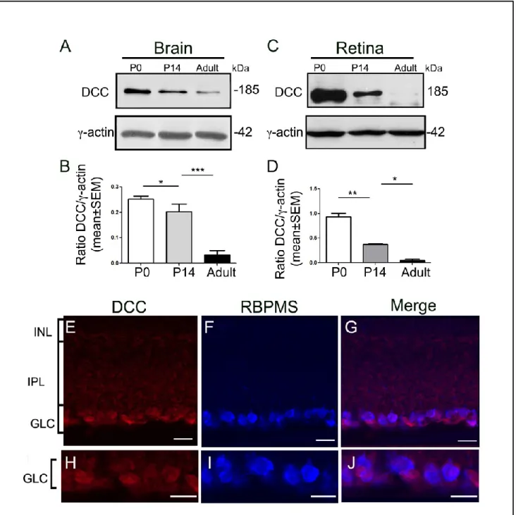
IV.1 Netrin-1 is expressed in the developing and adult mouse retina ... 49
IV.2 DCC is abundantly expressed in early developing mouse retina and it is downregulated in adulthood ... 52
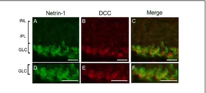
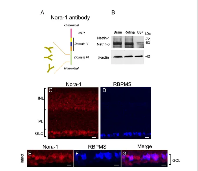
IV.3 Adult RGC soma abundantly express netrin-1 and DCC ... 54
IV.4 Netrin-3, the closest homolog of netrin-1 is expressed in the adult retina ... 55
IV.5 RGC survival in experimental optic nerve damage ... 57
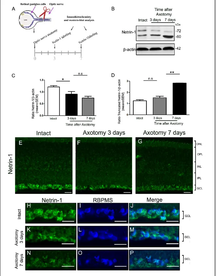
IV.6 Netrin-1 expression is downregulated after optic nerve axotomy ... 58
IV.7 DCC expression decreases after optic nerve axotomy ... 60
IV.9 Experimental protocol to study the neuroprotective effect of netrin-1 on axotomized
RGCs ... 64
IV.10 Recombinant netrin-1 does not extent RGC survival ... 65
IV.11 Netrin-1 gene transfer to the retina ... 67
IV.11.1 AAV.netrin-1 structure ... 67
IV.11.2 AAV-mediated netrin-1 is not expressed by adult RGCs ... 68
V. Discussion ... 71
V.1 Netrin-1 and its receptor DCC are expressed in the adult retina ... 71
V.2 Netrin-3 is expressed in the adult retina... 72
V.3 Possible roles of netrin-1 in adult retina ... 73
V.3.1 Netrin-1/DCC signaling may regulate synaptic integrity in the adult retina .. 73
V.3.2 DCC mediates an adhesive interaction with substrate-bound netrin-1 ... 74
V.4 Netrin-1, netrin-3 and DCC are downregulated after axotomy ... 75
V.4.1 Truncated netrin-1 gradually increases after axotomy ... 75
V.4.2 Netrin-1-dependent protein synthesis and RGC death ... 76
V.5 The effect of netrin-1 on RGC survival after axotomy ... 77
V.5.1 Netrin-1 supplementation: recombinant protein versus gene delivery ... 77
V.5.2 DCC downregulation may compromise the RGC response to recombinant netrin-1 ... 78
V.5.3 Netrin-1 might promote chemorepulsion after axotomy ... 78
V.5.4 Recombinant netrin-1 concentration ... 79
List of tables
Table.1: List of primary antibodies used for immunofluorescence and Western Blot Table.2: PCR (Invitrogen Platinum Taq)
List of figures
Figure I.1. A schematic diagram of the retina demonstrating the principal cell types involved in retinal signaling.
Figure I.2: The netrin family of proteins. Figure I.3: Canonical netrin-1 receptors.
Figure I.4: Netrin-1 is a bifunctional axonal guidance cue. Figure I.5: Netrin-1 signal transduction mechanisms.
Figure IV.1: Netrin-1 is expressed in the developing and adult mouse retina. Figure 1V.2: DCC is expressed in adult mouse retina.
Figure IV.3: DCC and netrin-1 colocalize.
Figure IV.4: Netrin-3, the closest homolog of netrin-1 is expressed in the adult retina Figure IV.5: After axotomy, RGCs in the adult mammalian retina die by apoptosis in a time-dependent manner.
Figure IV.6: Netrin-1 protein decreases after optic nerve axotomy. Figure IV.7: DCC levels are downregulated after optic nerve axotomy. Figure IV.8: Netrin-3 expression level declines after optic nerve axotomy.
Figure IV.9: Outline of experimental protocol used to test the effect of recombinant netrin-1 on retinal ganglion cell (RGC) survival and regeneration.
Figure IV.10: Recombinant netrin-1 does not promote the survival of injured RGCs. Figure IV.11.1: Schematic representation of recombinant AAV vectors.
List of abbreviations
ALS Amyotrophic lateral sclerosis
BDNF Brain derived neurotrophic growth factor
BRB Blood retinal barrier
cAMP Cyclic adenosine monophosphate
Cdc42 Cell division cycle 42 CGNs Cerebellar granule neurons
CNS Central nervous system
DB DCC-binding
DCC Deleted in colorectal cancer
DD Death domain
DSCAM Down syndrome cell adhesion molecule
ECM Extracellular matrix
EGF Epidermal growth factor
ERK Extracellular signal-regulated kinase
FAK Focal adhesion kinase
GCL Ganglion cell layer
ICD Intracellular domain
IHC Immunohistochemistry
INL Inner nuclear layer
IPL Inner plexiform layer
ITRs Inverted terminal repeats
kDa KiloDalton
LTP Long-term potentiation
MAPK Mitogen-activated protein kinase; MMP-9 Matrix metalloprotease 9
mTOR Mammalian target of rapamycin
N-WASP Neuronal Wiskott-Aldrich syndrome protein
OB Olfactory bulb
ONL Outer nuclear layer
OPL Outer plexiform layer
P0 Post-natal day 0
P14 Post-natal day 14
Pack1 Serine/threonine kinase p21 activated kinase 1
Pak1 P21-activated kinase
PI3K Phosphatidylinositol 3-kinase
PIP2 Phosphatidylinositol (4,5) bisphosphate; IP3, inositol triphosphate;
PKC Protein kinase C
PLCγ, Phospholipase Cγ
PNS Peripheral nervous system
Rac1 Ras-related C3 botulinum toxin substrate 1
RGC Retinal ganglion cells
RhoA Ras homologous member A
“If a little kid ever asks you just why the sky is blue, you look him right in the eye and say, "It's because of quantum effects involving Rayleigh scattering combined with a lack of violet photon receptors in our retinae.”
RBPMS RNA binding with multiple splicing RGCs Retinal ganglion cells
RPE Retinal pigmented epithelium
RT-PCR Reverse-transcription polymerase chain reaction siRNAs Short interfering RNAs
Src Tyrosine kinase sarcoma
TLR Toll like receptors
TSP-1 Thrombospondins type I
UNC-5 Uncoordinated locomotion-5
VDCCs Voltage-dependent Ca2+ channels in the plasma membrane VEGF Vascular endothelial growth factor
Philip Plait
Acknowledgement
I take this opportunity to thank and dedicate the following lines.
My special thanks go to my research director Dr. Adriana Di Polo for making this research possible. Her support, guidance, advice throughout the research project are greatly appreciated. I am also very thankful to Dr. Timothy Kennedy (Montreal Neurological Institute, McGill University) for his valuable advice and comments and also criticisms which allowed me to question myself and to go beyond my limits.
I would like to express my deepest gratitude to Dr. Nicolas Belforte for his guidance during the course of this project, helping me to develop an understanding of the subject. I am much obliged to him for sharing his ideas with me to complete the project successfully.
I would like to thank my colleagues, with whom I have had long and constructive discussions about the issues and obstacles that my research project could bring. Their technical support, suggestions and their good humor allowed me to go a long way in the laboratory: I am very grateful to Marius, Heberto, Yoko, Luis, Jessica, Jorge, Deborah, Hanane and Florence. I would also like to thank my parents and my sister for the constant support, unconditional love and humor. They were always supporting me and encouraging me with their best wishes. Finally, I thank the Department of Neuroscience for hosting me.
Introduction
Netrin-1 is an extracellular protein belonging to the family of netrin proteins that direct cell and axon migration during neural development. Netrin-1 is a laminin-related protein with dichotomous biological effects. Depending on the receptor it binds, netrin-1 can act as attractant for some cell types and repellent for others. Receptors for netrin-1 include the Deleted in Colorectal Cancer (DCC) family, the Down's Syndrome Cell Adhesion Molecule (DSCAM), the Uncoordinated-5 Homolog Family (UNC5A-D), and the recently described A2b. Canonical signaling through DCC leads to chemoattraction, while signaling through UNC5 mediates chemorepulsion [1].
Although originally recognized for its axon guidance during the development of the nervous system, it was later found that netrin-1 has a rich biology: it is expressed in multiple tissues outside the central nervous system (CNS) such as lung, pancreas, mammary gland, vasculature and muscle where it contributes to organogenesis by mediating cell migration and cell-cell adhesion [2]. Of interest, netrin-1 expression has also been reported in several regions of the adult CNS. For example: it plays an important role in regulating the migration of adult neural stem cells to sites of injury in the mature spinal cord [3]. It is also required for the maintenance of appropriate neuronal and axon–oligodendroglial interactions in the mature nervous system [4]. Most interestingly, netrin-1 and its receptor DCC are present in the adult forebrain and play an important role in regulating synaptic function and plasticity in pyramidal neurons [5].
The role of netrin-1 during retinal development is well established, however, its potential role in the mature retina remains unknown. In the developing mouse retina, netrin-1 is secreted by epithelial cells at the optic disk and guides the DCC expressing embryonic retinal ganglion cell (RGC) axons towards the optic nerve head [6]. It is also shown that the expression of DCC is essential for the survival of RGCs and amacrine cells during postnatal development [7]. However, previous studies provided conflicting results on the expression of netrin-1 in the adult retina. While original data showed that netrin-1 is downregulated to below detection levels in the adult rat retina, subsequent studies contradicted this by showing that netrin-1 is constitutively expressed in adult retinal neurons [8-10]. Therefore, to resolve these issues, we
investigated the expression of netrin-1 and its receptor DCC as well as netrin-3, the closest netrin-1 homolog, in the adult mouse retina using a variety of complementary techniques. Our data revealed that netrin-1 and DCC are present in adult mouse retinas and they are constitutively expressed by adult RGCs. In addition, we showed that netrin-3 is abundantly present in the ganglion cell layer and the inner nuclear layer of the adult mouse retina. We also demonstrated that the expression of netrin-1, netrin-3 and DCC are downregulated after axotomy. Taken together, our results suggest a potential role for netrin-1 in the mature visual system.
I.1. The retina: cellular and functional organization
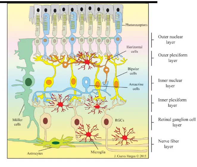
The retina is the innermost neural layer of the eye where the light energy is transformed into a neural signal. The retina forms a sheet of only ∼200 μm thick and is comprised of complex neuronal circuits and glial cells. More than 60 distinct types of neurons are arranged into distinct morphological and functional circuits working in parallel to encode visual information in the retina. In fact, the retina has a stereotypical laminar structure comprised of three nuclear layers, where retinal cell bodies are located, and two intermediate plexiform layers where synaptic interactions between these nuclear layers take place [11, 12]. The retinal neurons are grouped into five major classes of cells: the photoreceptors (rods and cones), three major classes of interneurons (horizontal, amacrine, and bipolar cells) and RGCs (Fig. 1)[13].
Light rays entering the eye cross through the entire retina to reach to the light sensitive outer segments of rods and cones in the outer nuclear layer (ONL) where they are converted into electrical signal. The signal is further relayed to the inner nuclear layer (INL) where bipolar, horizontal, and amacrine cells perform initial information processing. From the bipolar cells the signal is finally transferred to the innermost layer of the retina, the ganglion cell layer (GCL) where the projection neurons the RGCs are located and conveyed the visual image information via their axons bundles in the optic nerve to higher processing centers within the brain (Fig. 1). Dividing these nerve cell layers are two plexiform layers where synaptic connections occur: the outer plexiform layer (OPL), a single lamina where photoreceptors make synaptic connections with bipolar and horizontal cells, and the inner plexiform layer (IPL) where bipolar and amacrine axon terminals synapse onto RGCs [14].
Figure 1. A schematic diagram of the retina demonstrating the principal cell types involved in retinal signaling. The retinal neurons are photoreceptors cells, bipolar cells, horizontal cells, amacrine cells, and retinal
ganglion cells (RGCs). These cells are organized into five interconnected layers. Of the five layers, the ganglion cell layer (GCL), inner nuclear layer (INL) and outer nuclear layer (ONL) consist mainly of cell bodies while synaptic connections among the neurons are located in the inner plexiform layer (IPL) and the outer plexiform layer (OPL). The retina also contains glial cells that interact with neurons and blood vessels. The glial cells are categorized into Muller cells, astrocytes and microglia. Muller cells span across the entire thickness of the neural retina and provide an anatomical and functional link between retinal neurons and blood vessels. The astrocytes surround the blood vessels and microglial cells keep the central nervous system under surveillance for infections and injuries. Source of image: Jorge Luis Cueva Vargas.
I.1.1 Photoreceptors
The photoreceptors form a single sheet of regularly spaced light-sensitive cells comprised of rods and cones. Photoreceptors are conical-shaped structures consisting of a membranous outer segment containing photopigments (rhodopsin or cone opsins) and an inner segment containing the cell nucleus. The light absorbing pigments of photoreceptors contains 11-cis retinal which, upon absorption of light, undergoes conformational changes and dissociates itself from opsin. The opsin moiety can now bind and activate a trimeric G-protein called transducin. The activated α subunit of transducin further binds to cGMP phosphodiesterase or PDE, which converts the available supply of cGMP to its non-cyclic form 5'-GMP. The dropping cGMP levels leads to cation channel closure in the plasma membrane blocking sodium and calcium influx. Consequently, the photoreceptor outer segment hyperpolarizes generating a direct flow of electrical current along the membrane to the synaptic terminals promoting the release of the neurotransmitter glutamate [15].
Although both rods and cones respond to light with a slow hyperpolarizing response, they report quite different image properties. The fovea the central region of the retina exclusively contains cones which require high levels of light to generate signals, and therefore work best during the day time illumination. In addition, cones are most sensitive to specific wavelengths of light and are therefore responsible for high visual resolution and color vision. There are three types of cones: long (red or L-), middle (green or M-) and short (blue or S-) wavelength [16]. In the peripheral region of the retina, rod photoreceptors outnumber cone photoreceptors by about 20-fold in almost all mammalian retinas. In fact, after the cerebellar granule cells, ~125 million rod photoreceptors appear to be the second most numerous neurons of the human body. Rods have exquisite sensitivity to light with the resolution of a single photon detection and thus are specialized for vision in dim light [17].
I.1.2 Bipolar cells
Retinal bipolar cells comprise a diverse class of interneurons in the INL, transmitting the photoreceptor signals on amacrine and ganglion cells. There are at least 13 distinct types of bipolar cells which differentially collect and shape photoreceptor signals into parallel
information pathways, each encoding a highly processed feature such as motion, contrast or edges [18, 19]. Bipolar cells fall into two main groups based on their response to glutamate released by photoreceptor: ON (glutamate hyperpolarization) and OFF (glutamate depolarization) bipolar cells. In response to light increments, ON bipolar cells that have inhibitory glutamate receptors, depolarize. On the other hand, OFF bipolar cells that are depolarized in the dark will hyperpolarize. OFF bipolar cells contact OFF RGCs, and ON bipolar cells contact ON RGCs [11, 20].
I.1.3 Horizontal cells
Horizontal cells are the laterally interconnecting neurons in the INL that release inhibitory γ-aminobutyric acid (GABA) neurotransmitters and, as such, help integrate and regulate the input from multiple photoreceptor cells by providing inhibitory feedback. In fact, the horizontal cells measure the average level of illumination across the broad region of photoreceptors and subtract the signal from less illuminated ones, resulting in the reduction of redundancy of the signal transmitted to the RGCs. This selective suppression of certain neuronal signals is called lateral inhibition, and its overall purpose is to improve the contrast and definition and therefore the acuity of the visual stimulus [21].
I.1.4 Amacrine cells
Amacrine cells are important interneurons that establish synaptic contacts with bipolar cells and RGCs in the IPL. Due to the lack of clear polarity, which makes it difficult to differentiate the site of their input from output, they were named amacrine cells meaning axon less neurons [22]. Amacrine cells modulate the synaptic input to ganglion cells in three different ways: either directly by feedforward inhibition on RGC dendrites or by feedback inhibition of axon terminals of bipolar cells that drive them. They also provide lateral inhibition by making connection to other amacrine cells [11, 23]. They mediate their inhibitory functions largely by releasing two neurotransmitters: γ-aminobutyric acid (GABA) and glycine. Amacrine cells exhibit great structural diversity and complexity and are divided into 42 different
morphological subtypes and are thus considered the most diverse cell class in the retina. Amacrine cells are also grouped into four subtypes of narrow-field (30-150 μm), small-field (150-300 μm), medium-field (300-500 μm) and wide-field (>500 μm) based on the measurement of their dendritic fields [22, 24].
I.1.5 Retinal ganglion cells (RGCs)
RGCs are the largest retinal neurons with a density of 2,500 cells per square millimeter. RGCs are the output neurons of the retina by extending axons through the optic nerve to higher processing centers in the brain. The signals from bipolar cells are integrated by RGCs and are further transferred as a diverse set of parallel action potentials travelling down the optic nerve to the brain. RGC action potentials are generated spontaneously and change accordingly when stimulated by the appearance of light in the RGC receptive fields.
RGCs exhibit strikingly varied dendritic morphology and field size. Based on their patterns of dendritic arborization, RGCs were initially categorized in approximately 20 subtypes [25, 26]. However, in addition to their structural heterogeneity, RGCs can be distinguished by their diverse functions and connectivity. In fact, besides being a light detector due to the presence of the pigment melanopsin in an RGC subtype, these neurons can also behave as feature detectors: each neuron tuned to distinct visual features such as contrast, color, or specific motion directions [27, 28]. Therefore, the anatomical classification of RGCs thus far cannot encompass the functional diversity of these neurons and a further categorization of RGCs is necessary to better understand the visual outcome in both physiological and pathophysiological states. Recently, new molecular, genetic, and functional approaches have complemented the RGC classification scheme increasing the estimated number of RGC types to >30 [25, 27, 29]. RGC axons converge at the optic nerve head (ONH) to form the optic nerve which projects to four main targets in the human brain: 1) the lateral geniculate nucleus of the thalamus from where signals are relayed to the visual cortex, 2) the superior colliculus for orienting responses, 3) the pretectal nucleus for the pupillary light reflex, and 4) the suprachiasmatic nucleus for light entrainment of the sleep–wake cycle [30].
I.1.5.1 RGCs are the cellular target in many retinal diseases
RGC death is the final common pathway that leads to loss of vision in several retinal diseases such as glaucoma [31]. RGCs are the sole retinal neurons that generate action potentials transmitted over long distances along the optic nerve trajectory (∼50 mm) to the brain visual centers [32]. Therefore, RGCs are the most energetically demanding neurons in the retina. In addition, each RGC subcellular compartments: the dendrites, cell body, non-myelinated axon, and myelinated axon, are located in a different extracellular environments with different energy requirements. Therefore, intracellular energy distribution and consumption is different and specific to each RGC cell relative to other neurons. Accordingly, for the maintenance of optimal neuronal function, mitochondria are unevenly distributed within RGCs to meet the local energy demands of the cell [33]. All these factors render RGCs more vulnerable to minor disruptions of energy homeostasis and predispose them to respiratory chain dysfunction, oxidative stress and ultimately apoptosis [33, 34].
I.1.6 Retinal glial cells
In addition to the neuronal network, the retina contains three basic types of glial cell subdivided into macroglial cells, astrocytes and Muller cells, and microglial cells (Fig. 1). In general, retinal glial cells regulate the microenvironment and ensure optimal neuronal function. Glial cells not only provide the neurons with structural and metabolic support but are also involved in maintaining retinal homeostasis by forming blood-retinal barrier (BRB) and regulating local blood flow [35]. As such, they participate in retinal glucose metabolism, elimination of metabolic waste products, release of certain transmitters, trophic factors and potassium uptake [36].
Under pathophysiological conditions, glial cells also participate in local immune responses and protect retinal neurons through a process called reactive gliosis. Reactive gliosis involves both morphological and functional alteration of the glial cells and is associated with cellular hypertrophy, proliferation, migration and cytokine release with the aim to maintain retinal homeostasis [37]. However, glia cells can also undergo chronic gliosis which constitute
the primary pathogenic element exacerbating disease progression by increasing vascular permeability, infiltration of toxic compounds, and even neovascularization [35, 36, 38]
I.1.6.1 Muller glia
Muller glia are cylindrical, fiber-like cells that constitute the predominant glial cells of the retina. They are arranged in parallel spanning the entire width of the retina with their apical processes in intimate contact with photoreceptor cell bodies and their end feet enveloping RGCs and displaced amacrine cells. Muller cell are endowed with many side branches that are involved in the formation of the blood retinal barrier (BRB) allowing intimate association with both neuronal elements such as synapses as well as the blood vessels. In this way, they play a role in neurotransmission and support the neurons by providing neurotrophic factors and blood-derived nutrients as well as removing metabolic waste [36]. Furthermore, they are involved in maintaining the BRB by providing a permanent anti-proliferative environment for retinal vascular endothelial cells [39].
Müller cells act as living fibers guiding the light through the randomly oriented and irregularly shaped neurons providing a low-scattering light passage from the retinal surface to the photoreceptor cells. As a result, light arrives at individual photoreceptor with high intensity and minimal distortion. Interestingly, it has been shown that Müller cells can generate neural stem cells and as such could be considered as the primary source of new neurons in mammalian retinas [38, 40].
I.1.6.2 Astrocytes
Astrocytes represent the most abundant and heterogeneous retinal glial cell. Astrocytes are mostly located in the nerve fiber layer and support the function of RGC axons. They comprise almost 50% of the cells in the optic nerve head. It is believed that astrocytes migrate from the optic nerve to the retina along the blood vessels and locate themselves exclusively in the inner retina. Accordingly, the presence and distribution of retinal astrocytes is correlated with the presence and distribution of retina blood vessels. They are considered as the main
producer of vascular endothelial growth factor (VEGF) and, as such, are strongly implicated in retinal vascularization under both physiological and pathological states [41]. It is noteworthy that astrocytes communicate by gap junctions and play an important role in the maintenance of the extracellular pH and ion homeostasis as well as glutamate clearance in the retina [36].
I.1.6.3 Microglia
Microglia are distributed ubiquitously in different retinal layers. Microglia can exist in two different states: surveillance and activated states in the retina. In the healthy and homeostatic state, microglia have a small cell body with a rather large nucleus forming a dynamic and highly organized non-overlapping network. This configuration allows the microglia to survey the microenvironment and participate in cell-cell interactions with retinal neurons and other microglia. The presence of surface proteins such as receptors for cytokines, growth factors, purinergic ligands, toll like receptors (TLR) and complement components in addition to their intricate interactions with other neurons allows microglia to systematically sense their environment [42]. Microglia rapidly change their distribution and organization within the retinal layers in pathological states: they exhibit ameboid cell shapes with pseudopodia and contribute to the remodeling of neuronal circuits, as well as the elimination of cell debris and misformed synapses. In this regard, in the experimental model of axotomy, microglia containing RGC debris can be detected months after performing axotomy, indicating both the phagocytotic capacity of microglia and their relative longevity [43-45].
I.2. The retina as a model to study neurodegenerative diseases
The retina is an extension of the CNS and exhibits many structural similarities to the brain and spinal cord [46]. Furthermore, the eye is a very accessible organ providing the opportunity for non-invasive imaging of retinal neuron as well as the potential for drug discovery and delivery using methods such as eye drops or intraocular injections [47]. The intravitreal injection of short interfering RNAs (siRNAs), plasmids and recombinant proteins allow the selective manipulation of RGCs without confounding effects on other neighboringneurons or surrounding glia. Moreover, different retinal neurons and glial cells can be selectively targeted by the injection of specific viral vectors in the retina.
Diseases of the retina often share similarities and common neurodegenerative mechanisms with other disease of the brain or the spinal cord [48]. For example, accumulating evidence suggest an array of common physiological and pathological changes between glaucoma, the leading cause of blindness worldwide characterized by RGC loss, and Alzheimer disease. In this regard, Chiasseu et al. demonstrated that tau accumulation and altered phosphorylation, hallmarks of Alzheimer disease, also plays a neurotoxic effect in glaucoma [49]. These features render the retina a particularly attractive system to investigate pathophysiologic mechanisms underlying neurodegenerative diseases [13]. A better understanding of these mechanisms could have implications for the development of therapeutic approaches for retinal/optic nerve and brain diseases.
I.2.1 Optic nerve axotomy
RGCs are a well-characterized CNS neuronal population with cell bodies located in the inner retina and axonal processes along the optic nerve that reach specific targets in the brain. The optic nerve can be accessed within the orbit of the eye and completely transected or crushed producing a well characterized temporal course of RGC death. Optic nerve axotomy is considered a reproducible model of apoptotic neuronal cell death and has been widely used to study the response of CNS neurons to axonal injuries as well as to test neuroprotective therapies and regenerative strategies [50, 51]. The mechanisms of RGC death in axotomy are still poorly understood. It is believed that following axotomy the physical separation of the cell body from its target results in neurotrophic factor deprivation followed by oxidative stress [52]. Finally, calcium influx destabilizes the cytoskeleton and RGCs die by apoptosis as they do in glaucoma and other CNS diseases [30, 53]. Ultimately, 90% of the injured RGCs die within 14 days post-axotomy and the few remaining neurons are severely impaired and inactive. After post-axotomy, the pathways involved in the RGCs apoptosis as well as survival can be further investigated by intravitreal injections of modulatory factors. Strategies to promote cell survival in this system may be extrapolated to other neurodegenerative diseases and CNS trauma [54].
I.2.1.1 Optic nerve axotomy and dynamics of RGC death
RGC apoptosis represents a characteristic bi-phasic time-course after axotomy. In fact, RGC apoptosis has a well-defined onset at approximately 3 days post-axotomy providing a time window for experimental manipulations of the apoptotic pathways [55]. After this 3-day interval, the cells degenerate rapidly and only 50% of RGCs survive at 7 days and less than 10% remain at 14 days after injury. Approximately 5% of RGCs remain up to 20 months after transection, but the functional state of these neurons is unknown [56-58]. Interestingly, early morphological changes in dendrites (dendritic retraction) were shown to occur at 3 days post axotomy prior to overt RGC death suggesting that dendrite pathology is an early neurodegenerative event following axonal injury. The identification of pathways that contribute to RGC dendritic arbor maintenance and synaptic integrity will be useful to understand the molecular basis of pathological changes after axotomy [59].
The survival of RGCs after optic nerve axotomy can be tracked over time with ease and accuracy. In fact, because the ganglion cell layer is a monolayer (one cell thick), RGC densities can be quantified in flat-mounted tissue, without the need for stereology. For this, retinal whole-mounts are labelled with RGC specific markers such as brain-specific homeobox/POU domain protein 3A (Brn3a) [60] and RNA binding protein with multiple slicing (RBPMS) [61] and counted at specific time points.
I.2.1.2 Neuroprotective strategies to promote RGC survival after axotomy
Detailed knowledge of the temporal course of RGC death and the mechanisms that lead to neuronal loss is essential to develop efficient neuroprotective strategies after optic nerve injury. In this regard, axotomy-induced RGC loss has been mainly attributed to the withdrawal of trophic factors from target neurons. The focus of many studies has been to activate pathways that are involved in RGC survival, including TrkB signaling, the Erk1/2 pathway following the intravitreal administration of growth factors such as brain derived neurotrophic growth factor (BDNF) [62-64]. However, recent data suggest that axonal injury leads to early changes in RGC dendritic structure prior to cell death, compromise synaptic integrity, and lead to functional deficits [59, 65]. Accordingly, emerging data revealed that dendritic abnormalities and loss of
synapses contributes to the pathology of many psychiatric and neurodegenerative disorders [66, 67]. Therefore, the focus of recent studies is the elucidation of molecular mechanisms leading to RGC dendrite degeneration and synaptic rearrangements after axonal injury. The identification of new pathways that contribute to RGC dendritic arbor maintenance will be necessary for the development of the strategies to protect RGCs connectivity and enhance survival [51, 68, 69].
I.3. Netrin-1: structural organization and evolutionary history
During the development of the nervous system, neurons are produced in specialized regions and migrate through defined pathways until they reach their final location. Each neuron develops a dendritic arbor that is characteristic by its phenotype and an axon that extends to reach its synaptic target. The interaction of neuronal cell surface receptors with attractive and repulsive guidance cues secreted by intermediate and final cellular targets dictates a defined trajectory that the neuron will navigate [70, 71]. Until now, molecular studies have led to the identification of several families of guidance cues over the past 15 years including netrin and its receptors, Robo/Slit, and semaphorins/collapsins [72].Netrin proteins were originally identified in embryonic chick brain, where they direct commissural axons in a circumferential trajectory to the floor plate at the ventral midline of the spinal cord [73]. Later, netrin was found to be capable of repelling trochlear motor axons, that grow away from the floor plate underlining their role as bi-functional chemotropic guidance cues [74]. Netrins are phylogenetically conserved in all bilaterally symmetrical animals directing a wide range of outgrowing axons and migrating neurons. Subsequent work established that netrins have multivalent ability and are also involved in the development and maintenance of several non-neuronal tissues by regulating processes such as cell adhesion, migration, survival, differentiation and branch morphogenesis [2].
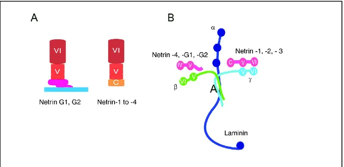
Netrin-1 is a member of the family of secreted netrins, which consists of 1, netrin-3, and netrin-4. Recently, netrin-5 was also identified [75]. In addition to secreted netrins, there are two membrane-tethered netrins, G1 and G2, which are attached to the plasma membrane by a glycophosphatidylinositol (GPI) anchor and are believed to have evolved independently of
secreted netrins. Netrin family proteins consist of an N-terminus laminin-like VI-V domain which is attached to a C-terminal netrin-like domain (NTR) in secreted netrins (Fig. 2A). The N-terminal domains of netrin 1 and netrin 3 share homology with the laminin γ1 short arm, and those of netrins 4, G1 and G2 are homologous to the laminin β1 chain (Fig. 2B). The C-terminal sequence of secreted netrin is rich in basic amino acid residues which interacts with heparin sulfate proteoglycans (HSPGs) which presents secreted netrins on cell surfaces or retains them in the extracellular matrix (ECM) (Fig. 1C). For this reason, the majority of netrin proteins are not freely soluble in vivo but are retained on the cell membranes in the ECM [2].
Figure 2. The netrin family of proteins. (A) Netrins 1 to 4 are secreted proteins that are attached to a C-terminal
domain whereas netrins G1 and G2 are linked to the plasma membrane by a GPI linker. (B) Laminin 1 is a heterotrimer composed of α (blue), β (green), and γ (turquoise) chains. The amino-terminal VI and V domains of netrins 1 to 3 (red) are homologous with the γ chain of laminin 1. These domains in netrins 4, G1 and G2 resemble β chain of laminin 1. Source of image: Rajasekharan and Kenned, 2009
I.3.1 Netrin-1: the prototypical member of the netrin family
Since their discovery, secreted netrins have been shown to have overlapping functions and receptor repertoires. However, the diverse functional roles of netrins have been best characterized in studies of netrin-1. In fact, netrin-1 has been extensively shown to be implicated in axonal outgrowth and neuronal migration during embryogenesis. The molecule’s cDNA encodes a 603 amino acid (aa) protein precursor of approximately 75 (kDa) [76]. Mouse netrin-1 shares 52% aa identity with mouse Netrin-3, and 98% and 87% aa identity with human and chicken. Netrin-1 and netrin-3 are essentially functionally equivalent: both bind to the same receptor proteins and evoke chemoattractant or chemorepellent responses from responsive cells [77]. Loss-of-function mutations in netrin-1 or in certain netrin-1 receptors are lethal in mice, highlighting the importance of netrin-1 signaling during development [2]. In the following chapters, I will summarize the current state of knowledge on netrin-1 and its receptors and discuss netrin-1 functions in the CNS both during development and in adulthood.
I.3.2 Multiple receptors for netrin-1
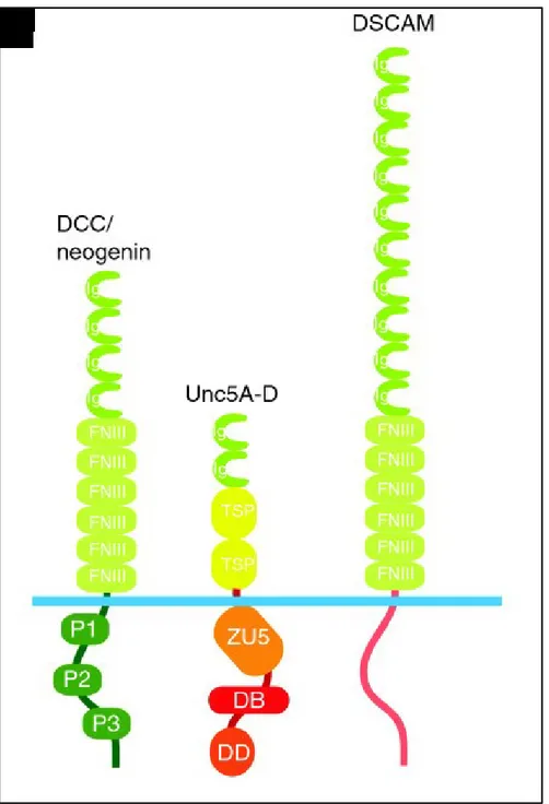
Netrin-1 exerts most of its regulatory functions through the signaling pathways downstream of its two main receptors: Deleted in Colorectal Cancer (DCC) and the Uncoordinated-5 homologous family (UNC5A, -B, -C, and –D). Other secreted netrins are known to interact with these receptors with different affinity. Recently Down’s syndrome cell adhesion molecule (DSCAM) and adenosine receptors (A2b) have also been classified as netrin-1 receptors. All netrin-netrin-1 receptors identified thus far are single-pass type I transmembrane proteins and are members of the immunoglobulin (Ig) superfamily (Fig. 3)[1].
Figure 3. Canonical netrin-1 receptors. The DCC receptor family, the UNC-5 homologs (Unc5A-D), and
DSCAM are receptors for netrin-1. DB, DCC-binding domain; DD, death domain; Ig, immunoglobulin domain; FNIII, fibronectin type III repeat; TSP, thrombospondin type-I module; ZU5, domain homologous to part of Zona Occludens-1. Source of the image: Rajasekharan and Kennedy, 2009.
I.3.2.1 The DCC receptors
The DCC receptor family was discovered in 1996 as a cell surface receptor encoded within a 370-kb region on chromosome 18q that is deleted in tumors. Therefore, initially it was proposed as a putative tumor suppressor gene whose absence on colon cells implied a malignant state of colon cancer [78]. Later, it was proposed as a receptor for netrin-1. The DCC receptor family is comprised of both DCC and neogenin with approximately 50% aa identity. Neogenin receptor functions are generally associated to cell adhesion rather than axon guidance. The interaction of neogenin with netrin-1 regulates cell-cell adhesion at paranodal junctions and synapses [2].
Extracellularly, the DCC receptor is composed of four Ig-like domains at its N-terminus, followed by six fibronectin (FN) type III domains, an approximately 50-residue long membrane-proximal stalk and a transmembrane segment. The cytoplasmic tail of DCC is catalytically inactive and comprises three highly conserved domains (P1, P2, and P3) that are involved in conducting netrin-1 downstream signal transduction pathways [79].
Several axon tracts in the spinal cord and the brain express DCC and the loss of DCC expression in mice causes defects in the formation of spinal and cerebral commissures that are comparable to those observed in netrin-1deficient mice [80]. Moreover, mutations in the DCC gene locus are associated with mirror movement disorders in humans and mice, a reported consequence of midline crossing defects [2].
Netrin-1 protein can simultaneously bind to two DCC molecules through a DCC-specific site and a unique generic receptor binding site. This characteristic two-site feature is the key for the interpretation of netrin-1 bi-functionality which will be recapitulated in detail in the chapter 3.3 [81].
I.3.2.2 The UNC5 receptor family
The family of UNC5A-D receptors are transmembrane protein with an extracellular domain consisting of two immunoglobulin repeats followed by two thrombospondins type I groups (TSP-1). The immunoglobin repeats of UNC5 proteins are required for netrin-1 binding.
The UNC5 intracellular domain is about 550 residues and encodes three distinct domains that are required for receptor signaling: (A) ZU-5 domain, a domain of yet undetermined function, named for its homology with a portion of Zona Occludens-1, (B) DCC-binding (DB) motif, and (C) Death Domain (DD), associated with apoptotic signaling [1]. UNC5A-D receptors are widely expressed across the nervous system including neurons that express DCC. In fact, in the presence of UNC5, DCC is replaced by UNC5A on the generic receptor binding site of netrin-1 switching the netrin-netrin-1 response from chemoattraction to repulsion [8netrin-1].
I.3.3 Netrin-1: bi-functionality and reverse signaling
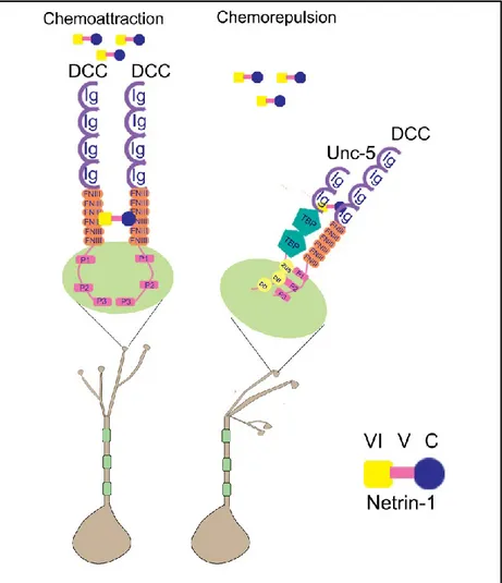
Netrin-1 is a guidance cue that depending on the receptor it binds to, it triggers either attractive or repellent effects on migrating neurons. In general, netrin-1 exerts its chemoattraction activity when it binds to DCC receptors. Upon binding, netrin-1 brings two DCC receptors in close proximity and mediates DCC homo-dimerization by bringing DCC cytoplasmic P3 motifs in close proximity. The dimerized P3 motifs serve as a structural unit for further signaling and recruit intracellular signaling complexes leading to the release of calcium, kinase activation and a rearrangement of the cytoskeleton [2]. On the other hand, netrin-1-mediated chemorepulsion is induced when netrin-1 binds to a DCC and a UNC-5 receptor. In the presence of netrin-1, a ternary complex is formed between netrin-1 and the extracellular portions of UNC-5 and DCC. This heterodimerization through a cytoplasmic interaction between the P1 motif of DCC and the DCC-binding domain (DB) of UNC-5 gives rise to an alternate signaling complex leading to chemorepulsion [81]. Therefore, netrin-1 appears to serve as a scaffold both for symmetric clustering of a single receptor, and asymmetric clustering of a pair of different receptors with remarkable versatility [79, 81, 82]. An important generalization drawn from these data is that the intracellular domain of netrin-1 receptors is determinant in netrin-1 signaling as an attractant or repellent factor. The signaling pathways downstream of DCC and UNC-5 receptors are explained in the next chapter.
Figure 4. Netrin-1 is a bifunctional axonal guidance cue. Netrin-1 can bind to the ecto-domains of two receptors
DCC/DCC or DCC/UNC5, and brings them close enough to initiate interactions between their cytoplasmic domains leading to either chemoattraction or chemorepulsion. Source of image: Pegah Chehrazi
I.3.4 Netrin-1 signaling mechanisms
Netrin-1 signaling has been largely studied in relation to axon guidance where axon motility is directed by orchestrating the dynamic reorganization of actin cytoskeleton in axonal growth cones. The growth cone, a fan-like structure located at the tip of an extending axon, contains the machinery necessary to detect and respond to guidance cues and provides the motor energy necessary for neurite outgrowth [83]. Upon netrin-1 binding, cytoplasmic signal transduction molecules in the growth cone link the activation of axon guidance receptors to the reorganization of the actin cytoskeleton [84]. In the case of chemoattraction, the
membrane extension toward the source of netrin-1 is derived by the insertion of DCC at the leading edge of growth cones, the stabilization of DCC in the plasma membrane and the linking of DCC to actin filaments [85]. However, DCC is a transmembrane protein without an obvious catalytic domain, and thus for a long time it remained unclear how it initiates downstream signaling to regulate axonal outgrowth. Recently, it was shown that DCC functions as a tyrosine kinase-associated receptor and the intracellular domain (ICD) of DCC is constitutively bound to the adaptor protein Nck1 and focal adhesion kinase (FAK). With the binding of netrin-1 to DCC, NCK1 and FAK phosphorylate DCC ICD resulting in the recruitment of a number of intracellular signaling components that activate several parallel cascades: Src family kinases, Rho GTPases, the release of Ca2+ stores and protein translation. All these pathways ultimately
converge on the rearrangement of the actin cytoskeleton [86, 87]. The signaling mechanisms that underlie netrin-1-induced chemorepulsion are considerably less well understood than those underlying chemoattraction.
Activation of small GTPases
Netrin-1 binding to DCC results in FAK activation which recruits SFKs, Src, and Fyn to the DCC ICD, leading to Nck1-dependent activation of p21 activated kinase1 (PAK1) as well as Rho GTPases [86, 88]. Rho GTPases, in particular Rac1 and Cdc42, are important signaling molecules involved in promoting actin polymerization at the extending edge of the growth cone. They further activate the actin nucleation factor Arp2/3 through the stimulation of the Wiskott-Aldrich syndrome protein (WASP) family members. Another member of Rho GTPases, RhoA, is inhibited during DCC activation leading to DCC endocytosis and thus promoting the trafficking of the DCC receptor on plasma membranes [1, 74, 89].
Regulation of mRNA translation machinery
Extending axons contain the machinery for local protein translation, providing the growth cone with a substantial level of functional autonomy from the cell body during embryogenesis. Netrin-1/ DCC signaling is shown to regulate protein translation via PI-3 kinase and rapamycin-sensitive mTOR protein synthesis. In particular, in response to actin-dependent
changes in growth cone motility, netrin-1 mediated DCC signaling regulates the local translation of β-actin mRNA by relieving its translational inhibition from Zipcode Binding Protein 1(ZBP1) [2, 90, 91].
DCC-dependent signaling also involves the activation of the mitogen-activated protein kinase (MAPK) cascade by extracellular signal-regulated kinase (ERK)-1 and 2 which are phosphorylated following netrin-1 receptor activation. The activation of MAPK pathway further results in the activation of specific transcription factors such as ELK1 underlining a potential mechanism by which netrin-1 may control transcription and protein translation [92].
Calcium in regulating neurite growth and motility
Notably, cytoplasmic Ca2+ participates in the concerted regulation of the actin
cytoskeleton to promote growth cone extension. The binding of netrin-1 to DCC promotes 1,4,5-trisphospate (IP3)-mediated Ca2+ release from internal stores, which is necessary to maintain
Ca2+ within the optimum range for neurite extension. The release of Ca2+ from intracellular
stores also activates transient receptor potential (TRP) channels to trigger a Ca2+ influx across
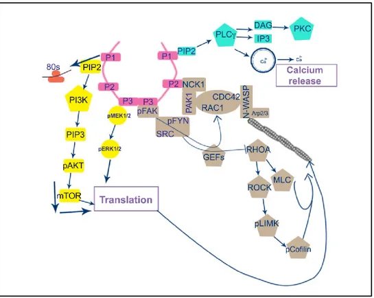
Figure 5. Netrin-1 signal transduction mechanisms. In response to netrin-1, DCC receptors homodimerize via
their P3 domains interaction. The DCC P3 domain is bound to FAK while DCC P2 domain is constitutively bound to NCK1. With netrin-1 binding, Nck1 and their downstream effector Pak1 serve as a scaffold for the recruitment of Rho family of small GTPases, in particular Rac1, and Cdc42 and RhoA. Furthermore, FAK recruits and activates the tyrosine kinases Src and Fyn, which also participate in regulating Rac1 and Cdc42 activity. Activated Cdc42 further recruits N-WASP, which promotes the nucleation of F-actin via the Arp2/3 complex modulating the dynamics of actin polymerization. In addition, PI3K and PLCγ are activated in response to netrin-1 binding. PLCγ hydrolyses PIP2 into IP3 and DAG. IP3 stimulates the release of intracellular calcium, and DAG activates PKC, which in turn result in cytoskeleton remodeling. Abbreviations: Pak1, p21-activated kinase; FAK, focal adhesion kinase; Src, tyrosine kinase sarcoma; ERK, extracellular-signal-regulated kinase; MAPK, mitogen-activated protein kinase; PLCγ, phospholipase Cγ; PI3K, phosphatidylinositol 3-kinase; PIP2, phosphatidylinositol (4,5) bisphosphate; IP3, inositol triphosphate; Rac1, Ras-related C3 botulinum toxin substrate 1; Cdc42, cell division cycle 42; N-WASP, neuronal Wiskott–Aldrich syndrome protein; Pack1, serine/threonine kinase p21 activated kinase 1; and Cdc42, cell division cycle 42; RhoA, Ras homologous member A; PKC, protein kinase C. Source of image: Pegah Chehrazi
I.3.5 Netrin-1: a role both as a long-range and a short-range cue
In addition to bi-functionality, netrin-1 has the dual characteristic of functioning at short or long distance. Netrin-1 functions at short-range when it induces signaling in the immediate vicinity of its cellular source. For example, in the mature nervous system, netrin-1 mediates cell-cell contacts between neurons at synapses, as well as between oligodendrocytes and axons at paranodal junctions [1]. By contrast, netrin-1 functions as a long-range cue when acting far from its secreting source. For example, netrin-1 guides the axonal projections across the midline in the embryonic brain and spinal cord [95]. Long-range netrin-1-induced repulsion, which requires higher sensitivity to netrin-1, is mediated by multimerization of UNC5 with DCC as a co-receptor; while the homodimerization of UNC5 homologues in the absence of DCC is enough to mediate short-range repulsion in response to netrin-1 [96].
I.3.6 Calcium modulates netrin-1 signaling
Cyclic adenosine monophosphate (cAMP) is a second messenger that is mainly recognized for its role on axon guidance and axon regeneration. Bi-directional turning responses of growth cones to netrin-1 depend on the relative activities of cAMP dependent pathways, which regulate the cytosolic level of Ca2+ signals in two different ways: 1) by regulating Ca2+
influx through voltage-dependent Ca2+ channels in the plasma membrane (VDCCs) [97], and 2)
by controlling the release of Ca2+ from intracellular stores. The intracellular level of Ca2+ in turn
regulates Rho GTPase activation and cytoskeletal dynamics [98, 99]. The netrin-1-induced chemoattractive activity is associated with a high level of cAMP, whereas the netrin-1-mediated chemorepulsive activity is related to a low cAMP level [100].
I.3.7 Other putative netrin-1 receptors
I.3.7.1 DSCAMThe Down’s syndrome cell adhesion molecule (DSCAM) receptor was originally identified as a candidate gene duplicated in Down’s syndrome. However, for a long time, it was considered as an orphan receptor without an identified ligand. The recent identification of
netrin-1 as a ligand for DSCAM, suggests that some of the deficits associated with this disorder might result from altered netrin-1 signaling. In this regard, Ly et al. showed that DSCAM is expressed on spinal commissural axons where it mediates axonal outgrowth and turning in response to netrin-1 both alone or in collaboration with DCC [80]. However, these data were contradicted by the analysis performed in DSCAM-null mice where axons outgrowth toward the floor plate were comparable to the level of axonal extension mediated by DCC alone [101]. However, signaling mechanisms activated by netrin-1 downstream of DSCAM have not yet been identified. Further investigation of the signaling molecules downstream of DSCAM will help clarify the discrepancy between the aforementioned studies.
It is interesting to mention that DSCAM is also shown to function as a repulsive netrin-1 receptor, which in collaboration with UNC5C, mediates netrin-netrin-1-induced growth cone collapse. Importantly, main signaling components involved in netrin-1-mediated attraction such as Fyn/FAK/PAK1 signaling were suggested to be required for the coordination of netrin-1/DSCAM and netrin-1/UNC-5-mediated repulsive signaling [102].
I.3.7.2 A2B
A2b is a member of the family of the adenosine-specific receptors that mediates adenylate cyclase activity in response to adenosine binding. A2b receptors are emerging as a regulator of netrin-1 receptor trafficking and thus modulating the response of axons to netrin-1.
As a growth cone extends toward its target, it modulates its direction in response to different guidance cues that are expressed in distinct multiple places along the path. For example, as RGC axons migrating from the retina reach the optic nerve head, a developmental switch from chemoattraction to chemorepulsion is required to convert the growth-promoting signal of netrin-1 into a repulsive one [91]. This allows the RGCs axons to leave the optic nerve head toward the optic chiasm [103]. It is shown that the activity of intracellular signaling intermediates, such as protein kinase C-alpha (PKCa) and adenylyl cyclase activated by A2b receptors, regulates cell surface levels of netrin-1 receptors underlining the versatile nature of the response of axons to netrin-1. The A2b receptor regulates multiple signaling pathways
including PKC and cAMP, with high levels of cAMP favoring chemoattraction while low levels of cAMP encouraging chemorepulsion [104, 105].
I.3.8 The functional role of netrin-1 in the nervous system during
development
I.3.8.1 Axon guidance
During development, a gradient of netrin-1 emanating from the floor plate directs the circumferential projection of commissural axons toward and across the ventral midline in the embryonic spinal cord. In contrast, it repels the trochlear motor neuron axons away from the floor plate towards the dorsal midline in the brain stem [106].
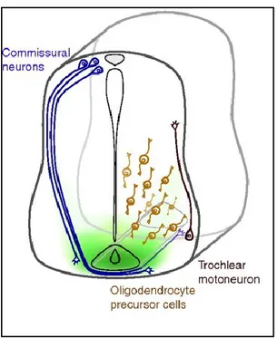
Figure 6. Netrin-1 orients axonal migration during development. In the developing spinal cord, commissural
neurons located dorsally extend axons toward a gradient of netrin-1 at the floor plate resulting in the development of the dorsal-ventral axis of the brain and spinal column. The chemorepulsive activity of netrin-1 was shown on the axons of trochlear motor neurons, whose cell bodies are located in the ventral neural tube and their axons innervate the extraocular muscles of the eye. Source of image: Lai Wing Sun et al, 2009
I.3.8.2 Neural precursor cell migration
Netrin-1 also acts as a bifunctional regulator of neuron migration. During cerebellar development, netrin-1 acts as an attractant for precerebellar neurons directing them to ultimately populate the pontine nuclei. Subsequently, during postnatal cerebellar development, netrin-1 acts as a repellent for the cerebellar granule neurons (CGN) precursors, which upregulate Unc5 expression as they exit the external granule layer [2]. Netrin-1 is also required for the development of the olfactory bulb at late embryogenesis where it directs the migration of neuronal cells to specific regions of the lateral olfactory tract where they act as guidepost cells for olfactory bulb axons [1].
I.3.8.3 Synaptogenesis
Although primarily recognized for its role as an axon guidance cue in the early stages of CNS development, accumulating data indicate that netrin-1 is a synapse-enriched protein with the capacity to regulate synapse formation and stability in two major ways. First, netrin-1 increases the probability of axon and dendrite contact by increasing the number and complexity of their arbors [107]. Second, at the site of contact, netrin-1 promotes synaptogenesis by increasing the focal accumulation of synaptic proteins through enrichment and reorganization of the actin cytoskeleton [108]. In this regard, it is suggested that netrin-1-related changes in synapse function might influence the development and progression of certain forms of human neurodegenerative diseases. For example, functional DCC heterozygosity and a subsequent reduction in DCC gene dosage were introduced as the main cause of neural circuit disruption in congenital mirror movements disease [2].
I.3.8.4 Netrin-1 in oligodendroglial development and maturation
Developing oligodendrocytes extend multiple branching processes over considerable distances, surveying the local environment to locate unmyelinated axons. Recently, it is shown that netrin-1 plays an important role in oligodendroglial maturation by facilitating the detection of target axons. At early stages of development, netrin-1 promotes the morphological maturation
of oligodendrocytes and thereby increases their chances of encountering with appropriate axonal targets [2]. At later stages of maturation, oligodendrocytes begin to express netrin-1 themselves which participates in the formation of large myelin membrane sheets via process extension and branching. Interestingly, the lamella elaborated by the tip of an extending oligodendrocyte process has been shown to be comparable with the neuronal growth cone. Notably, common intracellular signaling proteins are shown to be implicated in netrin-1-mediated axon guidance and oligodendrocyte development [109].
I.3.8.5 Retinal circuit development
Visual information is relayed from the eye to the brain via the axons of retinal ganglion cells (RGCs). Over 50,000 RGC axons in mice and over a million RGC axons in humans must be guided accurately into the optic nerve during retinal embryonic development. The extension of RGC axons along this precise path as well as synapse formation with appropriate target cells are shown to be highly dependent on netrin-1/DCC signaling [110]. At the time of optic fissure closure, netrin-1 is expressed strongly by the neuroepithelial cells that surround and extend into the optic disc while DCC is expressed by RGC axons. The interaction between netrin-1 and DCC locally guide the growing RGC axons to leave the retina and form the optic nerve. In mice lacking either netrin-1 or DCC, RGC axons navigate normally to the optic disc but fail to exit the eye resulting in smaller optic nerves known as optic nerve hypoplasia [111].
As mentioned above, processes of retinal interneurons (amacrine and bipolar cells) form synapses on dendrites of RGCs in the inner plexiform layer (IPL). The IPL is divided into at least 10 parallel sublaminae. Interestingly, subsets of interneurons and RGCs arborize and form synapses in only one or a few sublaminae. These lamina-specific circuits determine the visual features to which RGC subtypes respond. Interestingly, it has been shown that DSCAM is one of the netrin-1 receptors that plays an important role in this specific laminar targeting by directing correct process arborization and synapse formation between RGCs, amacrine cell and bipolar cells [112, 113].
I.3.9 The role of netrin-1 in the adult nervous system
Although originally identified for its developmental role in guiding axons and neurons to their appropriate target, important information about the expression and function of netrin-1 in adult CNS is beginning to emerge and will be discussed in the following sections.
I.3.9.1 Netrin-1 regulates synaptic function and plasticity in the adult brain
Increasing evidence suggests that many proteins that are essential for normal neural development are also expressed in the adult brain where they influence synapse formation and plasticity [114]. For example, Horn et al. have demonstrated that DCC is enriched in dendritic spines of mature forebrain neurons and is implicated in regulating synaptic function and plasticity. Notably, by regulating NMDAR dependent long-term potentiation (LTP), netrin-1-mediated DCC signaling plays a major role in spatial and recognition forms of memory [5].
I.3.9.2 Netrin-1 as an anti-apoptotic agent: the dependence-receptor hypotheses
In recent years, DCC and UNC-5 have been proposed to function as so-called ‘dependence receptors’ meaning that they are dependent on the availability of netrin-1 to ensure cell survival. Based on this theory, netrin-1-activated receptor pathways were suggested to play an important part in tumorigenesis where the loss of receptor expression or upregulation of netrin-1 expression is predicted to provide a selective advantage to tumor cell growth [115]. These studies were further validated by observations showing that netrin-1/DCC signaling is frequently inactivated in many human malignancies [116]. However, the ‘dependence receptor’ hypothesis remains controversial. Mice lacking netrin-1 do not exhibit increased apoptosis in the CNS, arguing that netrin-1 is not an essential dependence ligand in the developing CNS. DCC is also shown to be required for the survival of RGCs and amacrine cells in the developing mouse retina [7]. Considering these data, the formerly demonstrated role of netrin-1 as a cell survival factor is proposed to be restricted to specific cells within the nervous system.
I.3.9.3 Netrin-1 expression in the adult retina
While the mature and embryonic functions for netrin-1 have been extensively examined in many organs of rodents, to our knowledge, only few studies have analyzed the expression of netrin-1 in the adult retina. Indeed, while an initial study demonstrated that the expression of netrin-1 completely disappears in adult rat retina [9], later studies asserted that netrin-1 expression is maintained at the level of the ganglion cell layer and the inner nuclear layer in adult mouse and rat retinas [8, 10]. Further work is required to clarify the expression and role of netrin-1 in the adult visual system.