© Birkha¨user Verlag, Basel, 1998
X-ray studies of enzymes that interact with penicillins
J. A. Kellya, A. P. Kuzina, P. Charlierb,* and E. Fonze´baDepartment of Molecular and Cell Biology, Institute of Materials Science, University of Connecticut, Storrs,
Connecticut (USA)
bCentre d’Inge´nierie des Prote´ines, Unite´ de Cristallographie, Institut de Physique, Universite´ de Lie`ge,
Sart Tilman, B-4000 Lie`ge (Belgium), Fax +32 4 366 4741, e-mail: paulette.charlier@ulg.ac.be
Abstract. The technique of X-ray diffraction has been deal about the structures and catalytic mechanisms of penicillin-binding proteins andb-lactamases. An insight successfully applied to enzymes associated with
peptido-into the structural basis for antibiotic resistance is given. glycan biosynthesis. The technique has taught us a great
Key words. X-ray diffraction;b-lactamases; peptidases; penicillin-binding proteins; active-site serine.
Introduction
Over the last two decades, biophysical techniques have become increasingly important in the study of proteins. One extremely powerful method for investi-gating protein structure and function is X-ray diffrac-tion. This is the case despite the fact that there are serious hurdles that one must overcome to realize the rich results that can be obtained from the X-ray ap-proach. Chief among the difficulties is the need for significant quantities of pure protein sample. Jean-Marie Ghuysen and his colleagues at the University of Lie`ge have made extraordinary contributions to our understanding of the nature of bacterial cell walls and the enzymes associated with the peptidoglycan biosynthesis. They have also been responsible for the elegant characterization and purification work that has made available for biophysical studies many of the penicillin-binding proteins (PBPs) and b-lacta-mases that are key components of cell wall biochem-istry [1].
Penicillin binding proteins are enzymes involved in the final stages of bacterial cell wall synthesis. These en-zymes present a wide range of molecular weights from
27 to 120 kDa. The largest of these enzymes are bi-functional enzymes with a domain responsible for transglycosylation in the nascent glycan strands and a domain associated with the cross-linking of the pep-tide portions of the cell wall. The low molecular weight PBPs are strictly D-alanyl-D-alanine peptidases. The DD-peptidases can act as carboxypeptidases and
transpeptidases, catalysing the scission of the terminal
D-alanyl-D-alanine bond in the peptide portion of the
growing cell wall and the subsequent formation of a peptide bridge to an appropriate amino acceptor on an adjacent peptidoglycan strand. There are, however, examples of DD-peptidases that function solely or
pri-marily asDD-carboxypeptidases, limiting their reaction
to cleavage of the D-alanyl-D-alanine bond (e.g. the
DD-carboxypeptidase from Bacillus stearothermophilus [2]). There are also DD-peptidases that are character-ized as strict DD-transpeptidases such as the enzyme from Streptomyces K15 [3]. Penicillin-binding proteins are the targets in bacteria for b-lactam antibiotics (penicillins and cephalosporins). These drugs serve as potent antibacterial agents because they inhibit the PBPs in the growing bacteria, preventing the crucial cross-linking of the cell wall. Their effectiveness is the result of the structural analogy of the drugs to the normal D-alanyl-D-alanine peptide substrates of the
PBPs.
354 J.A. Kelly et al. Enzyme interactions with penicillins
Table 1. Penicillin-interacting enzymes whose structures have been determined.
Enzyme Source Catalytic entity MW PDB* Resolution Reference code
PBP S. albus G Zinc 18,000 1LBU 1.8 A˚ Wery, D. (1982) Nature 299: 5882, 469 – 470 S. K15 Serine 27,474 / 2.0 A˚ Fonze´, C. (1997) unpublished
S. pneumoniae PBP2x Serine 77,059 1PMD 3.5 A˚ Pares, D. (1996) Nature Stuct. Biol. 3: 284 – 289 S. R61 Serine 37,500 3PTE 1.6 A˚ Kelly (1995) J. Mol. Biol. 254: 223 – 236 b-Lactamase B. cereus 569/H Class B zinc 26,000 1BME 1.85 A˚ Carfi, D. (1995) EMBO J. 14: 20, 4919 – 4921
B. fragilis Class B zinc 26,000 1ZNB 1.85 A˚ Concha, H. (1996) Structure 4: 7, 823 – 836 1BMI 2.0 A˚ Carfi, D. (1997) Acta Cryst. D53: 485 – 487 B. cereus 5/B/6 Class B zinc 26,000 / 1.9 A˚ Fonze´, C. (1997) unpublished
S. albus G Class A Serine 28,500 / 1.7 A˚ Dideberg, C. (1987) Biochem. J. 245: 911 – 913 B. licheniformis Class A Serine 29,500 4BLM 2.0 A˚ Moews, K. (1991) J. Mol. Biol. 220: 435 – 455 S. aureus Class A Serine 28,500 3BML 2.0 A˚ Herzberg (1991) J. Miol. Biol. 217: 701 – 719 E. coli TEM1 Class A Serine 28,950 / 1.7 A˚ Strynadka, J. (1992) Nature 359: 700 – 705
1BTL 1.8 A˚ Jelsch, S. (1993) Proteins 16: 364 – 383 1XPB 1.9 A˚ Fonze´, C. (1995) Acta Cryst. D51: 682 – 694 C. freundii Class C Serine 39,000 / 2.0 A˚ Oefner, W. (1990) Nature 343: 284 – 288 E. cloacae P99 Class C Serine 39,000 2BLT 2.0 A˚ Lobkovsky, K. (1993) PNAS 90: 11257 – 11262 E. cloacae 908R Class C Serine 39,000 / 2.5 A˚ Fonze´, C. (1997) unpublished
*Protein Data Bank.
A second important class of enzymes that affects cell wall biosynthesis is that of the b-lactamases. These enzymes are believed to have evolved from the cell wall-synthesizing PBPs but with an important change in their catalytic mechanism. Instead of being inhibited by b-lactams, b-lactamases rapidly hydrolyse the b-lactam bond and release the inactive reaction product, thereby protecting the cell from the action of the drugs. b-Lac-tamases are grouped into four classes, A, B, C and D. Classes A, C and D rely on a reactive serine for cataly-sis, as do most PBPs. Their classification is based on amino acid sequence. Class B b-lactamases are metal-loenzymes that require zinc ions for activity.
X-ray diffraction studies
To date, X-ray diffraction has been carried out on 4 PBPs and 10 b-lactamases (see table 1) with many additional examples of complexes or mutants forms of the enzymes. A crucial problem in X-ray diffraction studies is the loss of phase information during the diffraction experiment. The phase problem has been solved primarily by multiple isomorphous replacement using heavy atom derivatives of the native enzymes (e.g.
Streptomyces R61DD-peptidase [4]), and more recently by molecular replacement using previously determined protein structures as search models (e.g. the TEM1 b-lactamase [5]). The biggest limitation of molecular replacement in the case of enzymes that exhibit a wide variation in their amino acid sequences is the low simi-larity with the probe structure. Up to now, only a few enzymes interacting with penicillin have been solved this way. However, with the increasing number of indepen-dently solved structures, we may expect more extensive use of this method.
The DD-peptidase domains of the bifunctional PBPs
and of most monofunctional PBPs, as well as the ma-jority of the b-lactamases known today, are serine enzymes that form acyl intermediates. Amino acid se-quence similarity between the different classes of PBPs (bi- and monofunctional) and between the three classes of serine-b-lactamases (A, C and D) is as low as bet-ween PBPs and b-lactamases, usually in the range of 10 to 15%. Among members of a given class, however, the similarity may range from more than 90% for the class C b-lactamases to 50% for the class A b-lacta-mases. It is interesting to note that for the S. R61
DD-peptidase, the highest primary sequence homology
is found with the class C b-lactamases (about 25 to 28%).
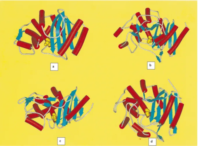
We have learned a great deal about these enzymes through structural studies. Both serine-mediated PBPs and b-lactamases share an overall two-domain struc-ture. One domain is all helical, and the second contains a b-sheet flanked by helices on both faces of the sheet (fig. 1a – d). The reactive serine is found at the interface of these two domains. There is higher tertiary struc-tural resemblance between the S. R61 DD-peptidase
and the class Cb-lactamases (as expected from the size of the molecules and their sequence similarity), and between the S. K15 DD-transpeptidase and the class A
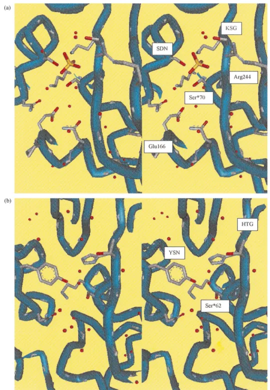
b-lactamases (even though it was not evident from the poor sequence similarity of 15%), than between the class A b-lactamases and the class C b-lactamases. There are only three brief sequence segments common to all PBPs andb-lactamases, each a key element of the active site (fig. 2a, b). These include SXXK, with the reactive serine followed by two variable residues and then an absolutely conserved lysine. This motif is found
Figure 1. Schematic drawings of the class Ab-lactamase of E. coli TEM1(a), class Cb-lactamase of Enterobacter cloacae908R (b), DD-transpeptidase/PBP of Streptomyces K15(c) andDD-carboxypeptidase-transpeptidase/PBP of Streptomyces R61(d). Theb-strands are drawn as blue arrows anda-helices as red cylinders. The reactive serine atoms are drawn as yellow spheres, at the amino-terminus of the central helixa2.
at the amino terminus of a helix generally within 70 residues of the amino terminus of the low molecular weight PBPs and theb-lactamases. The second region is a triad found on a loop in the active site below the reactive serine. The SDN motif in class Ab-lactamases is replaced by SGC in the S. K15 DD-transpeptidase, and by YXN in the S. R61DD-peptidase and the class Cb-lactamases. The hydroxyl group of the tyrosine in these enzymes is in the same position as the hydroxyl of the serine in the SDN enzymes. The last conserved region is KT/SG in all enzymes except the S. R61
DD-peptidase, which has an HTG triad. This motif is
found on the innermostb3 strand of the b-sheet, form-ing one edge of the active site pocket. It is this motif that orients the incoming peptide substrate or b-lac-tams, binding them as though they were extensions of the b-sheet of the protein. The class A b-lactamases have an additional active-site-defining motif EXELN, located at the entrance of the cavity near the bottom of theb3 strand. This motif defines the so-called V-loop and contains the essential residue Glu 166. This Glu 166, with Asn 170, positions a strictly conserved struc-tural water molecule, thus playing an important role in
the catalysis of penicillin hydrolysis by class A b-lacta-mases. In the case of the S. K15 dd-transpeptidase, a loop of different shape is positioned in the same direc-tion, but the possible acidic equivalent to Glu 166 is pointing away from the active site. A similar loop may be observed in the S. R61DD-peptidase and the class C b-lactamases, but the peptide chain is in the opposite sense, and there is no possible structural equivalent to Glu 166. Compared with the S. K15DD-transpeptidase and the class A b-lactamases, the S. R61DD-peptidase
and the class Cb-lactamases have additional loops and secondary structure elements away from the active site. Several crystallographic binding studies have been per-formed on these enzymes. In the case of the the class A b-lactamases, the first reported complex was the TEM Glu166Asn mutant acylated by penicillin G [6]. Com-plexes of the Staphylococcus aureus lactamase with clavulanate (PDB entry code 1BLC) and a methylphos-phonate monoester monoanion inhibitor (PDB entry code 1BLH) have been also studied at liquid nitrogen and room temperature, respectively [7, 8]. More re-cently, two novel designed inhibitors have been used for crystallographic binding studies on the TEM enzyme,
356 J.A. Kelly et al. Enzyme interactions with penicillins
Figure 2. Stereoviews of the catalytic cleft of the class Ab-lactamase of E. coli TEM1(a) and theDD-carboxypeptidase-transpeptidase/ PBP of Streptomyces R61(b), including the conserved active-site groups Ser70, Lys73, Ser130, Asn132, Glu166, Asn170, Lys234, Ser235 and Arg244 for the class A b-lactamase of E. coli TEM1, and Ser62, Lys65, Tyr159, Asn161, His298 and Thr299 for the DD-carboxypeptidase-transpeptidase/PBP of Streptomyces R61. The structural water molecules are drawn as small red spheres. One structural SO4ion is drawn in yellow in (a).
the 6a-(hydroxymethyl)penicillanate (PDB entry code 1TEM) [9] and the (1R)-1-acetamido-2-(3-carboxy-phenyl)ethane boronic acid [10]. Two complexed class C b-lactamase structures have been reported in the litera-ture, the Citrobacter freundii enzyme complexed with
the monobactam inhibitor aztreonam [11] and the
En-terobacter cloacae enzyme complexed with a
phospho-nate derivative (PDB entry code 1BLS) [12].
For the DD-peptidases, binding of the S. R61 enzyme
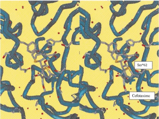
crystal-Figure 3. Stereoview of the catalytic cleft of the theDD-carboxypeptidase-transpeptidase/PBP of Streptomyces R61 complexed with cefotaxime, showing the theb-lactam antibiotic bound to the reactive serine Ser62.
lography [13]. This enzyme exhibits a remarkable stabil-ity with the cephalosporinyl intermediates. This stabilstabil-ity is said to be the result of the lack of a well-positioned water molecule in the active site that is observed in the b-lactamase structures. In the class A b-lactamases, this water molecule is thought to be activated by active site residues for nucleophilic attack on the acyl bond. Its absence would be consistent with the failure of the S. R61DD-peptidase to deacylateb-lactam intermediates,
which leads to their efficacy as antibiotics. Another interesting observation made on the structure with cefo-taxime, a clinically important b-lactam, is the confor-mational change in the side chain of Thr 301 (as shown in fig. 3). In all other b-lactamoyl and phosphonyl complexes studied, a hydrogen bond is made between the backbone of this residue or its equivalent and the drug side chain. The bulky, rigid oxime side chain of cefotaxime prevents this main-chain hydrogen bond from being formed. A change in the orientation of the threonine side chain positions the side chain hydroxyl group so that it can hydogen bond to the drug molecule. A cefotaxime-resistant laboratory mutant of
Streptococcus pneumoniae has been isolated that is
char-acterized by a Thr to Ala mutation at the equivalent site [14]. Such a mutation would not allow the substitution of a side-chain hydrogen bond for the lost main-chain hydrogen bond. It is clear that structural studies of these penicillin-interacting enzymes have been and will continue to be of significant importance for understand-ing the emergunderstand-ing resistance of the PBPs and
b-lacta-mases to the third-generation b-lactam antibiotic such as cefotaxime.
Conclusions
X-ray studies have demonstrated that the serine b-lac-tamases and PBPs are structurally related and that they must have evolved (and are still evolving) from a com-mon ancestor with preservation of much of the same serine-assisted acyl transfer machinery. However, the question of which structural features determine the dif-ferent functions, such as peptide bond transfer or hy-drolysis and penicillin binding or hyhy-drolysis, remains unclear.
Acknowledgement. The authors acknowledge the extensive work of our colleagues O. Dideberg, J. R. Knox and P. C. Moews on PBPs andb-lactamases. The work in Lie`ge was supported by the Belgian programme on Interuniversity Poles of Attraction ini-tiated by the Belgian State, Prime Minister’s Office, Services fe´de´raux des affaires scientifiques, techniques et culturelles (PAI no. P4/03).
1 Ghuysen J.-M. (1991) Serineb-lactamases and penicillin-bind-ing proteins. Ann. Rev. Microb. 45: 37 – 65
2 Despreaux C. W. and Manning R. F. (1993) The dacA gene of Bacillus stearothermophilus coding forD-alanine carboxypepti-dase: cloning, structure and expression in E. coli and Pichia pastoris. Gene 131: 35 – 41
3 Nguyen-Diste`che M., Leyh-Bouille M., Pirlot S., Fre`re J.-M. and Ghuysen J.-M. (1986) Streptomyces K15 DD-peptidase-catalysed reactions with ester and amide carbonyl donors. Biochem. J. 235: 167 – 176
358 J.A. Kelly et al. Enzyme interactions with penicillins
4 Kelly J. A. and Kuzin A. P. (1995) The refined crystallo-graphic structure of aDD-peptidase penicillin-target enzyme at 1.6 A˚ resolution. J. Mol. Biol. 254: 223–236
5 Fonze´ E., Charlier P., To´th Y., Vermeire M., Raquet X. and Fre`re J.-M. (1995) TEM1 b-lactamase structure solved by molecular replacement and refined structure of the S235A mutant. Acta Cryst. D51: 682 – 694
6 Strynadka N. C. J., Adachi H., Jensen S. E., Johns H., Sielecki A., Betzel C. et al. (1992) Molecular structure of the acyl-en-zyme intermediate inb-lactam hydrolysis at enzyme at 1.7 A˚ resolution. Nature (Lond.) 359: 700 – 705
7 Chen C. C. H. and Herzberg O. (1992) Inhibition of b-lacta-mases by clavulanate. Trapped intermediates in cryocristallo-graphic studies. J. Mol. Biol. 224: 1103 – 1113
8 Chen C. C. H., Rahil J., Pratt R. F. and Herzberg O. (1993) Structure of a phosphonate-inhibitedb-lactamase: an analog of the tetrahedral transition state/intermediate of b-lactam hydrolysis. J. Mol. Biol. 234: 165 – 178
9 Maveyraud L., Massova I., Birck C., Miyashita K., Samama J.-P. and Mobashery S. (1996) Crystal structure of a 6 a-(hy-droxymethyl)penicillanate complexed to the TEM1 b-lacta-mase from E. coli: evidence on the mechanism of action of a novl inhibitor designed by a computer-aided process. J. Am. Chem. Soc. 118: 7435 – 7440
10 Strynadka N. C. J., Martin R., Jensen S. E, Gold M. and Jones J. B. (1996) Structure-based design of a potent transition state analogue for the TEM-1 b-lactamase. Nature Struct. Biol. 3: 688 – 695
11 Oefner C., D’Arcy A., Daly J. J., Gubernator K., Charnas R. L., Heinze I. et al. (1990) Refined crystal structure of b-lacta-mase from Citrobacter freundii indicates a mechanism for b-lactam hydrolysis. Nature 343: 284–288
12 Lobkovsky E., Billings E. M., Moews P. C., Rahil J., Pratt R. F. and Knox J. R. (1994) Crystallographic structure of a phosphonate derivative of the Enterobacter cloacae P99
cephalosporinase: mechanistic interpretation of ab-lactamase transition state analog. Biochemistry 33: 6762 – 6772 13 Kuzin A. P., Liu H., Kelly J. A. and Knox J. R. (1995)
Binding of cephalothin and cefotaxime toD-Ala-D-Ala pepti-dase reveals a functional basis of a natural mutation in a low-affinity penicillin-binding protein and in extended-spec-trumb-lactamases. Biochemistry 34: 9532–9540
14 Coffey T. J., Daniels M., McDougal L. K., Dowson C. G., Tenover F. C. and Spratt B. G. (1995) Genetic analysis of clinical isolates of Streptococcus pneumoniae with high-level resistance to expanded-spectrum cephalosporins. Antimicrob. Agents Chemother. 39: 1306 – 1313
.


