Adhesion of endometrial cells labeled with
111lndium-tropolonate to
peritoneum: a novel in vitro model to study endometriosis
Aude Beliard, M.D., Agnes Noël, Ph.D., Frederic Goffin, M.D., Francis Frankenne, Ph.D., and Jean-Michel Foidart, Ph.D.
Laboratory of Biology of Tumors and Development, University of Liège, Sart-Tilman, Liège, Belgium
Objective: To evaluate, in a new original in vitro assay, putative factors that could modulate the adhesion of endometrial cells to peritoneum.
Design: Prospective, controlled in vitro study. Setting: Academic research laboratory.
Patient(s): Fourteen nonmenopausal women undergoing hysterectomy or laparoscopy for benign gynecologic indication.
Intervention(s): Endometrial cells obtained from women with regular cycles without endometriosis were labeled with 111Indium and confronted in vitro with mouse peritoneum in the presence of various cytokines and/or antiadhesive compounds.
Main Outcome Measure(s): Radioactivity in 111Indium-labeled endometrial cells.
Result(s): The adhesion of human endometrial cells to mouse peritoneum was increased by treatment with pro-inflammatory cytokines (interleukin IL-1 β, IL-6, TNF α, TGF-β1). Whereas heparan sulfate had no effect on cell adhesion, a gel of ferric hyaluronate (Intergel) was able to counteract the pro-adhesive effect of cytokines. Interestingly, the pretreatment of peritoneum with cytokines, 24 hours before cell seeding in the presence of the ferric hyaluronate gel, restored the cytokine-promoting effect on cell adhesion.
Conclusion(s): Proinflammatory cytokines promote the in vitro peritoneal adhesion of endometrial cells. An antiadhesive hyaluronate gel used in clinics decreases the adhesion in a dose-dependent manner and reduces cytokine bioavailability.
Key Words: Endometriosis, in vitro system, adhesion, and cytokines
INTRODUCTION
Endometriosis is characterized by the presence of ectopic endometrial tissue. One of the first steps in the pathogenesis of this disease is the attachment of endometrial cells to the peritoneal lining. The theory of retrograde menstruation with implantation of endometrial fragments in conjunction with peritoneal factors to stimulate cell growth is the most widely accepted explanation for peritoneal endometriosis (1, 2). Direct access to suitable endometriotic models is problematic because animals (with the exception of primates) do not develop endometriosis.
This prompted us to set up an in vitro model to study putative factors that could modulate the attachment of human endometrial cells to peritoneum. Cytokines present in the peritoneal fluid are among the factors believed to play important roles in the pathophysiology of endometriosis (3), especially in peritoneal endometrial cell adhesion (4) and in proliferation of stromal and epithelial cells in vitro (5). Peritoneal fluid of women with endometriosis is characterized by an increased number of peritoneal macrophages that secrete large amounts of cytokines and growth factors and could be, thus, regarded as proinflammatory (6). Increased concentrations of interleukin IL-1β, IL-6, tumor necrosis factor TNFα, and transforming growth factor TGFβ have been detected in women suffering from endometriosis as compared with healthy control subjects (3, 7, 8). It is not known yet whether these modifications of cytokine concentrations precede or follow endometriosis. However, it has been recently demonstrated that the presence of endometrial cells in the peritoneal cavity triggers inflammation (9). Despite this information, the role of specific cytokines such as IL-1β, IL-6, TNFα, and TGF-β in the progression of endometriosis and in the adhesion of endometrial cells to the peritoneum is still unelucidated.
To study the pathogenesis of endometriosis, co-cultures of mesothelial and endometrial cells have been used in vitro. However, mesothelial expression of integrins has been shown to be different in vivo and in vitro (10). To circumvent this problem, we developed an original in vitro model in which 111indium-labeled human endometrial cells were confronted with fresh intact mouse peritoneum. Endometrial cell adhesion was evaluated in the absence or in the presence of cytokines (IL-1β, IL-6, TNFα, TGF-β). Adhesion barriers have been used to prevent postoperative adhesion formation, especially in endometriosis. In these conditions, the influence of an
antiadhesive barrier (ferric hyaluronate, Intergel) used in clinics was also determined to gain an insight into the mechanism of action of this product and to evaluate whether it could also prevent cellular adhesion.
MATERIALS AND METHODS Tissue Samples
The institutional review board of the University of Liège, Belgium approved the collection and use of human tissues. Human endometrium was obtained by biopsy aspiration from 14 nonmenopausal women ranging from 32 to 45 years of age and undergoing hysterectomy or laparoscopy for benign gynecologic indication (subserosal leiomyomata, elective tubal ligation) but who did not receive any hormonal treatment. We verified that no endometriotic lesion was present in the peritoneal cavity. Endometrial samples were obtained during the proliferative phase of the menstrual cycle.
Cell Culture
Endometrial specimens were placed immediately in ice-cold Hanks' balanced salt solution (HBSS) supplemented with penicillin-streptomycin (100 IU/mL; Gibco Life Technologies, Gaithersburg, MD) for transport to the laboratory. The tissue was gently trimmed into small pieces (1 to 3 mm3) and washed in fresh medium, then incubated for 30 minutes at 37°C in a shaking bath in HBSS containing 0.1% colla-genase, type IA (Sigma, St. Louis, MO). At the end of the incubation, cell clumps were mechanically dispersed by aspiration through a Pasteur pipette. After centrifugation, cells were collected in Dulbecco's modified essential medium-F12 medium (DMEM-F12; Gibco Life Technologies, Gaithersburg, MD) supplemented with 10% desteroidized fetal bovine serum (FBS), penicillin-streptomycin (100 UI/mL), 5 µg/mL fungizone, 4 mM L-glutamine, 20 mM HEPES (Gibco Life Technologies), and with 10 µg/mL insulin, 5 × 10⁻⁶ M hydrocortisone (Sigma), and plated onto plastic 75-cm2 T-flasks (NUNC, Roskilde, Denmark) at 37°C in 95% air, 5% CO
2. Cells were also cultured in parallel on 13mmdiameter Thermanox coverslips (NUNC) in 24well plates and then fixed with methanol at -20°C and colored with hematoxylin eosin to assess the percentage of epithelial and stromal cells (epithelial cells accounted for 10 to 20% and stromal cells for 80 to 90%). Cells were maintained in DMEM-F12. After 2 to 4 days of culture, when cells became confluent, they were collected by trypsin-ethylenediaminetetraacetic acid treatment and diluted to obtain a final concentration of 1.105 cells per 100 µL of serum-free medium. Cell Labeling
A stock solution of tropolone (2-hydroxy-2,4,6-cyclohep-tatrienone; Fluka, Germany) at 2 x 10⁻¹ M, in 0.8% wt/vol NaCl, 20 mM HEPES buffer, pH 7.6 was prepared and adjusted to pH 7.0 with NaOH (11). 111 Indium-tropolonate was prepared by adding 111indium oxinate (Mallinckrodt Medical, Petten, Netherlands) to 30 µL of tropolone solution at a final concentration of 10-2 M just before use. mIndium-tropolonate was then added to 1 mL of medium containing cells. The radioactivity of the 111Indium was measured in a gamma counter, and the dilution was adjusted to obtain 2.106 counts per minute (cpm) for 1.106 cells in 1 mL of serum-free medium. Adhesion Assay
Peritoneum was obtained from the anterior abdominal wall of 8-week-old female C57BL/6 mice and placed between two concentric Teflon rings (10.5 and 8.5 mm, Renner GMBH, Germany) with the mesothelium facing upward (Fig. 1). As soon as the mouse peritoneal cavity was opened, peritoneum was removed and immediately was submerged in HBSS to avoid air-drying. It was always maintained in this medium during insertion between the two Teflon rings. Care was taken to avoid abrasion of the mesothelium surface, and the part of the
peritoneum that had been manipulated with forceps was cut out. Histological analysis was done on each sample to verify the integrity of the peritoneum. Rings were placed in four-well plates (Greiner, Wemmel,
Belgium).111indium-labeled cells (105 cells in 100 µL of serum-free culture medium) were seeded on the mouse peritoneum fragment (25 mm2). After incubation in serum-free medium at 37°C for the indicated period of time, the supernatant was discarded, and the peritoneum was detached using a punch. Nonadherent endometrial cells were then removed by three gentle washings at room temperature with HBSS. The amount of adherent endometrial cells was quantified by counting the radioactivity in a gamma counter in cpm. Cell adhesion in control conditions was calculated as follows: cpm from experimental condition × 100 divided by cpm from control; it was compared with 100%. Cell adhesion for the different experimental conditions was determined as follows: cpm from experimental condition × 100/ cpm from control. All experiments were performed in triplicate and with endometrial cells from five different patients (n = 5).
Figure 1: Peritoneal cell adhesion assay. Mouse peritoneum was maintained between two concentric Teflon
rings with the mesothelium facing upward. Endometrial cells labeled with 111Indium, as described in Materials
and Methods, were seeded on the peritoneum and incubated at 37°C. After harvesting the peritoneum by using a punch and gentle washings to remove nonadherent cells, radioactivity retained by adherent cells was counted.
Beliard. An in vitro model of endometriosis. Fertil Steril 2003.
Experimental Conditions
Cytokines (IL-10, IL-6, TNFα, TGF-β1; R&D, Abingdon, UK) were used at a final concentration of 1 nM. Intergel (Ethicon Inc, Johnson & Johnson, Sonerville, NJ), an adhesion preventive solution used in clinics that consists of a 0.5% ferric hyaluronate gel, was added at various final concentrations of ferric hyaluronate (0.05%, 0.10%, 0.15%, and 0.20%) in culture medium containing labeled cells. Agar dissolved in HBSS (Sigma) was autoclaved and then used at 1% final concentration. Heparan sulfate proteoglycan (Sigma) was added to the culture medium at a final concentration of 0.1 mg per 100 µL. At this concentration, all cytokines, when used at nanomolar concentration, are known to be complexed with heparan sulfate and biologically inactive. In some assays, peritonea were pretreated for 24 hours with cytokines (1 nM) and rinsed with HBSS before adding the endometrial cells in the absence or in the presence of Intergel, heparan sulfate proteoglycan, or 1% agar gel. Statistical Analysis
The results were analyzed using the Tukey-Kramer multiple comparison test. A P value of <.05 was considered statistically significant (12).
RESULTS
Influence of Intergel or Cytokines on Endometrial Cell Adhesion
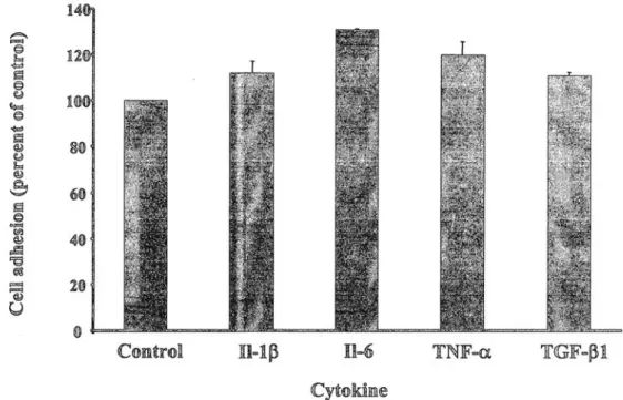
Different numbers of 111indium-labeled cells were first seeded on mouse peritoneum maintained between two concentric Teflon rings (Fig. 1) in serum-free medium and incubated for different periods of time (1 to 24 hours). Optimal cell adhesion was obtained when 105 cells were incubated for 24 hours (37 ± 1.4%). Addition of
cytokines enhanced endometrial cell adhesion to the peritoneum up to 112 ± 5.29% with IL-10, 131 ± 0.50 with IL-6, 120 ± 5.80% with TNFα, and 111 ± 1.60% with TGF-β1 (P<.001 for all cytokines; Fig. 2). When Intergel was added to the peritoneum simultaneously with cells, the adhesion was decreased in a dose-dependent manner. An inhibition of 16 ± 6.2%, 19 ± 7%, 36 ± 12.4%, and 70 ± 6.2% was observed at concentrations of 0.05%, 0.1%, 0.15%, and 0.2%, respectively. In further experiments, a 0.2% concentration of Intergel was used.
Combined Effect of Intergel and Cytokines on Cell Adhesion
Combination of Intergel with cytokines also resulted into cell adhesion inhibition but to a lesser extent in comparison with Intergel alone (70 ± 6.2% inhibition vs. 55 ± 1.6% with IL-1β, 54 ± 5% with IL-6, 46 ± 6% with TNFα, 54 ± 2.8% with TGFβ; P<.001). Therefore, cytokines were able to partially counteract the inhibitory effect of Intergel (Fig. 3, column 4 with IL-6). In some studies, peritonea were pretreated for 24 hours with each cytokine before Intergel and endometrial cells were added to assess whether Intergel acts as a nonspecific physical barrier. In the absence of Intergel, pretreatment of peritoneum with cytokines led to an enhancement of cell adhesion similar to that observed when cytokines were added concomitantly with cells (Fig. 3, columns 3 and 5 with 6; pretreatment with cytokine 24 hours before cell seeding: cell adhesion = 113 ± 6.59% with IL-10, 130 ± 5% with IL-6, 118 ± 4.9% with TNFα, and 114 ± 3.4% with TGFβ). However, cytokine pretreatment of peritoneum abolished completely the inhibition of cell adhesion elicited by Intergel (112 ± 7.50% with IL-1β, 131 ± 3% with IL-6, 120 ± 6.40% with TNFα, and 116 ± 5% with TGF-β1; P <.001; Fig. 3, column 6 with IL-6). Because it is well documented that heparan sulfate proteoglycan, another anionic biopolymer, can bind large amounts of cytokines, we next evaluated its effect on endometrial cell adhesion. However, heparan sulfate proteoglycan did not modulate endometrial cell adhesion (105 ± 4.20%, P >.05). The agar gel decreased the cell adhesion to peritoneum by 75 ± 3.1% whether they were pretreated or not with cytokines.
Figure 2: Effect of cytokines on endometrial cell adhesion to the peritoneum. Different cytokines (1 nM) were
added on the peritoneum, simultaneously with endometrial cells, and incubated for 24 hours. The different cytokines increased cell adhesion. Results are expressed in percentage of the values recorded in control conditions. Control corresponds to adhesion of endometrial cells alone (without Intergel and/or cytokines). The results are presented as the average ± SD of triplicates from a representative assay.
Beliard. An in vitro model of endometriosis. Fertil Steril 2003.
DISCUSSION
In the present study, we have set up an in vitro model suitable to evaluate the adhesion of human endometrial cells to peritoneum that takes place in the early development of endometriosis. Endometrial cells were labeled with 111Indium, a radionuclide widely used to trace leukocytes and platelets (11, 13, 14). Cultured endometrial cells were used rather than fresh tissue to seed exactly the same number of endometrial cells in each experiment and thereby to improve the reproducibility of the assay. To avoid the risk of phenotypic and functional changes that could occur in long-term primary cell culture, culture were always maintained for a maximum of 4 days before use.
The role of the several cytokines in the development of endometriosis has previously been investigated in animal models as well as in vitro. In animal models, a reduction in the size of endometriotic lesions has been reported after treatment with IL-12 (15), interferon-α-2b (16), or recombinant human TNFα binding protein-1 (17).
Different in vitro models have been used to study the initial steps of the disease using cells labeled with either 51Cr (4) or a fluorescent dye (18). These studies evaluated adhesion of endometrial stromal cells to peritoneal mesothelial cells or to purified fibronectin. These models suffer from an important drawback because they do not take into account the interactions between stromal and epithelial cells that influence the development of
endometriotic lesion (19, 20). Because stromal and epithelial cells have been demonstrated in peritoneal fluid during normal menstruation, it is important to evaluate the adhesion of both stromal and epithelial cells together (21). In addition, it has been demonstrated that the integrin expression pattern markedly differs between mesothelium in culture expiants and in monolayer (10). This differential expression is likely to influence the adhesion process of endometrial cells. Therefore, our model mimics more closely the pathophysiological conditions than models using monolayer of cultured mesothelial cell and allows a quantitative evaluation of cell adhesion.
Figure 3: Effect of cytokines with or without Intergel on endometrial cell adhesion. The adhesion assays were
performed by using peritoneum treated (columns 2, 4, and 6) or not (columns 1, 3, and 5) with Intergel. Cytokine is either preincubated for 24 hours with peritoneum before endometrial cell seeding (columns 5, 6) or added simultaneously with endometrial cells (columns 3, 4). Results are expressed in percentage of the values recorded in control conditions (column 1, control group—absence of Intergel and cytokine). The results are presented as the average ± SD of triplicates from a representative assay. The data presented illustrate the effect of IL-6. Similar results are obtained with IL-1β, TNFα, and TGFβ. (Column 1): Control group; (column 2): Intergel alone; (column 3): cytokine alone; (column 4): Intergel and cytokine; (column 5): Pretreatment with cytokine alone; and (column 6): Pretreatment with cytokine, Intergel 24 hours later.
Beliard. An in vitro model of endometriosis. Fertil Steril 2003.
Peritoneal fluid from patients with endometriosis was shown to contain high levels of peritoneal macrophages and inflammatory cytokines likely produced by these cells. These cytokines could then contribute to the development of peritoneal endometriosis (3). Our data demonstrate indeed a moderate increase of endometrial cell adhesion to the peritoneum in response to the addition of TGFβ, IL-1β, IL-6, and TNFα.
Endometriosis is associated with pelvic adhesions, which are a major source of morbidity. Experimental models in rats have pointed to a promoting role of TGF-β in postoperative adhesion formation (22, 23). Transforming growth factor-β1 could act by stimulating cell-cell and cell-matrix interactions (24). Antiadhesive barriers are widely used in pelvic surgery and proved to decrease the extent of adhesions. We therefore tested the effect of such an antiadhesive barrier (a gel of ferric hyaluronate (Intergel) in our in vitro model. Addition of ferric hyaluronate reduced significantly endometrial cell adhesion. This gel could act as a nonspecific physical barrier between endometrial cells and the peritoneum. If this is the case, cytokine treatment would be unable to overcome the Intergel inhibition. Simultaneous addition of ferric hyaluronate and cytokines resulted in a persistent decrease of endometrial cell attachment. However, this decrease was slightly less important. Alternatively, this gel could act by trapping cytokines and reducing their bioavailability. To investigate this hypothesis, we pretreated peritoneum for 24 hours with cytokines before seeding cells and adding the gel. Peritoneum pretreatment with cytokines abolished completely the effect of ferric hyaluronate (Fig. 3). The mechanism of action of hyaluronate is therefore not the consequence of a mere matrix effect but could be related to an impairment of cytokines' bioavailability. This effect is specific and cannot be reproduced with a 1% agar gel or heparan sulfate proteoglycan.
In healthy women, regurgitated endometrial tissue is eliminated (21). The incapacity of endometrial tissue suppression in endometriotic women and its subsequent implantation could result either from specific features of the endometrium itself or from abnormalities in the peritoneal environment or both. The present study has focused on the effects on the peritoneal compartment, showing that the inflammatory context could promote endometrial cell adhesion. It would be of great interest to use our model with menstruating endometrium instead of endometrial cells and to compare endometrium of endometriotic patients with that of healthy women.
In conclusion, our in vitro model provides a useful reproducible tool to test the effect of putative factors involved in endometrial cell adhesion to peritoneum-like pro-inflammatory cytokines. This model is also useful to screen for or to evaluate the mechanism of action of therapeutic agents.
Supported by grants from the Communauté Française de Belgique (Actions de Recherches Concertées), the Commission of European Communities, the Fonds de la Recherche Scientifique Médicale, the Fonds National de la Recherche Scientifique (FNRS, Belgium), the Fonds spéciaux de la Recherche (University of Liège), the Centre Anticancéreux près l’Université de Liège, FORTIS-Assurances, the Fondation Léon Frédéricq (University of Liège), the Direction Générale des Technologies de la Recherche et de L’Energie (D.G.T.R.E.) from the “Région Wallonne,” and the Fonds d’Investissements de la Recherche Scientifique (CHU, Liège, Belgium).
References
1. Sampson JA. The development of the implantation theory for the origin of peritoneal endometriosis. Am J Obstet Gynecol 1940;40:449-557.
2. van der Linden PJ. Theories on the pathogenesis of endometriosis. Hum Reprod 1996;11(Suppl 3):53-65.
3. Barcz E, Kaminski P, Marianowski L. Role of cytokines in pathogenesis of endometriosis. Med Sci Monit 2000;6:1042-6.
4. Zhang RJ, Wild RA, Ojago JM. Effect of tumor necrosis factor-alpha on adhesion of human endometrial stromal cells to peritoneal mesothelial cells: an in vitro system. Fertil Steril 1993;59:1196-201.
5. Hammond MG, Oh ST, Anners J, Surrey ES, Halme J. The effect of growth factors on the proliferation of human endometrial stromal cells in culture. Am J Obstet Gynecol 1993;168:1131-6.
6. Halme J, Becker S, Haskill S. Altered maturation and function of peritoneal macrophages: possible role in pathogenesis of endometriosis. Am J Obstet Gynecol 1987;156:783-9.
7. RanaN, Braun DP, House R, Gebel H, Rotman C, Dmowski WP. Basal and stimulated secretion of cytokines by peritoneal macrophages in women with endometriosis. Fertil Steril 1996;65:925-30.
8. Oosterlynck DJ, Meuleman C, Waer M, Koninckx PR. Transforming growth factor-beta activity is increased in peritoneal fluid from women with endometriosis. Obstet Gynecol 1994;83:287-92.
9. D'Hooghe TM, Bambra CS, Xiao L, Peixe K, Hill JA. Effect of menstruation and intrapelvic injection of endometrium on inflammatory parameters of peritoneal fluid in the baboon (Papio anubis and Papio cynocephalus). Am J Obstet Gynecol 2001;184:917-25.
10. Witz CA, Montoya-Rodriguez IA, Miller DM, Schneider BG, Schen-ken RS. Mesothelium expression of integrins in vivo and in vitro. J Soc Gynecol Investig 1998;5:87-93.
11. Danpure HJ, Osman S, Brady F. The labelling of blood cells in plasma with 111In-tropolonate. Br J Radiol 1982;55:247-9.
12. Motulsky H. Intuitive biostatistics. Oxford, United Kingdom: Oxford University Press, 1995.
13. Aktolun C, Ussov WY, Arka A, Glass D, Gunasekera RD, Peters AM. Technetium-99m and indium- 111 double labelling of granulocytes for kinetic and clinical studies. Eur J Nucl Med 1995;22:330-4.
14. Giannessi D, Bernini W, Puccetti P, De Caterina R. Platelet radiola-belling with 111 Indium for in vivo studies: a methodological reappraisal. Nucl Med Biol 1995;22:399-403.
15. Somigliana E, Vigano P, Rossi G, Carinelli S, Vignali M, Panina-Bordignon P. Endometrial ability to implant in ectopic sites can be prevented by interleukin-12 in a murine model of endometriosis. Hum Reprod 1999;14:2944-50.
16. Ingelmo JM, Quereda F, Acien P. Intraperitoneal and subcutaneous treatment of experimental endometriosis with recombinant human in-terferon-alpha-2b in a murine model. Fertil Steril 1999;71:907-11.
17. D'Antonio M, Martelli F, Peano S, Papoian R, Borrelli F. Ability of recombinant human TNF binding protein-1 (r-hTBP-1) to inhibit the development of experimentally-induced endometriosis in rats. J Reprod Immunol 2000;48:81-98.
18. Garcia-Velasco JA, Arici A. Interleukin-8 stimulates the adhesion of endometrial stromal cells to fibronectin. Fertil Steril 1999;72:336-40.
19. Wild RA, Zhang RJ, Medders D. Whole endometrial fragments form characteristics of in vivo endometriosis in a mesothelial cell co-culture system: an in vitro model for the study of the histogenesis of endometriosis. J Soc Gynecol Investig 1994;1:65-8.
20. Beliard A, Noel A, Goffin F, Frankenne F, Foidart JM. Role of endocrine status and cell type in adhesion of human endometrial cells to the peritoneum in nude mice. Fertil Steril 2002;78:973-8.
21. Bartosik D, Jacobs SL, Kelly LJ. Endometrial tissue in peritoneal fluid. Fertil Steril 1986;46:796-800.
22. Williams RS, Rossi AM, Chegini N, Schultz G. Effect of transforming growth factor beta on postoperative adhesion formation and intact peritoneum. J Surg Res 1992;52:65-70.
23. Lucas PA, Warejcka DJ, Young HE, Lee BY. Formation of abdominal adhesions is inhibited by antibodies to transforming growth factor-betal. J Surg Res 1996;65:135-8.
24. Dou Q, Williams RS, Chegini N. Inhibition of transforming growth factor-beta 1 alters the growth, anchor-dependent cell aggregation and integrin mRNA expression in human promonocytes: implications for endometriosis and peritoneal adhesion formation. Mol Hum Reprod 1997;3:383-91.