THE ROLE OF THE NUCLEAR RECEPTOR NR5A2
IN OVULATION IN MICE
par
KALYNE BERTOLIN
Département de biomédecine vétérinaire Faculté de médecine vétérinaire
Mémoire présenté à la Faculté de médecine vétérinaire en vue de l’obtention du grade de
Maître ès sciences (M.Sc.) en sciences vétérinaires
option reproduction
Août, 2010
Faculté de médecine vétérinaire
Ce mémoire intitulé
THE ROLE OF THE NUCLEAR RECEPTOR NR5A2
IN OVULATION IN MICE
présentépar KALYNE BERTOLIN
a été évaluépar un jury composé des personnes suivantes
Lawrence C. Smith, président-rapporteur Bruce D. Murphy, directeur de recherche
Résumé
Le récepteur nucléaire Nr5a2 est exprimé dans l’ovaire, plus spécifiquement dans les cellules de granulosa et lutéales. Une déplétion conditionnelle de Nr5a2 dans les cellules de granulosa au stade de follicule primaire par croisement de souris Nr5a2-flox et Amhr2-Cre (Nr5a2f/fAmhr2Cre/+) génère des problèmes au niveau de l’expansion du cumulus, de l’ovulation et de la lutéinisation. Ainsi, nous estimons que Nr5a2 régule les connexions intercellulaires dans le follicule ovarien via la connexine 43 (Cx43), une protéine de jonction impliquée dans l’expansion du cumulus. Le premier objectif de l’étude était de déterminer si l’absence d’expansion du cumulus chez les souris Amhr2Cre-cKO est liée à l’absence de communication intercellulaire adéquate entre les cellules de granulosa et de cumulus dans les follicules préovulatoires. À cette fin, des ovaires de souris immatures Amhr2Cre-cKO et non transgéniques ont été prélevés (n=3) après un traitement de superstimulation utilisant les gonadotropines eCG suivie de hCG afin d’induire l’ovulation. Nous avons ainsi démontré, par RT-PCR, une sous-expression de Cx43 avant et au moment du stimulus ovulatoire (0 h et 2 h) chez le groupe Amhr2Cre-cKO (P<0.01), ce qui pourrait mener à un problème dans l’acquisition de la compétence développementale de l’oocyte. D’un autre côté, au moment de l’ovulation (12 h), l’ARNm de Cx43 est surexprimé dans le groupe Amhr2Cre-cKO, ce qui pourrait prévenir les cellules du cumulus de se détacher l’une de l’autre. Nous avons ainsi conclu que Cx43 est un gène sous le contrôle de Nr5a2 et qu’une régulation erronée de ce gène est une cause possible du problème d’expansion du
cumulus chez les souris Amhr2Cre-cKO. Afin d’examiner le rôle de Nr5a2 dans l’ovulation et la lutéinisation à différents stades de la maturation folliculaire, nous suggérons que Nr5a2 module la séquence temporelle des événements menant à l’ovulation. En croisant des souris Nr5a2-flox et Cyp19-Cre (Nr5a2f/fCyp19Cre/+), l’expression de Nr5a2 a été interrompue dans les cellules de granulosa des follicules antraux et préovulatoires. Aucune portée n’a été obtenue de ces souris (n=4) durant un essai d’accouplement de 6 mois. Chez les souris Cyp19Cre-cKO on remarque la présence de structures s’apparentant à des cellules de type lutéales et les femelles âgées d’un an présentent des kystes folliculaires hémorragiques et une hypertrophie de l’épithélium en surface de l’ovaire. Les deux modèles transgéniques démontrent donc une absence de l’expansion du cumulus et de l’ovulation. En conclusion, Nr5a2 semble réguler différemment la folliculogenèse et l’ovulation dans les cellules de granulosa des follicules primaires et antraux.
Mots-clés : Nr5a2, ovulation, expansion du cumulus, connexine 43, souris knockout
Abstract
The nuclear receptor Nr5a2 is expressed in the ovary, exclusively in granulosa and luteal cells. Conditional disruption of Nr5a2 in granulosa cells beginning with primary follicles by means of Nr5a2-floxed and Amhr2-Cre mice (Nr5a2f/fAmhr2Cre/+) results in failure in cumulus expansion, ovulation and luteinization. We hypothesize that Nr5a2 regulates intercellular connections in ovarian follicles through connexin 43 (Cx43), a gap-junctional protein related to cumulus expansion. The first objective of this study was to determine whether the lack of cumulus expansion in Amhr2Cre-cKO mice is related to the absence of normal cell-to-cell communication in cumulus/granulosa cells of preovulatory follicles. To address this, immature ovaries of Amhr2Cre-cKO and non-transgenic littermates mice were collected (n=3) after superstimulation to induce follicle development and ovulation. Using RT-PCR, the Cx43 mRNA levels were shown to be downregulated prior to and at the time of the ovulatory stimulus (0 h and 2 h) in the Amhr2Cre-cKO group (P<0.01), which may lead to the failure in the acquisition of oocyte developmental competence. On the other hand, by the time of ovulation (12 h), mRNA levels of Cx43 are upregulated in Amhr2Cre-cKO group, which may prevent the cumulus cells to detach one from another. We conclude that Cx43 is one of the downstream genes under Nr5a2 control and its dysregulation can be one reason for the defect in cumulus expansion in Amhr2Cre-cKO females. To examine the role of Nr5a2 in ovulation and luteinization in different stages of the follicle maturation, we hypothesized that Nr5a2 modulates the events leading to ovulation in a temporal sequence. By crossing Nr5a2-floxed and Cyp19-Cre mice
(Nr5a2f/fCyp19Cre/+), Nr5a2 was disrupted in granulosa cells of antral and preovulatory follicles. No litters were born to Cyp19Cre-cKO females (n=4) during a 6 months breeding trial. Cyp19Cre-cKO enabled the development of a luteal-like structure, and 1-year-old females presented hemorrhagic follicular cysts and hypertrophic ovarian surface epithelium. Both knockout models display lack of cumulus expansion and ovulation. We conclude that Nr5a2 differentially regulates folliculogenesis and ovulation in granulosa cells of small and antral follicles.
Table of Contents
Résumé... ii
Abstract ... iv
Table of Contents ... vi
List of Tables... viii
List of Figures ... ix List of Abbreviations ... xi Dedication ... xiii Acknowledgments... xiv 1. Introduction ... 1 2. Literature Review... 3 2.1. Nuclear Receptors ... 3 2.1.1. Subfamilies... 4 2.2. Nr5a2... 6 2.2.1. Co-regulators... 7 2.2.2. Biological functions ... 9
2.3. Ovarian dynamics in mice... 12
2.3.1. Folliculogenesis... 12
2.3.2. Cumulus expansion ... 15
2.3.2.1. Gap junctions and the ovary ...17
2.4.1. Conditional knockout (cKO)... 24
2.4.2. Amhr2-Cre mouse line... 26
2.4.3. Cyp19-Cre mouse line... 27
3. Materials and Methods... 29
Animals and tissue collection procedures ... 29
Laser Microdissection (LMD)... 31 RNA analysis ... 32 Immunohistochemistry... 34 Hormone assay ... 35 Statistical analysis ... 35 4. Results ... 36
Females Amhr2Cre-cKO fail to undergo cumulus expansion following stimulation with exogenous gonadotropins... 36
Cx43 expression is dysregulated in Amhr2Cre-cKO mouse ovarian granulosa and cumulus cells... 37
Absence of Nr5a2 in granulosa cells of antral follicles leads to infertility... 39
Ovarian morphology in Cyp19Cre-cKO... 40
5. Discussion ... 44
6. Conclusion... 51
7. References ... 52
List of Tables
Results
TABLE I – Reproductive performance of Cyp19Cre-cKO and Cyp19Cre-control mice in a breeding trial. Females were housed with proven C57BL/6J males for 6 months. Conditional knockout females are infertile. ... 40
List of Figures
Literature Review
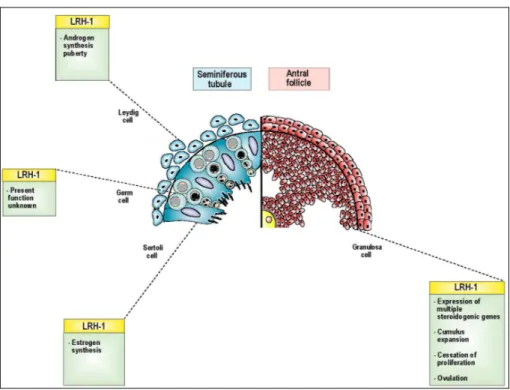
Figure 1 – Overview of the effects of Nr5a2 (LRH-1) on the constituents of the seminiferous tubule in the male and the antral follicle in the female (Bertolin, Bellefleur et al. 2010)... 11
Figure 2 – Classification scheme of mouse ovarian follicles and the relationship to gonadotropin (Gn) (Orisaka, Tajima et al. 2009)... 13
Figure 3 – Illustration of a model experiment in genetics using the Cre-loxP system (Zepper 2008). ... 25
Figure 4 – Scheme demonstrating the excision in different phases of the follicle development comparing Amhr2-Cre and Cyp19-Cre mice lines... 28
Results
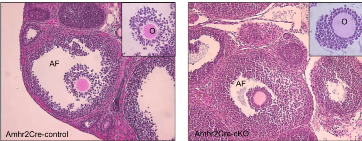
Figure 1 – HE staining of Amhr2Cre-control and Amhr2Cre-cKO ovaries collected at 7 h post-hCG. Injection of 10 IU of PMSG/hCG fails to rescue the cumulus expansion in Amhr2Cre-cKO mice. Inserts show cumulus cells around the oocyte from a preovulatory follicle. (AF) Antral follicle; (O) Oocyte (200x). ... 36
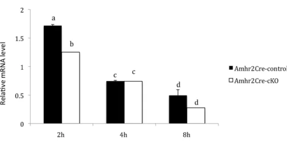
Figure 2 – Relative abundance of Cx43 mRNA (Means ± SEM) in ovarian granulosa and cumulus cells of immature mice stimulated with PMSG and hCG and collected 2 h, 4 h or 8 h after hCG as determined by qPCR. Females were Amhr2Cre-control (solid bars) and Amhr2Cre-cKO (open bars)... 37
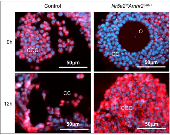
Figure 3 – Protein expression of Cx43 in mouse ovarian follicles focusing on cumulus cells. Amhr2Cre-cKO and Amhr2Cre-control ovaries were stained at time 0 h and 12 h after hCG. Images are merged of Cx43 and DAPI staining. (O) Oocyte; (COC) Cumulus-oocyte-complex; (CC) Cumulus cells. ... 38
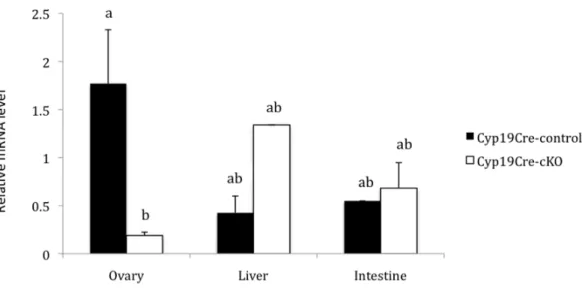
Figure 4 – Abundance of Nr5a2 mRNA in the ovary, liver and intestine of Cyp19Cre-control and Cyp19Cre-cKO adult females (n=3/genotype). P<0.01... 39
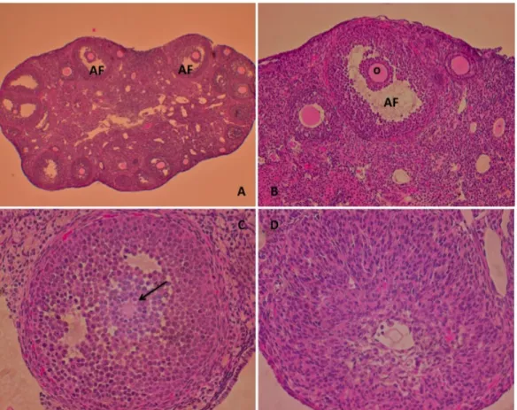
Figure 5 – HE staining of Cyp19Cre-cKO ovaries in 6-month-old females. A) Whole ovary (100x); B) Antral follicle lacking cumulus expansion (200x); C) Entrapped oocyte (400x); D) Luteal-like structure (400x); (AF) Antral follicle; (O) Oocyte... 41
Figure 6 – Immunofluorescent protein detection of SCARB1 in adult Cyp19Cre-cKO mouse ovaries. A) Luteal-like cells; B) Granulosa cells... 42
Figure 7 – Cyp19Cre-cKO ovarian structures in 1-year-old mouse. A) Ovarian morphology; B) HE staining of whole ovary (400x); C) Hemorrhagic cyst (400x); D) Hypertrophic ovarian surface epithelium (400x). ... 43
List of Abbreviations
AF-1 activation function-1ADAMTS-1 a disintegrin and metalloproteinase with thrombospondin-like motifs 1 αERKO estrogen receptor α knockout
AMH Anti-Müllerian hormone Amhr2 Amh type II receptor ANOVA analysis of variance ArKO aromatase knockout ATP adenosine-5’-triphosphate BSA bovine serum albumin
cAMP cyclic adenosine monophosphate
cDNA complementary DNA
cKO conditional knockout COC cumulus-oocyte-complex COX-2 cyclooxygenase 2
Cx43 connexin 43
CYP11a1 cytochrome P450 family 11 subfamily a polypeptide 1 CYP19 cytochrome P450 family 19
DAPI 4’,6-diamidino-2-phenylindole
DAX1 dosage-sensitive sex reversal adrenal hypoplasia congenital region on the X chromosome, gene 1
DBD DNA-binding domain
DNA deoxyribonucleic acid EGF epidermal growth factor ERRα estrogen related receptor α floxed flanked by loxP
FSH follicle-stimulating hormone Ftz-F1 Fushi tarazu factor-1
GDF9 growth differentiation factor 9 GJA1 gap junction protein a1
HAS-2 hyaluronic acid synthase 2 hCG human chorionic gonadotropin
HDL high-density lipoprotein HE hematoxylin and eosin IαI inter-α trypsin
LBD ligand-binding domain LH luteinizing hormone LMD laser microdissection LRH-1 liver receptor homolog-1 MAP mitogen-activated protein MIS Müllerian-inhibiting substance
mRNA messenger RNA
NCOR nuclear receptor co-repressor
Nr5a2 nuclear receptor subfamily 5 group A member 2 NRs nuclear receptors
OCT optimal cutting temperature
PAF paraformaldehyde
PBS phosphate-buffered saline PCR polymerase chain reaction
PK protein kinase
PMSG pregnant mare serum gonadotropin
PPAR peroxisome proliferator-activated receptor PTGS2 prostaglandin synthesis
RIP140 repressor interacting protein 140 RNA ribonucleic acid
RPS18 ribosomal protein S18
RT room temperature
SCARB1 scavenger receptor-B1
SE standard error
SF-1 steroidogenic factor-1 SHP short heterodimeric partner SRC-1 steroid receptor co-activator-1
StAR steroidogenic acute regulatory protein SUMO small ubiquitin-like modifier
TSG-6 tumor necrosis stimulated gene 6
To my parents, Ivani and Carlos Bertolin
Acknowledgments
First and foremost, I would like to gratefully acknowledge my supervisor Dr. Bruce D. Murphy. I appreciate all the opportunities presented to me since I started as a Vet student trainee. It has been an honor to be his student, especially due to the friendly environment that he prioritizes; the social visits, the jokes and the exchanged pairs of shoes lightened the burden of lost ovaries, degraded RNA and failed driving test. His support and contributions in time, patience and confidence in my work, made my Master’s a productive and stimulating experience.
For the financial support, I thank the Canadian Institute of Health Research, Operating Grant MT11018 for funding the original investigation from the laboratory of Bruce D. Murphy. Also, for the RQR/CREATE Scholarship from the Natural Sciences and Engineering Research Council of Canada.
I owe my deepest gratitude to Dr. Mario Binelli, who has made available his support, as a professional and as a friend. Also, I am greatly indebted to Dr. Valerio Portela, who gave me my first vote of confidence for a career in research.
Furthermore, it is a pleasure to thank the members of the BDM Laboratory, whose sources of friendship, advice and collaboration have contributed immensely to my professional improvement. I am especially grateful to Vickie Roussel for patiently teaching me the techniques required for my research and for her excellent partnership, to Anne-Marie Bellefleur for her teamwork and for carefully reviewing this thesis, to Debora Binelli
for her useful help with the mice job during the first part of this project, to Hamid Reza Kohan Ghadr for his significant help with the qPCR, and finally to Micheline Sicotte, who was always available and efficient in making life easy at the CRRA.
I would like to dedicate a special paragraph to Mira Dobias-Goff, for technical and moral support, for our long conversations and her wise advice that made me more mature during these years abroad and for showing us everyday, with chocolates and affection, how sweet life truly is. She has all my admiration. “Obrigada”.
My most joyful thanks to the “Brazilian Gang”, my dear friends and surrogate family, for making life wonderful, even away from home. Thanks to all the “single ladies” chez Micheline Beaudoin, for the special moments together in a pleasant home sweet home.
I would like to show all my gratitude to my family, to my brother Thiérre and especially to my parents Ivani and Carlos, for the unconditional love, for my education and for supporting the distance in favor of my dreams. For being my examples of dignity, honesty and persistence, and mainly for the indefatigable waiting to have me back home.
Last but not least, my lovely thanks to my supportive and encouraging boyfriend Eduardo for sharing my dreams, always taking on challenges by my side and for coming to Canada to build a future together.
1. Introduction
Reproduction is a fundamental feature of all known life, and it is an evolutionarily conserved process assembled to ensure the survival of species. Errors during the development of functional gonads and gametes can result in subfertility or infertility (Jorgez, Lin et al. 2005). According to the Center for Disease Control from USA, infertility, defined as the inability of a couple to conceive or carry a baby to term after one year of unprotected intercourse, is a major health issue. Among the infertility causes in humans, including problems with either the male or the female reproductive system, 20% are caused by the failure in ovulation (Eisenberg 2009; Brzakowski, Lourdel et al. 2009).
Ovulation is a biological phenomenon beginning when gonadotropic hormones stimulate mature ovarian follicles, whereby ovarian follicles reactivate oocyte meiosis, create a rupture pore in the apical follicle wall and liberate the oocyte into the oviduct (Espey 1980; Russell and Robker 2007; Espey and Richards 2006); following that, the tissue restructuring and differentiation will form the corpus luteum (Russell and Robker 2007). Normal oocyte development, ovulation, and fertilization are dependent on the critical process of cumulus expansion (Elvin, Clark et al. 1999).
To expand the understanding of fertility disorders in humans, animal models have been providing invaluable insights into reproductive physiology. The mouse has become an important animal model to study reproduction and other processes (Jorgez, Lin et al. 2005). Using the power of mouse genetics, a granulosa cell specific knockout mouse model led the discovery that an orphan nuclear receptor subfamily 5 group A member 2 (Nr5a2) is a critical and pleiotropic regulator of multiple mechanisms essential for female fertility
(Duggavathi, Volle et al. 2008). Nr5a2 is expressed in the ovary, exclusively in granulosa and luteal cells. Because germline ablation of Nr5a2 causes embryo lethality (Labelle-Dumais, Jacob-Wagner et al. 2006), Nr5a2 granulosa specific mutant mouse was generated by the Cre-loxP system, and the consequence was failure in cumulus expansion, ovulation and luteinization (Duggavathi, Volle et al. 2008).
A gap-junctional protein, known as connexin 43 (Cx43), participates in the network of cell-to-cell communication established in the cumulus/granulosa cells (Teilmann 2005), and the process of cumulus expansion is related to modifications of gap junctions (Dell'Aquila, Caillaud et al. 2004). Our preliminary electron microscopy revealed that the mural granulosa cells of preovulatory follicles in granulosa specific Nr5a2 knockout mice display an apparent reduction in normal cell-cell contacts in comparison with wild type (WT) animals.
The first objective of this study was to determine whether the lack of cumulus expansion in granulosa specific knockout mice is related to the absence of normal cell-to-cell communication in cumulus/granulosa cell-to-cells of preovulatory follicles. We hypothesized that Nr5a2 regulates intercellular connections in ovarian follicles through Cx43 in mammalian ovarian follicles. The second objective was to examine the role of Nr5a2 in ovulation and luteinization in different stages of the follicle maturation, by timed tissue specific disruption of the Nr5a2 gene in granulosa cells, and our hypothesis was that Nr5a2 modulates the temporal sequence events leading to ovulation.
2. Literature Review
In this section the physiological and molecular bases of the reproductive phenomena are described, focusing on cumulus expansion and ovulation, events that are regulated by the nuclear receptor Nr5a2.
2.1. Nuclear Receptors
Nuclear receptors (NRs) are intracellular transcription factors that work in concert with co-activators and co-repressors to regulate the activity of complex gene networks (Krasowski, Ni et al. 2010). The activity of many NRs is controlled by the binding of small lipophilic ligands, including hormones, metabolites and certain synthetic ligands (Fayard, Auwerx et al. 2004). In mammals, the NR superfamily is composed of approximately 50 functional members, with 48 genes identified in the human genome, 49 in mice and 47 in rats (Zhang, Burch et al. 2004).
In a strategic position in the cell to direct proliferation, differentiation and apoptosis, NRs are proteins that share a conserved modular structure, which includes a modulatory A/B domain (often containing a ligand-independent activation function-1, the AF-1); the highly conserved deoxyribonucleic acid (DNA)-binding domain (DBD or C domain); the binding domain (LBD or E domain), which contains an equally conserved ligand-dependent activation function-2, the AF-2 motif that mediates co-activator interaction; and the D domain serving as a flexible hinge between DBD and LBD (Fayard, Auwerx et al. 2004; Krasowski, Ni et al. 2010). NRs have amino acid sequence similarity for the two
highly conserved domains – the DBD and the LBD, responsible for binding specific DNA sequences and small lipophilic ligands, respectively. NRs bind specific DNA response elements in the regulatory regions of genes and regulate transcription in response to ligand binding (Sladek 2010).
NRs recruit positive and negative co-regulatory proteins, referred to as co-activators and co-repressors. In general, in the absence of ligand the nuclear receptor interacts with co-repressors. Activation of NRs following ligand binding and/or phosphorylation induces a conformational change, resulting in the dissociation of co-repressor complexes and recruitment of co-activator proteins by the NRs (Fayard, Auwerx et al. 2004), and subsequent transcriptional activation. Ligand-activated NRs serve first as adaptors between gene regulatory regions and the chromatin modifying enzyme complexes and then as activators of ribonucleic acid (RNA) polymerase II (Carlberg and Seuter 2010).
NRs control a wide array of biological processes, including eukaryotic development, reproduction and metabolic homeostasis (Bain, Heneghan et al. 2007). Because of their versatile signaling functions and their implication in many physiological processes, NRs have become important drug targets, receiving wide attention from both basic and applied biomedical researchers (Fayard, Auwerx et al. 2004).
2.1.1. Subfamilies
The NR superfamily is typically subdivided into seven subfamilies (NR0 – NR6), however three classes well characterize important diversity between them (Fayard, Auwerx
et al. 2004). The steroid receptor family (class I) includes the progesterone receptor, estrogen receptor, glucocorticoid receptor, androgen receptor and mineralocorticoid receptor. The thyroid/retinoid family (class II) includes the thyroid receptor, vitamin D receptor, retinoic acid receptor and peroxisome proliferator-activated receptor. The third family defines a set of proteins identified by comparative sequence analysis as belonging to the NR superfamily but for which the cognate ligand has not yet been identified and the molecular mechanisms of their transcriptional regulation remain unclear; they are therefore termed the orphan receptor family (class III) (Bain, Heneghan et al. 2007).
Although all NRs regulate gene expression, among the three classes there are subtle differences in the biochemical mechanisms by which the receptors carry out this function. Regarding the steroid receptor family, upon binding their hormonal ligand, the receptors release heat shock proteins to challenge the cell to respond rapidly (Morimoto, Kline et al. 1997), translocate into the nucleus and bind as homodimers to the response elements at upstream promoter sites (Beato and Klug 2000). The class II receptor proteins typically function as heterodimers. In the absence of ligand, gene activation is prevented by co-repressor interactions with the DNA-bound heterodimer. Evidence about the orphan nuclear receptors indicates that they either can heterodimerize or can bind as monomers at response elements to carry out their function (Bain, Heneghan et al. 2007).
Studies by Keith Parker demonstrated the presence of a cellular protein that regulated the expression of P450 steroid hydroxylases (Parker and Schimmer 1997). The complementary DNA (cDNA) that was cloned coded for the protein, described as gene
product that appeared to belong to the nuclear receptor family on the basis of their sequence identity, is now known as steroidogenic factor-1 (SF-1), also known as Nr5a1, and it proved to be the mammalian ortholog of the Drosophila Fushi tarazu factor-1 (Ftz-F1) (Lala, Rice et al. 1992), a regulator of embryogenesis and metamorphosis in this organism and the first NR5A member cloned (Sadovsky and Crawford 1998). A second factor, the liver receptor homolog-1 (LRH-1), recently designated as Nr5a2 (Kliewer, Lehmann et al. 1999), was later shown to interact with the same DNA sequence (Fayard, Auwerx et al. 2004). Since the first identification of these two factors, numerous studies have demonstrated that these two nuclear receptors of subfamily 5 are essential for the success of diverse reproductive processes in mammals (Bertolin, Bellefleur et al. 2010).
2.2. Nr5a2
The Nr5a2 (also known as LRH-1, α-fetoprotein transcription factor, FTZ-F1-related factor and CYP7A1 promoter binding factor) is an orphan member of the nuclear receptor superfamily (Galarneau, Pare et al. 1996). It is mainly expressed in tissues of endodermal origin, including the liver, pancreas, intestine, adrenal and testis (Sirianni, Seely et al. 2002); high levels of Nr5a2 are found in granulosa and luteal cells of the ovary (Fayard, Auwerx et al. 2004; Hinshelwood, Repa et al. 2003) and, at lower levels, in the placenta (Sirianni, Seely et al. 2002).
Members of the NR5A subfamily regulate target gene transcription by binding as monomers to DNA response elements (Fayard, Auwerx et al. 2004). Through the
crystallization of NR5A LBD, some groups have shown that small phospholipid ligands can bind to this structure and then regulating gene expression (Krylova, Sablin et al. 2005; Ortlund, Lee et al. 2005). Nr5a2 is closely related to Nr5a1 and both can bind the same hexameric elements (Hinshelwood, Repa et al. 2003).
Both Nr5a1 and Nr5a2 have transcriptionally active conformations, suggesting that they are constitutively active as well (Parker and Schimmer 1997). This notwithstanding, their transactivational activity appears to be regulated by multiple factors that include post-transcriptional mechanisms, binding of other orphan nuclear receptors and the action of transcriptional co-activators and co-repressors (Ohno, Komakine et al. 2010). Phosphorylation of Nr5a2, brought on by activation of the protein kinase-C pathway, increases its transcriptional activity (Lee, Choi et al. 2006). A second post-transcriptional modification is the conjugation of Nr5a2 (Yang, Pan et al. 2009) to the small ubiquitin-like modifier (SUMO), at lysine residues (sumoylation). This epigenetic event reduces the transcriptional activity of both nuclear receptors, by reducing binding of the receptors to their cognate DNA sequence (Campbell, Faivre et al. 2008), or by restricting translocation of Nr5a2 into nuclear bodies (Yang, Pan et al. 2009).
2.2.1. Co-regulators
There are two further orphan nuclear receptors, short heterodimeric partner (SHP, NR0B2) and dosage-sensitive sex reversal adrenal hypoplasia congenital region on the X chromosome, gene 1 (DAX1, NROB1) that play important roles in the expression of
Nr5a2. DAX1 binds to Nr5a2 (Suzuki, Kasahara et al. 2003), specifically in the co-activator groove, indicating competitive inhibition as the repressive mechanism (Sablin, Woods et al. 2008). There is also evidence that DAX1 recruits co-repressors, such as the nuclear receptor co-repressor (NCOR) and ALIEN, providing a further mechanism of inhibition of NR5 nuclear receptor activity (Iyer and McCabe 2004). SHP inhibits not only the transcriptional activity of a number of nuclear receptors, including Nr5a2 (Iyer, Zhang et al. 2006), but also appears to suppress expression of both NR5 orphan nuclear receptors (Goodwin, Jones et al. 2000). Nr5a2-induced transcription has its mechanism of inhibition resembling that of DAX1, and it appears to compete with co-activators for binding to Nr5a2 (Lee and Moore 2002). In contrast to other nuclear receptors, ligand binding does not effect a conformational change, rather it reduces the affinity of Nr5a2 for SHP (Burendahl, Treuter et al. 2008). By this means, ligand binding may thus increase transcription by reducing inhibition. The SHP-Nr5a2 interaction has biological significance, as studies of SHP in the testis reveal that its inhibition of Nr5a2 is a mechanism that prevents precocious puberty (Volle, Duggavathi et al. 2007).
In addition to the DAX1 and SHP, other co-activators and co-repressors participate in regulating Nr5a2 transactivation of target genes. The repressor interacting protein 140 (RIP140) is a negative transcriptional regulator of nuclear receptor action (Feige and Auwerx 2007). RIP140 is required for ovulation, and its null mutation in the mouse phenocopies aspects of ovarian disruption seen in the granulosa specific knockout of Nr5a2 mouse, in particular, the failure of cumulus expansion and of follicle rupture (Duggavathi,
Volle et al. 2008). RIP140 interacts with several other nuclear receptors, including estrogen related receptor α (ERRα) (White, Morganstein et al. 2008), and is therefore considered likely to regulate Nr5a2 as well. Steroid receptor co-activator-1 (SRC-1) (Xu, Liu et al. 2004) and the peroxisome proliferator-activated receptor (PPAR)γ-coactivator-1a (Yazawa, Inaoka et al.) potentiate the activity of Nr5a2, indicating that inhibition is not the sole regulatory mechanism of transactivation by this NR.
2.2.2. Biological functions
Nr5a2 regulates important steps of development, endocrine homeostasis and metabolism. Mice homozygous for a germline mutation in the gene encoding Nr5a2 die before embryonic day 7.5, indicating that it plays a crucial role in the development, as the gene is necessary for primitive streak formation (Pare, Malenfant et al. 2004; Labelle-Dumais, Jacob-Wagner et al. 2006).
In enterohepatic tissues and in the liver, Nr5a2 is involved in cholesterol and bile acid homeostasis by regulating several essential genes in the reverse cholesterol transport and bile acid synthesis pathways. It is also implicated in the regulation of epithelial cell renewal and the synthesis of glucocorticoids (Lee, Schmidt et al. 2008). Moreover, it is expressed in the stromal compartment of human breast, comprised of undifferentiated adipose tissue, possibly participates in the control of pre-adipocyte function and/or adipocyte differentiation, and may play a role in breast tumor development (Clyne, Speed et al. 2002).
Nr5a2 is expressed in the gonadotropin cells of the pituitary (Zheng, Yang et al. 2007). The hypothalamo-hypophyseal axis is another potential site where Nr5a2 might regulate reproductive processes. A study using in situ hybridization identified the Nr5a2 transcript throughout the mouse brain, including the hypothalamus (Grgurevic, Tobet et al. 2005). Nr5a2 is expressed in the pituitary gland and in gonadotropic cell lines, and transcriptional studies have shown that it upregulates promoter activity of the follicle-stimulating hormone (FSH)-ß, gonadotropin-releasing hormone the luteinizing hormone (LH)ß genes (Zheng, Yang et al. 2007).
Given the importance of Nr5a2 to induction of steroidogenic enzymes and factors (Fayard, Auwerx et al. 2004), it is not surprising that it is highly expressed in the Leydig cells of the testis (Pezzi, Sirianni et al. 2004). It appears not to be present in Sertoli cells, where Nr5a1 predominates (Pezzi, Sirianni et al. 2004). Mice heterozygous for Nr5a2 mutation, i.e. where only a single functional allele is present, have circulating testosterone levels that are less than half their WT littermates (Volle, Duggavathi et al. 2007). Precocious expression of Nr5a2 in mice leads to precocious induction of androgen synthesis and early puberty (Duggavathi, Volle et al. 2008) (Figure 1). The most abundant information about Nr5a2 regulation derives from studies of the ovary. High levels of expression are reported in mouse, human (Sirianni, Seely et al. 2002) and equine (Boerboom, Pilon et al. 2000) ovaries. The gene is expressed exclusively in granulosa cells and corpora lutea and distinctly absent in theca cells, oocytes and ovarian stroma; it is present in primary follicles and at all later stages of folliculogenesis. The expression of
Nr5a2 is induced significantly in the corpus luteum during pregnancy (Hinshelwood, Shelton et al. 2005). A number of studies over the last decade have described Nr5a2 regulation of target genes, including steroidogenic enzymes and proteins and inhibin-α (Zhao, Li et al. 2007) (Figure 1). As seen in the testis (Volle, Duggavathi et al. 2007), deletion of one allele compromises steroidogenesis, manifest in the female as luteal progesterone deficiency (Labelle-Dumais, Pare et al. 2007). Recent studies have focused on tissue specific knockout of the Nr5a2 gene using Cre specific to granulosa cells and a mouse line with loxP sites flanking the third and fourth exon of the gene, and results in infertility. In this case, the principal cause for sterility is the total failure to ovulate that cannot be redressed by gonadotropin stimulation (Duggavathi, Volle et al. 2008).
Figure 1 – Overview of the effects of Nr5a2 (LRH-1) on the constituents of the seminiferous tubule in the male and the antral follicle in the female (Bertolin, Bellefleur et al. 2010).
Mice ovaries bearing a granulosa cell-specific deletion of Nr5a2 resulted in a failure in expansion of the cumulus cells and the normal expression of a number of genes known necessary for steroidogenesis was disrupted, including the steroidogenic genes cytochrome P450 family 11 subfamily a polypeptide 1 (CYP11a1), cytochrome P450 family 19 (CYP19) and steroidogenic acute regulatory protein (StAR), the rate limiting gene in prostaglandin synthesis (PTGS2) and genes associated with cholesterol transfer such as scavenger receptor-B1 (SCARB1). The concomitant defects in cumulus expansion and luteinization lead to anovulation and thus infertility (Duggavathi, Volle et al. 2008). These data represent an important insight into the causes of female infertility.
2.3. Ovarian dynamics in mice
Mammalian ovaries have two principal functions: they produce sex steroids to prepare the adult female for reproduction, and they release eggs, referred to as ovulation, at appropriate intervals during the female reproductive life.
2.3.1. Folliculogenesis
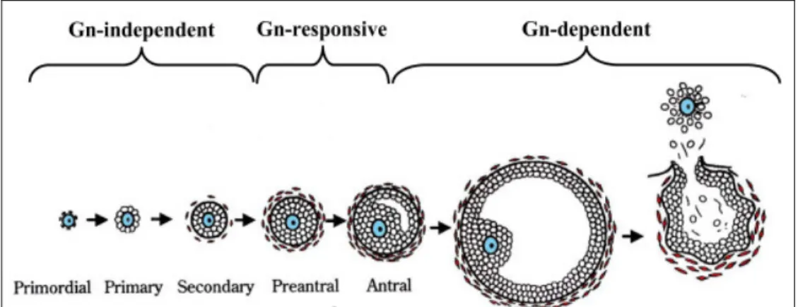
Folliculogenesis is the process by which the female germ cell develops within the somatic cells of the ovary and matures into a fertilizable egg. Folliculogenesis is dependent on signals within the ovary, as well as on endocrine hormones from the pituitary. It is a highly regulated developmental sequence resulting in the growth and differentiation of the oocyte and associated somatic cells (Binelli and Murphy 2010). The follicles are classified
as primordial, primary, secondary, preantral and antral, according to Pedersen and Peters classification (Pedersen and Peters 1968) (Figure 2).
Newborn mouse ovaries are densely packed with oocytes as clusters with no evidence of surrounding granulosa cells (Peters 1969). They are connected by intercellular bridges (Rajkovic, Pangas et al. 2006), and the majority of them enter meiosis during embryonic life, at which stage they will remain until meiosis is resumed shortly before ovulation (Park, Su et al. 2004). As reviewed in Rajkovic, Pangas et al. 2006, a number of primordial follicles appear 1 to 2 days after birth, when flat squamous pre-granulosa cells surround oocytes. By postnatal day 3, cuboidal granulosa cells and growth of the oocyte determines a class of primary follicles. At day 7, primordial follicles are the most abundant follicular type, but primary and secondary follicles, having more than 1 layer of granulosa
Figure 2 – Classification scheme of mouse ovarian follicles and the relationship to gonadotropin (Gn) (Orisaka, Tajima et al. 2009).
cells and an additional theca layer, are present in the medullar region. By day 21, multiple layers of granulosa cells that contain scattered areas of interstitial fluid surround the oocyte. This will form the antral cavity, and the granulosa cells will be divided into cumulus and mural granulosa cells (Rajkovic, Pangas et al. 2006).
Antral follicles are responsive to gonadotropins. In dominant follicles, LH stimulates thecal and androgen production, while FSH stimulates granulosa cells proliferation, aromatization of androgens to estrogens and LH receptor expression. This web of interaction between the oocyte, granulosa cells, thecal cells, pituitary and hypothalamus ultimately selects follicles for ovulation (Richards 1994).
The LH surge triggers release of the oocyte from meiotic arrest, breakdown of the follicle wall and extrusion of the cumulus-oocyte-complex (COC) (Rajkovic, Pangas et al. 2006). Oocyte progresses to the metaphase stage of the meiosis II, arresting again until fertilization. Granulosa cells remaining in the postovulatory follicle undergo luteinization and the resulting corpus luteum is a transient endocrine organ that produces progesterone, essential for uterine preparation and maintenance of pregnancy (reviewed in Rajkovic, Pangas et al. 2006).
The ovulation process begins when gonadotropic hormones stimulate mature ovarian follicles and it ends when the follicles rupture and release eggs into the oviduct (Espey 1980). The ovulatory surge in gonadotropins induces an acute inflammatory reaction, generating protease activity in the granulosa and/or thecal layers that degrades extracellular matrices within the connective tissue in the ovary (Chen, Wert et al. 1990).
The rupture occurs after the dissociation of the follicle wall under the force of a steady intra-follicular pressure, resulting in release of the COC. Ovulation is complete when the expanded COC is discharged, and the cumulus expansion is critical for successful release of fertilizable oocytes, hence successful ovulation (Espey and Richards 2006).
2.3.2. Cumulus expansion
Cumulus granulosa cells within the preovulatory follicle are compacted around the oocyte (Pedersen and Peters 1968). They are closely connected to the oocyte during follicular development and ovulation, forming a reciprocal functional interconnection (Eppig 1991). Following the surge of LH the cumulus cells produce an extracellular matrix along which they migrate outwardly from the oocyte (Espey and Richards 2006). Cumulus expansion is a critical process for normal oocyte development, ovulation, and fertilization (Elvin, Clark et al. 1999).
Cumulus cells have specific functions and exhibit specific patterns of gene expression in the ovulating follicle. Growth differentiation factor 9 (GDF9) was the first oocyte-specific factor shown to cause cumulus expansion (Elvin, Clark et al. 1999), through the regulation of enzymes involved in this event, creating a microenvironment optimal for acquisition of oocyte developmental competence (Pangas and Matzuk 2005). Genes that are induced following the LH surge and that are essential for proper expansion are cyclooxygenase 2 (COX-2) and hyaluronic acid synthase 2 (HAS-2), that control the synthesis of prostaglandins and hyaluronan, respectively (McKenzie, Pangas et al. 2004).
HAS-2 controls the production of hyaluronan, one of the main components of the extracellular matrix secreted by cumulus cells during this process (Camaioni, Hascall et al. 1993). The cumulus becomes expanded as a result of the deposition of a hyaluronic acid matrix between the cumulus cells (Chen, Wert et al. 1990). During the synthesis of this matrix the cumulus cells lose contact with one another and move outward from the oocyte allowing the expansion (Eppig 1980).
Recent studies have indicated that LH may mediate its effects on COX-2 expression in cumulus cells via the induction of the epidermal growth factor (EGF) related factors amphiregulin, epiregulin and betacellulin in granulosa cells (Sekiguchi, Mizutani et al. 2004; Shimada, Hernandez-Gonzalez et al. 2006). The matrix is comprised of several components, like the hyaluronan binding proteins, i.e. the proteoglycan versican (Hizaki, Segi et al. 1999), the serum-derived factor inter-α trypsin (IαI) (Chen, Mao et al. 1992) and the tumor necrosis stimulated gene 6 (TSG-6) (Mukhopadhyay, Hascall et al. 2001). Proteoglycan versican is known to be a preferred substrate for a disintegrin and metalloproteinase with thrombospondin-like motifs 1 (ADAMTS-1) (Richards 2005). Egf-like ligands, versican and ADAMTS-1 are produced by mural granulosa cells but specifically translocate to and act within the COC (Russell and Robker 2007). Detailed changes in gene expression and follicular structure occur with overlapping control and interdependent consequences in the theca, granulosa, cumulus and oocyte compartments of the ovarian follicle. Several mural granulosa cell gene products are required for successful
and optimal ovulation, in a combined endocrine and paracrine intra-follicular communication (reviewed in Russell and Robker 2007).
In the pre-ovulatory follicle, cumulus granulosa cells are tightly connected to each other and to the oocyte through intercellular membrane processes and gap junctions (Grazul-Bilska, Reynolds et al. 1996). The oocyte depend on the cumulus cells for metabolism of glucose and supply of pyruvate for energy production (Gardner, Pawelczynski et al. 1996).
2.3.2.1. Gap junction and the ovary
Communication between most cells in animal tissues is performed via intercellular cytoplasmic channels known as gap junctions (Phelan 2005). Gap junctions are defined as clusters of molecules that metabolically couple cells to allow the direct passage of small molecules between them. They are assembled from a large family of homologous proteins, the connexins (Herve, Phelan et al. 2005). Six connexins oligomerize to form a “connexon” or “hemichannel”, and two adjacent connexons from opposing cells dock end-to-end to form the intercellular channel (Harris 2001). Gap junctions allow the rapid exchange of ions and metabolites up to approximately 1 kDa in size, including adenosine-5’-triphosphate (ATP), sodium, chloride, calcium ions and second messengers as cyclic adenosine monophosphate (cAMP) (Goldberg, Valiunas et al. 2004).
Connexins are encoded by members of a multigene family that are defined by their molecular weight and share high homology (Granot and Dekel 2002). Connexin proteins
have a short half-life of only a few hours, probably to respond to physiological requirements, and they are pre-programmed to be continually biosynthesized and degraded (Fallon and Goodenough 1981). Connexin degradation is regulated either directly or indirectly by proteasomes (Laird 2006). Phosphorylation is implicated in the regulation of gap junctional communication at several stages of the connexin lifecycle (Gershon, Plaks et al. 2008).
Twenty members of the connexin gene family are likely to be expressed in the mouse genome (Sohl and Willecke 2004). Each connexin shows tissue- or cell-type-specific expression (Gershon, Plaks et al. 2008), and many cell types and most organs express two or more members of the connexin family, with overlapping distribution of connexins (reviewed in Laird 2006). This co-expression within the same cell type allows for possible compensatory mechanisms in some cases, but not always, of loss or mutation of one connexin family member (Houghton, Thonnissen et al. 1999). Two compatible connexins are co-expressed in the same cell and are capable of assembling both homomeric (composed of one type of connexin) and heteromeric (comprised of a mixture of two or more types of connexins) channels (Laird 2006).
Although gap junction channels composed of different connexins serve a common purpose of allowing the intercellular exchange of small molecules, they perform different developmental or physiological functions in vivo in any one cell type (Sohl and Willecke 2003). They play pivotal roles in a wide range of processes, for example regulating events in development of various organs (Gershon, Plaks et al. 2008); also in cell differentiation,
growth and proliferation (Lo 1999), electrical activation of the heart (Morley, Vaidya et al. 2000) and smooth muscles (Dora 2001) or neuronal signaling (Nagy, Dudek et al. 2004). Furthermore, they participate in hormone secretion and immune functions as tissue inflammation and tissue repair, and they have been shown to act as tumour suppressors (Gershon, Plaks et al. 2008).
Gap junctional communication appears to play a major role in several functions in both male and female reproduction. In the female reproductive tract, it mediates ovarian functions such as oocyte maturation and corpus luteum formation, preparation of the uterus for embryo implantation and in the regulation of trophoblast invasion and placental function (Gershon, Plaks et al. 2008). Mammalian oocytes develop within follicles through contact-dependent interactions, in which each oocyte is linked with the surrounding layers of granulosa cells via gap junctions, supplying the oocyte with essential nutrients from the granulosa cells, and transmitting signaling molecules between the two compartments, sustaining its growth and regulating oocyte meiosis (Li, Colley et al. 2007).
Several connexins have been detected in ovarian follicles of mammalian species including rat, mouse, porcine, ovine and bovine. In rodents, Cx43, also known as gap junction protein α1 (GJA1) is the most abundant connexin in the ovarian follicle; in addition, Cx32, Cx37 and Cx45 are also expressed in mouse ovaries (Gershon, Plaks et al. 2008). Cx37 is the isoform of the connexin family responsible for the junction between the oocyte and cumulus cells, while the Cx43 localization is restricted between cumulus and granulosa cells (Teilmann 2005), although Cx37 and Cx43 must form heterotypic channels
in the cumulus cells surrounding the oocyte (Gershon, Plaks et al. 2008). Cx43-deficient ovaries demonstrated interruption of folliculogenesis, retardation of oocyte development, failure of meiotic maturation (Ackert, Gittens et al. 2001), and granulosa cells were prevented to form more than one layer around the oocyte (Gittens and Kidder 2005).
The expression of Cx43 in rodent ovaries is controlled by gonadotropins, and it is involved in germ cell proliferation from early stages of folliculogenesis (Bruzzone, White et al. 1996). When serum concentrations of FSH are elevated, between metestrus and the morning of proestrus, an increase in the amount of Cx43 in the large antral follicles is demonstrated (Granot and Dekel 1997). The preovulatory surge of LH interrupts cell-to-cell communication in the follicle, and consequently a decline in the level of Cx43 may suggest a specific function of Cx43 for ovulation as well as for the early luteal phase in processes of angiogenesis and luteal cell differentiation (Berisha, Bridger et al. 2009).
Short exposure to LH induces a mitogen-activated protein (MAP) kinase-dependent phosphorylation of the protein, mediated by protein kinase A (PKA) and protein kinase C (PKC), resulting in conformational modification of the protein that is immediately manifested as decreased permeability of the gap junctions (Granot and Dekel 1998; Norris, Freudzon et al. 2008); longer exposure to LH eliminates the protein and decreases the messenger RNA (mRNA) encoding Cx43 (Granot and Dekel 1994). LH may also have an effect on Cx43 at the level of transcriptional, translational and post-translational modifications, and protein degradation, including post-translational conformational changes
in the Cx43 protein, which then may account for the closure of the gap junction channels (Granot and Dekel 2002).
The meiotic status of the oocyte is subjected to regulation by the somatic compartment of the ovarian follicle (Binelli and Murphy 2010). The channels between granulosa cells and the oocyte not only permit the transfer of metabolites for growth and development, but also play an important role in the maintenance of meiotic arrest of the oocyte (Tsai, Lan et al. 2003). The somatic cells contribute to the maintenance of elevated cAMP in the oocyte. The cAMP transferred from the cumulus cells via gap junction is an inhibitory factor involved in meiotic resumption and the regulation of meiotic progression beyond the metaphase I stage (Dekel 1988). Meiotic resumption is induced by the disruption of gap junctions within cumulus cells, and is associated with the reduction of Cx43 protein level, leading to a drop in intra-oocyte concentrations of cAMP (Dell'Aquila, Caillaud et al. 2004)
The oocyte does not only receive regulatory signals from the somatic compartment, but also provides molecules which control growth and differentiation of the follicle cells, in a complex and bidirectional communication (Vanderhyden, Caron et al. 1990). Granulosa cells provide nutrients and molecular signals that regulate oocyte development; the oocytes, on the other hand, promote the organization of the follicle, proliferation of granulosa cells and the differentiation and function of cumulus cells (Eppig 1991). Deficient Cx43 expression could decrease the transfer of energy substrates from the cumulus cells to the
oocyte, leading to adverse oocyte energy metabolism, correlating with poor oocyte quality and poor maturational competence (Ratchford, Esguerra et al. 2008).
Cumulus expansion appears to be essential for cytoplasmic maturation of the oocyte (Chen, Russell et al. 1993). The preovulatory LH surge leads to a concomitant loss of oocyte-cumulus cell gap junction communication, causing a decrease in cAMP transferred to the oocyte, thereby triggering meiotic maturation (Sasseville, Gagnon et al. 2009). The process of cumulus expansion is accompanied by modifications of gap junctions (Dell'Aquila, Caillaud et al. 2004). Expansion of cumulus cells and down regulation of Cx43 may more profoundly affect the acquisition of developmental competence of the oocyte, defined as cytoplasmic maturation, than regulate meiotic resumption (Hasegawa, Yanaihara et al. 2007).
2.4. Transgenic Mice
The term “transgenic” has been created to describe animals that have foreign sequences inserted stably into their genome through human intervention by laboratorial techniques. They can be generated by microinjection or viral infection of embryos, or through the manipulation of embryonic stem cells (Jorgez, Lin et al. 2005).
The concept of genetically generating mouse models with targeted mutations is possible based on the ability to exchange specific chromosomal DNA sequences in mammalian cells, by means of homologous recombination, and the manipulation of
embryonic stem cells, in a way that allows the introduction of specific gene modifications and the inheritance of these modifications (Manis 2007). Homologous recombination is a DNA repair mechanism that is employed in gene targeting to insert a designed mutation into the homologous genetic locus, and it is typically employed to create a “loss of function” mutation. The most common application of gene targeting is to produce knockout mice (Hall, Limaye et al. 2009). The manipulation of embryonic stem cells knocks out both alleles and causes complete absence of the gene (null) from the cells (Sun, Liu et al. 2008).
The mouse constitutes an important animal model to study reproduction and other processes due to its anatomy, physiology and genetic presenting similarity to humans (Brault, Besson et al. 2007). Using mouse in this kind of research has advantages like relatively short generation time, post-partum estrus and ability to manipulate its genome (Jorgez, Lin et al. 2005).
Conventional knockout technology is particularly useful to determine the in vivo function of tissue or cell type-specific genes (Jorgez, Lin et al. 2005; Sun, Liu et al. 2008). However, knockout of many genes may be lethal either embryonically or before sexual maturation (Jorgez, Lin et al. 2005), or they display developmental abnormalities including fertility defects, which prevents investigations into a number of important questions (reviewed in Sun, Liu et al. 2008).
2.4.1. Conditional knockout (cKO)
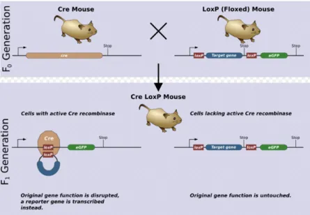
In the early 1990's a new method was developed to delete a specific portion of DNA in a specific location, such as a tissue or a cell type. The most widely used approach is the Cre-loxP system. Cre-recombinase is a 38 kDA protein that causes recombination of the bacteriophage P1 genome, recombining specific sequences of DNA with high fidelity (Jorgez, Lin et al. 2005). It efficiently catalyzes the recombination between two of its recognition sites, termed locus of crossover (x) in P1 (loxP), a 34 bp DNA sequence at which Cre catalyzes site-specific DNA recombination, in any cellular environment and on any kind of DNA (Nagy 2000), excising a part of the target gene, resulting in its inactivation (Sun, Liu et al. 2008).
Through insertion of loxP sites via homologous recombination into a gene of interest and by targeting the Cre-recombinase expression to a specific cell type using a tissue-specific promoter, it is possible to introduce tissue-restricted deletions into the mammalian genome (Lécureuil, Fontaine et al. 2002). A single recombinase molecule binds to each half of a loxP site and then splices together the two halves after the target DNA has been removed. At least two loxP sites are needed for Cre action. The loxP sites are inserted around a functionally essential part of the gene of interest, flanking critical exons, but the allele is kept completely functional in the targeted animal. Only after the Cre excision the gene will be knocked out (Nagy 2000) (Figure 3).
Cre-mediated recombination can be induced within the mouse by crossing mice carrying two loxP sites with a transgenic line expressing Cre, taking advantage of classical crossing-over (Brault, Besson et al. 2007). The flanked by loxP (floxed) mouse line bears the conditional allele of the gene to be deleted with recombinase-specific sites. The transgenic mouse line expressing the Cre-recombinase is guided by a promoter with a desired temporal and/or spatial pattern (Sun, Liu et al. 2008). After intercrossing to produce double transgenic offspring, the recombinase may delete or modify the conditional allele in expressing cells, while the unrecombined target gene remains intact and functional in the cells of other tissues where the recombinase is not expressed (Jorgez, Lin et al. 2005).
Cre-recombinase expression guided by a tissue or cell type-specific promoter exerts its functions, which allows examination of gene function in a specified tissue or cell type.
Figure 3 – Illustration of a model experiment in genetics using the Cre-loxP system (Zepper 2008).
These mutant mice usually have no evident developmental abnormalities and thus can be used for studies on a gene’s function in a specified tissue or cell type. It allows us to analyze the pathophysiological functions of a gene of interest without affecting survival of the animal (Sun, Liu et al. 2008).
2.4.2. Amhr2-Cre mouse line
Anti-Müllerian hormone (AMH), also known as Müllerian-inhibiting substance (MIS) is a gonad-specific glycoprotein produced and secreted only by ovarian granulosa cells and testicular Sertoli cells (Hirobe, He et al. 1992). In Sertoli cells it is produced from the time of testicular differentiation in fetal life until puberty and it is responsible for the regression of Müllerian ducts (Rey, Sabourin et al. 2000), which are the primordia of the uterus and oviducts (Lécureuil, Fontaine et al. 2002). In granulosa cells of the ovary, it is specifically expressed postnatally in human, rodents and ruminants (Rey, Sabourin et al. 2000).
The gene encoding the Amh type II receptor (Amhr2) has been isolated in several mammalian species. Its expression pattern is very restricted and overlaps with the Amh expression pattern in fetal Sertoli and postnatal granulosa cells (Jamin, Arango et al. 2003). Mice expressing Cre-recombinase driven by Amhr2 promoter are available. Amhr2-Cre is expressed in granulosa cells of small and growing ovarian follicles as well as ovarian surface epithelium cells (Fan and Richards 2010).
2.4.3. Cyp19-Cre mouse line
Aromatase enzyme, encoded by the Cyp19a1 gene, is a member of the cytochrome P450 superfamily, and it is important in the normal progress of the menstrual/estrous cycle in the ovary (Simpson 2004). Aromatase is the enzyme responsible for converting androgens (C19 steroids) to estrogens (C18 steroids). Under the control of FSH, it plays a role in transporting testosterone or androstenedione, produced by theca cells in response to LH, to the granulosa cells where aromatisation to estrogen occurs (Britt, Drummond et al. 2001).
In rodents, aromatase is restricted to the gonads and the brain. In the ovaries of sexually mature animals, aromatase expression is controlled in a cell-specific, temporal and spatial manner, limited only to mural granulosa cells of healthy large antral follicles, preovulatory follicles and luteal cells (Stocco 2008). The mouse line expressing Cre-recombinase driven by Cyp19 promoter is also available (Fan, Shimada et al. 2008). According to the aromatase expression, Cyp19-Cre is absent in ovarian surface epithelium cells, but is highly expressed in granulosa cells of antral and preovulatory follicles and induces 90 – 100% DNA excision of target genes (Fan and Richards 2010).
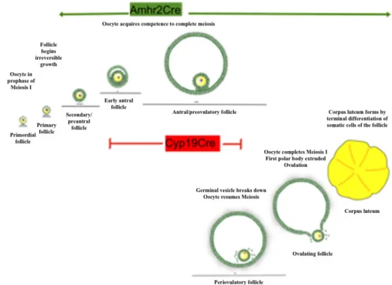
Cre strains in which granulosa cells express constitutively active Nr5a2 at specific stages of follicular growth are necessary to generate the Nr5a2 conditional knockout mice and specifically delete target genes in granulosa and cumulus cells. To study follicle growth and ovulation in granulosa cells of small and growing follicles, it is possible to cross mice carrying floxed Nr5a2 gene (Duggavathi, Volle et al. 2008) (described above) with mice
expressing Cre-recombinase driven by Amhr2 promoter (Amhr2-Cre). The double transgenic offspring is deficient in Nr5a2 in the granulosa cells of the ovary where Amhr2 and Nr5a2 are co-expressed (Duggavathi, Volle et al. 2008). On the other hand, by crossing Nr5a2-floxed and Cyp19-Cre mice, Nr5a2 is disrupted in granulosa cells of antral and preovulatory follicles. These conditional knockout models are important tools to the study of Nr5a2 at different stages of the follicle maturation in mouse (Figure 4).
Figure 4 – Scheme demonstrating the excision in different phases of the follicle development comparing Amhr2-Cre and Cyp19-Cre mice lines.
3. Materials and Methods
Animals and tissue collection procedures
All animal experiments were approved by the Université de Montréal Animal Care Committee and conducted according to guidelines of the Canadian Council of Animal Care. Animals were housed in a barrier facility with light/dark cycles of 14:10 h and provided food and water ad libitum.
Nr5a2-floxed mice have been described previously (Coste, Dubuquoy et al. 2007). To generate granulosa-specific Nr5a2 mutant mice, animals expressing Cre-recombinase from the Amhr2 promoter (gift of Dr. Richard Behringer) were crossed with Nr5a2-floxed mice (Nr5a2f/fAmhr2Cre/+, here referred as Amhr2Cre-cKO) to achieve the first objective of the experiment; non-transgenic littermates (Nr5a2f/fAmhr2+/+) were used as control group (Amhr2Cre-control). Animals expressing Cre-recombinase from the Cyp19-Cre promoter (gift of Dr. Jan Gossen) were crossed with Nr5a2-floxed mice (Nr5a2f/fCyp19Cre/+, here referred as Cyp19Cre-cKO) to develop the second part of the experiment; non-transgenic littermates (Nr5a2f/fCyp19+/+) were used as control group (Cyp19Cre-control). The sequences of the primer sets used for genotyping are listed in the Annex 1.
Stages of the estrous cycle were determined in mature (8-week-old) WT C57BL/6J mice and Cyp19Cre-cKO (n=4/genotype). Examination of exfoliative cytology of the vagina in phosphate-buffered saline (PBS) was undertaken on samples taken daily (between 7 and 8 a.m.) on glass slides for each mouse. For the breeding trial, 8-week-old Cyp19Cre-control and Cyp19Cre-cKO (n=4/genotype) females were housed with reproductively
proven C57BL/6J males for 6 months. Cages were inspected daily, and the day of parturition and number of pups in the litter from each female was noted. The control group for the breeding trial was composed of WT C57BL/6J couples (n=4), inspected in the same conditions as the Cyp19Cre-cKO females.
The superstimulation was performed using immature (22- to 25-day-old) Amhr2Cre-cKO (n=3) and Amhr2Cre-control (n=3) mice were injected with the hormones pregnant mare serum gonadotropin (PMSG; 5 IU i.p.) and human chorionic gonadotropin (hCG; 5 IU i.p.) to stimulate the growth and recruitment of ovarian follicles and to induce ovulation, respectively, 44 to 48 h of interval between each injection. Mice were euthanized 2 h, 4 h or 8 h post-hCG injection (n=3/time point/genotype) for gene expression evaluation; and 0 h and 12 h post-hCG (n=3/time point/genotype) for protein immunolocalization.
For histological analysis of the ovaries, prepubertal (22- to 25-day-old) females mice Amhr2Cre-cKO (n=3) and Amhr2Cre-control (n=3) were superstimulated with 10 IU of PMSG/hCG (44 to 48 h of interval between each injection). Ovaries were collected 7 h post-hCG and stained with hematoxylin and eosin (HE). Moreover, Cyp19Cre-cKO ovaries from 6-month-old females (n=4) and 1-year-old (n=4) were collected and stained with HE to evaluate the ovarian morphology related to the aging.
Before the euthanasia, female mice were anesthetized with isoflurane (USP, Pr AErrane, Baxter Corporation, Mississauga, ON, Canada). Blood was collected by cardiac puncture for hormone assay and euthanasia was achieved by cervical dislocation. The
ovaries were dissected; one ovary was snap frozen and stored at -80°C and the other ovary was fixed in paraformaldehyde (PAF) 4% for histological examination, and then paraffin-embedded, cut at 5 µM, and stained with HE. Ovaries, liver and intestine were collected from mature Cyp19Crecontrol and Cyp19CrecKO females (n=3 genotype) and stored at -80°C for RNA analysis of Nr5a2 in these tissues comparing both groups.
Laser Microdissection (LMD)
Gene expression was quantified in granulosa cells of antral follicles obtained by laser microdissection. Ovaries (n=3 mice in each of 2 h, 4 h or 8 h post-hCG injection) were collected and embedded in optimal cutting temperature (OCT) compound (Tissue-Tek, Sakura Finetek USA, Inc., Torrance, CA), snap frozen in liquid nitrogen, and stored at -80°C. Frozen whole ovaries were cut using a Leica CM3050 Cryostat in 25 µM thick slices at -16°C and adhered to a polyethylene naphthalate-coated glass slide (Leica MicroDissect GmbH, Herborn, Germany). Tissues on slides were subsequently washed in PBS to remove OCT, stained in toluidine blue, dehydrated by washing in ethanol at increasing concentrations (70 – 100%), and incubated for 1 h at 37°C. The granulosa cell compartment was laser-microdissected using Leica AS LMD System at 20x magnification. From each animal, ovarian structures of one ovary were obtained for total RNA extraction by collection of microdissected fragments in RLT buffer (Qiagen RNeasy Micro Kit, Qiagen, Valencia, CA).
RNA analysis
Total RNA was extracted from microdissected granulosa cells with RNeasy Micro Kit (Qiagen) as per manufacturer’s instructions. Total RNA was extracted from whole ovaries, liver and intestine using QIAshredder (Qiagen) followed by RNeasy Mini Kit (Qiagen) as per manufacturer’s instructions. For each sample, concentration of isolated total RNA was estimated using a NanoDrop apparatus (NanoDrop Technologies, Wilmington, DE), and the volume of extract containing 300 ng of RNA was submitted to DNase treatment and reverse transcription into cDNA was realized with the SuperScript II First-Strand Synthesis System (Life Technologies) and random hexamer primers.
The resulting cDNA was used in subsequent Real Time quantitative Polymerase Chain Reaction (PCR) using a 7300 real-time PCR system (Applied Biosystems, Foster City, CA) conducted in triplicate, and each reaction contained 10 µl Power SYBR® Green PCR Master Mix (Applied Biosciences, Warrington, UK), 4 µl of a sense-antisense primer mix, and 6 µl cDNA sample for a final volume of 20 µl. Common thermal cycling settings were used to amplify each transcript (2 min at 50°C, 10 min at 95°C, then 40 cycles of 15 s at 95°C and 60 s at 60°C). Melting curve analyses were performed to verify product identity, adding a dissociation step to the PCR run (15 s at 95°C, 60 min at 60°C, 15 s at 95°C, and 15 s at 60°C).
Cx43 expression was evaluated on microdissected granulosa cells of immature superstimulated Amhr2Cre-cKO and Amhr2Cre-control females; Nr5a2 expression was
evaluated on whole ovaries, liver and intestine of mature non-transgenic and Cyp19Cre-cKO females. The sense and anti-sense sequences of primers for Cx43 were 5′-TCCAAGGAGTTCCACCACTT-3′ and 5′-TGGAGTAGGCTTGGACCTTG-3′, each used in a final concentration of 300 nM. The sense and anti-sense sequences of primers for
Nr5a2 were 5′-TCATGCTGCCCAAAGTGGAGA-3′ and
5′-TGGTTTTGGACAGTTCGCTT-3′, each used in a final concentration of 300 nM (Duggavathi, Volle et al. 2008).
The ribosomal protein S18 (RPS18) was used for the normalization of Cx43 and Nr5a2 abundance. The RPS18 primer sequences for sense and antisense primers were 5′-GTGGTGTTGAGGAAAGCAGACA-3′ and 5′-TGATCACACGTTCCACCTCATC-3′, each used in a final concentration of 300 nM. To test the efficiency of amplification of primers of each gene, a cDNA pool was made using equal volumes of cDNA solution from each sample. The cDNA pool was serially diluted in autoclaved ddH2O from 1:7.5 to 1:240,
and samples were analyzed using the qPCR procedure described above. Efficiency of amplification of each sample was estimated using the LinReg software. Next, cDNA from each sample was diluted 1:10 in autoclaved ddH2O and analyzed by qPCR for RPS18 and
Cx43, and RPS18 and Nr5a2. Relative abundance of Cx43 and Nr5a2 transcripts was calculated by the ΔΔCt method corrected by the efficiency of amplification, as described by Pfaffl (Pfaffl 2001). The relative abundance of Cx43 and Nr5a2 was analyzed by analysis of variance (ANOVA) using the software JMP.
Immunohistochemistry
The distribution and protein expression of Cx43 on cKO and Amhr2Cre-control ovaries at 0 h and 12 h (n=3/time point/genotype) after hCG injection was verified by fluorescence immunohistochemistry (Annex 2). Immunohistochemistry was performed on 5 µM cryosections of the ovaries. Briefly, paraffin sections of ovaries were rehydrated, boiled in 10 mM sodium citrate (pH 6.0) for 20 min, and cooled to room temperature (RT). Subsequently, sections were blocked in PBS containing bovine serum albumin (BSA) 5% for 30 min at RT, incubated with rabbit polyclonal IgG connexin 43 (Santa Cruz Biotechnology) diluted 1:100 in PBS containing BSA 5% overnight at 4°C. As a negative control, some sections were incubated with BSA in the place of Cx43 antibody. Sections were washed, incubated with Alexa 594 donkey anti-rabbit IgG (Invitrogen Molecular Probes), diluted 1:600 in PBS for 1h at RT. Slides were then washed, and sections were counterstained with 4’,6-diamidino-2-phenylindole (DAPI, Sigma, St. Louis, MO), diluted 1:1000 in PBS, for 5 min. Slides were mounted in Permafluor (Lab Vision Corp., Fremont, CA). Ovarian distribution of Cx43 was observed by fluorescent microscopy using the microscope Leica DC-500.
The same protocol was applied on Cyp19Cre-cKOovaries (n=3) from mature (6-month-old) females to study this protein expression and immunolocalization of SCARB1 in granulosa and luteal-like structures described in the Cyp19Cre-cKO ovaries. In this case, the first antibody was rabbit polyclonal anti-mouse SCARB1 (Novus Biologicals), and the