Université de Montréal
Correlates of Protective Immunity against
Hepatitis C Virus
Par
Mohamed S. Abdel-Hakeem
Département de Microbiologie et Immunologie Faculté de Médecine
Thèse présentée à la Faculté des études supérieures en vue de l’obtention du grade du Doctorat (Ph.D.)
en Microbiologie et Immunologie
Mars, 2014
Université de Montréal Faculté des études supérieures
Cette thèse intitulée :
Correlates of Protective Immunity against Hepatitis C Virus
Présentée par :
Mohamed S. Abdel-Hakeem
A été évaluée par un jury compose des personnes suivantes :
Dre Cécile Tremblay, Présidente-rapporteuse Dre Naglaa Shoukry, Directrice de recherche
Dr Daniel Kaufmann, Membre du jury Dr Alain Lamarre, Examinateur Externe Dr Roger Lippé, Représentant du Doyen de la FESP
Résumé
Le virus de l’hépatite C (VHC) infecte ~185 millions d’individus dans le monde. Malgré le développement des nouvelles thérapies dirigées contre le VHC, on compte deux millions de nouvelles infections chaque année, en particulier dans les pays sous-développés et les populations marginalisées. Comme pour la plupart des virus à infection chronique, le développement d’un vaccin prophylactique efficace est limité par le manque de caractérisation des déterminants de la mémoire immunitaire protectrice lors des épisodes de réinfection naturelle chez les êtres humains. Le VHC représente un modèle unique au sein des virus à infection chronique pour étudier l’immunité protectrice. En effet ~30% des patients infectés par le VHC peuvent être guéris suite à un premier épisode d’infection spontanément.
Dans cette thèse, nous avons étudié l’immunité protectrice contre le VHC dans une cohorte d’utilisateurs de drogues par injection qui sont à risque d’être infectés ou réinfectés. Notre hypothèse est que la majorité des patients qui ont résolu une première infection par le VHC sont protégés contre le développement d’une infection chronique s’ils sont réexposés. Cette immunité protectrice est associée à la présence des cellules T CD4 et CD8 polyfonctionnelles qui possèdent des fréquences, magnitudes et avidités élevées. La capacité protectrice des cellules T mémoire contre les séquences variables du VHC est dépendante de la diversité et flexibilité du répertoire de leurs récepteurs de cellules T (TCR), qui reconnaissent les séquences variables des épitopes ciblés.
Notre premier objectif était de définir et détailler les déterminants de l’immunité protectrice conférée par les cellules T spécifiques du VHC. Nos résultats ont montré que la protection pendant l’épisode de réinfection était associée à une augmentation de la magnitude et du spectre des réponses spécifiques par les cellules T CD4 et CD8 polyfonctionnelles, ainsi que par l’apparition d’une population de cellules T tétramère+ CD8+ effectrices qui expriment faiblement le marqueur CD127 (CD127lo) lors du pic de la réponse. Chez les patients qui ont développé une infection chronique pendant l’épisode de réinfection, nous avons observé une expansion très limitée des cellules T CD4 et CD8. Le séquençage des épitopes ciblés par les cellules T CD8 chez ces patients qui sont non-protégés a montré que les
séquences de ces épitopes sont différentes des séquences de référence qui étaient utilisées pour tous les essais immunologiques de cette étude.
Le deuxième objectif était d’analyser la dynamique du répertoire des TCRs des cellules T CD8 spécifiques chez les patients protégés versus les patients non-protégés. Nos résultats ont montré que le répertoire des cellules T CD8 spécifiques est plus focalisé que chez les patients protégés. En plus, nous avons observé que les clonotypes qui forment le répertoire chez les patients protégés sont distincts de ceux chez les patients non-protégés. Ces clonotypes chez les patients protégés ont montré de plus grandes avidité et polyfonctionnalité que leurs homologues chez les patients non-protégés.
En conclusion, nos résultats suggèrent que la protection contre le développement d’une infection chronique pendant l’épisode de réinfection par le VHC est associée à une augmentation de la magnitude, du spectre et de la fonctionnalité des réponses des cellules T spécifiques, ainsi qu’à un répertoire des TCRs plus focalisé composé des clonotypes distincts qui possèdent de plus grandes avidité et polyfonctionnalité que chez les patients non-protégés. L’homologie des séquences des souches virales entre les différents épisodes de l’infection est un déterminant majeur de l’établissement d’une protection efficace. Ces résultats ont donc des implications très importantes pour le développement d’un vaccin prophylactique contre le VHC et d’autres virus à infection chronique.
Mots-clés : Virus de l’hépatite C (VHC) – Mémoire immunitaire – Réinfection par le VHC –
Abstract
Hepatitis C virus (HCV) currently infects ~185 million individuals worldwide. Despite the development of new effective antivirals against HCV, it is estimated that two million new infections occur annually, especially among marginalized populations and in developing countries. As with many chronic viral infections, the development of an effective prophylactic vaccine is hampered by the limited knowledge of determinants of a protective immune response in humans upon natural exposure to the virus. HCV represents a unique model to study protective immunity in chronic viruses, since ~30% of the patients are able to clear the primary infection spontaneously.
In this thesis, we proceeded to study protective immunity against HCV in cohorts of injection drug users (IDUs) who are continuously at risk of infection and reinfection by HCV. We hypothesized that the majority of spontaneous resolvers of a primary HCV infection are protected against chronicity of infection upon re-exposure. Protective immunity would likely be associated with the highest breadth, frequency and functional avidity of HCV-specific polyfunctional CD4 and CD8 memory T cells. The protective capacity of memory T cells upon infection with novel HCV variants will depend on the diversity and flexibility of the T cell repertoire that can recognize these viral variants and/or the capacity to generate de novo T cell responses specific for these variants.
The first aim of the project was to define and dissect correlates of protective immunity conferred by HCV-specific T cells in reinfected individuals. Our results showed that protection from chronicity upon reinfection was associated with an increased magnitude and breadth of the HCV-specific polyfunctional CD4 and CD8 T-cell responses, as well as the appearance of a CD127lo tetramer+ CD8+ effector T-cell population at the peak of the response. Individuals who developed persistent viremia upon HCV reinfection demonstrated very limited or no expansion of HCV-specific T cells. Sequencing of the cytotoxic T-lymphocyte (CTL) epitopes targeted by the memory immune response in unprotected individuals revealed that the sequence of the autologous reinfecting virus was different from the reference sequence used in our immunological assays.
with variant viral sequences. Our results demonstrate that for epitope-specific CD8 T cells a more focused TCR-repertoire of distinct clonotypes was associated with protection. These T-cell populations showed higher functional avidity and polyfunctionality compared to their counterparts in non-protected patients. The clonotypes forming the effector T-cell population during reinfection were recruited from the memory pool, rather being de novo responses generated from the naïve pool.
In conclusion, our results suggest that protection from persistence upon HCV reinfection is associated with an enhanced magnitude, breadth and quality of the HCV-specific T-cell response, as well as a more focused TCR-repertoire of distinct clonotypes with high functional avidity. The degree of homology between viral strains causing the consecutive episodes of infection is a critical determinant for protection. These findings provide a first insight into the correlates of protective immunity against HCV and have important implications for the rational design of effective prophylactic vaccines against HCV and other chronic viruses.
Keywords:
Hepatitis C Virus (HCV) – Memory immune response – HCV reinfection – Protective immunity – T-cell receptor (TCR) repertoire
Table of contents
CHAPTER 1: ... 1
INTRODUCTION ... 1
1.1. Immunity against primary viral infection ... 3
1.2. Immunological memory and vaccines against viruses ... 7
1.2.1. Generation of memory immune responses ... 7
1.2.2. Maintenance of immunological memory ... 10
1.2.3. Characteristics of a memory immune response ... 11
1.2.4. Memory T cell subsets ... 15
1.2.5. Immunological memory at the clonal level ... 17
1.2.5.1. Generation of TCR diversity ... 17
1.2.5.2. Impact of TCR diversity on the viral immune response ... 19
1.2.6. Immunological memory and vaccination ... 21
1.2.7. Challenges for developing vaccines against chronic viruses ... 22
1.2.8. Protective CMI mechanisms for successful vaccines ... 23
1.2.9. Human models for studying protective immunity against chronic viruses ... 24
1.3. Hepatitis C Virus ... 26
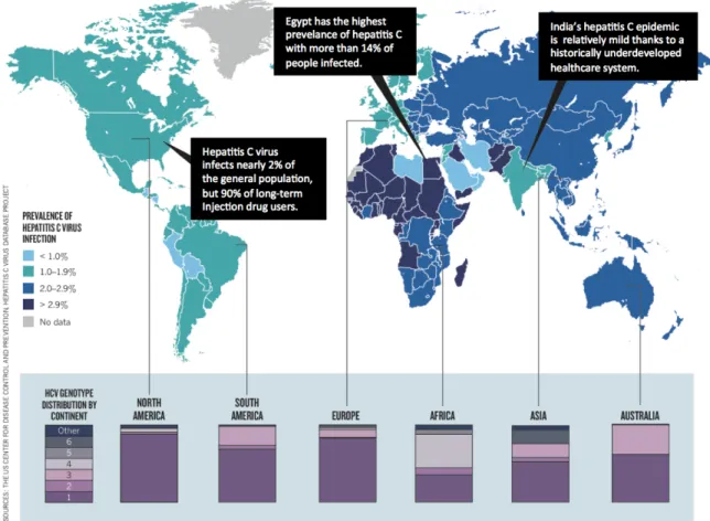
1.3.1. HCV epidemiology and transmission ... 26
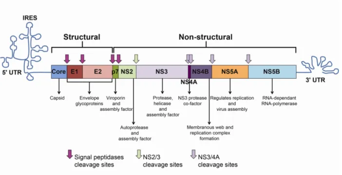
1.3.2. HCV genome and proteins ... 28
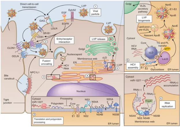
1.3.3. HCV life cycle ... 31
1.3.4. HCV infection and its outcome ... 34
1.3.5. Treatment of HCV infection ... 35
1.3.5.1. Available treatments ... 35
1.3.5.2. Novel HCV treatments ... 36
1.4. Immunity against primary HCV infection ... 38
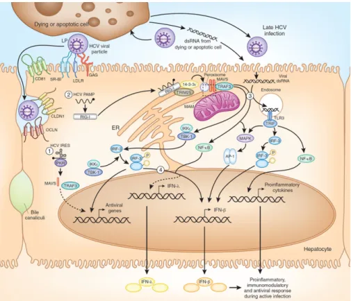
1.4.1. Innate sensing of HCV ... 38
1.4.2. Innate immune response against HCV ... 40
1.4.4. Failure of immune responses against HCV ... 47
1.4.4.1. Primary failure of the HCV-specific immune response ... 47
1.4.4.2. Viral evasion strategies ... 49
1.5. Protective immunity against HCV and vaccine trials ... 54
1.5.1. Memory immune responses against HCV ... 54
1.5.1. HCV vaccine trials ... 57
CHAPTER 2: ... 61
HYPOTHESIS AND OBJECTIVES ... 61
CHAPTER 3: ... 67
MANUSCRIPT 1: ... 67
Signatures of Protective Memory Immune Responses during HCV Reinfection ... 67
CHAPTER 4: ... 117
MANUSCRIPT 2: ... 117
Dynamics of Virus-Specific CD8 T-cell Repertoire during HCV Reinfection ... 117
CHAPTER 5: ... 169
DISCUSSION AND CONCLUSION ... 169
5.1. Correlation between total magnitude and breadth of T-cell responses and protective immunity upon HCV reinfection ... 172
5.2. Correlation between T-cell proliferative capacity and protective immunity ... 173
5.3. Functionality of HCV-specific T-cells is associated with protective immunity ... 175
5.4. Frequency and Phenotype of epitope-specific CD8 T-cells associated with protective immunity ... 177
5.5. Effect of viral-epitope variation on protection upon HCV reinfection ... 179
5.6. Avidity of HCV-specific T cells and protective immunity ... 180
5.7. Memory versus de novo CD8 T-clonotypes in protective immune responses ... 182
5.9. Role of specific CD8 T-clonotypes in protective immunity ... 186
5.10. Shifting epitope dominance upon HCV reinfection ... 190
5.11. Correlation between specific HLA-alleles and protective immunity ... 190
5.12. Conclusions, limitations and future studies ... 191
5.12.1. General conclusions ... 191
5.12.3. Probable causes of failure of protective immunity ... 194
5.12.3.1. Defects in signaling pathways and transcription of HCV-specific T cells ... 194
5.12.3.2. Suppression by Tregs ... 195
5.12.3.3. Absence of neutralizing Antibodies ... 195
5.12.3.4. Lack of particular T-cell clonotypes ... 195
5.13. Significance of the study ... 196
CHAPTER 6: ... 201
PERSPECTIVES AND FUTURE DIRECTIONS ... 201
6.1. Transcriptome analysis to dissect underlying causes of failure of protective immunity . 204 6.2. Defining the role of different CD4 T-cell subsets in protective immunity against HCV 205 6.3. Defining the role of neutralizing antibodies in protective immunity against HCV ... 208
6.4. Sequencing the HCV genome during the different episodes of infection ... 208
6.5. Dissecting the qualities of individual clonotypes associated with protection ... 209
6.6. Determining whether specific public clonotypes associate with protection ... 210
6.7. Concluding remarks ... 210
APPENDECIES ... 213
APPENDIX I: The candidate’s contribution to the articles ... 215
APPENDIX II: Review article ... 216
Bibliography ... 239 Curriculum Vitae ... XVIII
List of figures
Figure 1: Innate and adaptive immune responses in humans using hepatitis C virus (HCV) as a
model ………..……….... 5
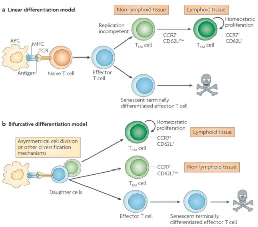
Figure 2: Possible models for memory T-cell differentiation ……….... 9
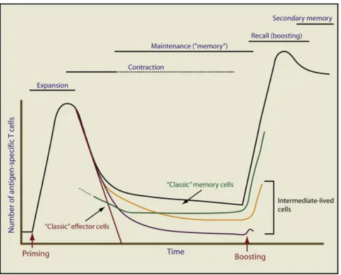
Figure 3: Generation and maintenance of T-cell memory ………... 14
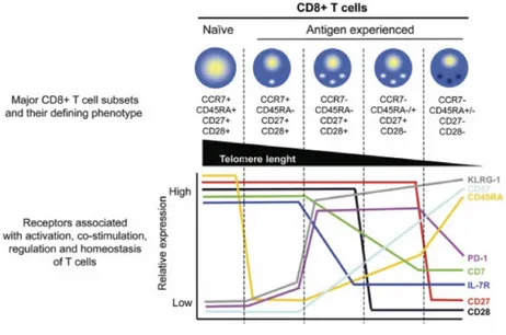
Figure 4: Different subsets of human T cells and their phenotypic and functional attributes ………... 16
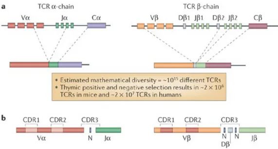
Figure 5: Generation of TCR diversity by recombination of TCR gene segments ………... 18
Figure 6: Prevalence of HCV and genotype distribution worldwide ………... 27
Figure 7: HCV genome and polyprotein ……….. 30
Figure 8: HCV viral life cycle ………...……….….. 33
Figure 9: Sensing HCV in hepatocytes ………..……….. 39
Figure 10: Successful versus unsuccessful immune responses during acute HCV infection ……….……….. 46
Figure 11: HCV proteins subvert intracellular signaling ………...……….. 50
Figure 12: Mechanisms of HCV-protein interference with the immune system …...……….. 53
Figure 13: Hypothetical model for protective and non-protective immunity upon HCV reinfection ………...………..…. 188
List of abbreviations
Ab Antibody
Ad Adenovirus
ADCC Ab-dependent cellular cytotoxicity
Ag Antigens
AIDS Acquire immunity deficiency syndrome
AP-1 Activator protein 1
APC Antigen presenting cells
Bcl-2 B-cell lymphoma-2 regulator protein
BrdU Bromodeoxyuridine
CCL C-C motif chemokine ligand
CCR7 C-C motif chemokine receptor type 7
CDR Complementarity-determining regions
CLDN Claudin
CMI Cell-mediated immunity
CMV Cytomegalovirus
CTL Cytotoxic T lymphocyte
CTLA-4 Cytotoxic T lymphocyte antigen-4
CypA Cyclophylin A
DAA Direct-acting antiviral agents
DC Dendritic cells
dsRNA Double-stranded RNA
EBV Epstein–Barr virus
EGFR Epidermal growth factor receptor
Fab Ag-binding fragment of the antibody
Fc Constant fragment of the antibody
FDA Food and Drug Administration
fDC Follicular DCs
FoxP3 Forkhead transcription factor 3
GATA3 GATA-binding protein 3
GC Germinal centre
GSEA Gene set enrichment analysis
GWAS Genome-wide association studies
HCC Hepatocellular carcinoma
HCV Hepatitis C virus
HCVpp HCV pseudo-particles
HCVcc Cell-cultured HCV
HEV High endothelial venules
HIV Human immunodeficiency virus
HPV Human papilloma virus
HSV Herpes simplex virus
HVR Hypervariable region-1
IDU Injection drug users
IFN Interferon
Ig Immunoglobulin
IKKε IkB kinase-ε
IL Interleukin
IL7Rα IL-7 receptor alpha chain
IRES Internal ribosome binding site
IRF Interferon regulatory factor
ISG Interferon-stimulated genes
JAK Janus kinase
JFH1 Japanese fulminant hepatitis-1
kb Kilo base-pairs
KIR Killer cell immunoglobulin-like receptors
LCMV Lymphocytic choriomeningitis virus
LDL Low density lipoproteins
LN Lymph nodes
LTNP Long-term non-progressors
LVP Lipoviroparticles
MAVS Mitochondrial antiviral signaling protein
mDC Myeloid DCs
MHC Major histocompatibility complex
MVA Modified vaccinia Ankara
nAb Neutralizing Abs
NANB Non-A, non-B
NCR Non-coding regions
NK Natural killer cells
NKG2D Natural killer group 2, member D
NR Non-response
NS Non–structural
OCLN Occluding
ORF Open reading frame
PAMP Pathogen Associated Molecular Patterns
pDC Plasmacytoid DCs
PEG Polyethylene glycol
pMHC Peptide-MHC
p.ri. Post reinfection
PRR Pattern recognition receptor
RdRp RNA-dependant RNA-polymerase
RIG-I Retinoic-acid-inducible gene I
RORγt Retinoic acid receptor-related orphan receptor-γt
SNP Single-nucleotide polymorphisms
SR-BI Scavenger receptor class B type I
ssRNA Positive single-stranded RNA
STAT Signal transducer and activator of transcription
SVR Sustained virological response
TANK TRAF family member-associated NFκB
TBK TANK-binding kinase 1
TCM Central-memory T cells
TCR T-cell receptor
Teff Effector T cells
TEM Effector-memory T cells
TEMRA Terminally-differentiated T cells
Tfh Follicular helper T cells
TGF Transforming growth factors
Th T helper cells
Tim-3 T-cell immunoglobulin and mucin domain 3
TJ Tight junction
TLR Toll-like receptors
TNF Tumor necrosis factor
TRAF TNF receptor–associated factor 3
TRAIL TNF-related apoptosis-inducing ligand
Tregs Regulatory T cells
TRIF TIR domain-containing adapter inducing IFN-β Tyk Tyrosine kinase
UTR Untranslated region
VLDL Very-low-density lipoproteins
VLP Virus-like particles
VV Vaccinia virus
WHO World Health Organization
WT Wild-type
To freedom fighters all over the world, especially heroes of The Egyptian Revolution,
January 25th, 2011. To Mom, family & friends in Egypt.
Acknowledgement
… and you (mankind) have been given but a little knowledge Quraan (17 :85)
« Seek knowledge even at the farthest place » Prophet Muhammad (PBUH)
The past five years were the most influential in my life. The experiences I gained during my PhD journey had a great impact on the evolution of my character and way of thinking, as much as it impacted me as a researcher. This manuscript represents a piece of collective work, not only of my lab supervisor and colleagues, but also of my family, friends, professors and humans that I encountered throughout my life-path. I learned something from each and every one of you, and I hope that I can show my gratitude by conveying this to others. I was fortunate to spend this time in one of the most beautiful and inviting cities in the world, Montréal, a city that became my second home after Cairo.
It is very confusing when it comes to giving special thanks to each person by name, as the space is never enough to thank all those who had a direct or an indirect input in the creation of this thesis. I will start with my mentor, supervisor and friend, Naglaa Shoukry, who taught me how to be a better researcher and scientist, but also taught me many things in life. Thank you for your trust, support and guidance throughout the years. I would like to thank all members of my
lab-family during my PhD; especially Nathalie and Sandy (who were there all the way!), Hassen, Marion, Elias, Thomas, Camille, Julie, Sarah, Anna and JF. I would also like to thank my
mentors/colleagues/friends at the CRCHUM –St-Luc; Dre.Ancuta, Dre. Grandvaux, Dr.Mourad, Dr.Finzi and Dr.Kaufmann, members of their labs; esp. Amal & Ghada, Dr.Mohamed Elfar, YuWei, les Manons, Norma, Myriam, Dominique, Vero and Jean-Luc, at the new CRCHUM; esp. Dr.Lamarre and his lab members & Nirmin, at UdeM; esp. Dr.Hugo Soudeyns and at the
National CIHR Research Training Program – Hepatitis C (NCRTP– HepC). I would also like to thank our collaborators; esp. Dr.Willems, professors who gave me training in their labs;
Dr.Arash Grakoui and Dr.Michael Houghton, my committee and jury professors, and researchers
who worked for the last 25 years to unravel the mysteryof HCV, whom I call the
“HCV-superfamily” J. I would like to extend my thanks to the agencies/organizations who offered me
fellowships and awards. Last, but not least, I would like to thank our volunteer-patients. I was honoured and lucky to get to know all of you and work with you.
All the gratitude to each and every member of my family & friends in Egypt, who gave me their unconditional support at difficult times and when most needed, especially, my small family who had to endure the nature of my work/passion; Reham & Malek, Mom & Baba
Farouk, my sisters; Shaza & Shadw, their husbands; Ehab & Kamal, my uncles; Moustafa & Sameh, my cousins (esp. Ahmed) my nieces & nephews (all 9+1 of you J), Fofa & Sheri.
I would like to thank people who inspired me and set an example for me with their dedication and passion for what they do, their integrity and their code of ethics; Mom, you were and will always be my role model, my late Grandma Thorayya, I wish I had finished my PhD earlier to share my success with you, but I hope I made you proud, Baba Farouk, your encouragement and prayers made all the difference, Naglaa, my CRCHUM & UdeM mentors;
esp. Petronela, Nathalie, Hugo, Daniel and Andrés, my NCRTP mentors; especially, Dr.Marc Bilodeau, Dr.M. Houghton, Dr.Lorne Tyrell and Dre.Julie Bruneau, and Egyptian officials I
was proud to know; HE Wael Abo-Elmagd, Dre. Maha Kamel, Dr. Elsayyed Mahfouz.
I would like to extend my gratitude to: my dear friends M.Sarhan, Tamer I. Mahmoud,
Gamal Badr, M.Darwish, Hatem Elshabrawy and Amin Ismail for their time and interesting
debates, all of my professors throughout the years, my colleagues/friends at the Egyptian Student Association in North America (ESANA); esp. M.Sarhan, Sahmel, Tamer Awad,
M.Afifi, Maha Farid and A. Osman, as well as ESAs UofT, UofAlberta, Kingston, Ohio SU
and my ESA-UdeM family, esp. Wessam & Bakry, Noha & Hussein, other friends who made life in Montréal an even more pleasant experience; Mme.Amal, Dre.May, Dre.Fikria, Rim &
Bourhan, Mona & Farouk, Ola & Sakka, Dina & Hazem, Olfat & Tamer, Marwa & Omdah,
the Jundis, Eman, Nabil, A.Salah, A.Fahmy, M.Kaitar, Ossama Allam, Moomen, Ayman, Sheikh/Metwally & Haj/Usama and other friends at Al-Ummah and Fatimah mosques, Sally, Julie, Jen, Vanessa, Valérie, Alex, Mélissa, Maria and Kelly, friends in Canada & the US; esp. Zoeiby, Bob, Riham & Ximo, Mah.Sarhan, M.Sallam, W.ElRayess and Hawwary, my friends all over the world who kept in touch (esp. from school & college days), twitter friends and
finally colleagues/friends at the Faculty of Pharmacy, Cairo University (FOPCU) who inspire me with their perseverence to teach new generations of pharmacists and perform meaningful
CHAPTER 1:
INTRODUCTION
(Parts of this introduction were used in the review article “Protective Immunity against Hepatitis C: Many Shades of Gray”,
The rational design of prophylactic vaccines against chronic viral infections necessitates dissection of the correlates of protective immunity that remain undefined to date. Hepatitis C virus (HCV) represents a unique model among chronic viruses where two dichotomous outcomes of infection are observed: spontaneous resolution or chronic infection. Thus it represents an ideal model for understanding determinants of the memory immune response that can confer protection upon re-exposure to the virus in a human cohort.
This introduction discusses what is known about primary and memory immune responses against chronic viral infections, followed by a more specific review of the literature about the primary and memory immune responses against HCV infection. It also gives a brief account of different aspects of HCV; characteristics of the virus, the infection caused by HCV, treatment strategies and HCV-vaccine trials.
1.1. Immunity against primary viral infection
The human immune system is composed of two interconnected lines of defence; innate immunity and adaptive immunity. Innate immunity is non-specific, as it recognizes common motifs shared by interrelated pathogens known as “Pathogen Associated Molecular Patterns” (PAMPs), while adaptive immunity is highly specific and recognizes specific pathogen derived peptides known as antigens (Ags) or epitopes. Furthermore, adaptive immunity is privileged by its ability to generate immunological memory, which has the capacity to respond more rapidly to subsequent exposures to an Ag that has been previously encountered (also known as, the recall response).
Innate immunity is the first line of defence triggered by foreign pathogens including viruses. Cells of innate immunity include monocytes, natural killer cells (NKs) and dendritic cells (DCs) [1]. NKs have direct cytotoxic action and can kill infected cells. In addition, they possess antiviral action through secretion of inflammatory cytokines such as interferon-gamma (IFN-γ) and tumor necrosis factor-alpha (TNF-α) [2], as well as having the ability to stimulate maturation of DCs that represent the link between innate and adaptive immunity (Figure 1) [3]. NK cell activity is triggered by reduced expression of the major histocompatibility complex (MHC) class I molecules on the surface of infected cells, known as the “missing self” hypothesis. The extent of NK cell activation is determined by the balance of interaction between the activating and inhibitory receptors on NK cells and their corresponding ligands expressed on target cells. These receptors include members of the killer cell immunoglobulin-like receptors (KIRs) family and the NKG2 family [4, 5]. DCs are the main professional antigen presenting cells (APCs) involved in priming naïve adaptive immune cells known as T cells [6]. There are two main populations of DCs: myeloid DCs (mDCs) and plasmacytoid DCs (pDCs). The mDCs are the major APCs involved in antigen uptake and presentation, while pDCs mainly produce IFN-α and act as overall orchestrators of the immune response. DCs become activated upon recognition of PAMPs by cell surface and/or intracellular receptors [7].
B cells that produce antibodies (Abs) that mainly target extracellular microorganisms (e.g. bacteria), but also neutralize intracellular pathogens (e.g. viruses) by targeting surface epitopes to prevent viral binding and entry into target cells. Abs also help in the destruction of infected cells by Ab-dependent cellular cytotoxicity (ADCC), where specific Abs bind to cell surfaces using their variable Ag-binding fragment (Fab) recognizing a specific Ag and their constant portion (Fc) binds to the Fc-receptors on NK cells and/or monocytes and activates their cytotoxic functions [6]. Cell-mediated immunity mainly targets intracellular pathogens (e.g. viruses) and abnormal cells like cancer cells, and it is comprised of CD8 T cells, known as cytotoxic T lymphocytes (CTLs) and CD4 T cells that act as helper T cells. CD8 T cells recognize and kill cancer cells and virus infected cells expressing foreign Ags/epitopes on their cell surface in the form of peptides bound to MHC class I molecules. CD8 T lymphocytes kill infected/cancerous cells by secreting pore-forming molecules inducing cell lysis (granzymes and perforin) or through ligand-mediated initiation of programmed cell death (e.g. Fas-FasL interactions). CD8 T cells also secrete cytokines (e.g. IFN-γ and TNF-α) that have direct antiviral effects and can activate other effector cells [8]. CD4 T cells, were previously divided into two major helper subsets; T helper 1 cells (Th1), that help priming and
sustaining CTL responses, and Th2 that secrete specific cytokines that help activate B cells and
promote them to differentiate into plasma cells and produce Abs [9]. More recently other CD4 T-cell subsets were identified that regulate different aspects of the immune response and include Th17 cells that mediate inflammation and response to microbial pathogens, regulatory
T cells (Tregs) that regulate different immune responses and follicular helper T cells (Tfh) cells
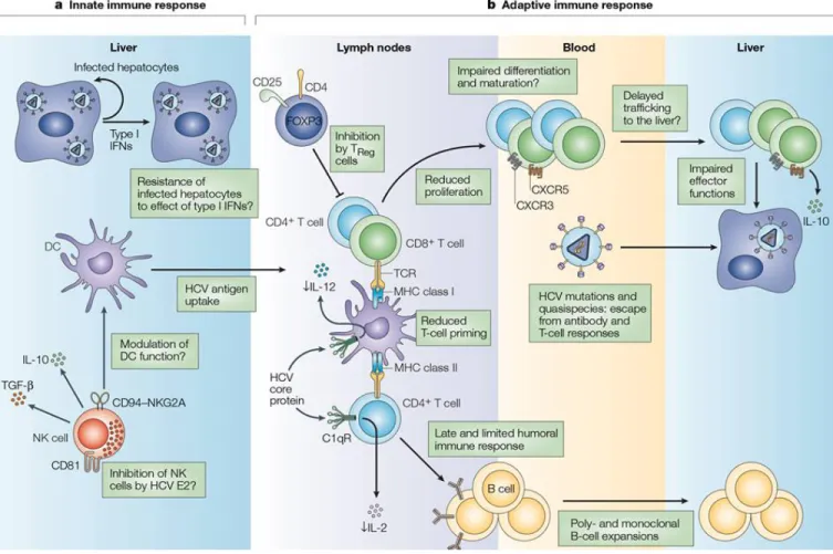
Figure 1: Innate and adaptive immune responses in humans using hepatitis C virus (HCV) as a model.
Reproduced with the permission of Frontiers scholarly communications: Frontiers in Immunology [10], author copyrights 2014.
Legend: (1) Pathogen entry and replication induces the production type I IFNs that are also
secreted by pDCs and create a defensive state and stimulate NK cells that kill infected
hepatocytes. (2) APCs (especially DCs) uptake apoptotic bodies from destroyed infected cells and present epitopes to both CD4 and CD8 T cells in the context of MHC class II and MHC class I, respectively. (3) The cross-talk between DCs and NKs regulates the function of both cells enhancing antigen presentation and priming, as well as NK-mediated killing. (4) CD4 helper T cells support responses of CD8 T cells and B cells through production of Th1 and Th2 cytokine, respectively. (5) Abs produced from B cells could have an essential role in pathogen clearance by directly neutralizing the pathogen or by mediating ADCC. Tfh cells play an essential role in mediating the selection and survival of B cells. (6) CD8 T cells eliminate pathogen-infected cells through direct cytotoxic mechanisms (cytolytic granules, as perforin and granzyme) or non-cytolytic mechanisms through secretion of the antiviral cytokines IFNγ and TNFα or through ligand-mediated initiation of programmed cell death (e.g. Fas-FasL interactions). (7) CD8 T cell functions are sustained and enhanced by IL-21 mainly produced by Th17 cells. IL-21 is also essential to rescue virus-specific T cells from exhaustion caused by persistent exposure to HCV antigens. Reduced IL-21 production or Th17 cell numbers results in an increased exhaustion status and expression of exhaustion markers like PD-1, Tim-3, CTLA-4, and others. (8) Regulatory T cells might be a cause in the failure of the primary immune response by secreting the regulatory cytokines TGFβ and IL-10, or secretion of Gal-9 that enhances apoptosis of Tim-3+ CD4 and CD8Tcells.
ADCC, antibody-dependentcellular cytotoxicity; APC, antigen-presenting cell; CTLA-4, cytotoxic T lymphocyte antigen-4; DC, dendritic cell; FcR, receptor for the constant fragment of the antibody; Gal-9, galactin-9; Gz, granzyme; HCV, hepatitis C virus; IFN, interferon; IL, interleukin; NK, natural killer; PD-1, Programed death-1; pDC, plasmacytoid DC; Tfh, T follicular helper cells; TGF, transforming growth factor; Th17, T-helper 17 cell; Tim-3, T cell immunoglobulin and mucin domain 3;TNF, tumor necrosis factor;Treg, regulatoryT cell.
1.2. Immunological memory and vaccines against viruses
Immunological memory is the ability to mount a specific accelerated recall response by antigen experienced T- and B-lymphocytes against a previously encountered pathogen [11-13]. The memory immune response is also characterized by the increased frequency of antigen-specific lymphocytes with improved recognition and function [14, 15], as well as enhanced trafficking, migration and localisation at the site of infection [16, 17].
Most of our current knowledge about immunological memory against viruses is based on studies of the lymphocytic choriomeningitis virus (LCMV) infection in mouse models. LCMV is an excellent infection model, since its different strains have distinct courses of infection; where the Armstrong strain causes an acute infection cleared within a few days, as opposed to LCMV-clone 13 that leads to chronic infection and serves as a model that would correspond to human chronic viral infections (e.g. HIV and HCV) [18]. It is only recently that these findings were validated in human viral infections [19].
1.2.1. Generation of memory immune responses
Naïve B cells are activated and proliferate when they encounter their specific antigen for the first time. Activated B cells either remain in the marginal zone of T cells within lymph nodes and differentiate into short-lived plasma cells or migrate to B cell follicles initiating a germinal centre (GC) reaction. Within the GC, B cells undergo somatic hypermutation, affinity maturation and selection leading to the generation of high affinity memory B cells. The GC formation requires help from Tfh CD4 T lymphocytes [20, 21].
Development of memory T cells (Tmem) includes three phases (Figure 3);
Phase I (Expansion): activation and expansion of Ag-specific T-cell clones that have recognized their specific Ag within the context of MHC molecules, and with adequate costimulation these T cells differentiate into effector T cells (Teff).
Phase II (Contraction): after resolution of infection the majority (~90%) Teff die by apoptosis.
Ag-Two models for the development of Tmem were proposed, the linear/progressive model
and the asymmetric/bifurcative/stochastic model. Evidence supporting both models is equivocal, thus it remains unclear whether the pathway is linear or asymmetric, and whether it is one-way or reversible. The linear model proposes that activated naïve T cells develop into Teff then a proportion of those Teff differentiate into Tmem that are either effector-memory T
cells (TEM) or central-memory T cells (TCM) (Figure 2). This was supported by microarray
analysis of the gene-expression program of T cells in the LCMV model showing that acquisition of Tmem cell traits was gradual and was still occurring several weeks after viral
clearance. This suggests that the 5-10% of the surviving Teff are promoted to Tmem by
completing a gradual program of differentiation and acquisition of all memory properties [23]. An interesting study of SIV-infected monkeys treated with bromodeoxyuridine (BrdU) demonstrated the progression of labelled cells, from TCM to TEM and finally to
terminally-differentiated T cells (TEMRA) representing the end-stage of T-cell development [24] (different
T-cell subsets are explained in detail in section 1.2.4.). These results suggest that memory subsets are not independent, but rather share a common differentiation pathway, supporting the linear model [25]. On the other hand, the asymmetric model suggests that a single naïve T cell once primed gives rise to a heterogeneous population of Ag-specific T cells, and that the different populations of T cells of all phases are programmed for their final fate shortly following antigenic stimulation [26]. This early imprinting of a specific developmental program is reflected in changes in the gene expression that occur shortly after priming of naïve T cells [27]. Asymmetric T-lymphocyte division during early steps of initiation of the adaptive immune response were demonstrated in a mouse model of Listeria monocytogenes [28].
Important issues concerning generation of immunological memory remain to be resolved. First, what factors determine which cells would express memory T-cell markers versus those that become short-lived effector cells? Second, are those factors intrinsic, extrinsic or both? Finally, is it predetermined for CD8 T cells to become Tmem from the earliest
Figure 2: Possible models for memory T-cell differentiation.
Reproduced with the permission of Macmillan Ltd: Nature Reviews in Immunology [29], author copyrights 2009.
Legend: (A) The linear model versus (B) the asymmetric model for the generation and
1.2.2. Maintenance of immunological memory
The Tmem population remains stable at a nearly constant number for many years. BrdU
labelling of T cells in a mouse model showed that Tmem cells maintain their numbers by
continuously dividing [30]. Homeostatsis of Tmem is achieved through slow turnover balanced
by both survival and death [11]. Survival of Tmem will depend on intrinsic properties of T cells,
but also on the space available in the anatomic niche that is accessible to memory cells [31]. The dogma of the space limitation is challenged by a recent study showing that the memory niche is subject to expansion [32].
Homeostatsis of Tmem is regulated by common γ-chain cytokines as interleukin-7
(IL-7), IL-15 and IL-21 [33]. IL-7 is essential for the generation and basic survival of Tmem. This
was partially attributed to its ability to sustain the expression of B-cell lymphoma-2 regulator protein (Bcl-2), which is an important anti-apoptotic protein [34, 35]. Expression of the IL-7 receptor alpha chain (IL-7Rα/CD127) was not sufficient for the formation of a CD8 Tmem
population during viral infection [36], but it was necessary for Tmem generation and identifies
T cells that give rise to a long-lived memory population [37]. IL-15 is another important cytokine for homeostasis, but mainly for the stage of maintenance of Tmem [38]. This was
demonstrated in mice deficient in IL-15 or IL-15Rα, since they could still generate LCMV-specific CD8 Tmem, but they declined in number faster than in normal mice and regained their
proliferative capacity when transferred to wild-type (WT) mice with intact IL-15/IL-15Rα [39-41]. Under steady-state conditions, Tmem slowly turn over, even in the absence of the Ag,
depending on the constitutive production of IL-7 and IL-15 [42]. IL-21 was also shown to be important for maintenance of Tmem by preventing their exhaustion [43].
Whether maintenance of B and T cell memory requires continuous Ag stimulation (or even intermittent stimulation) is still controversial. In several mouse models it has been shown that T cells may persist indefinitely in the absence of Ag [44, 45]. This was confirmed by observations in humans, where specific memory was preserved despite lack of exposure to the pathogen for many years. Long-term immunological memory was reported for measles on the Faroe Islands (where the population was not exposed to the pathogen for 65 years) [46], yellow fever in Virginia (75 years) [47], or polio in remote Eskimo villages (40 years) [48].
However, these studies did not specify whether those memory cells would be protective [49, 50]. In contrast, results from other human studies suggest that the presence of Ag plays a role in maintaining memory (reviewed in [13]). Epidemiological studies confirm this concept by demonstrating that the levels of memory responses are higher in people who continue to live in endemic regions than in individuals who leave those areas [11]. To reconcile those seemingly contradicting findings, it was proposed that even in the absence of reinfection and re-exposure to Ag there is maintenance of Ag presence on follicular DCs (fDCs) trapped in immune complexes in the GCs for months or even years after the resolution of infection [51, 52]. Another possible mechanism is that lymphocytes might undergo low affinity interactions with self-Ags [53]. A third explanation could be that there is periodic re-exposure to the antigen(s) due to asymptomatic exposures to the same pathogen (or a closely similar pathogen).
Longevity of memory also depends on inherent properties of the lymphocytes, as their division potential. Telomere shortening is an important mechanism for limiting the number of cell-divisions, where rapidly dividing cells/populations reach senescence before slowly dividing ones [54]. Despite up-regulation of telomerase to limit telomere shortening, it will ultimately reach the Hayflick limit and cell division will be arrested [55]. Different proliferation rates are required for continuous replenishment of memory pools according to their life spans; where TEM have a proliferation rate of 4.7% per day compared to 1.5% for
TCM [56].
1.2.3. Characteristics of a memory immune response
The memory immune response is characterized by having more rapid kinetics upon re-exposure to the same Ag. Many studies in animal models reported this finding; for example, rechallenging chimpanzees with HCV showed that memory immune cells responded in a much shorter time than that required for naïve cells to respond to primary infection [57].
A second important characteristic of the memory immune response is the increased frequency of the specific lymphocytes targeting the Ag. It was shown in the LCMV model that
reach ~107 cells. After the resolution of infection, a memory pool of ~ 5x10⁵ cells was present
[45]. Other reports confirmed that Ag-specific cells are 20- to 1000-fold more prevalent after primary exposure than in naïve mice [14].
Third, memory immune cells show improved recognition and responsiveness to their specific Ags upon re-exposure. The nature and magnitude of stimulatory and co-stimulatory signals required for activation of lymphocytes differ between naïve and memory populations. Memory B cells were shown to respond more effectively than naïve B cells and to lower concentrations of Ag and with less help from helper CD4 Tcells [58]. This was suggested to be due to somatic mutations of immunoglobulin (Ig) genes that take place upon primary exposure [59]. Memory T cells also have enhanced proliferation and more rapid differentiation into effectors in response to lower doses of Ag when compared to naïve T cells [15]. Some of these changes have been attributed to alterations in T-cell signalling, thus leading to less stringent requirements for Ag presentation, costimulation and help [60].
Fourth, altered trafficking contributes to the accelerated secondary response. Where Tmem, TEM in specific, are more predominant in non-lymphoid organs and preferentially
migrate through different peripheral tissues enabling them to perform surveillance where infections usually start and concentrate at the initial site of Ag encounter which is the most probable site of reinfection [61]. In contrast, naïve cells mainly circulate the blood and lymph nodes (LNs) [62]. This is attributed to lower level of expression of the LN-homing receptors such as the C-C motif chemokine receptor type 7 (CCR7) and L-selectin (CD62L) (both detailed in section 1.2.4) [16].
Fifth, memory T cells were shown to have improved effector functions than naïve cells. As naïve cells get primed during the primary immune response they are educated through the signals they receive, and as they differentiate into memory cells their gene expression is reprogrammed to enable the production of more cytokines. CD4 T cells that are able to produce high levels of one cytokine only (IL-2) when they are naïve, gain the capacity to produce other cytokines at high levels as they develop into differentiated Tmem, becoming
polyfunctional rather than monofunctional like their naïve counterparts [17]. Similarly, genes encoding IFNγ and cytotoxic proteins are not expressed in naïve CD8 T cells, but constitutively expressed in CD8 Tmem [23].
Sixth, differences in proliferation kinetics have been observed for Tmem in comparison
to naïve T cells. This could be attributed to the difference in telomere length, which is shorter in Tmem than naïve cells, and is reflected in the more rapid turnover of Tmem and their limited
number of cell divisions [12]. Nevertheless, heterogeneity of proliferative capacity exists among the different subpopulations of both Tmem and naïve T cells [56, 63].
Finally, the relative contributions of humoral versus cell-mediated immunity (antibodies versus CD8 and CD4 T cells) during primary and secondary viral infections seem to be different. Using smallpox as a model, it was shown that mice lacking Ab responses had comparable rates of viral clearance during primary infection as WT mice indicating that CD8 T cells had the predominant role [64]. In contrast, in a reinfection setting pre-existing neutralizing Abs (nAbs) represented the first line of defense against the virus [50].
Figure 3: Generation and maintenance of T-cell memory.
Reproduced with the permission of Cell Press: Immunity [65], author copyrights 2009.
Legend: The diagram depicts the dynamics of Ag-specific T cells at various stages after
priming and boosting the immune response. The total number of T cells (black line) is the sum of the effector T cells activated during the primary response (red line) and the memory population generated following the clearance of the Ag (green line). Some proposed intermediate populations are also represented (yellow and violet lines).
1.2.4. Memory T cell subsets
Memory T cells represent a heterogeneous population. The different subsets of Tmem
are associated with the expression of specific surface receptors and intracellular molecules/transcription factors, and this is linked to their differential requirements for stimulation, survival, homing and some effector functions [66].
These subsets are mainly classified according to the expression of the surface molecules involved in homing to lymph nodes; CCR7 and CD62L. CD62L mediates attachment and rolling to walls of high endothelial venules (HEVs). CCR7 binds to the C-C motif chemokine ligand 19 (CCL19) and CCL21 on endothelial cells causing arrest of movement and extravasation [67]. Combined expression of adhesion molecules and chemokine receptors allow tissue-specific targeting of T cells [68]. CD62Lhi CCR7hi Tmem
cells are located predominantly in the lymph nodes and constitute the central memory T population. Effector memory T cells are CD62Llo CCR7lo and mainly circulate the blood and peripheral tissue [69]. Distinct CD8 T-cell subsets have differential functional profiles, where TCM mainly produce IL-2 and TEM produce effector cytokines as IFNγ and TNFα [70].
Additional markers better distinguish the different subsets. These include markers involved in T-cell activation (e.g. CD45RA or CD45RO), costimulation/maturation markers (e.g. CD27 or CD28), regulation markers (e.g. programmed death-1 (PD-1)) and cytokine receptors (e.g. CD127/IL-7Rα). General phenotypic profiles emerge within the heterogeneity of T-cell population (Figure 4). TCM (CCR7+ CD45RA- CD27+ CD28+) express low levels of
PD-1, but high levels of CD127 and CD62L, in contrast, to terminally-differentiated cells (TEMRA) having an opposite phenotype (CCR7– CD45RA+ CD27– CD28–) [66].
Ag-specific CD8 T cells display unique profiles depending on their viral specificity; where HCV-specific CD8 T cells are predominantly CCR7+ CD27+ CD28+, whereas for human immunodeficiency virus (HIV) they are mainly CCR7– CD27+ CD28– and for cytomegalovirus (CMV) they are mainly CCR7– CD27– CD28– [71]. Interestingly, the phenotypic profiles of CD4 T cells specific for the corresponding viruses are also present with
Figure 4: Different subsets of human T cells and their phenotypic attributes.
Reproduced with the permission of John Wiley & Sons publications: Cytometry Part A [66], author copyrights 2008.
Legend: Different homing and activation markers that distinguish the various stages of
differentiation of CD8 T cells (upper half), and the level of expression of these markers, as well as markers of activation, co-stimulation and regulation at the different phases of differentiation (lower graph).
1.2.5. Immunological memory at the clonal level
Another level of complexity is that Tmem subsets and even T-cell populations specific
for a single epitope are formed of heterogeneous clones of T cells bearing different T-cell receptors (TCRs). It is the additive function of these clones and their differential capacity to recognize antigen that would finally determine the efficacy of the memory response [13]. However, some Ag-specific CD8 T-cell clones can dominate the total effector CD8 response, especially during the initial phase of the response [74-76]. The clonal repertoires of Tmem
subsets could be different (e.g. the repertoire of TEM could be distinct from the TCM-repertoire)
[77].
1.2.5.1. Generation of TCR diversity
The adaptive immune system has evolved to generate a large number of T-cell clonotypes, each expressing a unique TCR. The TCR is formed of a heterodimer of the α and β chains (Figure 5). The variable region of both chains is generated by somatic recombination of genes encoded by the variable (V) and junction (J) gene segments, in addition to the diversity (D) gene segment for the β chain. Regions of hypervariability are encoded within the V gene segments, these are known as complementarity-determining regions (CDRs). CDR3 of the β-chain is formed by combinatorial and junctional variation and is characteristic for a specific clonotype. During T-cell development, gene segments recombine and are spliced to form a unique TCR for each T-cell clone [78]. To add to the diversity, the recombination process involves random addition and deletion of nucleotides to/from the ends of V, J and D gene segments. Mathematically, this would lead to the generation of a TCR repertoire of > 1015 different TCRs in humans. However, positive and negative selection in the thymus leaves only ~107 T-cell clonotypes with unique TCR amino acid sequences forming the human repertoire [79].
Figure 5: Generation of TCR diversity by recombination of TCR gene segments.
Reproduced with the permission of Macmillan Ltd: Nature Reviews Immunology [78], author copyrights 2006.
Legend: (A) TCRs are formed of heterodimers of both α and β chains. They are generated by
gene recombination of the V and J gene segment (and for the β-chain, also the D gene segment). During T-cell development, genes segments recombine and are spliced together to form a single functional αβ TCR unique for each T-cell clonotype (B) Regions of hypervariability (CDRs) are encoded in the V gene segments. The diversity of the TCR repertoire is further increased due to lack of precision during gene rearrangements.
1.2.5.2. Impact of TCR diversity on the viral immune response
Given the huge number of different T-cell clonotypes with unique TCRs present in the repertoire, it would not be expected that the same clonotype could be found in different individuals. A rarer event would be that a specific clonotype would be responding to a particular epitope in several individuals. Surprisingly, specific T-cell clonotypes were detected recurrently in different individuals against the same viral epitopes, referred to as a “public” T-cell clonotype [79]. Several models and explanations have been proposed to explain this phenomenon [80, 81]. A decisive factor in the selection of the clonotype(s) that would participate in the initial response against a specific epitope is the TCR fitness measured by its functional avidity. Functional avidity is a determinant of T-cell efficacy that reflects the ability of specific T-cell clonotypes to recognize peptide-MHC (p-MHC) complexes. Higher avidity of a clonotype/TCR would mean the recognition of p-MHC at lower densities on cell surfaces, leading to more rapid proliferation and more rapid induction of effector responses. This is usually associated with prompt elimination of virus-infected cells [82]. Consequently, these T cells with high avidity have a selective advantage over other clonotypes and would proliferate more rapidly shaping the dominance of clonotypes in the responding T-cell pool. Thus, despite the presence of a great variety of T-cell clonotypes with TCRs that could interact with a specific Ag, not all of them would be present in the pool of the responding Teff population
[81].
Memory T-cell populations are composed of the clones that expanded during the initial exposure to the pathogen. The longevity of specific memory and the stability of the repertoire are limited by the senescence of the different clones, which might be still replenished by homeostasis. Senescence of the individual clone is governed by a number of factors including: avidity to the epitope and competition from other clones. Other general factors include the nature of the pathogen and the microenvironment of the clone [13]. Longevity of persistence of an individual clone in the memory repertoire was measured in the context of Epstein–Barr virus (EBV) infection. Many clones were detected for at least a year and some persisted for over 3 years [74, 83], and the overall repertoire was stable for 19 months [77]. Nevertheless,
largest responses are the ones with high avidity. Nevertheless, dominant clones show the highest proportional reduction over time [85], as their accelerated exhaustion was observed in mouse models [86]. Another factor affecting clonotype survival is the persistence of the corresponding epitope. Persistence of epitopes in the chronic phase of viral infections (e.g. HIV, EBV and CMV) can result in deletion of their specific highly reactive clonotypes [18]. Antigen decay was associated with dynamic changes in the TCR repertoire and gain of function within HIV-specific CD8 T populations attributed to functional improvement of the clonotypes originally present and/or recruitment of additional clonotypes with higher functionality [87].
The lack of consistent correlations between the quantitative parameters of the overall specific CD8 T-cell responses and control of viral infection indicated that not all specific T cells have equivalent efficacies. Dissecting the responses according to the fittest clonotypes provided better correlations with the control of the viral replication, indicating that it is more precise [66]. T-cell avidity, diversity and cross-recognition were shown to be crucial for superior control of HIV replication by Ag-specific T cells and their polyfunctionality [88]. Furthermore, it was shown that both avidity and polyfunctionality are interrelated, since Ag-sensitivity is a major determinant of CD8 T-cell polyfunctionality and HIV-suppressive activity [89]. A study rechallenging chimpanzees with HCV after clearance of a primary infection demonstrated that rapid resolution of viremia after rechallenge temporally coincided with the expansion of the dominant Tmem clonotype that was associated with the clearance of
the primary infection [90].
Another important difference between T-cell clonotypes is their ability to tolerate mutations in the targeted peptides [91]. HIV-positive patients who are long-term non-progressors (LTNPs) to AIDS (i.e. had better long-term control on the virus) possessed highly cross-reactive clonotypes that recognize the different variants of targeted viral epitopes efficiently [92]. Studies analysing HCV-specific immune responses at the clonotypic level showed that one of the mutations in a specific epitope (NS3-1406) was aimed at exploiting a virtual gap or a “hole” in the TCR repertoire, thus avoiding recognition [93]. Rechallenging a chimpanzee that spontaneously resolved a primary HCV infection demonstrated that higher diversity within the TCR CDR3 region correlated with viral clearance and better control on the
evolution of escape mutations within the targeted epitope. Despite preferential expansion of the dominant clonotypes at the peak of the immune response to reinfection, the repertoire was diverse in the memory phase following primary infection and following clearance of the secondary viral infection. This indicates that focusing/narrowing the repertoire at the peak of the recall response was only temporary [94]. Altogether, these findings underscore the importance of studying the protective memory immune response at the clonal level.
1.2.6. Immunological memory and vaccination
Vaccination is the most efficient and cost-effective intervention strategy for prophylaxis against pathogens. It represents the main pillar for public health measures to contain infectious diseases. The only infectious diseases that were successfully eradicated are those against which an effective vaccine was developed and accompanied with successful vaccination campaigns worldwide (e.g. Smallpox). Ironically, successful vaccines were developed without understanding the molecular basis for protection or immunological memory. We realize now that vaccines are an effective and well-controlled alternative to a primary infection for inducing immunological memory, without suffering from the high rates of morbidity and mortality associated with many pathogens.
The majority of available antiviral vaccines are protective against lytic viruses causing acute infections. The main correlate of protection conferred by these vaccines is the generation of nAbs that inhibit viral binding and entry into target cells [95]. Another factor contributing to the success of these vaccines is the presence of only a few closely related strains in nature, thus the inclusion of few cross-reactive strains in the vaccine provided broad protection against the majority of strains (e.g. polio, human papilloma virus (HPV) and rotavirus) [96-100]. This memory immune response provides sterilizing immunity, where the immune response neutralizes the pathogen before establishing an infection. However, for chronic viruses the realistic goal would be to have protective immunity that could prevent the establishment of persistent infection.
1.2.7. Challenges for developing vaccines against chronic viruses
Early vaccines were the result of an empirical design based on clinical observations of patients infected with the corresponding pathogen. However, several challenges impede the successful design of vaccines against chronic viruses, since major questions remain unanswered. First, how to design a vaccine for viral infections where the CD8 T-cell response is the major determinant of viral clearance and protection upon re-exposure? Second, how to overcome the high variability of viral-sequences for viruses that continually mutate, as HIV and HCV? Third, is the memory immune response always protective in case of chronic viruses? The answers to these critical questions are essential for the rational design of an effective prophylactic vaccine [101]. But, the most challenging issue remains the definition of the characteristics of the protective immune response, specifically, the properties of the Tmem
that would be protective upon exposure and/or re-exposure to chronic viruses.
Whether the memory immune responses observed for the different chronic viruses would be protective upon re-exposure is debatable, since strong memory responses do not necessarily provide protective immunity, as it was shown for different human pathogens [102]. The discrepancy between the demonstration of immunological memory and protective immunity may reflect the time needed to produce sufficient levels of nAbs or effector T cells following exposure to the pathogen versus the incubation period of the pathogen [103]. Thus, an important factor for memory immune cells to be protective would be their ability to differentiate into effector cells upon re-exposure fast enough to outpace the virus and prevent the establishment of persistent infection [104]. It is also difficult to verify whether the memory immune response would be protective for some viruses upon re-exposure, since the virus is never eliminated from the body of the host after the primary infection due to being chronic or latent (e.g. HIV and herpes simplex virus (HSV), respectively). For other viruses as HCV (where ~30% of patients do clear the virus spontaneously and with no latency), it has been very challenging to study a reinfection setting.
The setback caused by the STEP trial for developing an HIV vaccine by Merck & co. (New Jersey, USA) underscores the importance of defining the correlates of memory immune responses, especially for complex chronic viruses like HIV and HCV [105]. This vaccine was tested in a population at risk and aimed at inducing CMI to reduce viremia, consequently
slowing disease progression and transmission [106]. Despite being immunogenic in 77% of the vaccinees, it did not prevent infection or reduce viremia in the vaccinated group, and there were even more infection cases in the vaccinated group compared to the placebo group [107, 108].
1.2.8. Protective CMI mechanisms for successful vaccines
The Food and Drug Administration (FDA) defines a correlate of protection as a parameter that has been shown to be associated with protection from clinical disease [109]. Deeper understanding of correlates of protection of successful vaccines would ensure better design of next-generation vaccines [110]. With the recent advances in flow cytometry, genomics, proteomics and other immunology and molecular biology techniques, it is inevitable to revise our strategies for vaccine design based on lessons learned from successful vaccines on how to trigger an efficient memory CMI response. More important, what are the correlates of this memory immune response associated with protection against the different pathogens with their varying disease courses and pathologies?
Early protection provided by most available vaccines has been mainly attributed to the presence of nAbs or antibody-dependent mechanisms [95]. The contribution of CMI to the long-term protection conferred by those vaccines has not been thoroughly studied [103]. Studies using yellow fever (YF) 17D vaccine as a model were able to shed light on some of the determinants of protection of CMI. Yellow fever 17D vaccine is an ideal example of a successful vaccine where 540 million doses have already been administered [111]. Despite existing in seven different genotypes differing by 25% at the nucleotide sequence level, the YF 17D vaccine provides effective protection against all of them [111]. The nAb response is a correlate of protection, yet, the CD8 T cell response seems to contribute by mediating viral clearance. A system-biology approach demonstrated that both innate and adaptive immunity collaborate in providing protection. Both humoral and CMI are implicated in the protection orchestrated by transcription factors as signal transducer and activator of transcription-1 (STAT1) and interferon regulatory factor-7 (IRF7) [112]. The vaccine was shown to activate multiple DC subsets via several toll-like receptors (TLRs) (specifically, TLR-2, 7, 8 and 9),
1.2.9. Human models for studying protective immunity against chronic
viruses
The LCMV model was invaluable in shaping our knowledge about anti-viral immunity against chronic viruses and characteristics of the memory immune response. However, these findings require validation in a human model due to caveats of the LCMV model that hinder applying this knowledge directly for developing human vaccines. First, the mouse is a short-lived species when compared to humans, thus, whether a successful memory generated in the mouse would be paralleled by a similar memory in humans is questionable. Second, many differences exist between the immune system and the cell biology of mice versus humans. Third, mice used for these studies are inbred and they do not reflect the genetic diversity found in human populations. They are also kept in a restricted semi-sterile environment with limited exposure to environmental factors and other pathogens that shape the natural immune response. Finally, LCMV causes systemic infection and affects many organs [114, 115]. Whether similar findings would be observed for chronic viruses causing localized infections needs verification. One example reflecting these discrepancies is the observation of experiments for blocking inhibitory receptors. In human HCV infection cytotoxic T lymphocyte antigen-4 (CTLA-4) blocking synergized with PD-1 blocking in restoring CD8 T cell functions, versus LCMV infection in mice where there was no synergism [116].
A better understanding of the mechanisms and correlates of the protective memory against chronic viral infections in humans would enable the rational design of a vaccine capable of stimulating such protective memory. With more elaborate immunological tools available, we need to define these correlates, not only at the cellular level (phenotype and function) but also at the molecular level (signalling and transcriptome), and not only at the cell-population level but also at the clonotypic level.
We remain in a quest for a model of a human chronic virus that would permit studying the correlates of protective immunity under real-life settings. HCV represents an excellent model for studying protective memory immune responses in a natural human infection. First, the infection with HCV has the advantage that the natural immune response in humans can clear the virus spontaneously [117]. Second, the infection has a clear dichotomous outcome
during the acute phase of infection (first 6 months of infection); either clearance or persistence. Third, epidemiological studies have reported that in populations at risk, such as injection drug users (IDUs), patients who were able to clear the first infection could be infected for a second time (reinfected) as they continued their high-risk behaviour. Among those reinfected patients, many successfully cleared the second infection [118, 119]. These cohorts represent a unique opportunity to study protective memory immune responses in a real-life human infection context, where humans (rather than in-bred laboratory animals) are infected with variant quasispecies (rather than single reference sequences) and are under natural environmental conditions (rather than controlled laboratory conditions). Therefore, HCV represents a unique setting for identifying correlates of protective immunity against chronic viruses in humans.