HAL Id: tel-01298428
https://tel.archives-ouvertes.fr/tel-01298428
Submitted on 6 Apr 2016
HAL is a multi-disciplinary open access
archive for the deposit and dissemination of sci-entific research documents, whether they are pub-lished or not. The documents may come from teaching and research institutions in France or abroad, or from public or private research centers.
L’archive ouverte pluridisciplinaire HAL, est destinée au dépôt et à la diffusion de documents scientifiques de niveau recherche, publiés ou non, émanant des établissements d’enseignement et de recherche français ou étrangers, des laboratoires publics ou privés.
Role and prognostic importance of regulatory T cells in
lung cancer patients, according to the presence of
tertiary lymphoid structures
Priyanka Devi
To cite this version:
Priyanka Devi. Role and prognostic importance of regulatory T cells in lung cancer patients, according to the presence of tertiary lymphoid structures. Immunology. Université Pierre et Marie Curie - Paris VI, 2015. English. �NNT : 2015PA066345�. �tel-01298428�
1
Université Pierre et Marie Curie
Ecole doctorale ED394 : Physiologie et Physiopathologie et
thérapeutique
UMRS1138
Thèse de doctorat d’immunologie
Présentée par :
Priyanka DEVI
Pour obtenir le titre de Docteur de l’université Pierre et Marie Curie
Role and prognostic importance of regulatory T cells in lung cancer
patients according to the presence of tertiary lymphoid structures
Présentée et soutenue publiquement le 5 Octobre 2015
Devant un jury composé de :
Prof. François Lemoine : Président du Jury Prof. Eric Tartour : Rapporteur
Dr. Bruno Lucas : Rapporteur Dr. Christine Caux : Examinatrice
Prof. François Ghiringhelli : Examinateur Prof. Catherine Sautés-Fridman : Invitée
2
Acknowledgement
First, I would like to thank Prof. Herve Fridman and Current director Mr. Pascal Ferre, for accepting me to the Centre de recherche de Cordeliers.
I would like to thank you, Prof. Herve Fridman, for his scientific advices and valuable discussions during the lab-meetings which helped me to get insights of tumor immunology and helped this work to improve qualitatively.
I thank Prof. Catherine Sautes-Fridman for giving me an opportunity to join this laboratory as PhD student through the Erasmus Mundus scholarship program. I thank her for the kindness and scientific discussions during the lab meetings. I thank her for giving me an opportunity to talk in the departmental day. Since, my first day in the lab, I am so pleased with your and Herve’s love about Indian food and culture. I thank their affection about India and I hope they will continue loving India.
I thank Prof. François Lemoine for accepting to be president of the jury. I thank Prof. Eric Tartour and Dr. Bruno Lucas for reviewing the thesis manuscript. I thank Dr. Christine Caux and Prof. François Ghiringhelli for accepting to be examiners of the thesis.
I would like to sincerely acknowledge Dr. Marie-Caroline Dieu-Nosjean, who accompanied me throughout this journey. I thank her, for her patience and the confidence she showed in me. Her scientific parenthood helped me to understand the tumor immunology, which was quite a foreign field for me on my arrival in lab. I thank her for giving me an opportunity to learn so many new techniques and helping me to be an independent in performing the experiments and understanding the results. I cannot forget the long meetings with the designing experiments or discussing the results (especially writings on the white board!). This thesis could not have been better without her motivation and immense knowledge which widen my research from various aspects.
My sincere thanks also go to Dr. Jean-Luc Teillaud and Prof. Isabelle Cremer, who integrated me in this laboratory and allowed me to access the laboratory and research facilities. I also thank them for the kind follow up my work during these four years and their scientific contributions which helped this work to improve. I especially
3 thank Jean-Luc for sharing not only scientific interests, but also the political, geographic, historic and artistic views with me. I would like to thank Dr. Sophie Siberil for her contributions in the lab meeting and her kindness.
I thank Dr. Audrey Lupo, Dr. Diane Damotte, Dr. Marco Alifano and the team of clinicians and pathologists at Hotel Dieu and Cochin Hospital; also to the Dr. Pierre Validire and the team at the Institute Mutualiste Montsouris hospital for their valuable help for the surgical specimen and the clinic-pathological datas of the cancer patients. Without their help this project could not have been moved ahead.
I would like to thank CICC team for their grand technical support. My thanks goes to Hélène and Estelle for their great help for the flow cytometry and I thank Christophe for his help in the imaging. I wish Estelle a very good luck for end of her thesis.
I thank my dear friends (“100% women’s team Marie Caroline”) i.e. Claire, Samantha, Helene, Claudia and Myriam for their love and support throughout this thesis. I would like to thank a lot of my dear post-doc Claire, for her incredible help during the experiments and her kindness. Thanks for sharing the Calopix pain with me in all these years! Your hard work and perfectionism have always been inspiring for me! I would like thank Sam for her kindness and motherhood during the molecular biology experiments. Thank you for motivating me with your kind words “ca vas aller” during stressful times! I would like to thank my dear friend Helene for her happy and humorous nature. Thank you for making me laughs all the time! I wish you a very good luck for your thesis. I would like to thank you Claudia, my English speaking companion in the team for your help and support. Thank you for teaching me the Ingenuity software and it was nice to discuss with you a lot about gene expression data. Gracias mi amiga peruana! I would like to thank Myriam for her help in the last few months of my thesis and I hope you will continue liking the Indian food. I would like to thank my former DC-Lamp companion, “Mr. DOC GOC” for his help and support during the initial phase of my thesis. I hope he will be successful in his adventures in New-York.
I would also like to thank other post-docs Sarah, Jerome, Pauline in the lab. Your presence in the lab was helpful for me! I thank you Sarah for introducing the nanostring technology and most importantly for giving pleasure to hear some American
4 English in the lab! I thank you Jerome for always helping me with antibodies and also thank you for your curiosity with the Tregs project. I thank you Pauline for your kindness and help. I wish a good luck to new postdocs Mihaela and Angella for their projects in the lab.
I would like to thank the IHC “super girls” Ben Ben, Tick Tick, Laetitia, Hanane, Estephania and Marion, for their “super energy” to organize parties and fun times. I thank them for keeping the IHC lab always in a full energized mode. I thank you a tennis girl Tessa for your kindness and your help in the molecular biology experiments. Thanks for sharing and teaching me how to break correctly the “Noix”! I hope you will visit the Himalayas (Mt. Everst) and of course me:P one day! I also thank the Lucie, Nathalie Josseaume, Tania, Kris and Melanie for their help. I would like to thank Gabriela and Bernhard for their advice during the bioinformatics data analyses.
I would like to thank my dear Indian friend Saradiya for accompanying me in the initial years of the thesis. I was always lucky, to have you before and later Shambhu, to share Indian food and to speak in Hindi while learning French! I wish you very good luck Shambhu, to end of your thesis! Also, I would like to thank other PhD students in the lab who were sailing in the same boat with me! I would like to thank Nicolas, Etienne, Nicolas Merle, Claire Deligne, Mikael, and Benoit for their friendship throughout my thesis.
I would like to thank Dr.Lubka Roumenina, Dr. Véronique Fremeaux-Bacchi and Marie-Agnes Dragon-Durey for their scientific discussions in lab meetings.
I would like to thank the past and present M2 students in lab for keeping the lab young and fresh! Thanks to the past members of the lab Jeremy Cosette, Romain Remark, Caroline, Claire Galand and Duy for their help.
Je vous remercie de Nathalie et Eliane pour être mère à nous tous! La manière vous soignez pour moi a toujours été incroyable! Je remercie Johanna et Lamia pour toute l'aide administrative que vous avez fait pendant toutes ces années! Je remercie Jasmina pour amener l'humour pendant le déjeuner et en général aussi! Sans vous tout le laboratoire ne peut pas fonctionner sans problèmes !
I would like to thank our collaborators in this project Sadrine Katsahian and Sylvain Leveugle from team 22 CRC for their help in the statistical analyses. I thank
5 David Gentien and Benoit Albaud for their hospitality at the Curie institute. I also thank Emmanuel Donadieu and Houcine Bougherara for their help and hospitality in Cochin institute.
I would like to thank my professor Dr. Kanchanganga Gandhe for her enormous support and love during all this phase of my work. Her kind words have always motivated me all these years. I thank my dearest friend Rhucha for her incredible love and support. I would like to thank my friends in India Tejashree, Revatee, Sharvari, Kalyani, Mihir for their priceless love, friendship and their continuous support, although we were far from each other in all these thesis years.
I would like to thank my parents, parent’s in-laws and my younger brother for their precious love, their kindness and their continuous support throughout this phase of my life. Words feel shorter to thank them!
Last but not least, I thank deep from my heart to love of my life, my husband, Swanand who made this journey pleasant for me. I was lucky to have you with me in this beautiful and romantic city, Paris. There comes a time in PhD when you have to constantly remind yourself that not everyone is running the same race, and that you're only competing with who you were yesterday. In all those times, I thank you for being with me, for showering your relentless love and tremendous support.
6
“Life is not easy for any of us.
But what of that? We must have
perseverance and above all
confidence in ourselves. We
must believe that we are gifted
for something and that this
thing, at whatever cost, must be
attained”
-Marie Curie
7
To my parents,
8
Table of contents
Acknowledgement ... 2 Index of figures ... 11 Index of tables ... 12 Abstract 13 Abbreviations ... 14 A. Introduction ... 171. Immune system and tumors: a complex discourse ... 17
1.1. Origin of the concept of tumor microenvironment... 17
1.2. Origin and concept of immune surveillance ... 19
1.2.1.Immunoediting: 3 `E’ concept ... 20
1.3. Tumor microenvironment: a complex interactome ... 22
1.3.1.Characteristics of contexture ... 24
1.3.2.Cancer associated TLS ... 25
1.3.2.1. General characteristics of TLS ... 25
1.3.2.2. Formation of TLS ... 26
1.3.3.TLS in anti-tumor immune response ... 28
1.4. Infiltration of immune cells in solid tumor: a strong prognostic marker ... 31
1.4.1. Prognostic importance of the TLS in cancer ... 32
2. Tregs: Key Regulators of anti-tumor immune response... 35
2.1. Discovery and features of regulatory T cells ... 35
2.2. Regulatory T cell subsets... 38
2.3. Regulatory mechanisms exerted by Tregs ... 41
2.3.1.Inhibitory cytokines ... 42
2.3.2.Suppression by cytolysis ... 43
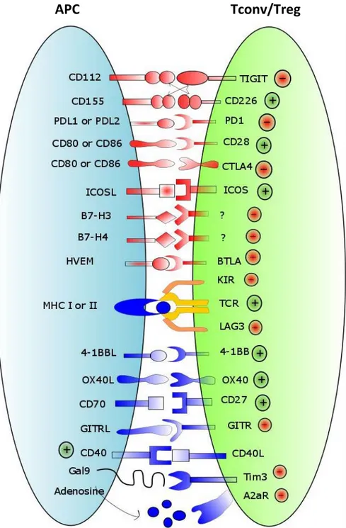
9 2.3.4.Cell to cell contact dependent suppression: Involvement of co-stimulatory and
co-inhibitory signals... 45
2.4. How many mechanisms do Tregs need? Treg plasticity ... 51
2.5. Infiltration, differentiation and activation of Tregs in tumor microenvironment 53 2.5.1.Infiltration of Tregs in tumor microenvironment ... 53
2.5.2.Expansion and activation of Tregs ... 55
2.5.3.Antigen specificity of Tregs in cancer ... 55
2.6. Tregs in cancer: ambiguity in prognostic importance ... 56
3. Tregs and immunotherapy: a blessing in disguise? ... 60
4. Lung cancer: a study model ... 66
4.1. Etiology and histology of the lung cancer ... 67
4.2. TNM classification and survival of patients ... 68
4.3. Treatment of lung cancer patients ... 71
4.4. Era of combined therapies: promising for NSCLC ... 72
B. Hypothesis and objectives ... 75
C. Results 80 References 106 Tregs in advanced stage chemotherapy treated lung cancer patients ... 141
D. Discussion ... 146
TLS in lung tumors: centers of the protective immune responses ... 146
Infiltration, activation of Tregs in cancer microenvironment ... 147
Anti-inflammatory role of Tregs in lung cancer ... 150
Tregs in TLS: Proponents or opponents?... 152
Expansion of the specific subsets of Tregs in TLS ... 156
Modulation of the Treg phenotype in the neoadjuvant chemotherapy treated lung cancer ... 157
10
E. Limitations of this study ... 161
F. Conclusion and perspectives ... 162
G. References ... 165
Publication bibliography ... 165
11
Index of figures
Figure 1: The hallmarks of cancer ---18 Figure 2: The cancer immunoediting concept ---22 Figure 3: Components of the tumor microenvironment (core of the tumor, invasive margin and the TLS) ---24 Figure 4: Proposed model describing the principle events of the TLS neogenesis ----28 Figure 5: Association of immune cell infiltrates with prognosis in various types of cancer ---34 Figure 6: Basic immunoregulatory mechanisms used by Tregs. ---41 Figure 7: Immune checkpoints in regulating T cell responses ---48 Figure 8: Molecules expressed by the Tregs used for the immunotherapy trials in cancer patients ---62 Figure 9:Age‐standardized lung cancer incidence rates by sex and world area. Source: GLOBOCAN 2008. ---66 Figure 10: Histological types of lung cancer. ---68 Figure 11: Characterization of Ti-BALT in NSCLC. ---76 Figure 12: Evaluation of DC-Lamp as a marker of Ti-BALT, and its prognostic value. ---77 Figure 13: Hypothesis about the role of Tregs in lung Tumor microenvironment. ----79 Figure 14: comparison of the Tregs in NAC treated and NAC untreated patients --- 143 Figure 15: Activation and immunosuppression profile of the Tumor Tregs in the NAC treated and untreated patients --- 144 Figure 16: Activation and immunosuppression profile of the Blood circulating Tregs in the NAC treated and untreated patients --- 145
12
Index of tables
Table 1: The comparison among the general characteristics of SLO and TLS ---26 Table 2: Prognostic value of TLS-associated biomarkers in naïve and vaccinated cancer patients (Dubois et al., submitted) ---30 Table 3: The regulatory T cell subsets and their suppressive mechanisms (Mougiakakos et al. 2010) ---38 Table 4: Main differences between nTreg and iTreg cells ---40 Table 5: Study of prognostic importance of Tregs in types of cancers ---60 Table 6: Co-stimulatory and co-inhibitory molecules, activating or blocking antibodies in anti-cancer treatment ---65 Table 7: TNM classification in human lung cancer (7th edition) ---69
13
Abstract
Tumor comprise complex niche of the immune and non-immune components. The complex interaction between the tumor cells with its environment turns into either eradication or the growth and metastasis of the tumors. In our team we have previously demonstrated the role of TLS (tertiary lymphoid structures) in lung tumors, in the generation of the protective anti-tumor responses. These TLS act as foci of the generation of the humoral, Th1 and CD8 cytotoxic T cell responses. High densities of the mature DC, B cells in TLS and CD8+ T cells are associated with the good clinical outcome in the lung cancer patients. Despite of the anti-tumor responses generated by the immune system tumors do develop via exploiting the regulatory mechanisms. And one of these mechanisms includes the infiltration of the Tregs (regulatory T cells) in the tumor microenvironment.
The aim of thesis was to study the putative role of Tregs in regulating the immune responses in lung cancer. This study strongly demonstrates the presence of FoxP3+ Tregs in the TLS as well as non-TLS areas of the lung tumors. Ti-Tregs (tumor infiltrating Tregs) mainly exhibit central memory and effector memory phenotype expressing vast repertoire of the activation and ICP (immune checkpoint) molecules. The gene expression and flow cytometry experiments showed that Tregs express the co-stimulatory and co-inhibitory markers constituting the ICP which are known to be involved in the immune suppression. Tregs expressed peculiar set of genes in comparison to the CD4+ conventional T cells. At the end I showed that high density of the Ti-Tregs either in TLS or in nonTLS areas is associated with the poor survival of the NSCLC patients. When combined with the density of TLS mature DC or TLS B cells or conventional T cells or CD8+ T cells, it was observed that a group of patients with the low DC, B cells and CD8+ T cells but high Tregs densities, had the worst clinical outcome. Combination of the TLS mature DC or TLS B cells or conventional T cells or CD8+ T cells densities with Tregs allowed to identify the NSCLC patients with highest risk of death.
Thus, it be concluded that the Tregs create the immunosuppressive environment in the lung tumors by acting in both TLS and nonTLS areas of the tumors and thus could be possible reason for the reduced survival of the lung cancer patients.
14
Abbreviations
APC Antigen presenting cell
BALT Bronchus-associated lymphoid tissue
Bcl-2 B-cell lymphoma 2
BMDC Bone marrow-derived cells
CTLA-4 Cytotoxic T-lymphocyte-associated protein 4
DC Dendritic cell
ER Estrogen receptor
GITR Glucocorticoid-induced TNFR family related gene
GC Germinal center
iBALT Induced BALT
IDO Indoleamine 2,3-dioxygenase
iTreg Induced regulatory T cell
LAG-3 Lymphocyte activation gene 3
Lti Lymphoid tissue inducer cell
mAB Monoclonal antibody
MALT Mucosa-associated lymphoid tissue
MDSC Myeloid-derived suppressor cell
MHC Major histocompatibility complex
NK Natural killer cell
NKT Natural killer T cell
NOTCH Neurogenic locus notch homolog protein
NSCLC Non-small-cell lung cancer
nTreg Naïve regulatory T cell
PD-1 Programmed death 1
SCLC Small-cell lung cancer
SLO Secondary lymphoid organ
TAA Tumor-associated antigen
TCM Central memory T cell
TCR T cell receptor
TEM Effector memory T cell
TFH Follicular helper T cell
Th1 T helper type 1cell
Th17 T helper type 17 cell
Th2 T helper type 2 cell
Th22 T helper type 22 cell
Th9 T helper type 9 cell
Ti-BALT Tumor-induced BALT
TIGIT T cell immunoreceptor with Ig and ITIM domains
Tim-3 T-cel limmunoglobulin domain and mucin domain 3
TLS Tertiary lymphoid structure
TNF Tumor necrosis factor
TNFR1 TNF receptor 1
TRAIL TNF-related apoptosis-inducing ligand
Treg Regulatory T cell
15 CD137/4-1BB Tumor necrosis factor receptor super family member
9 (TNFRSF9)
mAB Monoclonal antibody
GrA/GrB Granzyme A/B
16
Introduction
17
A. Introduction
Large amount of research is going on, to know, how the normal cells from body get transformed into the cancerous cell. Not only the oncologists but tumor immunologists are also participating to understand the microenvironment shaped around these tumor cells. It has been understood now that tumors modulate this microenvironment and escape the counteracting immune responses. Immune regulating factors (includes regulatory cells) also serve as a tool for this escape. In this study, we tried to decipher the immune regulation of the lung cancer microenvironment by regulatory T cells and especially their role in the tumor-induced TLS. Study of the phenotype of Tregs and their association with the prognostic importance in the lung may help to search new biomarkers for immunotherapy modulating Tregs in cancer patients.
1. Immune system and tumors: a complex discourse
1.1. Origin of the concept of tumor microenvironment
It all started with the discovery of tumor development as abnormal proliferation of cells which was summarized by reductionist’s as is a simple aggregate of malignant cell proliferation. This long prevailed concept later changed in year 2000 and it is now clearly established that tumor growth is accompanied by the formation of a complex niche that plays an important role in the progression of the malignancy (Hanahan, Weinberg 2000). With rapid advances in research about cancer revealed it as a disease with dynamic genomic changes. Complexities about this disease described in laboratories and clinical studies transformed into an underlying principle. These basic principles are termed as “hallmarks of cancer”.
In 2000, Hanahan and Weinberg suggested that malignant growth is accomplished through the different cancer cell genotypes and six essential alterations in cell physiology. These six <<Pillars>> of cancer growth are illustrated in the Fig. 1.
Each of these capabilities are acquired by the tumors to escape the recognition by the host cells (Hanahan, Weinberg 2000). Approximately all tumors may show in
18 common these six hallmarks. After 10 years of the first theory of “Hallmarks of cancer”, the two new pillars were added to this list that is <<deregulating cellular energetic and avoiding immune destruction>>(Hanahan, Weinberg 2011). The later one is particularly found to be important in tumor development because along with the transformed cells tumors also contain the stromal cells, matrix fibers, blood and lymph vessels and most importantly immune cells and interaction of these components leads to the development of the tumors.
Figure 1: The hallmarks of cancer
A. The primary six pillars of the cancer proposed in year 2000 (Hanahan, Weinberg 2000). B. The addition of the two emerging hallmarks and two enabling characteristics of cancer growth in year 2011 (Hanahan, Weinberg 2011).
A
19
1.2. Origin and concept of immune surveillance
The concept of relation between the inflammation and cancer is ancient. This was noted first time by Rudolf Virchow in 1863. He first proposed that the leucocytes in the neoplastic tissues are related with the inflammation and cancer (Balkwill, Mantovani 2001). In contrast, the concept of immune system to control the tumor progression was demonstrated later in 1909, when Paul Ehrlich postulated that cancer occurs spontaneously in vivo and that the immune system is able to both recognize and protect against it. In the late 1950s, Lewis Thomas introduced the theory of immunosurveillance, which was subsequently developed by Sir MacFarlane Burnet in 1964.
With the functional demonstration of mouse tumor-specific antigens supporting the ideas of all these scientists the cancer immunosurveillance hypothesis was put forth. It stated that protective thymus-dependent cells of the body constantly survey host tissues for transformed cells. Despite subsequent challenges to this hypothesis over the next several decades, new studies in the 1990svalidated the cancer immunosurveillance concept and expanded it to include the contributions of both innate and adaptive immunity (Smyth, Trapani 2001; Dunn et al. 2002; Dunn et al. 2004).
In 1990’s improved mouse models of immunodeficiency allowed the scientists to reassess the role of immunity in controlling the cancer. Along with lymphocytes, IFN-γ was also found to be promoting immunological rejection of the transplanted tumor cells. Experiments in mice lacking IFN-γ receptors or STAT-1 transcription factor which is required for the IFN receptor signaling or mice lacking adaptive immunity were susceptible for the spontaneous formation of the primary tumors. Soon other laboratories started reporting similar findings which supported the concept that immune system functions as an intrinsic tumor suppressor (Shankaran et al. 2001; Schreiber et al. 2011).
20
1.2.1.
Immunoediting: 3 `E’ concept
The immune system plays three primary roles in the prevention of tumors. First, it can protect the host from virus-induced tumors by eliminating or suppressing viral infection. This prevents the inflammatory environment which may be lead to tumor formation. Second, the immune system can specifically identify and eliminate tumor cells expressing tumor-specific antigens or stress molecules. The third process is tumor immune surveillance, where the immune system identifies cancerous and/or precancerous cells and eliminates them before they can cause harm (Swann, Smyth 2007). Despite this, tumors do develop in the presence of immune system, and therefore the updated concept of tumor immunoediting was discovered to explain the role of the immune system in tumor development.
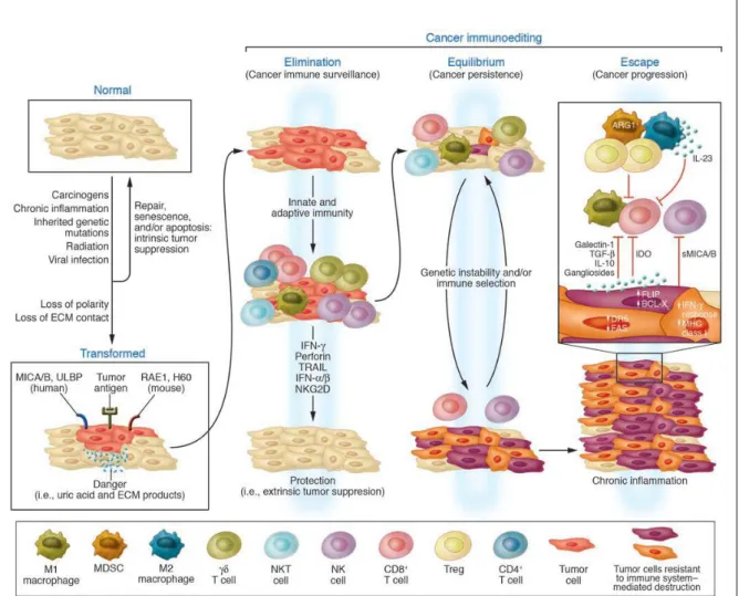
The discovery in 2001, that immune system controls not only the quantity, but also the quality (immunogenicity) of tumors gave rise to revision to the cancer immunosurveillance theory. The study in Schreiber’s laboratory revealed that the tumors growing in the mice that lacked an intact immune system were more immunogenic (unedited) whereas, the tumors from immunocompetant mice were less immunogenic (edited). This notion of, immune system protects against tumors and also shapes tumor immunogenicity is the basis of the immunoediting theory. Cancer immunoediting passed through the three different “E” phases: elimination, equilibrium, and escape (Schreiber et al. 2011) (Fig 2).
Elimination: in elimination phase immune system recognizes and destroys the
tumor. Initiation of the antitumor response occurs when the innate immune cells first receive alert signal due to the presence of the tumor cells. Pro-inflammatory molecules secreted by tumor cell itself act as danger signal for the innate immune cells. In the second step, innate immune response is amplified and more cells are recruited in the affected site due to the chemokine milieu produced locally. Tumor-infiltrating macrophages produce IL-12 which stimulates infiltration of NK cells. Chemokine production is amplified by positive feedback loop and results into the IFN-γ dependent killing of tumor. In the third step, DC comes into the scenario. Tumor antigens liberated by effects of innate immunity are engulfed by the
21 immature DC’s recruited at tumor site. Activated, antigen bearing DC’s migrated to the draining LN and induces the activation of the naïve tumor specific CD4+ Th1 cells. Th1 facilitate the development of the tumor specific CD8+ T cells via cross-presentation of the antigenic tumor peptides on DC MHC class I molecules. These tumor specific CD4+ and CD8+ T cells then home in the tumor site and participate in the killing of the antigen positive tumor cells.
Equilibrium: In this phase, the immune system and tumor cells that have survived
the elimination phase enter into equilibrium; here lymphocytes and IFN-γ exert potent and rigorous pressure on the tumor cells that is enough to limit but not to fully quench genetically unstable and mutating tumor cells. The equilibrium phase is probably the longest phase among the three, and may occur over a period of many years in human (Dunn et al. 2004).
Escape: In the escape phase, tumor cell variants selected in the equilibrium phase
can grow in an immunologically intact environment. This occurs when genetic and epigenetic changes in the tumor cells confer the resistance to the immune detection allowing the tumors to expand and become clinically detectable. Since, innate and adaptive immunity work hand in hand to eliminate the tumor, tumor circumvents either of the two or both arms of immunity in order to achieve the growth. It may employ the immune-evasive strategies to elude the responses against it.
It is now recognized that tumors can impede the antitumor responses through production of the immunosuppressive cytokines (such as TGF-β and IL-10) or via mechanisms involving immunoregulatory cells (i.e. Tregs and MDSC). Immune escape also occurs through loss of tumor antigen expression, loss of MHC components, shredding NKG2D ligands and development of IFN-γ insensitivity. Discovery of the escape mechanisms by tumors gave a better knowledge of the tumor immunology to the scientists and initiated a research in several different aspects of the tumor immunology.
22
Figure 2: The cancer immunoediting concept
The three ‘E’ phases of the immunosurveillance theory is depicted in this figure (Swann, Smyth 2007).
1.3. Tumor microenvironment: a complex interactome
In physiological conditions, tissues bear the large number of cells which work in harmony to perform the normal functioning of the body. Due to mutations in proto-oncogenes or tumor suppressor genes or genes related to the growth and survival of the cells, some of these normal cells lose the constraint and become cancerous cells. Although cancers have altered identity it does not loses interaction with surrounding environment. These interactions may lead to the infiltration of the different immune cells through chemokine and cytokine cocktail. Complex structure composed of the tumor cells along with the stromal components, immune cells, vascular and
23 lymphatic vessels results into the interacting tumor microenvironment. A continuous seesaw game between tumor and immune compartment is a characteristic of this microenvironment and it may go through changes throughout the 3 “E” phases of the immunoediting. In some cases, the defense signals exerted by the immune system is circumvented by tumors to exploit the surrounding cells, proliferate and finally invade to metastasize (Joyce, Pollard 2009).
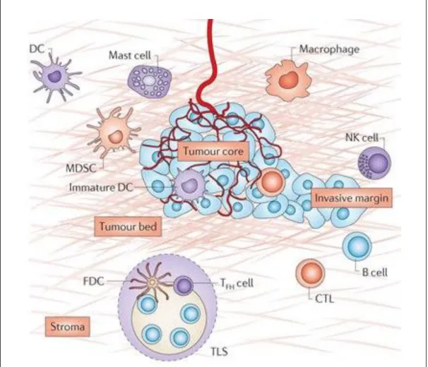
In general, tumor microenvironment consists of immune cells with different functional properties (Fig.3). It consists of the antigen-presenting cells (B cells, DCs, and macrophages), T cell subsets, NK cells, neutrophils, and mast cells. Even within individual cell types, different subsets may have adverse functions. For example different subsets of CD4+ T cells, NK cells and macrophages may have either tumor suppressing or tumor promoting properties. From patient to patient and from tumor to tumor, heterogeneity can be observed with respect to the numbers, localization of the tumor-infiltrating immune cells. These immune cells can be located in mainly in the invasive margin compared to the center of the tumor, either in the stroma among with TLS or in the tumor nests. Analysis of this immune contexture allows the determination of the beneficial or deleterious effects on the cancer patients.
24
Figure 3: Components of the tumor microenvironment (core of the tumor, invasive margin and the TLS)
The tumor microenvironment consists of numerous cells including the tumor cells, endothelial cells of blood and lymphatic vessels, stromal fibroblasts, bone marrow-derived cells, tumor associated macrophages (TAM), and myeloid-derived suppressor cells (MDSC) (Fridman et al. 2012).
1.3.1.
Characteristics of contexture
Immunohistochemistry, gene expression, and the clinical techniques have helped to study the presence of the various cell substrates infiltrating the different areas of tumors. Lymphocytes are not randomly distributed but are specifically localized in tumor microenvironment. Macrophages, granulocytes including the mast cells and MDSC are mainly found in the tumor beds and at the invasive margin. NK cells are mostly found in the stroma of the tumor (Platonova et al. 2011). Most B cells are organized in TLS which are mainly located in the invasive margin of the tumors
25 (Germain et al. 2015). Cytotoxic and memory CD8+ T cells infiltrate in the stroma and sometimes in the tumor beds (Goc et al. 2014a). TLS are highly organized lymphoid follicles consisting of the B cell area and T cell area. B cells area consists of the several subsets of the B cells, Follicular DC and Follicular helper T cells whereas T cell area is composed of mature DC and T cells (Dieu-Nosjean et al. 2008). Immune cell infiltration is guided by various events in the tumor microenvironment. The chemokines, adhesion molecules and cytokines are important architects of orientation of immune cells and thus are integral part of immune contexture. Complex crosstalk among the receptors and chemokines expressed by the cells is responsible for the build of tumor microenvironment. If this balance is lost, it will result into the loss of co-ordination and inefficiency of the immune system in controlling the tumor.
1.3.2.
Cancer associated TLS
1.3.2.1.
General characteristics of TLS
TLS are the ectopic lymphoid structures which can be defined as the highly organized aggregates of T cells and B cells in the form of distinct zones adjacent to the HEV.PNAD+ HEV help in the extravasation of the CD62L+ immune cells from blood. The B cell area of the TLS acts as the active germinal centers with antibody producing plasma cells and memory B cells whereas the T cell area of the TLS shows the presence of the mature DC and stromal cells in T cell zone which can secrete the chemokines CCL19 and CCL21 involved in the attraction of the CCR7+ mature DC and T cells to the TLS. These structures are considered as transient structures which may develop in chronic infections, allograft rejection, autoimmune diseases and chronic inflammation like cancers and probably disappear after the infection or inflammatory conditions are resolved. TLS show considerable morphological, cellular, chemokine and vasculature resemblance to secondary lymphoid organs particularly to lymph nodes. It has been shown that same processes and molecules governing LN development are also basis of the TLS formation (Kratz et al. 1996). The following table shows the characteristics and comparison between SLO and TLS.
26
SLO TLS
Localization Spleen, lymph nodes and MALT Non lymphoid tissues like lung, pancreas, breast, thyroid, salivary glands etc.
Structure Encapsulated (Spleen and lymph node) non encapsulated (MALT)
non encapsulated
Ontogeny Detected Pre or post-nascent Post-nascent
Development Programmed during embryogenesis
Development during inflammatory conditions
Plasticity Constitutive presence and remodeling during inflammatory
conditions
Uniquely developed during chronic inflammation and may regress after
termination of the same.
Table 1: The comparison among the general characteristics of SLO and TLS
1.3.2.2.
Formation of TLS
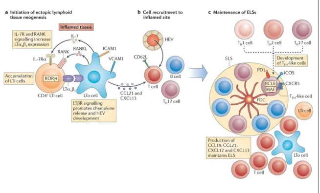
Although TLS resemble the SLO, it is not identical to SLO. Its organization resembles the loose non encapsulated organization of MALT. In TLS, cells like T cells, B cells and DCs are organized in various zones alongside the HEVs. Three critical events promote the TLS development: inflammatory cytokine production like TNF; lymphoid chemokine production by stromal cells, and HEV development (Drayton et al. 2006). The cascade of events that occurs prenatally in SLO development shows similarities to the cellular interactions that are involved in the formation of the TLS. Paracrine interactions between the mesenchymal cells and hematopoietic cells are thought to be the basis of the TLS development. LTβR signaling on the endothelial cells induces the lymph node and the HEV formation (Onder et al. 2013). The interaction between LTi (lymphoid tissue inducer) cells and the LTo (lymphoid tissue organizer) cells through lymphotoxin signaling has found to be crucial in the initiation of the TLS formation. The mouse studies show that LTi cells upregulate the Id2 and RORγt which induces the lymphoid neogenesis (Cherrier et al. 2012). The fibroblasts producing the CXCL13 are thought to be attracting the LTi cells at the site of the TLS formation. Chemokines and cytokines, adhesion molecules like VCAM1, ICAM1, MADCAM1 produced locally by the LTo cells after the interaction with the LTi cells induce the TLS formation in the affected areas (in case of the inflammation) (Neyt et al. 2012). In pancreatic mouse model, it is found that LTα expressed in inflammatory
27 lesions promote expression of CXCL13 and CCL21, which appear to organize the B and T cell zones (Kratz et al. 1996; Hjelmström et al. 2000). In this model, it was observed that the expression of chemokines was dependent on the TNFR1 and not on the LTβR signaling. In other study, in case of LTαβ signaling it was observed that, LIGHT also signals through the LTβR and plays important role in the expression of homeostatic chemokines (Gommerman, Browning 2003). B cells, T cells, NK and DCs are found to be the important source of the LTβ.
The role of the LTi cells in the TLS formation in humans is poorly discovered. It is thought that RORC positive ILCs especially ILC3 (group 3 innate lymphoid cells) are the LTi cells. These cells induce the chemokines and adhesion molecules by stromal cells responsible for the recruitment of the immune cells in the TLS (Lochner et al. 2011; Noort et al. 2015).Mesenchymal cells also express the additional chemokines like CCL19, CCL21 and CXCL13 which attract the different lymphocytes into the T and B-cell zones of TLS, respectively. HEVS express the CCL21 which carries the circulating lymphocytes in and out of the T cell zone of the TLS (Ohmatsu et al. 2007). NK cells, naïve and memory T cells and activated DC express the CCR7 which signals through CCL19 and CCL21 for recruitment, activation and functioning through the TLS. Stromal cells, DCs and macrophages can produce the CXCL13 on lipopolysaccharide stimulations which can attract the B cells to the TLS through CXCR5 signaling (Neyt et al. 2012; van de Pavert, Serge A, Mebius 2010).CD4+ CXCR5+ Tfh cells expressing the ICOS, PD-1 and BCL-6 and producing IL-21 are considered important in the germinal center formation. Tfh cells are considered important in the plasma cell and memory B cell differentiation and thus important for the functional TLS formation (Chevalier et al. 2011).
28
Figure 4: Proposed model describing the principle events of the TLS neogenesis
Following events take place during the TLS formation. A. IL-17 producing CD4+ Lti (lymphoid tissue inducer) cells and lymphoid tissue organizer LTo cells are thought to be involved in the process. B. The inflammatory tissues give the chemokines signals which recruit the T cells, B cells via CXCL13 and CCL21. The resident stromal cells allow the organization of the aggregates of the T and B cells via IL-7 and LTα2β2 signaling. C. The continuous antigen presentation by the FDC and B cells and the T follicular helper cells are required for the maintenance of the TLS (Pitzalis et al. 2014).
1.3.3.
TLS in anti-tumor immune response
TLS were first discovered and are extensively discussed in the infection and autoimmunity conditions. But the presence of these structures in tumors has been recently found. Reminiscent to the TLS in the autoimmunity, TLS in tumors show the well-organized follicles of B and T cells. Since along with the inflammation, tumors bear immunosuppressive environment due to the presence of the macrophages and regulatory T cells, it is thought that, this induces the formation of these follicles in invasive margins of some solid tumors.
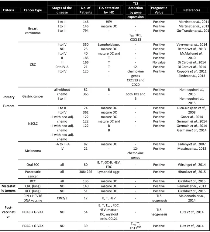
There are several studies demonstrating the importance of the TLS in different types of primary as well as metastases for example, breast cancer, colorectal cancer, lung
29 cancer, lung metastasis of the renal and colorectal cancer, gastric cancer which shows that occurrence of TLS in cancer is broad phenomenon (Table 2). Along with the primary tumors, vaccination strategies also show the lymphoid neogenesis in cancer patients. In the HPV-16 vaccinated cervical cancer patients the functional TLS (containing the ki-67+ proliferating cells, CD20+ B cells and CD3+ T cells) were formed with the increased Th1 and CD8+ T cell responses (Maldonado et al. 2014). Post GVAX treatment, formation of the lymphoid aggregates and increase in the Th17 signatures with decrease in Tregs associated signature results into the better survival in pancreatic cancers(Lutz et al. 2014).
In case of tumor microenvironment, TLS formation has been found to be influenced by presence of Tregs. Tregs depleted in the mouse bearing MCA induced tumor lead to the increased development of the HEV and T cell infiltration and tumor destruction (Hindley et al. 2012). This effect was also observed in breast tumors where, it was observed that although the HEV can be developed in presence of Tregs, their density is influenced by the ratio of the Tregs to T cells infiltrating the tumors (Martinet et al. 2013). But so far, it is scarcely demonstrated that, the infiltration of Tregs in lymphoid aggregates is associated with worst clinical outcome of the cancer patients.
Although presence of TLS in tumors is associated with the large infiltration of the immune cells TLS among the tumors and patients to patient heterogeneous. Also, immune cells may found not in well-developed TLS, but in the less organized lymphoid aggregates like in the breast cancer, metastatic melanoma (Gobert et al. 2009; Cipponi et al. 2012).Until now, detailed mechanisms of the TLS formation and their maintenance have not completely discovered.
In summary, TLS formation in the tumor microenvironment represents the generation of the protective anti-tumor immune responses. TLS are involved in the infiltration, local education of the T cells and creation of the adaptive as well as humoral responses. This privileged functioning of TLS in the immunosuppressive microenvironment generated by tumors thus help in the better survival of the cancer patients.
30
Criteria Cancer type Stages of the disease No. of Patients TLS detection by IHC TLS detection by gene expression Prognostic Value References Primary Tumors Breast carcinoma I to III I to III I to III 146 146 794 HEV mature DC - - - - TFH, Th1, CXCL13 Positive Positive Positive Martinet et al., 2011 Martinet et al., 2013 Gu-Trantienet al., 2013 CRC I to IV ND I to IV II III 0 to IV-A I to IV 350 25 40 185 166 21 125 Lymphoidaggr. mature DC mature DC and T T T - - - - - - - 12-chemokine genes CXCL13 and CD20 Positive Positive Positive Positive No value Positive Positive Vayrynenet al., 2014 Remarket al., 2013 McMullenet al., 2010 Di Caro et al., 2014 Di Caro et al., 2014 Coppola et al., 2011 Bindeaet al., 2013 Gastric cancer all without chemo I to III 82 365 B - - both Th1 and B Positive Positive Hennequinet al., 2015 Hennequinet al., 2015 NSCLC I to II I to IV III with neo-adj.
chemo III with neo-adj.
chemo III with neo-adj.
chemo 74 362 122 122 122 mature DC mature DC mature DC mature DC and B B - - - - - Positive Positive Positive Positive Positive Dieu-Nosjean et al., 2008 Gocet al., 2014 Germain et al., 2014 Germain et al., 2014 Germainet al., 2014 Melanoma I-A to III-A IV 82 21 mature DC - - 12-chemokine genes Positive Positive Ladanyiet al., 2007 Messinaet al., 2012
Oral SCC all 80 B, T, GC-B, HEV,
FDC - Positive Wirsinget al., 2014 Pancreatic
cancer
all 308+226 Lymphoid aggr. - Positive Hiraokaet al., 2015 RCC all 135 mature DC - Positive Giraldoet al., 2015
Metastat ic tumors
CRC (lung) ND 140 mature DC - Positive Remark et al., 2013 RCC (lung) ND 51 mature DC - Positive Giraldoet al., 2015
Post- Vaccinati
on
CIN + HPV16
DNA vaccine CIN2/3 12 B, T, HEV -
TLS neogenesis Maldonado et al., 2014 PDAC + G-VAX ND 54 B, T, Treg,, FDC, HEV, mature DC, myeloid cells, CCL21 - TLS
neogenesis Lutz et al., 2014
PDAC + G-VAX ND 39 - Treg
Low,
Th17High Positive Lutz et al., 2014
Table 2: Prognostic value of TLS-associated biomarkers in naïve and vaccinated cancer patients (Dubois et al., submitted)
31
1.4. Infiltration of immune cells in solid tumor: a strong
prognostic marker
Distribution of the immune cells in different areas of the tumors suggests that these cells may have different role in tumor control. Correlation between the immune infiltration and clinical outcome was investigated in the large number of cases in literature. Although high infiltration of the immune cells in good for the patient’s survival, the important fact is that, different immune subsets have different impact on the prognosis of patients. It is observed that, high density of CD3+ T cells, CD8+ cytotoxic T cells and CD45RO+ memory T cells are associated with longer disease-free and overall survival (Galon et al. 2006; Goc et al. 2014b). In NSCLC patients, it has been observed that high density of the CD8+ T cells with low mature DC density is associated with high death rate compared to the high density of both CD8 and mature DC. It is also observed that high mature DC tumors bear the beneficial antitumor Th1 and cytotoxic T cell contexture than the low mature DC tumors. Mature DCs are required to license the positive prognostic value to CD8+ T cells (Goc et al. 2014b). Exception for this, is Hodgkin’s lymphoma (Scott et al. 2013), ocular melanoma and renal carcinoma (Osamu et al. 2001), where Th1 cells and CD8+ T cells are associated with poor clinical outcome respectively.
In contrast to CD8+ T cell population, effect of CD4+ T cell population on clinical outcome has contradictory results. Due to the presence of different subtypes among CD4+ T cells with opposite immune function, impact of total CD4+ T cell population is hard to describe. The striking example of this is Th2 cells, Th17 cells and Tregs. High Treg infiltrate has poor clinical outcome in ovarian (Curiel et al. 2004), whereas increase in density of Tregs is associated with good clinical outcome head and neck cancer (Badoual et al. 2006), follicular lymphoma(Carreras et al. 2006) and colorectal cancer (Salama et al. 2009b). Surprisingly, there are also cases where Tregs are not associated with clinical outcome (Grabenbauer et al. 2006; Heimberger et al. 2008; Jacobs, Joannes F M et al. 2010). The discrepancies about the prognostic importance and different theories about it are discussed in detail in the chapter about the Tregs.
32 Th1 cells are associated with good clinical outcome in majority of the cancer types including breast, ovarian, colorectal ad pancreatic cancers, renal and lung carcinomas (Fridman et al. 2012). Intra-tumoral number of DCs have often but not always associated with the good clinical outcome. This is especially associated with the accumulation of T cells in the esophageal cancer and melanoma. In breast and lung cancers, the DC infiltration is of independent prognostic relevance (Galluzzi et al. 2012). B cells act as antigen-presenting cells (Bergwelt-Baildon 2002; Zirakzadeh et al. 2013; Marits et al. 2013)and therefore may be important for inducing CD4+ T cell dependent CD8+ memory T cells that help to control tumor invasion. In lung cancer, the high density of TLS-B cells correlated with early-stage as well as
advanced-stage chemotherapy patients in Non-small-cell lung cancer
(NSCLC)(Germain et al. 2014a). The density of B cells is found to be associated with good clinical outcome in breast cancer and epithelial ovarian cancer (Coronella et al. 2001; Milne et al. 2009).Immunoglobulin K chain expression and the CD138+ plasma cells found to be associated with the good clinical outcome of the NSCLC patients(Lohr et al. 2013).
1.4.1. Prognostic importance of the TLS in cancer
The development of the local antitumor response is found to be associated with the presence of TLS. In primary tumors, the presence of the mature DC in the T cell zone is the bona fide feature of these TLS and is found to be a beneficial factor influencing survival of the patients with lung, breast, colorectal carcinoma, colorectal cancer lung metastases and melanoma (Dieu-Nosjean et al. 2008; Ladányi et al. 2007; Treilleux et al. 2004; Remark et al. 2013). An exception to this is renal cell carcinoma where the mature DC is not associated with well-organized lymphoid follicles and the high density of DC outside the TLS is associated with poor survival of patients (Giraldo et al. 2015). Mature DC in these TLS may involve in the education of the T cells, boosting the Th1 and cytotoxic T cell response. Center of these TLS are ruled by the B cells, follicular DC and TFH. B cells in TLS are involved in generation of local
humoral response to the tumor antigens (Reuschenbach et al. 2009; Germain et al. 2014a, Germain et al. 2015),But their exact role in the tumor-induced TLS and the
33 impact on the clinical outcome is debatable. In NSCLC, the plasma cells in TLS has found to be secreting the high levels of the IgG and IgA against the tumor antigens such as LAGE-1, p53 and NY-ESO-1 and the high density of the B cells is associated with the better survival of the early stage and late stage NSCLC patients(Germain et al. 2014a).. In breast tumor-associated TLS, it has been found that CXCR5+ CXCL13 producing follicular helper T cells are involved in the formation and stability of the lymphoid follicles are associated with the better survival of the patients (Gu-Trantien et al. 2013a).
It has been observed that Tregs can infiltrate in the lymphoid aggregates. In colonic mucosa, FoxP3+ Tregs in lymphoid follicles is associated with poor clinical outcome. The size of germinal center and number of Tregs observed to be inversely correlated (Salama et al. 2012). In primary breast tumors, the CCL17/CCR4 mediated recruitment of Tregs in lymphoid aggregates is associated with poor clinical outcome whereas, presence of Tregs in the tumor beds is not well associated with the prognosis of the patients (Gobert et al. 2009). Although the infiltration of Tregs in tumor-induced lymphoid aggregates is demonstrated in few examples, the exact mechanisms of the regulation of TLS formation by Tregs in not completely clear.
34
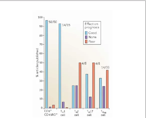
Figure 5: Association of immune cell infiltrates with prognosis in various types of cancer
The analysis of 124 published articles studying the impact of cytotoxic T cells, memory T cells, Tregs and THcell subpopulations with regard to prognosis of cancer patients (20
different cancer types were analyzed) is represented. ‘Good ‘means that cells associated with good prognosis. ‘None’ means that there was no correlation and ‘poor’ means that cells are associated with poor prognosis (Fridman et al. 2012).
In summary, Presence of different immune subsets and clinical outcome in cancer patients is context dependent. Although, cancer is an unobvious outgrowth of body’s own transformed cells, it has been thought to play an important role in shaping its environment. The translation of this large study on the correlation between immune infiltrate and clinical outcome has led to the concept of the IMMUNOSCORE (Fridman et al. 2013). Since, a high density Th1 and CD8+ T cells correlate with positive prognosis thus the concept of immune score developed considering two markers among CD3, CD8 and CD45RO in two regions (tumor core and invasive margin), grading from 0-4 (Galon et al. 2006; Pagès et al. 2010). This IMMUNOSCORE appears to be strongest tool to prognosticate in colon cancer patients and now is
35 being studied in other cancer types worldwide (Galon et al. 2012). Defining the group of high risk patients with help of the immune infiltrate status will be eventually strong tool for treatment of these patients.
2. Tregs: Key Regulators of anti-tumor immune
response
2.1. Discovery and features of regulatory T cells
For several decades, the concept of the cells which can suppress the immune responses has been debated. At present time, regulatory T cells are integral part of the immunology but their discovery in 1970’s, then fall in between time period and renaissance 20 years after has been an amusement ride.
Gershon and Kondo, in 1970, first time showed that, lymphocytes can suppress antigen specific T cell responses. They showed that, induction of tolerance and immunity in thymus dependent bone marrow derived cell population seems to require the co-operation of thymus derived cells (Gershon R.K. and Kondo K. 1970). Also, they observed that transfer of antigen encountered T cells to naïve mice can lead to antigen specific tolerance by arresting activity of T cells (Gershon R.K. and Kondo K. 1971). These observations showed that immune system not only eradicated the pathogens but simultaneously prevents the autoimmune conditions through the “suppressor cells”.
Despite great significance of this discovery, the research in this area was paused for the next 20 years when immunologists failed to define this cell population equivocally. Inability to find out markers for distinguishing suppressor T cells from other T cells, ambiguity in the molecular mechanisms of suppression and difficulty in designing antigen specific T cell clones suitable for cellular and molecular analyses were the obstacles in the study of this cell population.
36 In 1995, Sakaguchi and colleagues initiated the renaissance of these “suppressive cells” (Sakaguchi et al. 1995). They showed that when CD4+ cells from BALB/c nu/+ mice LNs and spleen depleted of CD25+ cells by adding mAB and inoculated into athymic nude (nu/nu) mice (BALB/c), all recipients spontaneously developed autoimmune diseases. Reconstitution of CD4+CD25+ cells maintained the self-tolerance by down regulating immune response to self and non-self-antigens in an antigen nonspecific manner. Also, the CD25 molecule was first promising candidate in phenotypic definition of suppressive cells that were further named as thymus derived naturally occurring regulatory T cells (nTregs). Approximately 10% of peripheral CD4+ cells and less than 1% CD8+ T cells in normal unimmunized adult mice expressed the IL-2 receptor α-chain (CD25) molecule (Sakaguchi et al. 1995). In 2003, Forkhead Box P3 (FoxP3) was identified as unique marker for Tregs as it was predominantly expressed within CD25+ CD4+ T cells (Sakaguchi 2003; Fontenot et al. 2003a). The majority of the CD4+FoxP3+ T cells were found to be CD25high (Roncador et al. 2005).Natural Tregs express high affinity hetero-trimeric receptor for IL-2 composed of CD25, CD122 and CD132 chains.
Mutation in the FoxP3 gene in the CTLA-4 null mice displayed the multi-organ disease and lack of conventional CD4+CD25+ Treg cells (Schubert et al. 2001). Inhuman, CD4+ CD25+ Treg cells express FoxP3 but TCR stimulated CD4+CD25- human T cells also express FoxP3 and acquire Treg function (Walker et al. 2003). FoxP3 allele deletion, cells lose their suppressive function and acquire the ability to produce the large amount of IL-2 and Th1 pro-inflammatory cytokines. Thus, it was observed that, FoxP3 acts as a Treg lineage specification factor and mediator of the genetic mechanism of tolerance. Irrespective of CD25 expression, FoxP3 correlates to the suppressor activity (Fontenot et al. 2003b; Fontenot et al. 2005b; Fontenot, Rudensky 2005). Continuous FoxP3 expression is essential for maintenance of the developmentally established suppressive program in mature Tregs in the periphery (Williams, Rudensky 2007).
Tregs rely on the expression of the FoxP3 and availability of IL-2 for transcriptional program and functionality. By modifying signaling and cell surface molecules, and
37 also by repressing cyclic PDE (cyclic phosphodiesterase), FoxP3 maintain Treg lineage stability (Gavin et al. 2007). When FoxP3 gene attenuated in Tregs in mice, showed less FoxP3 expression and autoimmune syndrome similar to that of Scurfy mice (Wan, Flavell 2007) which suggests the importance of the FoxP3 in maintaining the Tregs stability. In mice expressing mutant FoxP3, it was observed that EGFP+CD4+ T cells lacked regulatory function and the mice developed autoimmune disease (Lin et al. 2007).
Although FoxP3 is presently considered as a reliable marker for nTregs major concerns arose when it was found that FoxP3 is expressed in low levels on conventional CD4+ and CD8+ T cells upon activation. Eventually, various co-stimulatory molecules expressed on Tregs like CTLA-4 and GITR were discovered as Treg markers (Roncador et al. 2005; Ermann, Fathman 2003). Furthermore, two studies demonstrated that expression of IL-7R α-chain (CD127) is useful marker in discriminating the Tconv and nTregs. Majority Tregs found to be negative or weekly positive for CD127 whereas conventional T cells were found to be positive for CD127 upon activation (Liu et al. 2006).All these discoveries speed up the research in Treg biology and it eventually became one of the glamorous fields of study in immunology (Collison et al. 2007).
38
2.2. Regulatory T cell subsets
Although Tregs could be integrated into overall T cell population with suppressive properties, there are increasing evidences of the different subsets of this cell type with distinct development, phenotype and function. These are summarized in the Table 3.
Cell type Origin Phenotype Suppressive mechanism References
Naturally occurring Tregs CD4+
nTregs
Thymus CD4+CD25+FoxP3+CD127-/low
CTLA-4+ LAG-3+ GITR+
Contact dependent, cytotoxicity mediated, IL-10, TGF-β (Fehérvari, Sakaguchi 2004) CD8+ nTregs
Thymus CD8+CD25+FoxP3+CTLA-4+
CD122+
Contact dependent Fontenot et al.
2005 Adaptive/induced Tregs
CD4+nTreg like
Periphery CD4+CD25+FoxP3+ CTLA-4+
GITR+
Contact dependent (requires IL-2 and TGF-β)
(Apostolou, Boehmer 2004)
Tr1 Periphery CD4+ CD25-/low FoxP3-/low IL-10 (Groux et al.
1997)
Th3 Periphery CD4+CD25+FoxP3+ IL-10, TGF-β (Chen et al.
1994) CD8+ iTregs
CD8+ iTreg Periphery CD8+CD25+FoxP3+ IL-10, TGF-β (Chaput et al.
2009)
CD8+ iTregs Periphery CD8+CD25+ CD28-
FoxP3+CTLA-4+ GITR+
Contact dependent, IL-10, ILT3, ILT4
(Cortesini et al. 2001)
Table 3: The regulatory T cell subsets and their suppressive mechanisms (Mougiakakos et al. 2010)
Most CD4+ nTregs produced by the normal thymus constitutively express FoxP3and CD25 which represent functionally mature population responsible maintenance of immune self-tolerance and homeostasis by suppressing excessive immune responses harmful for host (Ohkura et al. 2013). Expression of CTLA-4 and GITR is also important hallmark of the nTreg development (Wu et al. 2006; Bettini, Vignali, Dario A A 2009). Many co-stimulatory signals have been implicated in the development and lineage commitment of nTregs. This includes: CD28 ligation by CD80/CD86, IL-2R, thymic stromal-derived lymphopoietin receptor,CD154, GITR, and STAT5 signaling (Salomon et al. 2000; Burchill et al. 2006; Spence, Green 2008). Initial reports have demonstrated the expression of the transcription factor Helios (Ikaros family ) limited to the thymic Tregs in mice and humans (Thornton et al. 2010). But further it was found that in humans natural Tregs consists of both the Helios + and –
39 Tregs (Himmel et al. 2013). There was no difference found in the suppressive activity and the expression of the CD39 and CTLA-4 between the Helios+ and – Tregs (Himmel et al. 2013). Although, there is a debate regarding the Helios as a marker for the thymic Tregs vs induced Tregs it has been observed that Tregs infiltrating the tumors and in periphery express high levels of the Helios (Scurr et al. 2014). Also, it is observed that in mice majority of the Tregs in the spleen and lymph node of the mice ,expressing Helios transcription factor also express the Neuropilin 1 and thus Neuropilin 1 can also be considered as the specific marker for the thymic Tregs (Shevach, Thornton 2014).
IL-2 is critically important cytokine for nTreg generation and normal activity in vivo, but nTregs itself do not produce IL-2 upon TCR ligation. In addition to IL-2R signaling, ligation of transforming growth (TGF-β),IL-4, IL-7, and IL-15 also appears to be involved in development, suppressive activity, and maintainace of nTregs (Bettini, Vignali, Dario A A 2009). The anti-apoptotic and pro-apoptotic molecules Bcl-2 and Bim also appear to have a hand in Treg development (Pandiyan, Lenardo 2008). Natural Tregs suppress activation and expansion of cells from adaptive as well as
innate immunity affecting cellular and humoral immune responses;
CD4+CD25+FoxP3+ nTregs hamper effector and memory CD4+ and CD8+ compartments, NK and NKT cells, DCs with respect to their activation, proliferation and function (Ghiringhelli 2005). Furthermore, proliferation, immunoglobulin (Ig) production and Ig class switch of B cells can be suppressed by nTregs (Iikuni et al. 2009).
While nTregs play a critical role in regulating self-tolerance, iTreg are responsible for regulating the immune response to wide variety of microbial and tissue antigens. They develop in SLO from naïve T cells, giving immune system an environmental adaptability. Antigenic stimulation insufficient for the generation of effector T cells is considered a prerequisite for iTreg development after TCR triggering of naïve T cells. The circumstances under which iTregs develop are wide ranging and may include presence of certain cytokines mostly, high levels of IL-2, IL-10 or TGF-β, low dose of antigens and APC exhibiting alterations in maturation and function. The local microenvironment is stimuli to the generation of iTregs (Curotto de Lafaille, Maria A,
40 Lafaille 2009). The iTreg cell appear in the mesenteric LNs during induction of oral tolerance (Mucida et al. 2005), in the lamina propria of the gut in response to microbiota and food antigens. They are also generated in chronic inflammation (Curotto de Lafaille, Maria A et al. 2008), tumor and transplanted tissues (Cobbold et al. 2004). Tumors can directly stimulate iTregs through several factors like CD70, Cox-2, IDO, IL-10, and TGF-β (Bergmann et al. 2007; Curti et al. 2007; Li et al. 2007).
Characteristics nTreg iTreg
Tissue Thymus GALT, spleen, LN, inflamed
tissue Co-stimulatory requirement CD28 CTLA-4 Cytokine requirement
TGF-β, IL-2, IL-15 TGF-β, IL-2
Specificity Self-antigens Allergens, commensal
microbiota, neoantigens (tumor), alloantigens
Table 4: Main differences between nTreg and iTreg cells
Although, CD4+ Tregs have been a main focus of Treg research, CD8+ Tregs are also increasingly emerging as a crucial component of immune regulation. Besides initial difficulties in identifying CD8+ Tregs, these types of cells were outshone by the discovery of CD4+CD25+ Tregs. This discovery of CD4+ Tregs by Sakaguchi et al. kickoff the intense research in this domain and thus CD8+ Tregs remained a little ignored. The number of CD8+ Tregs are relatively small, <1% in peripheral circulation, though much higher in intestine. A number of markers of CD8+ Tregs have been studied including CD28low, CD122+ and CD8αα expression compared with the normal CD8αβ (Smith, Trevor R F, Kumar 2008). Similar to CD4+ Tregs, CD8+ Tregs may develop in thymus as well as in peripheral tissue. CD8+CD25+FoxP3+CTLA-4+ nTregs have been identified in several studies in rodents and humans and which may act in cell to cell contact dependent manner (Cosmi 2003). CD8+ Tregs are also reported in the cancer patients. In prostate cancer patients, CD8+ Tregs were described to be CD25+ CD122+ FoxP3+ and partly GITR+. Their activity was found to be IL-10 and TGF-β dependent (Kiniwa et al. 2007). CD8+ CD25+ FoxP3+ TGF-β expressing Tregs are observed in colorectal cancer and these cells demonstrated the immunosuppressive function in vitro (Chaput et al. 2009).
41
2.3. Regulatory mechanisms exerted by Tregs
Either nTreg or iTreg, they are critical in maintaining the steady state condition in normal physiological situation to avoid the autoimmunity. Understanding Treg function is to determine how they suppress other lymphocytes at the molecular level. These suppressive mechanisms act together depending on the pathogenic consequences and type of immune response. Based on functional aspect, the various mechanisms used by Treg cells can be grouped into four basic “modes of action”: 1. Suppression by inhibitory cytokines
2. Suppression by cytolysis
3. Suppression by metabolic disruption
4. Suppression by contact dependent mechanisms
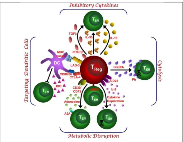
Figure 6: Basic immunoregulatory mechanisms used by Tregs.
Depiction of the various regulatory T (Treg)-cell mechanisms centered around four basic modes of action (Vignali, Dario A A et al. 2008).
42
2.3.1.
Inhibitory cytokines
Inhibitory cytokines such as IL-10 and TGF-β have considerable attention as mediators of Treg cell induced suppression. IL-10 suppresses T cell proliferation and cytokine production and maintains peripheral tolerance to allergens, auto-antigens, transplantation and tumor antigens. It can suppress proliferation of Th1 and Th2 cells (Groux et al. 1997; Cottrez et al. 2000).It is not only produced by Tr1 cells but also by Th1 and Th2 cells (Cottrez et al. 2000). It acts on DC, monocytes and macrophages by inhibiting production of pro-inflammatory cytokines such asIL-1α, IL-1β, IL6, IL-12, IL18, GM-CSF, TNF, and IL-12 and blocking cell maturation and up regulation of co-stimulatory molecules (Trinchieri et al. 1993).
On one hand, the capacity of IL-10 and Tregs in the inflammatory tumor microenvironment to impair anticancer Th1 immunity makes them attractive target for cancer immunity, but, paradoxically, IL-10 and Tregs also suppress Th17 activity, inflammation and anticancer response. It has been shown that IL-10 and Type I IFN signaling play overlapping role in limiting Th17 inflammation that may otherwise drive tumor growth and autoimmunity (Wilke et al. 2011; Zhang et al. 2011; Stewart et al. 2013).
Along with IL-10, TGF-β is another member of immunosuppressive cytokine which play important role in regulation of cell proliferation, differentiation. TGF-β is believed to be important in regulation of the immune system by FoxP3+ Tregs and the differentiation of both FoxP3+ Tregs and Th17 cells(Yamagiwa et al. 2001). It blocks the activation of lymphocytes and monocytes. TGF-β is expressed on the surface of the murine and human Tregs making it possible to reconcile TGF-β expression with cell contact dependent suppression (Shevach et al. 2008). Treg membrane TGF-β is also involved in NK cell suppression. TGF-β mediated suppression of NK cells leads to lower NKG2D expression, lower NK cell cytotoxicity and NK sensitive tumor growth (Ghiringhelli et al. 2005). NOTCH is involved in the immunosuppressive mechanism mediated by TGF-β. Membrane expression of TGF-β