UNIVERSITÉ DE MONTRÉAL
BIOMECHANICAL SIMULATOR FOR THE SURGICAL CORRECTION OF SAGITTAL BALANCE IN ADULT SPINAL DEFORMITY
DAVID BENOIT
DÉPARTEMENT DE GÉNIE MÉCANIQUE ÉCOLE POLYTECHNIQUE DE MONTRÉAL
MÉMOIRE PRÉSENTÉ EN VUE DE L’OBTENTION DU DIPLÔME DE MAÎTRISE ÈS SCIENCES APPLIQUÉES
(GÉNIE MÉCANIQUE) MAI 2019
UNIVERSITÉ DE MONTRÉAL
ÉCOLE POLYTECHNIQUE DE MONTRÉAL
Ce mémoire intitulé :
BIOMECHANICAL SIMULATOR FOR THE SURGICAL CORRECTION OF SAGITTAL BALANCE IN ADULT SPINAL DEFORMITY
présenté par : BENOIT David
en vue de l’obtention du diplôme de : Maîtrise ès sciences appliquées a été dûment accepté par le jury d’examen constitué de :
Mme VILLEMURE Isabelle, Ph. D., présidente
M. AUBIN Carl-Éric, Ph. D., membre et directeur de recherche M. DAMMAK Maher, Ph. D., membre
DEDICATION
REMERCIEMENTS
Je voudrais remercier en premier lieu mon directeur de recherche, le professeur Carl-Éric Aubin pour m’avoir donné l’opportunité de réaliser ce projet et d’avoir fait preuve d’une grande pédagogie tout au long de celui-ci. Malgré son énorme implication dans le domaine de la recherche, il aura toujours été disponible pour répondre à mes questions et me guider dans mon projet. Merci de m’avoir fait confiance en me donnant autant d’autonomie dès mon premier stage que j’ai complété dans ce laboratoire, alors que j’étais encore au baccalauréat.
Plusieurs personnes ont contribué à l’accomplissement de ce projet d’une manière ou d’une autre. Merci à Christiane Caouette qui a toujours été disponible pour la résolution de problèmes ou pour l’ajout de modules concernant la création du modèle multi-corps flexible. Ses connaissances avancées en Matlab m’auront aussi beaucoup aidé à réaliser l’interface de planification chirurgicale. Merci à Xiaoyu Wang pour son aide à la présentation des résultats ainsi qu’à la rédaction et la révision de l’article scientifique. Merci aux associés de recherche Nathalie Bourassa pour la grande aide à réaliser les reconstructions 3D de patients et Christian Bellefleur pour l’aide à la résolution de problèmes informatiques. Merci à Jeremy Rawlinson pour les nombreux conseils offerts tout au long du projet ainsi qu’à Maher Dammak pour les commentaires sur l’article scientifique.
Merci à Dr Dennis G. Crandall et Jan Ravela de Sonoran Spine d’avoir collaboré au projet en nous fournissant les radiographies de patients.
Merci au Conseil de Recherches en Sciences Naturelles et en Génie du Canada (CRSNG) et à Medtronic qui ont financé ce projet de recherche.
Un grand merci à tous mes collègues du laboratoire, autant pour le support scientifique que pour le support moral. J’aurai développé de belles amitiés que j’espère garder longtemps. Un merci particulier à Maeva Lopez Poncelas, ma collègue de modélisation multi-corps flexible. J’espère t’avoir aidé autant que tu m’as aidé!
Un énorme merci à ma famille qui aura toujours été là pour me soutenir dans tous les projets que j’entreprends. Elle m’aura permis de m’investir dans mon projet à un point que je n’aurais pas pu atteindre sans sa précieuse aide.
RÉSUMÉ
Pour maintenir une posture érigée minimisant les dépenses énergétiques, l’alignement de la colonne vertébrale dans le plan sagittal est d’une grande importance. Dans le contexte des déformations de la colonne vertébrale chez l'adulte, un mauvais alignement dans le plan sagittal demande une dépense énergétique plus élevée et est associé à la douleur et à une perte de fonction. Le maintien d'une posture érigée dans de telles conditions implique une activation accrue des muscles du tronc et l'utilisation de mécanismes compensatoires pour contrebalancer le débalancement antérieur du haut du corps. L'instrumentation chirurgicale est indiquée chez les patients souffrant de grandes douleurs et de handicaps lorsque les traitements non chirurgicaux ne sont plus suffisants. Cette procédure consiste à insérer des vis dans les pédicules des vertèbres et à redresser la colonne vertébrale à l’aide de tiges métalliques, ce qui conduit à la fusion permanente de la colonne vertébrale. Pour la correction de déformations importantes et manquant de flexibilité dans le plan sagittal, l'ostéotomie de soustraction pédiculaire (OSP) est une procédure souvent utilisée pour rétablir le profil sagittal normal de la colonne lombaire. Cette technique implique la résection des éléments postérieurs de la vertèbre ainsi qu’un coin d’os dans le corps vertébral pour créer une forte angulation de la colonne vertébrale. C'est une procédure très exigeante en raison des risques de complications mécaniques. De nombreux facteurs de risque ayant une incidence sur les taux de complications mécaniques après une instrumentation chirurgicale avec OSP ont été identifiés dans le cadre d’études cliniques. Les patients ayant eu des complications mécaniques avaient reçu une correction significativement plus grande de l’axe vertical sagittal, un cintrage plus grand des tiges dans le plan sagittal et une ostéotomie réalisée à un niveau plus caudal. Il a également été démontré que jusqu'à 40% des patients gardaient un alignement sagittal antérieur après une chirurgie avec OSP et qu'un alignement sagittal non neutre était associé à des taux plus élevés de révision chirurgicale. Même si des objectifs chirurgicaux globaux ont été définis avec la classification SRS-Schwab pour la correction du déséquilibre sagittal, la stratégie chirurgicale optimale spécifique au patient reste mal définie. En outre, malgré les études cliniques et biomécaniques, les relations entre les contraintes mécaniques dans l'instrumentation et les différents paramètres de correction dans le plan sagittal (degré de correction sagittale par variation de l'angle de l’OSP et de l'angle de cintrage des tiges, niveau vertébral de l’OSP et nombre de tiges) sont encore mal comprises. Les connaissances biomécaniques sur les facteurs de risque et leurs effets sur les complications mécaniques liées aux OSP telles que le bris des tiges sont encore
limitées et une meilleure compréhension de l'impact biomécanique des OSP pourrait être un excellent outil pour aider les chirurgiens dans leur planification préopératoire de la correction du déséquilibre sagittal.
Ce projet vise donc à répondre à la question de recherche suivante : « Comment l’angle de résection
de l’OSP, le cintrage des tiges, le niveau vertébral de l’OSP et le nombre de tiges impactent-ils biomécaniquement la correction de l’équilibre sagittal et les forces dans l’instrumentation, et comment doivent-ils être ajustés pour réduire les risques de défaillance mécanique dans le contexte des difformités de la colonne vertébrale chez l’adulte? »
Pour répondre à la question de recherche, les objectifs suivants ont été définis :
• Développer un modèle biomécanique multi-corps personnalisé de la colonne vertébrale intégré dans une plateforme de simulation pour simuler la correction chirurgicale de l’équilibre sagittal chez l’adulte;
• Exploiter le modèle biomécanique pour évaluer les effets des paramètres de la correction dans le plan sagittal sur la distribution des forces et des moments dans la colonne vertébrale et l’instrumentation.
Un modèle biomécanique multi-corps flexible de la colonne vertébrale spécifique au patient a été développé pour simuler la chirurgie d'instrumentation avec OSP pour la correction des déformations dans le plan sagittal chez l’adulte. Les vertèbres et le bassin étaient considérés comme des corps rigides. Ceux-ci étaient reliés par des ressorts à 6 dimensions représentant les disques intervertébraux, les ligaments et les facettes dont les propriétés mécaniques étaient issues de la littérature. Les vis pédiculaires, les tiges et les manœuvres chirurgicales ont finalement été modélisées pour chaque cas.
Le modèle biomécanique a ensuite été intégré à une plateforme de simulation. Cette plateforme de simulation permet de définir graphiquement les principales étapes de la planification chirurgicale telles que différentes configurations d'ostéotomies et de paramètres d'instrumentation. Plusieurs scénarios chirurgicaux ont été simulés afin de comparer relativement les différentes stratégies en termes de correction géométrique et des efforts dans la colonne instrumentée.
Enfin, le modèle biomécanique et la plateforme de simulation ont été utilisés pour simuler les chirurgies d’instrumentation de trois patients adultes ayant un déséquilibre sagittal fixe avec OSP
dans la région lombaire. L'instrumentation réelle a été simulée pour vérifier le modèle, puis trois paramètres ont été simulés en alternance : la quantité de correction sagittale en variant l'angle de résection de l’OSP et l'angle de cintrage des tiges (± 7.5°), le niveau vertébral de l’OSP (± 1 niveau) ainsi que le nombre de tiges (2 vs 4). Les différents scénarios chirurgicaux ont ensuite été comparés sur la base de trois variables biomécaniques : forces axiales dans les vis pédiculaires, moments de flexion dans les tiges et les forces de compression vertébrale. Ces variables ont été plus spécifiquement étudiées près du niveau de l’OSP, où la plupart des complications mécaniques sont rapportées.
Dans les trois cas, la différence maximale entre l'instrumentation chirurgicale simulée et réelle était inférieure à 4° pour les courbes sagittales et coronales et inférieure à 8 mm pour la distance de l’axe vertical sagittal (SVA), valeurs inférieures ou égales au seuil défini par la variabilité intra et inter-observateur. L'augmentation (ou la diminution) de l'angle de résection de l’OSP de 7.5°, concomitamment au cintrage des tiges, a modifié la force axiale moyenne dans les vis de + 38% (-19%) et les moments de flexion des tiges de + 28% (-11%) autour de l’OSP, respectivement. Les moments de flexion dans les tiges étaient inférieurs de 31% au site de l’OSP pour une OSP performée à un niveau supérieur et de 20% supérieurs pour l’OSP à un niveau inférieur. L'ajout de tiges satellites a diminué les moments de flexion dans les tiges de 24% au niveau de l'OSP et les forces axiales moyennes dans les vis de 22% autour de l'OSP. Pour tous les paramètres étudiés, aucune tendance particulière n’a été trouvée pour les forces de compression vertébrale. Enfin, une étude de sensibilité a été réalisée pour évaluer l’effet sur les résultats de cette étude des paramètres dont les valeurs étaient incertaines. L'angle de résection de l’OSP (± 1.5°), la rigidité intervertébrale (± 15%) et la rétroversion pelvienne postopératoire (± 5°) ont été étudiés. La différence de pourcentage relative des efforts dans la colonne vertébrale instrumentée pour différents degrés de correction sagittale a été évaluée pour tous ces paramètres (<6% pour l'angle de résection de l’OSP, <7.8% pour la rigidité intervertébrale et <5% pour la rétroversion pelvienne postopératoire) et il a été constaté que les résultats de cette étude n'étaient pas affectés.
En conclusion, au cours de ce projet de maîtrise, un modèle biomécanique de la colonne vertébrale simulant la chirurgie instrumentée pour la correction de l’alignement sagittal a été développé. Ce modèle biomécanique a ensuite été intégré à une plateforme de simulation afin de planifier les principales étapes de la chirurgie. Enfin, les effets biomécaniques de différents paramètres de correction sagittale et d'instrumentation sur les charges supportées par la colonne vertébrale et
l'instrumentation ont été évalués. Une correction sagittale plus importante grâce à l'angle de résection de l’OSP et au cintrage des tiges a entraîné des forces axiales plus élevées dans les vis et des moments de flexion sagittaux dans les tiges au niveau de l’OSP. L’OSP réalisée à un niveau plus caudal était associée à des moments plus élevés soutenus par les tiges au niveau de l’OSP. L'utilisation d’instrumentation à 4 tiges a permis de réduire les charges exercées sur les vis et les tiges, réduisant ainsi potentiellement les risques de défaillance mécanique. Les connaissances acquises grâce à ce projet peuvent aider à mieux comprendre les différents facteurs de risque de complications mécaniques après une OSP et, éventuellement, aider les chirurgiens dans leur planification préopératoire de la correction du déséquilibre sagittal.
Mots clés : Modélisation biomécanique, Ostéotomie de soustraction pédiculaire, Instrumentation spinale, Déformation de la colonne vertébrale chez l'adulte, Balance sagittale
ABSTRACT
To maintain an erect posture minimizing energy expenditure, the alignment of the spine in the sagittal plane is of great importance. In adult spine deformity (ASD), sagittal misalignment requires higher energy expenditure and is associated with pain and loss of function. Maintaining an erect posture in such conditions involves increased trunk muscles activation and the use of compensatory mechanisms to counter balance the shift of the upper body. Surgical instrumentation is indicated for patients with high pain and disabilities when non-surgical treatments are not sufficient. This procedure consists in inserting screws in the pedicles of the vertebrae and straightening the spine with metal rods connected to the pedicle screws, leading subsequently to the permanent fusion of the spine. For the correction of large and rigid deformities in the sagittal plane, pedicle subtraction osteotomy (PSO) is a procedure used to restore normal sagittal profile of the lumbar spine. This technique involves a wedge-shaped resection of the vertebral body along with all posterior elements of the vertebra to locally increase the lumbar lordosis. It is a highly demanding procedure due to the risks of mechanical complications. Patients with mechanical complications after PSO had a significantly greater correction of the sagittal vertical axis, higher sagittal contour of the rods, and osteotomy performed at a more caudal level.
It was also reported that up to 40% of patients kept an anterior sagittal alignment after surgery with PSO and a non-neutral sagittal alignment is associated with higher rates of revision surgery. Even though global surgical objectives have been defined through the SRS-Schwab ASD classification for the correction of sagittal imbalance, patient-specific optimal surgical strategy is still poorly defined. Also, despite clinical and biomechanical investigations, relations between stresses in the instrumentation and different sagittal correction parameters (amount of sagittal correction through varying PSO wedge angle and rod sagittal contouring angle, vertebral level of the PSO and number of rods) is still not well understood. Biomechanical knowledge of the reported risk factors and their effects on mechanical complications related to PSO such as rod breakage are still limited and a better understanding of the PSO’s biomechanical impact could be a great tool to assist surgeons in their preoperative planning of sagittal imbalance correction.
Therefore, this project aims to address the following research question: « How do PSO resection
correction of sagittal balance and loads in the construct, and how should they be adjusted to reduce the risks of mechanical failure in adult spinal deformity? »
To answer the research question, the following objectives were defined:
• Develop a personalized multi-body biomechanical model integrated in a simulation platform to simulate the surgical correction of sagittal balance with osteotomy for adult spinal deformity.
• Exploit the biomechanical model to evaluate the effects of the tested sagittal correction parameters on the distribution of forces and moments in the instrumented spine.
A patient-specific multi-body biomechanical model of the spine was developed to simulate the instrumentation surgery for sagittal correction of ASD with PSO. The vertebrae and pelvis were considered as rigid bodies. They were connected by 6-dimensional springs representing intervertebral discs, ligaments and facets and mechanical properties were derived from the literature. The pedicle screws, rods and surgical maneuvers were finally modeled for each case. The biomechanical model was then integrated into a simulation platform. This simulation platform allowed us to graphically define the main steps of surgical planning such as different configurations of osteotomies and instrumentation parameters. Multiple surgical scenarios may be simulated with the help of the biomechanical model to relatively compare different strategies in terms of geometrical and biomechanical outputs.
Finally, the biomechanical model and simulation platform were used to simulate three adult patient surgeries for fixed sagittal imbalance with PSO at L2 or L3. The actual instrumentation was simulated to verify the model, and then three parameters were alternately simulated: amount of sagittal correction through varying the PSO wedge angle and rod sagittal contouring angle (± 7.5°), vertebral level of the PSO (± 1 level) and number of rods (2 vs. 4). The different surgical scenarios were then compared on the basis of three biomechanical variables: axial forces in the pedicle screws, bending moments in the rods and vertebral compressive forces. These variables were more specifically studied near the PSO level, where most of the mechanical complications have been shown to happen.
For the three cases, the maximum difference between simulated and actual surgical instrumentation was below 4° for sagittal and coronal curves and below 8 mm for SVA, which are under or equal
to the threshold defined by the intra- and inter-observer variability. Increasing (or decreasing) the PSO wedge angle by 7.5°, concomitantly to the sagittal rod contour, modified the average screw axial force by +38% (-19%) and the rods bending moments by +28% (-11%) around the PSO, respectively. The bending moments in the rods were 31% lower at the PSO site for a PSO done one level above, and 20% higher for a level below. The addition of satellite rods lowered the bending moments in the rods by 24% at PSO level and lowered the average screw axial force around the PSO by 22%. For all the sagittal correction parameters, no particular trend was found for the vertebral compressive forces. Finally, a sensitivity study was performed to assess the effect of parameters whose values were uncertain on the findings of this study. PSO wedge angle (±1.5°), intervertebral stiffness (±15%) and postoperative pelvic tilt (PT) (±5°) were investigated. Relative percentage difference of the loads in the instrumented spine for different degrees of sagittal correction was evaluated for all those parameters (<6% for PSO wedge angle, <7.8% for intervertebral stiffness and <5% for postoperative PT) and it was found that the conclusions of this study were not affected.
In conclusion, during this master’s project, a biomechanical model of the spine to simulate the instrumentation surgery for the correction of sagittal alignment was developed. This biomechanical model was then integrated into a simulation platform to easily plan the major steps of the surgery. Finally, the biomechanical effects of different sagittal correction and instrumentation parameters on the loads sustained by the spine and instrumentation was assessed. Larger sagittal correction through PSO wedge angle and sagittal rod contour resulted in higher screw axial forces and sagittal bending moments in the rods at the level of the PSO. PSO performed at a more caudal level was associated with higher moments sustained by the rods at the PSO level. Using 4-rod constructs reduced the loads sustained by the implants and the rods, thus potentially reducing the risks of mechanical failure. The knowledge acquired from this project may help better understand the different risk factors of mechanical complications after PSO and eventually help to assist surgeons in their preoperative planning of sagittal imbalance correction.
Keywords: Biomechanical modeling, Pedicle subtraction osteotomy, Spinal instrumentation, Adult spinal deformity, Sagittal balance
TABLE OF CONTENTS
DEDICATION ... III REMERCIEMENTS ... IV RÉSUMÉ ... V ABSTRACT ... IX TABLE OF CONTENTS ...XII LIST OF TABLES ... XV LIST OF FIGURES ... XVII LIST OF SYMBOLS AND ABBREVIATIONS... XIX LIST OF ANNEXES ... XX
CHAPTER 1 INTRODUCTION ... 1
CHAPTER 2 LITERATURE REVIEW ... 3
2.1 Descriptive and functional anatomy of the spine ... 3
2.1.1 Anatomical landmarks ... 3
2.1.2 Anatomy of the spine ... 4
2.2 Adult spinal deformity ... 8
2.2.1 Sagittal alignment ... 9
2.2.2 Sagittal malalignment ... 12
2.2.3 Adult scoliosis ... 13
2.3 Surgical correction of sagittal alignment by posterior instrumentation ... 14
2.3.1 Posterior instrumentation surgery: biomechanical principles and instrumentation used ………15
2.3.2 Use of osteotomies for the posterior instrumentation surgery ... 17
2.4 Biomechanical simulation of posterior instrumented spinal surgery ... 24
2.4.1 3D reconstruction techniques ... 24
2.4.2 Biomechanical models of the human spine for surgical instrumentation ... 25
CHAPTER 3 RATIONALE, OBJECTIVES AND RESEARCH QUESTION ... 34
2.1 Summary of the problem ... 34
3.1.1 Research question ... 34
3.1.2 Specific objectives ... 35
CHAPTER 4 BIOMECHANICS OF THE SURGICAL CORRECTION OF SAGITTAL BALANCE………….. ... 36
4.1 Situation and decription of article 1 ... 36
4.2 Article 1: Biomechanical analysis of sagittal correction parameters for surgical instrumentation with pedicle subtraction osteotomy in adult spinal deformity ... 36
4.2.1 Abstract ... 37 4.2.2 Introduction ... 38 4.2.3 Methods ... 39 4.2.4 Results ... 44 4.2.5 Discussion ... 47 4.2.6 Conclusions ... 49 4.2.7 References ... 50
4.3 Complementary methodological aspects ... 53
4.3.1 Biomechanical model of the spine ... 53
4.3.2 Modeling of instrumentation maneuvers and postoperative functional loading ... 54
4.4 Verification, validation and uncertainty quantification ... 59
4.4.1 Comparison of the simulated results to the actual surgery ... 61
4.5 Complementary studies ... 66
4.5.1 Sagittal moment on the pelvis ... 66
CHAPTER 5 GENERAL DISCUSSION ... 67
5.1 Interpretation of the biomechanical simulations and of the parametric study ... 67
5.2 Interpretation of the biomechanical simulator approach planning of sagittal correction surgery………70
CHAPTER 6 CONCLUSIONS AND RECOMMENDATIONS... 74
BIBLIOGRAPHY ... 76
LIST OF TABLES
Table 1-1: Mean sagittal radiographic features of asymptomatic subjects ... 11 Table 3-1: Patient demographic data, geometric indices and instrumentation specifications ... 40 Table 3-2: % of body weight applied on each vertebra ... 59 Table 3-3: Comparison of the geometrical indices for the actual surgery and the simulated results
for case #1 ... 62 Table 3-4: Comparison of the geometrical indices for the actual surgery and the simulated results
for case #2 ... 62 Table 3-5: Comparison of the geometrical indices for the actual surgery and the simulated results
for case #3 ... 62 Table 3-6: Vertebra-implant axial forces (N) adjacent to PSO level for different degrees of sagittal
correction when varying the reference PSO wedge angle (28°) by ±1.5° (% increase or decrease from Reference) ... 63 Table 3-7: Bending moments (Nm) in the rods adjacent to PSO level for different degrees of sagittal
correction when varying PSO wedge angle (28°) by ±1.5° (% increase or decrease from Reference) ... 64 Table 3-8: Vertebra-implant axial forces (N) adjacent to PSO level for different variations of the
degrees of sagittal correction when varying intervertebral stiffness by ±15% (% increase or decrease from Reference) ... 64 Table 3-9: Bending moments in the rods (Nm) adjacent to PSO level for different variations of the
degrees of sagittal correction when varying intervertebral stiffness by ±15% (% increase or decrease from Reference) ... 65 Table 3-10: Vertebra-implant axial forces (N) adjacent to PSO level for different degrees of sagittal
correction when varying postoperative PT (23°) by ±5° (% increase or decrease from Reference) ... 65 Table 3-11: Bending moments in the rods (Nm) adjacent to the PSO level for different degrees of
sagittal correction when varying postoperative PT (23°) by ±5° (% increase or decrease from Reference) ... 66
Table 3-12: Sagittal moment on the pelvis in relation to the simulated sagittal correction (simultaneous variation of the PSO wedge angle and rod contour) ... 66
LIST OF FIGURES
Figure 1.1: Anatomical planes ... 3
Figure 1.2: Vertebral column and its different regions ... 5
Figure 1.3: Anatomy of the thoracic and lumbar vertebrae ... 6
Figure 1.4: Anatomy of the pelvis ... 7
Figure 1.5: Intervertebral joint ... 8
Figure 1.6 : Cone of economy of Dubousset ... 9
Figure 1.7 : Main sagittal plane spinal measurements ... 10
Figure 1.8: Main pelvic measurements ... 11
Figure 1.9: SRS – Schwab ASD classification system ... 13
Figure 1.10 : Example of adult idiopathic kyphoscoliosis ... 13
Figure 1.11: Multiaxial pedicle screws ... 16
Figure 1.12 : Osteotomy classification grades 1 to 6 ... 18
Figure 1.13 : PSO ... 19
Figure 1.14 : 4-rod construct covering the zone of the PSO ... 21
Figure 1.15 : Simulation of a PSO with Surgimap ... 24
Figure 1.16 : ADAMS multibody model (left) and SM2S finite element model (right) ... 26
Figure 3.1: Main steps of the simulated instrumentation (case #2) ... 43
Figure 3.2: Preoperative (1st row) and postoperative (2nd row) lateral radiographs and simulated instrumented spine (3rd row) ... 45
Figure 3.3: Average results for the three cases and for the different parameters tested. ... 46
Figure 3.4: Comparison of the sagittal bending moments in the rods in the lumbar region for the 2-rod constructs and 4-2-rod constructs (case #2). ... 47
Figure 3.5: Flexible element connected to the center of mass of adjacent vertebrae along with their respective axis system ... 53
Figure 3.6: Initial boundary conditions ... 54
Figure 3.7: Insertion of the rods in the implants proximal to the PSO ... 55
Figure 3.8: Simulation of the PSO ... 56
Figure 3.9: Boundary conditions for the closure of the PSO ... 57
Figure 3.10: Auxiliary rods covering the segment of the PSO ... 58
Figure 4.1: Different scenarios of surgical planning for the same patient ... 71
Figure A.1: Main window of the simulation platform ... 87
Figure A.2: Osteotomy window of the simulation platform ... 88
Figure A.3: Geometric model of the vertebra before (left) and after (right) PSO ... 89
Figure A.4: Implant window of the simulation platform ... 90
Figure A.5: Rods definition window of the simulation platform ... 91
LIST OF SYMBOLS AND ABBREVIATIONS
2D Two Dimensions
3D Three Dimensions
ASD Adult Spinal Deformity
CRSNG Conseil de Recherches en Sciences Naturelles et en Génie du Canada
k Stiffness LAT Lateral LL Lumbar Lordosis Max Maximum mm Millimeter N Newton PA Posteroanterior PI Pelvic Incidence PT Pelvic Tilt
PSO Pedicle Subtraction Osteotomy RoM Range of Motion
SPO Smith-Petersen Osteotomy SRS Scoliosis Research Society
SS Sacral Slope
SVA Sagittal Vertical Axis
TK Thoracic Kyphosis
LIST OF ANNEXES
Appendix A Biomechanical simulator for the surgical correction of sagittal balance in adult spinal deformity ……….87
CHAPTER 1
INTRODUCTION
The ideal alignment of the spine in the sagittal plane maintains the center of gravity between the feet and minimizes energy expenditure. Sagittal malalignment with loss of lumbar lordosis is often associated with pain and loss of function and maintaining an erect posture in such conditions involves increased activation of trunk muscles and use of compensatory mechanisms to counter balance the upper body. Surgical instrumentation with pedicle screws and rods is indicated for patients with high pain and disabilities when other treatments are not sufficient.
The SRS-Schwab classification was established to define the overall objectives of surgical correction (Schwab, Ungar, et al., 2012). Based on the correlations between spinopelvic parameters and health-related quality of life scores, deformity thresholds have been defined to predict disability. Radiographic parameters most correlated with pain and disability were a relationship between pelvic incidence (PI) and lumbar lordosis (LL), pelvic tilt (PT) and sagittal vertical axis (SVA).
Pedicle subtraction osteotomy (PSO) is a procedure commonly used to restore normal sagittal profile of the lumbar spine and is indicated for the correction of large and rigid deformities in the sagittal plane. This type of osteotomy involves resection of all posterior elements and removal of the pedicles at the chosen vertebral level, as well as a wedge in the vertebral body, offering a correction of up to 35° on a single level (Schwab et al., 2014).
However, it was reported that up to 40% of patients were under-corrected after surgery with PSO (Blondel et al., 2013) and poorer sagittal alignment is associated with higher rates of revision surgery (Maier et al., 2014). It is also a highly demanding procedure due to the high loads and risk of mechanical complications. Multiple risk factors impacting the rates of mechanical complications after surgical instrumentation with PSO have been identified in clinical studies, such as greater sagittal alignment correction (Smith et al., 2016), highly contoured rods (Barton et al., 2015; Smith et al., 2017; Tang et al., 2013) and osteotomy performed at a more caudal level (Ferrero et al., 2017).
Despite clinical and biomechanical investigations, the relationships between stresses in the instrumentation and the degree of sagittal correction, the level of the osteotomy, and the use of different instrumentation constructs are not yet well understood. Biomechanical knowledge of the
reported risk factors and their effects on mechanical complications related to PSO are still limited to assist surgeons in their preoperative planning of sagittal imbalance correction.
This master’s project first aimed at developing a personalized biomechanical model integrated in a simulation platform to simulate the surgical correction of sagittal balance with PSO for adult spinal deformity (ASD). Then, the biomechanical model was exploited to evaluate the effects of sagittal correction parameters on the distribution of forces and moments in the instrumented spine. Chapter 1 presents a detailed review of knowledge, which aims to define the scientific framework of the research project. Chapter 2 presents the rationale and objectives of the project. The developed biomechanical model to simulate the surgical correction of sagittal balance, the scientific article, and the complementary results are presented in Chapter 3. A general discussion of the project and a conclusion are presented in Chapters 4 and 5, respectively. The numerical platform for surgical planning of sagittal balance with osteotomy is presented in Appendix A.
CHAPTER 2
LITERATURE REVIEW
2.1 Descriptive and functional anatomy of the spine
The spine is a complex structure made of multiple vertebrae stacked on top of each other, maintained together by muscles, tendons and ligaments. It provides support for the upper body and allows to stand upright or perform functional movements such as flexion, extension, lateral bending and axial rotation. The spine also protects the spinal cord, which goes down along the back of the body of the vertebrae.
2.1.1 Anatomical landmarks
Three orthogonal planes have been defined in order to study the anatomy of the spine (Figure 2.1): • The coronal or frontal plane vertically divides the body in an anterior ventral part and a
posterior dorsal part.
• The sagittal or lateral plane vertically divides the body in a left half part and a right half part.
• The transverse or axial plane horizontally divides the body in a cranial (upper) part and a caudal (lower) part.
The orthogonal reference frame is defined so that the sagittal axis (x) is headed towards the dorsoventral direction, the coronal axis (y) is headed towards the left and the longitudinal axis (z) is headed towards the caudocranial direction.
2.1.2 Anatomy of the spine
This section contains essential notions to understand the descriptive and functional anatomy of the spine. The concepts presented mostly come from : (Kim et al., 2013; Steinmetz & Benzel, 2016). The vertebral column is composed of 24 mobile vertebrae divided into three spinal segments, and between 8 and 10 fixed vertebrae into the sacrum and coccyx (Figure 2.2):
• The cervical segment is composed of seven vertebrae (C1 to C7), forming the neck; • The thoracic segment is composed of twelve vertebrae (T1 to T12), forming the upper part
of the back;
• The lumbar section is composed of five vertebrae (L1 to L5), forming the lower part of the back;
• The sacrum is composed of five fused vertebrae (S1 to S5) linking the spine to the pelvis and the coccyx is composed of three to five fused vertebrae (Co1 to Co3/Co5).
When viewed in the coronal plane, the healthy spine is straight. On the other hand, when viewed in the sagittal plane, the spine has four natural curves: the cervical region with a lordosis curve (posterior concavity), the thoracic region with a kyphosis curve (posterior convexity), the lumbar region with a lordosis curve and the sacrum with a kyphosis curve. Those curves allow to maintain a stable and energetically efficient erect posture by keeping the gravity line over the feet and avoiding buckling of the spine under mechanical loads.
Figure 2.2: Vertebral column and its different regions (adapted from Flickr LiverPoolHLS, copyright free image)
To understand the anatomy and functions of the vertebral column, it is necessary to describe its major components. The spine is divided into rigid components (vertebrae) allowing to support the upper body, and the flexible elements (intervertebral discs, muscles and ligaments) allowing functional movements and stability.
Vertebrae
The anatomy of the vertebrae differs depending on the region of the spine in which they are located. The cervical vertebrae are the smallest, and their size gradually increases to the lumbar region to sustain the increased weight corresponding to the anatomical structures above the respective vertebra. The outer layer of the vertebrae is composed of dense and compact cortical bone and the interior is composed of cancellous trabecular bone.
They are all composed of two major sections: the vertebral body and the neural arch, which includes the posterior elements and the vertebral canal (Figure 2.3).
Figure 2.3: Anatomy of the thoracic and lumbar vertebrae (adapted from Servier Medical Art, copyright free image)
The vertebral body is positioned anteriorly to the spine and its function is mainly to support the weight of the upper body. The neural arc has several functions while allowing to protect the spinal cord. The pedicles link the posterior to the anterior part. The superior and inferior articular facets prevent certain movements and ensure a good connection with the other vertebrae. The transverse and spinous process allow muscle and ligament attachment. The thoracic vertebrae also have costal and transverse costal facets, which allow to fix the ribs.
The pelvis links the vertebral column to the lower limbs. It is formed with three main parts (Figure 2.4): the sacrum containing S1 to S5 fused vertebrae, the iliac bones, and the coccyx, which contains three to five fused vertebrae. The fused vertebrae of the sacrum provide strength and stability to the pelvis while the coccyx serves as a site of attachment for the muscles located in the pelvic area.
Figure 2.4: Anatomy of the pelvis (adapted from Flickr LiverPoolHLS, copyright free image)
Intervertebral joint
The components of the intervertebral joint, presented in the following paragraphs, allow the vertebrae to articulate with each other to achieve different functional movements (Figure 2.5). The intervertebral discs are located between each pair of adjacent vertebrae. It is composed of a fibrous outer ring, called the annulus fibrosus, connecting the vertebrae above and below it. An incompressible gel-like substance, called the nucleus pulposus, is located inside the annulus. While separating the vertebrae, the intervertebral disc transmits the mechanical loads between them, absorbs shocks, and restricts intervertebral range of motion (RoM).
Multiple ligaments are included in the intervertebral joint. The ligaments are strong fibrous bands, linking every pair of vertebrae. Their functions are to hold the vertebrae together and stabilize the spine. The anterior and posterior longitudinal ligaments span the entire spine, attaching themselves to the anterior or posterior part of the vertebral body. The ligamentum flavum binds to the lamina between each vertebra. The interspinous and supraspinous ligaments bind the spinous processes together. Finally, the intertransverse ligaments bind the transverse processes together.
Between each pair of vertebrae are facet joints, covered by lubricated cartilages. These facet joints guide and limit the movement between pairs of vertebrae. The facets have different orientation depending on whether they are located in the thoracic or lumbar region. In the thoracic region, they limit the flexion and extension movement and in the lumbar region, they limit the axial rotation.
Figure 2.5: Intervertebral joint (adapted from Wikimedia Commons, copyright free image)
2.2 Adult spinal deformity
Adult spinal deformity (ASD) is a complex pathology of the musculoskeletal system that covers various clinical presentations. The aging of the spine can lead to an altered spinal alignment (V. Lafage et al., 2008) and deformity can be present in both sagittal and coronal planes. Indeed, ASD includes scoliosis, sagittal malalignment, kyphosis, spondylolisthesis, rotatory subluxation and axial plane deformity (Ames et al., 2016). The prevalence has been reported to be higher than 60% in the older population (Schwab et al., 2005). In the current section, spinal alignment and deformities will be described with a focus on the sagittal plane for adult population. Cases of altered sagittal alignment are often due to a kyphotic deformity of the lumbar or thoracic region following inflammatory, degenerative or post traumatic disorders (Roussouly & Nnadi, 2010). Individuals suffering from ASD demonstrate functional limitations, pain, and disability. As the magnitude of the deformity increases, health related quality of life measures were also shown to worsen (Glassman, Berven, et al., 2005; V. Lafage et al., 2009; Schwab et al., 2006). To understand sagittal malalignment and its effects, it is first necessary to understand sagittal alignment and its evaluation methods.
2.2.1 Sagittal alignment
For the maintenance of an upright posture while minimizing energy expenditure, normal spinal alignment in the sagittal plane is of great importance. The cone of economy (Dubousset, 1994), a concept on the fundamentals of standing balance, describes a conical zone surrounding an individual from the ground to the head (Figure 2.6). As the body deviates from the center and approaches the side of the cone, higher energy expenditure is required to maintain an erect position. A study using force plate analysis revealed that the center of gravity of the upper body passes through the sacrum and lies in-between a small area around the feet, offering quantitative support to the cone of economy concept (Schwab et al., 2006).
Figure 2.6 : Cone of economy of Dubousset
Normal spinal alignment in the sagittal plane is obtained by a complex relationship between the physiologic curvatures of the spine (cervical, thoracic, lumbar) and the morphology of the pelvis (Savage & Patel, 2014). The different spinal curves allow efficient mechanical loads distribution while increasing efficiency of the spinal muscles.
The main radiographical measurements used to analyze the sagittal spinal alignment are the lumbar lordosis (LL), the thoracic kyphosis (TK) and the sagittal vertical axis (SVA) (Figure 2.7). The LL is the angle between L1 superior vertebral endplate and L5 inferior endplate and TK is the angle between T4 superior vertebral endplate and T12 inferior endplate, but variants also exist (LL between L1 and S1, TK between T2 and T12). The SVA, a parameter to describe the global spine alignment, is the sagittal offset of a plumb line from the vertebral body of C7 and the posterior corner of S1 endplate. The cervical lordosis, the angle between C2 inferior vertebral endplate and
C7 inferior endplate, is also important to maintain a balanced upright posture and horizontal gaze of the head.
Figure 2.7 : Main sagittal plane spinal measurements (adapted from Wikimedia commons, copyright free image)
In more recent years, multiple authors have highlighted the importance of assessing pelvic parameters in the context of sagittal plane alignment. The main pelvic parameters are pelvic incidence (PI), pelvic tilt (PT), and sacral slope (SS) (Figure 2.8). The PI is the angle measured between the line connecting the center of the femoral head and the middle of the S1 endplate and the line orthogonal to this endplate. PI is a morphological parameter since it does not vary depending on the posture of the patient and is considered to be constant in the evaluation of sagittal alignment (Le Huec et al., 2011). The PT is the angle between a vertical line and the line connecting the center of the femoral head and the middle of the S1 endplate. SS is the angle between the sagittal projection of S1 endplate and a horizontal line. PT and SS are not morphological parameters since they depend on the orientation of the pelvis. They are both interdependent and their sum is equal to the PI (Legaye et al., 1998): 𝑃𝐼 = 𝑃𝑇 + 𝑆𝑆.
Figure 2.8: Main pelvic measurements (adapted from Wikimedia commons and LiverPoolHLS, copyright free image)
Contrary to the coronal plane, where the normal shape of the spine is in a straight line, there is no unique arrangement of the sagittal alignment in the physiological erect posture. The sagittal plane indices may be affected by multiple variables: age, gender, weight, and the pelvic morphology of the patient (Vialle et al., 2005). Lafage et al. later demonstrated, based on health-related quality of life questionnaires, that normal spino-pelvic parameters vary with age (R. Lafage et al., 2016). In this study, older patients were more likely to have higher positive SVA, compared to the original spino-pelvic parameters. A study analyzed the radiographic indices of 300 asymptomatic subjects aged from 20 to 70 years in order to determine the normal range of sagittal parameters (Vialle et al., 2005). The results are summarized in Table 2-1.
Table 2-1: Mean sagittal radiographic features of asymptomatic subjects
Radiographic measures Mean (°) Range (°) Standard deviation (°)
TK (T4-T12) 41 0 to 69 10
LL (L1-L5) 43 14 to 69 11
SS 41 17 to 63 8
2.2.2 Sagittal malalignment
As the body ages, changes in the normal spinal alignment can occur after inflammatory, degenerative or post traumatic disorders. These disorders often lead to a loss of LL or thoracic hyperkyphosis, inducing a forward shift of the upper body. To maintain an upright posture, the body needs to use increased activation of the trunk muscles and adapt its posture.
In order to counteract the anterior shift of the upper body, compensatory mechanisms will often occur in the spine, pelvis and lower limbs (Barrey et al., 2011). The goal of the compensation is to keep a balanced erect posture. A more regional compensation is the reduction of the TK through muscle activation, which can be observed in cases with flexible spine. Global compensation, such as retroversion of the pelvis (an increase of PT), results in a backward shift of the upper body and allows to translate posteriorly the anterior gravity line. For more severe spine deformity and when pelvic retroversion is not enough to compensate, recruitment of the lower limbs through knee flexion is observed (Obeid et al., 2011). Ambulatory function may be greatly affected by those compensatory mechanisms.
Global alignment (measured by the SVA) may be used to describe the sagittal spinal alignment. However, SVA is not a morphologic parameter and is dependent on the posture and the extent of compensatory mechanisms. Patients may develop pelvic retroversion in order to correct the anterior global alignment. In cases where there are no soft tissues issues or hip anteversion, pelvic retroversion may be enough to bring the head back over the pelvis and hide any underlying spinal malalignment. For this reason, it is important to consider all the major spinopelvic parameters while assessing the degree of the deformity.
Patients with sagittal malalignment may suffer from back or leg pain from increased trunk muscles activation and usage of compensatory mechanisms. Symptoms also include leg weakness and numbness, difficulty standing upright, and general disability in daily tasks. As the deformity progresses, health related quality of life measures worsen (Glassman, Bridwell, et al., 2005; V. Lafage et al., 2009; Schwab et al., 2006). The radiographic parameters most correlated with pain and disability are sagittal parameters and thresholds of deformity have been defined to predict the disability, based on correlations between spinopelvic parameters and health-related quality of life scores (Schwab et al., 2013) (Figure 2.9). These radiographic parameters are SVA, PT, and mismatch between PI and LL.
Figure 2.9: SRS – Schwab ASD classification system
For cases with small or medium deformity and limited pain and disability, the management of sagittal deformities may start with medical and interventional treatments (Ames et al., 2016). However, most studies in the literature are small cases series and expert opinion and the efficacy of those treatments has not been yet demonstrated (Berven et al., 2018; Everett & Patel, 2007). Surgical management may be considered when cases suffer from deformity progression, pain and functional limitations after medical and interventional treatments have not shown significant improvements (Fu et al., 2014).
2.2.3 Adult scoliosis
Scoliosis is a three dimensional deformity of the spine (Stokes, 1994), comprising a major curvature in the coronal plane (Figure 2.10).
Adult scoliosis may present itself under different forms. It can be present since childhood and progress into adult life or may appear in adult life without any preceding deformity. Multiple types of scoliosis exist: primary degenerative scoliosis (de novo), progressive idiopathic scoliosis in adult life, or secondary degenerative scoliosis following metabolic bone disease with asymmetric arthritic disease or vertebral fractures (Aebi, 2005).
A classification of the curve type determined on the basis of maximal coronal angle measured with the standard Cobb technique was done by Schwab et al. (Schwab, Ungar, et al., 2012):
• Type T: Thoracic major curve of more than 30°
• Type L: Lumbar or thoracolumbar major curve of more than 30°
• Type D: Double major curve of more than 30°
Contrary to adolescent scoliosis, where treatment is mainly guided by the deformity, older patients with adult scoliosis rather seek treatment for pain and disability (Bess et al., 2009).
2.3 Surgical correction of sagittal alignment by posterior instrumentation
ASD surgeries are complex and lead to an important cost and resource allocation (Waldrop et al., 2015). Between the years 2000 and 2014 in the US, data documented by the Healthcare Costs and Utilization Project show an increase of 280% in number of discharges reporting one or more diagnosis of abnormal spinal curvature for adults (Healthcare Costs, Utilization Project) and recent evaluation of annual cost of spine care is estimated to be over $86 billion (Arutyunyan et al., 2018). The complexity of the surgeries for ASD has also increased since the years 2000s, as data show an increase in complicated wedge osteotomies performed to treat severe and rigid spinal deformities (Ames et al., 2016).The goals of the surgical correction of sagittal alignment are radiographic correction of the deformities, prevention of the progression, neural decompression and improvement of health-related quality of life scores (such as reduction of pain and functional limitations and improvement of mental health) (Berven et al., 2018). Sagittal plane deformities are the main driver of disability; the most important improvement brought by surgical procedure is the reduction of pain and disability by correcting those deformities (Fujishiro et al., 2018; Schwab et al., 2013). Biplanar radiographs of the standing posture are mandatory for the analysis of the spinal alignment and
preoperative planning of the surgical correction. Radiographic measures, such as PI, SVA, LL, TK and PT, are used to evaluate the posture and the extent of compensatory mechanisms. A proper restoration of sagittal alignment through surgical instrumentation should probably consider the previously mentioned SRS-Schwab ASD classification parameters.
The surgery may be performed by a posterior approach or more recently by a minimally invasive surgery. There are advantages and disadvantages for both type of surgeries but minimally invasive surgery has been reported to result in suboptimal correction in the sagittal plane in cases with more severe deformities (Mummaneni et al., 2014). In this work, focus will be placed on surgery by posterior instrumentation of the spine.
2.3.1 Posterior instrumentation surgery: biomechanical principles and instrumentation used
Evolution of the instrumentation
The instrumentation used for posterior surgery has evolved considerably since the 1960s. The first type of instrumentation, called Harrington’s instrumentation, used a concave-distraction and a convex-compression stainless steel rod. Those rods had a ratchet and a collar end and were fixed to the spine with hooks at the top and bottom ends. This instrumentation led to frequent revisions for reasons such as pseudarthrosis, implant corrosion and breakage and hook dislodgements. Unidirectional distraction also often led to flattening of the back in the sagittal plane (Hasler, 2013). In the 1970s, Luque introduced the segmental spinal instrumentation with fixation on multiple vertebrae (Hasler, 2013; Luque, 1982). Sublaminar wires were used to connect the custom contoured rods to the vertebrae. This technique provides a better stability because of the multiple fixation sites on the vertebrae and the improved prevention of deformity within the instrumented levels (Wenger et al., 1982). Cotrel-Dubousset instrumentation, which aimed at correcting deformities in 3D with the concept of rod derotation maneuver, appeared in the 1980s. This maneuver consists of rotating the rod from the frontal to the sagittal plane, and was first introduced during this period (Hasler, 2013). This type of instrumentation provided more flexibility to create custom-made constructs by incorporating laminar hooks, pedicle hooks and pedicle screws. The rods were also contoured to match the deformity. This maneuver improved the correction obtained in the coronal and sagittal planes but 3D retrospective analysis later showed that there were no notable differences found for the correction in the transverse plane (Kadoury et al., 2009).
Instrumentation with pedicle screws became more and more popular in the 1990s and 2000s (Suk et al., 2001), and still is the most utilized technique. The screws allow for stable and safe manipulation of vertebral bodies by their resistance to pullout (Liljenqvist et al., 2001) and provide superior 3D correction, compared to previous fixation methods (Asghar et al., 2009).
Contemporary instrumentations
Nowadays, a few types of pedicle screws can be used for surgery of spinal deformities. Monoaxial screws have the head of the screw rigidly fixed to the shaft, uniaxial screws have a head that can freely rotate in one plane and multiaxial screws have a spherical link between the head and shaft, allowing rotation in all planes (Figure 2.11). The screws are generally inserted through the pedicles and into the vertebral body.
Figure 2.11: Multiaxial pedicle screws (from Wikimedia commons, copyright free image) The choice of which type of pedicle screw to use is left to the surgeon. It was shown that using multiaxial over monoaxial pedicle screws greatly reduced loads at the implant-vertebra interface, reducing the odds of screw pullout (X. Wang et al., 2011; X. Wang et al., 2012) and can also facilitate rod seating into the screw head saddle. However, monoaxial screws might still be the preferred choice in some cases for their lower cost. Lower implant density may also be used to lower the cost. A study found significant heterogeneity in number of screws, rods wires and cages used between different centers for ASD (Hostin et al., 2016). Another study found significant variability in implant distributions used to treat adolescent idiopathic scoliosis and proposed best regions for planned screw dropout (Le Naveaux et al., 2015).
Metal rods are used to connect same sided screws that were inserted into vertebral bodies. The rods stabilize the spinal segments, prevent motion, and eventually allow fusion of the disc space. To offer different stiffness, they can be made of different materials (stainless steel, titanium, and cobalt
chrome) and diameters (usually between 5 mm and 6.35 mm). The contour of the rod can be modified to fit the desired postoperative spinal curvature.
To restore LL angle and offer immediate postoperative stability to a vertebral segment, a prosthesis called a cage can be placed in the disc space. Cages are usually cylindrical or rectangular-shaped and their surface is porous to allow bone graft from the vertebrae to fuse with it. Cages geometry can vary in height, lordotic angle, and footprint.
2.3.2 Use of osteotomies for the posterior instrumentation surgery
In cases with flexible spinal curves, instrumentation alone may be sufficient to achieve proper correction of sagittal alignment. However, for severe and rigid curves, osteotomies of the spine may be needed in order to properly restore sagittal alignment (Diebo et al., 2014; Savage & Patel, 2014). Accordingly, assessment of the flexibility of the deformity is an important step of the preoperative planning and can be done by evaluating supine radiographs of the spine with fulcrums or bolsters, CT scans, or MRI scans (Ottardi et al., 2018).
Over the years, a wide variety of osteotomies have been developed. Amongst the most common techniques described in the literature to treat ASD are the Smith Petersen osteotomy (SPO), pedicle subtraction osteotomy (PSO) and vertebral column resections (Bridwell, 2006; Takahashi et al., 2017). In the literature, Smith Petersen (partial facet joint resection) and Ponte (complete facet joint resection) osteotomies are often described as the same technique or used interchangeably and confusion surrounds the nomenclature (Dorward & Lenke, 2010).
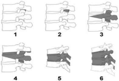
A classification of spinal osteotomies based on the extent of bone resection was developed to standardize the descriptions of techniques and interpretation (Schwab et al., 2014). Six grades of osteotomies are defined in the classification:
- Partial (1) and complete (2) facet joint removal;
- Partial (3) and complete (4) pedicle and body resection;
Figure 2.12 : Osteotomy classification grades 1 to 6 (Image reused with permission of: F. Schwab, B. Blondel, E. Chay et al. The comprehensive anatomical spinal osteotomy
classification. Neurosurgery. 2014; 74[1]:112-120)
A description of SPO and PSO are presented in the next sections. Publications have shown that both of those osteotomy techniques may provide satisfactory correction (Liu et al., 2015), but indications are different. In this work, focus is put on pedicle subtraction osteotomy, which is the most commonly used technique for the treatment of fixed sagittal imbalance (Ottardi et al., 2018). 2.3.2.1 Smith Petersen osteotomy (SPO)
SPO (Smith-Petersen et al., 1945) is a grade 1 osteotomy and a commonly used technique originally used to treat ankylosing spondylitis. It is associated with the resection of the inferior facets, joint capsules, laminae, and posterior ligaments of a certain spinal level. The osteotomy site is then closed, shortening the posterior column and lengthening the anterior column. This technique often involves multiple levels and can achieve 5° to 10° of correction per level (Cho et al., 2005; Schwab et al., 2014). Limited deformity correction and prerequisite anterior column mobility make this type of osteotomy suitable for mild to moderate and flexible deformities of the spine. Best indications for using SPO technique were described as having a long, rounded and smooth kyphosis (Bridwell, 2006) or lack of lordosis.
2.3.2.2 Pedicle subtraction osteotomy (PSO)
PSO (Thomasen, 1985) is a grade 3 osteotomy and is the most commonly used technique to treat fixed sagittal imbalance. It involves a wedge-shaped resection of the vertebral body along with all
posterior elements of the vertebra (Figure 2.13). The osteotomy site is then closed, shortening the posterior column with no anterior column lengthening. The large contact area of the superior and inferior parts of the vertebra helps the fusion of the PSO body (Dorward & Lenke, 2010). It is possible to achieve between 25° and 35° of correction at the osteotomy level (Bridwell, 2006). The PSO is most often performed to increase LL in cases of ASD but may also be performed in the thoracic curve (Bakaloudis et al., 2011). Indications for using PSO technique were described as having degenerative changes in the spine and a sharp and severe deformity with little or no flexibility (Barrey et al., 2014; Berjano & Aebi, 2015).
Figure 2.13 : PSO (adapted from Wikimedia commons, copyright free image) 2.3.2.3 Clinical and biomechanical studies of ASD surgery with osteotomies
Osteotomies are an effective technique used to restore normal sagittal profile of the spine. However, they are highly demanding procedures due to the risk of complications, especially for the pedicle subtraction osteotomy. A retrospective study (Smith et al., 2011) on 578 patients who underwent surgical treatment of thoracolumbar fixed sagittal deformity showed that a higher complication rate was present for procedures including an osteotomy, compared to cases not including an osteotomy (34.8% vs 17%). Between the 215 cases that underwent an osteotomy, cases with a PSO had higher complication rate (39.1%) than cases with a SPO (28.1%). Another study compared the results of executing three SPO and one PSO for the correction of fixed sagittal imbalance (Cho et al., 2005). They found that the correction in kyphosis between the two methods was nearly identical but there was a significantly greater risk of decompensation with SPO, whereas there was substantially greater blood loss with the PSO technique. Another study documented a 19% reoperation rate for patients treated with PSO and a 16% rate for patients not treated with this procedure (Scheer et al., 2013). The authors reported that the most common indications for reoperation were instrumentation complications and radiographic failure.
The rate of mechanical complication after surgical correction of ASD with an osteotomy is relatively high. Barton et al. (Barton et al., 2017) investigated the rate of mechanical complication on a cohort of 88 patients and found an incidence of 43.6%. The prevalence of pseudarthrosis after lumbar PSO was assessed on a retrospective cohort of 171 patients (Dickson et al., 2014). The authors found that the overall prevalence of pseudarthrosis was 10.5%. Of those, 61% occurred at the PSO site and the most common radiographic finding was rod breakage. Smith et al. (Smith, Shaffrey, et al., 2012) evaluated symptomatic rod fracture for ASD on a cohort of 442 patients. It was found that rod fracture occurred in 6.8% of all cases, while it occurred in 15.8% of cases with a PSO. Failure occurred at the PSO site in 89% of those cases. Furthermore, the rate of rod fracture in patients with a PSO based on rod material was lower for cobalt chrome (7%), compared to stainless steel (17%) and titanium (25%). A prospective assessment of the rates for rod fracture following surgery for ASD was conducted on a cohort of 287 patients (Smith et al., 2014). They found a rate of rod fracture of 9% for all patients and a rate of 22% for patients with a PSO with a minimum of 1-year follow-up. 91% of the failures were adjacent to the PSO level. Patients with rod fracture were older, had greater BMI and a greater baseline sagittal malalignment. In a later study by the same author (Smith et al., 2017) on complication rates associated with 3 column osteotomies (3CO), 82 patients with a 2-year follow-up were included. The most common complication was rod breakage (32%) and the most common indication for reoperation was also rod breakage (n=14). The authors, in accordance with a biomechanical study on the fatigue life of the rods after PSO (Tang et al., 2013), suggested that the risk of rod breakage may derive from the severity of rod contour across the 3CO level. Another study on 75 patients reported an incidence rate of rod breakage of 16.2% when PSO was performed (Barton et al., 2015). Factors significantly associated with rod fracture were: fusion construct crossing thoracolumbar and lumbosacral junctions, sagittal rod contour >60°, the presence of dominos and/or parallel connectors, and pseudarthrosis at ≥1-year follow-up.
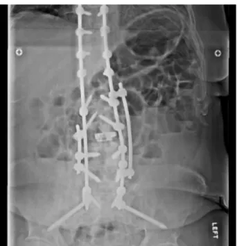
Multiple authors have proposed using 4-rod constructs in order to reduced rod breakage and pseudarthrosis (S. Gupta et al., 2017; Hyun et al., 2014; Luca et al., 2014; Smith et al., 2014) (Figure 2.14). A study investigated effects of a 4-rod construct on motion and surface rod strain of a PSO model to reduce incidence of rod fracture (Hallager et al., 2016). The authors found that addition of accessory rods significantly reduced flexion-extension motion at the PSO level and use of cobalt-chrome material significantly reduced rod strain. Similarly, La Barbera et al. (La Barbera
et al., 2018) conducted tests on six human cadaveric spine segments to compare constructs of 2, 3 and 4 rods and measured strains on the primary rods with strain gauge rosettes. The authors found that adding two accessory rods after PSO was an effective strategy to significantly reduce rod strains. A finite element model was used to investigate the biomechanical performance of different hardware constructs and found that using satellite rods was a good method to reduce stresses on the spinal fixators, near the site of the PSO (Luca et al., 2017). In a similar study, Januszewski et al. studied rod stress after PSO for different surgical constructs (Januszewski et al., 2017). The authors found that 4-rod constructs were effective at reducing stress in the rods near the zone of the PSO by up to 50%.
Figure 2.14 : 4-rod construct covering the zone of the PSO
Although the risks associated with osteotomies are high, a recent study investigated if the performance of surgeries including a 3CO improves with years of surgical practice (Diebo et al., 2017). The authors found that despite being performed on a more disabled population and offering a greater correction at the site of the osteotomy, surgical revisions and complication rate are decreasing.
2.3.3 Preoperative planning of the surgery
To help define surgical objectives, two major classifications have been developed. These classifications may help surgeons to consistently characterize the deformities and the associated surgical treatment. The SRS-Schwab classification is the most recognized one. It relies on studies correlating health-related quality of life scores and radiographical outcomes to define thresholds of correction during surgical procedures (Schwab, Ungar, et al., 2012). In the sagittal plane, three parameters have been defined to help define surgical objectives: PI-LL < 10°, SVA < 4cm, and PT < 20°. Roussouly’s classification has been developed from the geometrical analysis of the variation of sagittal curvatures of asymptomatic population (Roussouly et al., 2005). This classification initially included four patterns of sagittal alignment and was defined based on the SS and spinal shape:
• Type 1 SS smaller to 35° and apex of the lumbar lordosis located at the center of L5 • Type 2 SS smaller to 35° and apex of the lumbar lordosis located at the base of L4
• Type 3 SS between 35° and 45° and apex of the lumbar lordosis located at the center of L4 • Type 4 SS greater than 45° and apex of the lumbar lordosis located at the base of L3 Roussouly’s classification was later completed to include type 3 anteverted (Laouissat et al., 2018). Differently from the SRS-Schwab classification which is based on the magnitude of the restauration of lumbar lordosis, Roussouly’s classification also considers the geometrical shape of the spinal curves.
However, an important proportion of patients do not report a clinically significant change in health care related quality of life and/or still have poor sagittal alignment after surgery. Moal et al. analyzed the radiographic outcomes of ASD correction of 161 patients and found that only 23% of patients had a complete radiographic correction in both sagittal and coronal planes (Moal et al., 2014). Sagittal deformity with pathological SVA or PI-LL was corrected in 50% of the cases whereas pathological PT was only corrected in 24% of the cases. Another study reported failed realignment after ASD surgery with PSO in 23% of the cases (Schwab, Patel, et al., 2012). Patients with failed realignment had significantly larger preoperative deformity but received a similar amount of correction as the patients with successful realignment. An important factor to consider while planning the surgery is postoperative alignment changes through spinal segments that are not included inside the instrumentation construct. A clinical study evaluated how the compensatory
changes in the unfused part of the thoracic spine after PSO impacted global spinal alignment (V. Lafage, Ames, et al., 2012).The authors found that for patients with reciprocal changes, the majority was unfavorable and risk factors were larger PI, inadequate postoperative LL and older age. To illustrate the importance of preoperative planning, a study assessed the ability of surgeons to predict postoperative alignment of the spine after being shown preoperative radiographs and surgical plan (Ailon et al., 2016). The authors found that one-third of the time, surgeons were not able to predict the adequacy of the surgical plan.
The first steps of surgical planning of sagittal imbalance consists of finding the drivers of deformity (loss of LL being the most common one) and how the patient adapted to the deformity through compensatory mechanisms. After finding the amount of correction needed to obtain a good postoperative alignment, surgical technique can be selected depending on the etiology of the deformity. For more rigid deformities, PSO is the preferable technique. Different tools have been developed in order to help the preoperative planning of the surgery.
Predictive mathematical formulas
Mathematical formulas have been developed to help preoperative planning of sagittal correction with PSO and predict postoperative radiographic parameters (Y. J. Kim et al., 2006; V. Lafage et al., 2011; Ondra et al., 2006; Rose et al., 2009; Schwab et al., 2009) but are not all equally accurate. Smith et al. (Smith, Bess, et al., 2012) evaluated the ability of those formulas to predict SVA after PSO and found that formulas incorporating pelvic alignment were better at predicting SVA. Formulas that do not account for PT and expected changes in the unfused segments did not accurately predict sagittal correction and may predispose to residual postoperative deformity. Based on the preoperative variables PI, age, max LL and max TK, the Lafage formulas used to calculate the postoperative PT and SVA had the greatest accuracy (89%) in predicting postoperative SVA.
Computer assisted predictive methods
Current computer assisted methods used to predict postoperative alignment first require identification of anatomical landmarks. The surgical objectives are defined by the user and the objectives are then simulated to predict final alignment (R. Lafage et al., 2018).
Surgimap is a computer program integrating spinal measurements and tools for surgical planning through a graphical method with radiographs (Akbar et al., 2013). Surgeons may rotate the