Université de Montréal
Systemic sclerosis immunoglobulin induces growth and a pro-fibrotic state
in vascular smooth muscle cells through the epidermal growth factor
receptor
par Monique Arts
Faculté de pharmacie
Mémoire présenté en vue de l’obtention du grade de MSc en Sciences Pharmaceutique option Pharmacologie
Août 2018
Résumé
La sclérose systémique (SSc) est une maladie auto-immune qui se caractérise par la
dysfonction immunitaire, la fibrose et les vasculopathies. Il a été suggéré que les autoanticorps des patients atteints de la SSc peuvent induire la différentiation des fibroblasts en
myofibroblasts via l'activation du récepteur du facteur de croissance dérivé des plaquettes (RPDGF). Notre étude a pour but de caractériser les effets des IgG des patients atteints de la SSc sur les cellules musculaires lisses vasculaires (CMLV) et de déterminer s'il est possible de détecter la présence d'autoanticorps dirigés contre le RPDGF, et s'ils induisent une réponse profibrotique chez les CMLV primaires cultivées.
Des CMLV ont été exposées à différentes fractions d’IgG purifiées du sérum des patients atteints de la SSc (IgG SSc) et d’individus non atteints (IgG témoins). La
phosphorylation des protéines kinases ERK1/2 et AKT, la prolifération cellulaire, la synthèse protéique et l’expression de gènes profibrotiques ont été étudiées chez les CMLV stimulées, de même que le potentiel des IgG d'immunoprécipiter le RPDGF des lysats de CMLV.
L'activité stimulatrice des IgG SSc était plus grande que chez les IgG contrôles (p<0,05). Puis, les IgG SSc ont provoqué une immunoprécipitation plus importante du
RPDGF que les IgG contrôles. Il était intéressant de constater que l'activation d'évènements de signalisation en aval du RPDGF, comme la phosphorylation d'Akt et d'ERK1/2, étaient
indépendante de l'activité du RPDGF, mais nécessitaient la fonctionnalité du REGF. La stimulation des CMLV avec des IgG SSc, comparé aux IgG témoins, a induit une
augmentation de la synthèse protéique (p<0,001) ainsi que la modulation pro-fibrotiques de certains gènes (Tgfb1 +200%; Tgfb2 -23%; p<0,001).
Comparé aux IgG contrôles, les IgG SSc avaient un index de stimulation plus élevé chez les CMLV. Même si les IgG SSc interagissaient avec le RPDGF, la signalisation passe par le REGF chez les CMLVs. Par conséquent, nos travaux soutiennent un modèle de
transactivation du REGF par des autoanticorps anti-RPDGF provenant des patients atteints de la SSc et suggèrent que les inhibiteurs du REGF devraient être utilisés dans les études futures visant à identifier de cibles thérapeutiques pour la SSc.
Mots-clés : sclérose systémique, sclérodermie, autoanticorps, PDGFR, EGFR, cellules musculaires lisses vasculaires, inhibiteurs des protéines kinases, transactivation des récepteurs
Abstract
Systemic sclerosis (SSc) is an autoimmune disease characterized by the presence of autoantibodies, fibrosis and vasculopathy. It has been suggested that autoantibodies in systemic sclerosis may induce the differentiation of cultured fibroblasts into myofibroblasts through platelet-derived growth factor receptor (PDGFR) activation. The present study aims to characterize the effects of SSc IgG on vascular smooth muscle cells (VSMCs) and to
determine if stimulatory autoantibodies directed to the PDGFR can be detected, and whether they induce a profibrotic response in primary cultured VSMCs.
Cultured VSMCs were exposed to IgG fractions purified from SSc-patient or control sera. VSMC responses were then analyzed for ERK1/2 and Akt phosphorylation, cellular proliferation, protein synthesis, and pro-fibrotic changes in mRNA expression. The capacity of the IgG fractions to immunoprecipitate the PDGFR from VSMC lysates was also tested.
Stimulatory activity in IgG fractions was more prevalent and intense in the SSc samples than in the controls (p<0.05), and SSc IgG immunoprecipitated the PDGFR with greater avidity than control IgG. Interestingly, activation of downstream signaling events (Akt, ERK1/2) was independent of PDGFR activity, but required functional epidermal growth factor receptor (EGFR). We also detected increased protein synthesis (p<0.001) and pro-fibrotic changes in gene expression (Tgfb1 +200%; Tgfb2 -23%; p<0.001) in VSMCs treated with SSc IgG.
When compared to control IgG, SSc IgG has a higher stimulation index in VSMCs. Although SSc IgG interact with the PDGFR, the observed remodeling signaling events occur through the EGFR in VSMC. Our data thus favour a model of transactivation of the EGFR by
SSc-derived PDGFR autoantibodies and suggest the use of EGFR inhibitors in future target identification studies in the field of SSc.
Key words: Systemic sclerosis, scleroderma, vascular smooth muscle cells, epidermal growth factor receptor, EGFR, receptor transactivation, autoantibodies, platelet-derived growth factor receptor, PDGFR, protein kinase inhibitors
Table of Contents
Résumé ... i
Abstract ... iii
Table of Contents ... v
List of Figures ... x
List of Tables ... xii
List of Abbreviations ... xiii
Dedication ... xvii
Acknowledgments ... xviii
Chapter 1. Literature Review ... 1
1.1. Systemic Sclerosis (SSc) ... 2 1.1.1. Clinical Manifestations ... 2 1.1.2. Classification / subtypes ... 4 1.1.2.1. Diffuse cutaneous SSc (dcSSc) ... 5 1.1.2.2. Limited cutaneous SSc (lcSSc) ... 5 1.1.2.3. Other subtypes ... 6 1.1.2.4. Classification Criteria ... 6 1.1.3. SSc Etiology ... 10 1.2. SSc Pathophysiology ... 11 1.2.1. SSc Vasculopathy ... 13
1.2.1.1. Decreased Capillary Density ... 13
1.2.1.2. Endothelial Injury ... 14
1.2.1.3. Endothelin-1 ... 16
1.2.1.4. Insufficient Angiogenesis ... 16
1.2.1.5. Vascular Endothelial Growth Factor (VEGF) ... 17
1.2.1.6. Endothelial Progenitor Cells & Mesenchymal Stem Cells ... 18
1.2.1.7. Vascular Wall Remodeling in SSc ... 18
1.2.2. Immune Dysregulation ... 19 1.2.2.1. Inflammation ... 20 1.2.2.2. Chronic Inflammation ... 22 1.2.2.3. Immune Dysfunction in SSc ... 23 1.2.2.4. Cytokines ... 25 1.2.2.5. B Cells in SSc ... 27 1.2.3. Autoimmunity in SSc ... 28 1.2.3.1. Anti-PDGFR autoantibodies ... 30 1.2.4. Fibrosis in SSc ... 33
1.2.4.1. Fibroblasts and Myofibroblasts... 34
1.2.4.2. Transforming Growth Factor-β (TGF-β). ... 36
1.2.4.3. Platelet-Derived Growth Factor (PDGF) and its Receptors ... 38
1.2.4.4. Epidermal Growth Factor (EGF) and its Receptors ... 40
1.3. Animal Models ... 41
Chapter 2. Published Original Manuscript ... 43
2.2 Author Contributions ... 46
2.3 Title Page ... 47
2.4 Abstract ... 48
2.5 Introduction ... 49
2.6 Patients and Methods ... 51
2.6.1 Ethics Statement. ... 51
2.6.2 Patients and biological samples. ... 51
2.6.3 Immunoglobulin purification. ... 52
2.6.4 Reagents, antibodies and pharmacological inhibitors. ... 53
2.6.5 Cell culture. ... 53
2.6.6 Immunoblot analyses. ... 54
2.6.7 Immunoprecipitation assays. ... 55
2.6.8 RT-qPCR analysis. ... 55
2.6.9 [3H]-leucine and [3H]-thymidine incorporation assays. ... 56
2.6.10 Statistical analyses. ... 57
2.7 Results ... 57
2.7.1 Patients. ... 57
2.7.2 Purified IgG from SSc sera have a greater stimulation index in VSMC than normal IgG. ... 58
2.7.3 PDGFR-β interacts more avidly with SSc IgG than control IgG but does not mediate the increase in ERK1/2 and Akt signaling. ... 59
2.7.4 Epidermal growth factor receptor (EGFR), but not the AT1R, plays a key role in the VSMC response to SSc IgG. ... 60
2.7.5 SSc IgG causes growth and pro-fibrotic responses in VSMCs. ... 61
2.7.6 Relationship to disease phenotype. ... 62
2.8 Discussion ... 62
2.9 Acknowledgments ... 66
2.9.1 CSRG Recruiting Rheumatologists ... 66
2.10 References ... 68
2.11 Tables & Figures ... 76
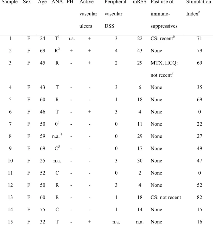
Table 1. Characteristics of our SSc cohort (N = 23). ... 76
Figure 1. ... 77 Figure 2. ... 78 Figure 3. ... 79 Figure 4. ... 81 Figure 5. ... 83 2.11 Supporting Information ... 84
Table S1. Details of individual patient samples. ... 84
Figure S1. ... 86 Figure S2. ... 87 Figure S3. ... 88 Figure S4. ... 89 Chapter 3. Discussion ... 90 3.1 General Discussion ... 91
3.1.1. Anti-PDGFR Autoantibodies - Recent Developments ... 92
3.2 Conclusion ... 95 References ... 97
List of Figures
Chapter 1 - Literature Review
Figure 1. Overview of SSc pathogenesis... 11 Figure 2. Nailfold video capillaroscopy images... 13
Chapter 2 - Published Original Manuscript
Figure 1. Effects of purified scleroderma (SSc) and control (Ct) IgG on signaling activity in vascular smooth muscle cells (VSMCs)... 74 Figure 2. IgG from systemic sclerosis (SSc) patients bind more to PDGFR than control IgG... 75 Figure 3. The phosphotransferase activity of EGFR plays a key role in SSc IgG-induced early signaling events in VSMCs... 76 Figure 4. Systemic sclerosis (SSc) IgG causes growth and profibrotic responses in
vascular smooth muscle cells (VSMCs)... 78 Figure 5. Proposed model of SScIgG signaling through PDGFR-EGFR heterodimeric
complexes... 80 Figure S1. SSc IgG causes increased ERK phosphorylation in quiescent vascular smooth muscle cells in a dose-dependent manner... 83 Figure S2. Effects of purified scleroderma (SSc) and control (Ct) IgG on signaling
Figure S3. The platelet-derived growth factor receptor (PDGFR) inhibitor AG1296 does not inhibit the phosphorylation of ERK or Akt in vascular smooth muscle cells (VSMCs) stimulated with IgG from systemic sclerosis (SSc) patients... 85 Figure S4. Constitutive heterodimerization of epidermal growth factor receptor (EGFR) and platelet-derived growth factor receptor (PDGFR) in quiescent vascular smooth muscle cells (VSMCs)... 86
List of Tables
Chapter 1 - Literature Review
Table 1. Summary of limited cutaneous and diffuse cutaneous SSc phenotypes... 7
Table 2. The ACR-EULAR Criteria for the classification of systemic sclerosis... 13
Table 3. Examples of cytokines classed according to their primary functions... 26
Table 4. Antinuclear autoantibodies relatively specific for SSc... 30
Table 5. Functional (putatively pathogenic) autoantibodies reported in SSc... 31
Chapter 2 - Published Original Manuscript Table 1. Characteristics of our SSc cohort (N = 23)... 73
List of Abbreviations
ACA Anti-centromere antibody
ACR American College of Rheumatology ADAM17 A disintegrin and metalloproteinase 17 AECA Anti-endothelial cell antibody
ANA Anti-nuclear antibody
ATA Anti-topoisomerase I antibodies AT1R Angiotensin type I receptor BAFF B cell activating factor
CREST Calcinosis, Raynaud's, esophageal dysfunction, sclerodactyly, telangiectasia CSRG Canadian Scleroderma Research Group
CTD Connective tissue disease CTGF Connective tissue growth factor DAMP Damage-associated molecular pattern dcSSc Diffuse cutaneous systemic sclerosis dsDNA Double-stranded DNA
dsRNA Double-stranded RNA
EC Endothelial cell
ECM Extra-cellular matrix EGF Epidermal growth factor
EMT Epithelial-mesenchymal transition Endo-MT Endothelial-mesenchymal transition EPC Endothelial progenitor cell
ERK Extracellular regulated kinase
ET-1 Endothelin-1
ETAR Endothelin type A receptor ETBR Endothelin type B receptor
EULAR European League Against Rheumatism Fra Fos-related antigen
Fli1 Friend leukemia integration 1 GAVE Gastric antral vascular ectasia GERD Gastroesophageal reflux disease GVHD Graft-versus-host disease
HB-EGF Heparin-binding EGF-like growth factor
IFN Interferon
IgG Immunoglobulin type G
IL Interleukin
ILD Interstitial lung disease IRF Interferon regulatory factor LAP Latency-associated peptide Klf5 Krüppel-like factor 5
lcSSc Limited cutaneous systemic sclerosis
MAPK Mitogen-activated protein kinase
MSC Mesenchymal stem cell
MVEC Microvascular endothelial cell NF-κB Nuclear factor-κB
NLR Nucleotide-binding oligomerization domain (NOD)-like receptor
NO Nitric oxide
PAH Pulmonary arterial hypertension PAMP Pathogen-associated molecular pattern PBMC Peripheral blood mononuclear cell PDGF Platelet-derived growth factor
PDGFR PDGF receptor
PF Pulmonary fibrosis
PI3K Phosphoinositide 3-kinase
PLC Phospholipase C
PKC Protein kinase C
PM Polymyositis
PRR Pathogen recognition receptor
qRT-PCR Quantitative reverse-transcriptase polymerase chain reaction RA Rheumatoid arthritis
RLR Retinoic acid inducible gene-I-like receptor
RNAP RNA-polymerase
ROS Reactive oxygen species
SLE Systemic lupus erythematosus SRC Scleroderma renal crisis
SS Sjögren's syndrome
SSc Systemic sclerosis
Th Helper T (cell)
TIMP Tissue inhibitors of metalloproteinases TGFα Transforming growth factor alpha TGFβ Transforming growth factor beta
TLR Toll-like receptor
TNF-α Tumour necrosis factor Treg Regulatory T (cell) TSK1 Tight-skin (mice)
TSP-1 Thrombspondin-1
UCD University of California Davis
U1snRNP U1-small nuclear ribonucleic particles VEGF Vascular endothelial growth factor VSMC Vascular smooth muscle cell
Dedication
For Remy, my mother.
Acknowledgments
This MSc thesis is the result of a somewhat convoluted journey. It is not what I had planned, as I began this journey with the goal of completing a PhD. Many factors contributed to my decision to switch to a Master of Science degree, which I like to summarize as life having gotten "in the way". Thus, this journey involves much more than a Master's thesis, and numerous people have contributed to my experience in a positive way. I am deeply grateful to all those who have accompanied me on this adventure, even if I forget to mention you here.
First, I thank Dr. Marc Servant for having invited me to join his lab and embark on this project, for his mentorship, and for his patience, and for having introduced me to the
wonderful world of cell signaling. And, I would not have met Marc, had it not been for Dr. Murray Baron. I am grateful to Dr. Baron for the opportunity to work in Systemic Sclerosis research and for setting me on the path to further graduate studies in this field. Also, I am grateful to the Canadian Scleroderma Research Group (or CSRG), under Dr. Baron's direction, for their student training program. This journey would have been impossible without the funding from the CSRG.
Thank you also to Dr. Baron's team, especially Maura Buchignani and Suzanne Taillefer. Importantly, I must recognize the patients and control subjects who participate in the CSRG. I know that much time and effort go into completing the stacks of questionnaires, not to mention giving your biological samples. I do not take any of this for granted!
I am grateful also for the support from my family - my parents, brother, sister-in-law, aunts, uncles, cousins. I have in my mind a cloud of miscellaneous words of encouragement that you have informally spoken to me over the years. They've stayed with me, and they helped me to keep going. Above all, I am grateful for my mother's interest, pride, and
insistence. I know you would have been proud to see that I produced something at the end of all this, though of course I wish I could have done more. Thank you to Léa, Wesley and Marco for the day-to-day support.
And last but not least, at the Faculté de Pharmacie: Thank you to my lab colleagues and friends. I hold dear many memories of thought-provoking conversations, laughter, and general good times :) and I cherish the friendships that "happened" while we were doing science. Thank you to Dr. Denis deBlois and Dr. Daniel Levesque for taking the time to read, evaluate and provide feedback on my thesis. Un gros merci to Andrée Mathieu for answering my many questions and always being helpful and encouraging. And I appreciated so, so much Dr. Céline Fiset for your time, your kindness, and for wanting to help me move forward.
1.1. Systemic Sclerosis (SSc)
Systemic sclerosis (SSc), also called “scleroderma”, is a devastating multi-system disease of the connective tissue, with features of autoimmunity, inflammation, fibrosis of the skin and internal organs, and vasculopathy1. It is a relatively rare chronic progressive disease with a general prevalence reported to range from 30 to 489 per million2-7 and an incidence of
0.6 to 122 per million per year7. Women are affected approximately 5 times more frequently than men2,5, but men who have SSc tend to have more severe disease symptoms and increased mortality8. The etiology of SSc is complex and not well understood. There is no cure for this disease, although disease survival and morbidity have improved over the past decades due to improved diagnosis and early detection and management of serious complications like
scleroderma renal crisis (SRC), gastro-esophageal reflux disease, digital ulcers and pulmonary arterial hypertension9,10. SRC was once the main cause of mortality, but since the advent of angiotensin-converting enzyme inhibitors, this is now a controllable complication11. Today, the main causes of death in SSc are pulmonary arterial hypertension and interstitial lung disease12.
1.1.1. Clinical Manifestations
SSc is clinically a very heterogeneous condition. Disease manifestations and severity, as well as disease course progression vary greatly from one patient to another. A diagnosis of SSc is typically based on the judgment of a rheumatologist and the presence of particular signs associated with SSc, like skin tightness and hardness, Raynaud’s phenomenon, and the
presence of disease-specific anti-nuclear antibodies (ANAs). Since SSc is rare and complex, it sometimes takes a long time before patients receive a correct diagnosis.
Raynaud’s phenomenon (RP) refers to transient ischemia in the extremities, for example, in the fingertips, due to vasospasm of the small blood vessels in response to cold or stress. RP is usually the first sign of a problem in SSc, and unlike other symptoms, virtually all patients (>95%) experience it13. Primary or idiopathic Raynaud’s is not uncommon, however, so screening for other markers such as ANAs and abnormal nailfold capillaries is required if SSc is suspected. Unlike primary RP, which is thought to be due to a functional problem, namely an exaggerated central and/or peripheral nervous system mediated vasospasm14,15, SSc-related Raynaud’s results at least in part from structural abnormalities in the
microvasculature and digital arteries14,15. SSc-related RP is severe, compared to primary RP which is reversible and does not lead to other complications.
Scleroderma, literally “hard skin”, due to dermal inflammation and fibrosis, is another prominent symptom of SSc. In most cases of SSc, skin fibrosis first develops on the fingers and hands.Eventually, hard skin also appears on the lower extremities, forearms, face and neck. In the diffuse cutaneous subset of patients, the skin of the trunk, arms and legs may also become fibrotic. In addition to the tightening and thickening of the skin, patients experience intense pruritis, areas of hyper- and hypo-pigmentation, reduced hair follicles and reduced sweating in the affected skin.
Many SSc patients develop distressing changes to their appearance. Scleroderma of the face gives the skin an abnormal shiny and smooth appearance. Conspicuous pigmentary changes may occur, and distinct telangiectasias - red spots due to dilation of subcutaneous blood vessels - are also common on the face. The lips become thin and furrowing appears around the mouth. The mouth opening is severely constricted, which can hinder effective oral hygiene and professional dental care. The nose can become narrower and pinched16,17.
The hands also undergo important changes. In addition to Raynaud’s, stiffness and puffiness of the hands and fingers are another early sign of SSc. Later, almost half of SSc patients develop digital ulcers, a painful complication that seems to appear spontaneously, heals exceedingly slowly, sometimes over the course of weeks or months, and can lead to impaired hand function, osteomyelitis, autoamputation and gangrene18. Calcinosis (calcium
deposits) in the soft tissues of the hands, as well as on the elbows and knees, is also common, and may lead to skin ulceration and infection19. Patients also experience joint pain and stiffness in their hands. Sclerodactyly (hard skin of the digits) is characterized by shiny, tight skin of the fingers, resulting in fingers or toes that are difficult to move or straighten. This may lead to flexion contractures, which refers to the digits becoming fixed in a flexed position. These complications in the hands result in pain and loss of function, and can be debilitating.
Internally, multiple organs are often affected by fibrosis and vascular problems, including those of the gastro-intestinal tract, lungs, heart, and kidneys. Patients experience a range of symptoms related to the extent and severity of internal organ involvement, such as: dry cough, dyspnea, chest pain, difficulty swallowing, bloating, weight loss, cachexia,
malabsorption, hypertension, pain and fatigue, to name but a few20. In short, patient quality of life is greatly affected by serious complications that range from esophageal dysmotility and bacterial overgrowth, to potentially fatal problems like lung fibrosis, pulmonary arterial hypertension and renal failure with severe hypertension (SRC).
1.1.2. Classification / subtypes
The diverse clinical manifestations of SSc, and the extent of organ involvement and pattern of disease progression vary widely among patients. However, homogeneous groups of
patients can be identified. Depending on the particular constellation of symptoms and signs that are present, patients can be classified into distinct disease subtypes. These are primarily defined by the extent of skin fibrosis, which is also associated with specific patterns of visceral involvement21. The two main subsets are limited cutaneous SSc and diffuse cutaneous SSc (summarized in Table 1). Patient autoantibody profiles are also used to distinguish between the two subtypes. There are certain important vascular complications and organ involvement that occur in both groups, which affect disease severity and mortality5.
1.1.2.1. Diffuse cutaneous SSc (dcSSc)
The diffuse cutaneous SSc (dcSSc) subtype is characterized by severe skin fibrosis on the torso as well as the extremities (hands, arms, face, and legs), and the presence of
antitopoisomerase antibodies (ATA)22. This form of SSc carries a worse prognosis, with more extensive and more severe complications, and tends to have a more rapid onset and
progression4. Diffuse SSc patients are more likely to have serious kidney and heart
involvement, and such complications most often occur early in the course of the disease23.
Scleroderma renal crisis is more likely to occur in this subgroup.
1.1.2.2. Limited cutaneous SSc (lcSSc)
Limited cutaneous SSc (lcSSc) is defined by skin fibrosis being distal from the elbows and knees22, but often limited to the fingers (sclerodactyly). The skin of the face may also be
fibrotic. This SSc subtype is sometimes referred to as CREST syndrome, named for the symptoms that these patients present: calcinosis, Raynaud’s phenomenon, esophageal dysmotility, sclerodactyly and telangieactases21. The presence of anticentromere antibodies
(ACAs) is also a marker for this subset21,22. Patients with lcSSc are more at risk of developing fingertip ischemia and ulcers, and pulmonary arterial hypertension (PAH), but despite
significant morbidity their overall prognosis is better than patients with the diffuse form21.
1.1.2.3. Other subtypes
Approximately 10% of SSc patients also express features of another connective tissue disease (CTD)24, such as systemic lupus erythematosus (SLE), polymyositis (PM), rheumatoid arthritis (RA) or Sjögren’s syndrome (SS). Patients with so-called SSc-CTD overlap syndome have been proposed to constitute a distinct subset24,25, because their SSc resembles a version intermediate to lcSSc and dcSSc, and they possess autoantibody markers distinct from those ascribed to the lcSSc and dcSSc subgroups. These overlap phenotypes are associated with a unique gene signature26 and with anti-U1-small nuclear ribonucleic particles (U1snRNP) antibodies27,28.
1.1.2.4. Classification Criteria
Given the heterogeneous nature of SSc, some experts suspect that “systemic sclerosis” comprises a group of distinct diseases with shared features29. Although SSc may be considered to have two main subtypes - lcSSc and dcSSc - further subtyping of patients may be important for managing patients and their treatments, and for research purposes which often require objectively relatively "homogeneous" groups of patients. In order to do meaningful SSc research with sufficient statistical power, it is often necessary to involve patients from an expanded population. Patient data and biological samples need to be collected at different geographical centers by different researchers. (For example, the Canadian Scleroderma
Research Group (CSRG) was created for this reason, allowing rheumatologists from across Canada to establish a bank of Canadian patient biosamples and data, which may be shared among its members for research purposes.) To reduce the possibility of variability in
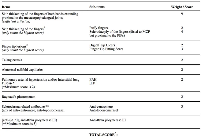
definitions and judgments used by the diagnosing specialists, classification systems have been developed in order to establish criteria that will allow identification of homogeneous groups of SSc patients and comparisons among these groups. These criteria may inform diagnosis, although that is not their intended purpose30,31 (Table 2). An earlier version had been used for many years, since 198030. A new criteria definition was created and accepted in 2013 jointly by the American College of Rheumatology (ACR) and the European League Against
Rheumatism (EULAR) 31. Studies in molecular subset definitions may be useful in clarifying
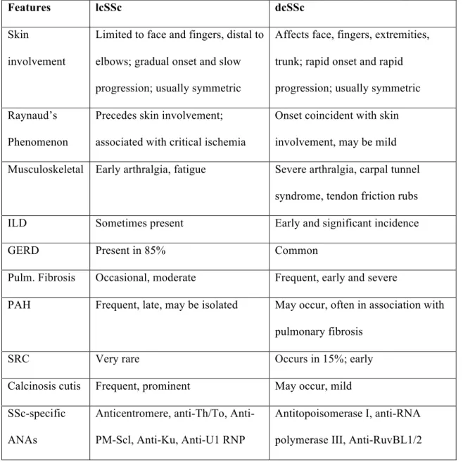
Table 1. Summary of limited cutaneous and diffuse cutaneous SSc phenotypes21,32-36
Features lcSSc dcSSc
Skin
involvement
Limited to face and fingers, distal to elbows; gradual onset and slow progression; usually symmetric
Affects face, fingers, extremities, trunk; rapid onset and rapid progression; usually symmetric Raynaud’s
Phenomenon
Precedes skin involvement; associated with critical ischemia
Onset coincident with skin involvement, may be mild
Musculoskeletal Early arthralgia, fatigue Severe arthralgia, carpal tunnel
syndrome, tendon friction rubs
ILD Sometimes present Early and significant incidence
GERD Present in 85% Common
Pulm. Fibrosis Occasional, moderate Frequent, early and severe
PAH Frequent, late, may be isolated May occur, often in association with
pulmonary fibrosis
SRC Very rare Occurs in 15%; early
Calcinosis cutis Frequent, prominent May occur, mild
SSc-specific ANAs
Anticentromere, anti-Th/To, Anti-PM-Scl, Anti-Ku, Anti-U1 RNP
Antitopoisomerase I, anti-RNA polymerase III, Anti-RuvBL1/2
1.1.3. SSc Etiology
Although the etiology of SSc is not known, it is believed to involve some
environmental trigger in genetically susceptible individuals. In the general population, the SSc incidence rate is 0.026%, while in families with a history of SSc, the incidence is 1.5-1.7%37. Different genetic variants can be linked to specific clinical patterns. For example, African American SSc patients are more likely to have pulmonary fibrosis and anti-topoisomerase I, anti-fibrillarin, and anti-RNP autoantibodies, compared with other groups38, and in the case of the Choctaw Native American population, which has a higher SSc prevalence than any other group39, SSc tends to be relatively homogeneous for the dcSSc subtype with pulmonary fibrosis and ATAs39. However, concordance among monozygotic twins is low. Genome-wide
association studies have identified an HLA II region that associates strongly with SSc40, and single nucleotide polymorphisms that confer susceptibility to SSc have been identified in the genes coding for interferon regulatory factor 5 (IRF5)41-43, IL-1α44, TGF-β45, fibrillin-146, tumour necrosis factor-α (TNF-α)47, connective tissue growth factor (CTGF)48, and others.
A number of environmental triggers are suspected as potential causative agents of SSc. Exposure to certain chemicals has been associated with SSc development, including silica dusts, benzene, trichloroethylene, vinyl chloride, epoxy resins, and others49. The cancer drug bleomycin, which is also used to induce skin and pulmonary fibrosis in mice, also has been implicated49. Exposure to and/or infection by specific microbial pathogens has been linked with SSc50, based on high titres of antibodies against parvovirus B1951, human
cytomegalovirus52and Helicobacter pylori53 in SSc patients. A connection with cancer has also been reported, in that antitumour immunity may bring about SSc in patients with anti-RNA-Polymerase III autoantibodies26.
Another hypothesis, which may explain the origin of autoimmunity in SSc, lies in the concept of microchimerism54, which refers to the presence of cells in one individual that originate from another genetically different individual. This may occur naturally by the persistence in maternal tissues of fetal cells that have passed the placenta during pregnancy
54-56, conversely by maternal cells that persist in the offspring long after birth54,57, or in the case
of a transplant or transfusion. Evidence of a greater degree of michrochimerism has been found in female SSc patients than in controls58,59. The persistence of fetal cells and DNA in the mother may relate to the increased incidence of SSc in women in the years after
childbearing60 (peak age at onset is 20-50 years61).
1.2. SSc Pathophysiology
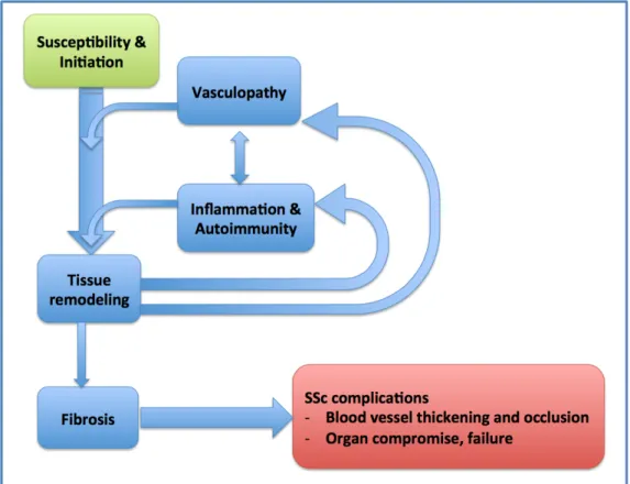
The clinical manifestations of SSc result from underlying pathologies that can be categorized according to four interrelated "hallmarks" of SSc, namely fibrosis, vasculopathy, inflammation, and autoimmunity. Aberrant connective tissue metabolism, culminating in fibrosis of skin or major organs, has traditionally been considered to be the primary problem in SSc.Currently, despite the lack of a single unifying theory that explains all of the typical SSc features, it is now hypothesized that SSc pathogenesis follows a typical sequence of events, possibly initiated by vascular dysfunction, which progresses to inflammation, immune cell activation and autoantibody production, all of which then contribute to profibrotic processes62. However, the interplay of these events is also important. For example, chronic inflammation and cytokine release cause further microvascular damage63, and fibroblasts, the cells that drive fibrosis, can also have proinflammatory activity64,65 (Fig. 1).
Interestingly, there is much more widespread inflammation and consequent fibrosis in dcSSc than in the limited cutaneous form. On the other hand, SSc vasculopathy is much more prominent in lcSSc, as evidenced by the earlier onset of Raynaud’s and the vascular
complications associated more frequently with this subtype, such as digital ulcers, PAH and SRC. This suggests that the pathogenesis of the two subtypes may be subtly distinct, although the sequence of events appears to be common.
What follows is a description of the pathophysiology underlying each hallmark feature and the corresponding cellular and molecular mechanisms involved. Although these four disease components are described individually, the separation into the four hallmarks is artificial, as these processes are intimately interrelated.
1.2.1. SSc Vasculopathy
The vascular manifestations of SSc, which constitute a major identifying feature, tend to occur early, prior to any signs of fibrosis. The vascular system, especially the
microvasculature is compromised in several important ways67. Specifically, SSc patients have a marked decline in capillary density, insufficient angiogenesis, and intimal proliferation. Endothelial cell (EC) dysfunction appears to be a ubiquitous early event in SSc. It has been suggested that some form of microvascular injury may be the initiating factor in SSc pathogenesis68.
1.2.1.1. Decreased Capillary Density
EC injury and apoptosis occur early and are speculated to be initiating events in SSc69 pathogenesis. Patients have progressive changes in their capillary beds, ranging from
abnormal capillaries to avascular areas. This decreased density of capillaries has been reported in the skin and other organs affected by SSc, including lungs, heart, kidneys, muscles67,70-74. Structural abnormalities and progressive reduction in capillary density increase with disease progression to the extent that more than 50% of expected capillaries have been reported as absent75,76. In the nailfold (the base of the finger nail) it is possible to observe non-invasively the capillaries near the surface of the skin. Distorted and micro-hemorrhaging capillaries as well as areas of low capillary density observed by nailfold video capillaroscopy (Fig. 2), are characteristic of SSc. Indeed, abnormal nailfold capillaries are a sign pointing to an SSc diagnosis in patients presenting with RP. It is possible that these abnormalities near the skin surface are also representative of the blood vessel pathology at the internal organs, which show reduced blood flow and tissue hypoxia77.
Figure 2. Nailfold video capillaroscopy images depicting normal capillaries compared to the capillary abnormalities typically observed in SSc. (magnification x200) (Images available online from http://www.the-rheumatologist.org/article/capillaroscopy-a-safe-and-direct-method-for-ssc-diagnosis/).
1.2.1.2. Endothelial Injury
Endothelial dysfunction plays an important role in SSc vasculopathy. The endothelium is a complex organ consisting of a single layer of endothelial cells which make up the capillaries and the inner lining of other blood vessels. It has important roles in regulating blood flow and homeostasis of the vascular wall. Its endocrine, paracrine and autocrine activities include regulating coagulation and fibrinolysis, vascular permeability, vascular tone, metabolism and
nutrition of surrounding cells66. The endothelium reacts to and produces a variety of mediators important for vasodilation (like nitric oxide (NO)), vasoconstriction (like endothelin), and other functions. ECs also are important for maintaining VSMCs in a quiescent state78. Endothelial dysfunction, refers to an abnormal “activation" in which there is a disturbance in the balance of vasoactive factors, resulting in increased vasoconstriction, and also a
proinflammatory and pro-coagulant state.
A variety of agents have been proposed that potentially induce injury and/or activation of the microvascular endothelium early in the SSc disease process, such as pathogens50-53, immune-mediated cytotoxity, ischemia/reperfusion and generation of reactive oxygen species (ROS)79-81, and anti-endothelial-cell autoantibodies (AECAs)82-84. AECAs have been reported
in a significant proportion of SSc patients82,83,85, and can upregulate EC adhesion molecules and induce apoptosis82,83,86,87. Similarly, ROS may cause vascular endothelial damage, and SSc patients were noted to have a significant reduction in plasma antioxidant capacity80.
Subsequent to microvascular injury, the ensuing events include endothelial and smooth muscle cell proliferation, damage to the internal elastic lamina, inflammatory infiltration of the vessel wall and fibrosis, leading to obliterative vasculopathy and significant tissue hypoxia88. Oxidative stress and ischemia further exacerbate vessel wall damage. Once endothelial dysfunction ensues, the delicate balance of EC-derived vasoactive mediators is disturbed, promoting an anti-angiogenic, pro-inflammatory and profibrotic state in SSc. For example, thrombospondin-1 (TSP-1), an antiangiogenic cytokine that induces EC apoptosis and inhibits their proliferation, is released in high concentrations by hypoxic ECs, and has accordingly been reported to be present at elevated levels in SSc samples compared to controls89.
1.2.1.3. Endothelin-1
Endothelin-1 (ET-1) is an important mediator of vasoconstriction, which acts on the type A and B endothelin receptors (ETAR and ETBR), and is primarly produced by ECs. SSc
patients have an imbalanced expression of endothelin receptors66,90. In addition, ET-1 is
overexpressed in SSc skin, lungs, liver and kidney tissues and in the circulation91-96. ET-1 also
has been shown to induce a profibrotic phenotype in fibroblasts, suggesting a possible role of ET-1 in both SSc vasculopathy and fibrosis90. The use of the dual endothelin receptor
antagonist, bosentan, has confirmed the importance of this target in SSc pathology. Bosentan was shown to improve the impaired vasodilation of tight-skin (TSK1) mice97, an experimental mouse model of SSc.
ET-1 also acts on VSMCs, inducing contraction and proliferation, which may
contribute to SSc PAH and digital ulcers. In clinical studies, bosentan appeared to have some ability to prevent digital ulcers and to treat SSc-related PAH98-100, and to slow the progression of changes to the nailfold microvasculature over a 3-year period101.
1.2.1.4. Insufficient Angiogenesis
Related to the problem of decreased capillary density is an insufficient angiogenic and vasculogenic response102-105. Normally, the hypoxic conditions resulting from capillary damage and reduced capillary density spur an increased production of proangiogenic mediators, which would stimulate the repair and regeneration (angiogenesis) or de novo generation (vasculogenesis) of blood vessels. In SSc, vascular regeneration processes are defective102-104. Studies in SSc peripheral blood mononuclear cells (PBMCs), SSc PBMC supernatants and SSc sera have demonstrated severe disturbance in angiogenic and
vasculogenic factors103,106-108. However, the lack of new blood vessel formation is not due to a diminished production of proangiogenic mediators, as many proangiogenic factors are
upregulated in SSc, including sVCAM-1, sE-selectin, E-selectin, endothelin-1, VEGF and VEGF receptors103,105,109,110.
1.2.1.5. Vascular Endothelial Growth Factor (VEGF)
Vascular endothelial growth factor (VEGF, also called VEGF-A) is the prototype member of the angiogenic VEGF cytokine family. VEGF signals through two related receptor tyrosine kinases, VEGFR-1 and VEGFR-2. This growth factor has important implications in both physiological and pathological angiogenic processes111.
In SSc, VEGF has been detected in blood and skin at greater levels than in a healthy population at different stages of disease progression109,110,112, and both 1 and VEGFR-2 are overexpressed in SSc110,113. Interestingly, the increased levels of VEGF correlate with the severity of blood vessel loss and damage, and SSc disease progression, and not with increased blood vessel repair and regeneration. This may be explained by the fact that translation of VEGF-A mRNA can result in different splice variants, some of which are pro- and others anti-angiogenic114,115. Indeed, Manetti et al. have shown a greater amount of antiangiogenic isoform VEGF165b in SSc circulation and skin than in healthy or other disease
controls116 and that the increased amount of VEGF165b correlates with the degree of capillary
abnormality and disappearance117. They have also reported that microvascular endothelial cells (MVECs) isolated from SSc patients constitutively express higher levels of
antiangiogenic VEGF165b in culture than do control MVECs116. The two VEGF165 splice
complete receptor phosphorylation, and results in attenuated downstream signaling. Thus the VEGF165b isoform acts as an endogenous inhibitor. Interestingly, the alternate splicing of the
antiangiogenic VEGF isoform may be driven partly by TGF-β116 (a cytokine considered to be the master regulator of fibrosis, discussed in Section 1.2.4.2).
1.2.1.6. Endothelial Progenitor Cells & Mesenchymal Stem Cells
In addition to deficient angiogenic pathways, the amounts of circulating vasculogenic cells are also abnormal. In SSc, bone-marrow derived endothelial progenitor cells (EPCs), which participate in vascular repair, have been found to be decreased in some studies105, and increased in others118-120. The significance of the contradicting reports is not clear, but
diminished numbers of EPCs would result in reduced vasculogenesis, while an expanded presence may be explained as EPCs homing to increased levels of angiogenic factors, but then having reduced capacity to regenerate. Indeed, SSc EPCs, including mesenchymal stem cells, have impaired migratory responses to VEGF, in addition to early senescence121 The giant and other abnormal capillaries observed in patient nailfolds (Fig. 2), as well as other commonly observed SSc features like telangiectasia of the skin and of the gastrointestinal mucosa (including gastric antral vascular ectasia (GAVE))122, are likely the result of failed efforts to build new vessels in response to the proangiogenic stimuli103.
1.2.1.7. Vascular Wall Remodeling in SSc
Vascular smooth muscle cells (VSMCs) constitute the majority of cells in the medial wall. VSMCs of the media are normally present in a differentiated contractile state, and
in this quiescent state123. Infiltrating leukocytes and ECM components also influence VSMC activation124,125. Following endothelial dysfunction, VSMCs dedifferentiate into a synthetic phenotype. In this state, they proliferate and secrete collagens and other ECM proteins126,127. Intimal proliferation and fibrosis of blood vessel walls, a striking feature of SSc vasculopathy, further exacerbate the diminished blood flow due to absent and damaged microvessels128,129.
In larger blood vessels, there is evidence of luminal narrowing, and sometimes complete obliteration, due to VSMC proliferation in the intima and the deposition of collagen and other ECM components76. In addition, normal intimal functions are disrupted, as evidenced by increased coagulation and decreased fibrinolysis130, again impeding blood circulation. These features clinically manifest as the vasculopathic complications typical of SSc: RP,
telangiectasias, DUs, GAVE, PAH, myocardial dysfunction, SRC.
1.2.2. Immune Dysregulation
Dysregulation of the immune system is another hallmark feature of SSc. The immune system has classically been divided into two parts: the innate immune system which provides
immediate non-specific defenses against disease; and the adaptive immune system, which acquires memory of encountered infections and can launch enhanced defenses to target specific pathogens or foreign entities. The innate immune system also has important functions in regulating and activating players of the adaptive immune system, and likewise, adaptive immune cells and their products can recruit and influence activity of innate immune cells. This makes for an intricate but powerful system of defense, and a complicated pathology in the case of SSc.
Chronic inflammation and autoimmunity are both significant and important
components of SSc. In early stages of SSc there is evidence of inflammatory cell infiltrates, degranulation of mast cells and peripheral blood mononuclear cells with an activated
phenotype131,132. As inflammation in skin precedes fibrosis, and given the normal role of inflammation in immune responses and wound healing, inflammatory processes likely are important in driving the pathogenesis of SSc, although the underlying mechanisms are not entirely clear. In addition, pathologic activation of the adaptive immune system is indicated by both B and T-cell abnormalities.
1.2.2.1. Inflammation
Inflammation is a complex protective mechanism that is an important component of the innate immune system’s response to injury or infection. An injury or sign of a pathogen will often trigger an inflammatory response, which is initiated by cellular effectors of the innate immune system when they recognize pathogen-associated molecular patterns (PAMPs) and/or danger-associated molecular pathogens (DAMPs) through their pattern recognition receptors (PRRs). There are different classes of PRRs, and they include membrane-bound, cytosolic and secreted proteins. Toll-like receptors, nucleotide-binding oligomerization domain (NOD)-like
receptors, and retinoic acid-inducible gene I (RIG-I)-like receptors (TLRs, NLRs, and RLRs, respectively) are all examples of PRR families. Specific PAMPs (such as lipopolysaccharide (LPS), or double-stranded RNA (dsRNA), for example, which betray the presence of gram-negative bacteria, or replicating virus, respectively) or DAMPs (e.g. cellular contents not normally present in the circulation, signaling tissue injury) will be detected by their cognate PRRs (TLR4, TLR3, TLR2 and TLR4, respectively). Activation of these receptors typically
causes a coordinated cascade of cellular events leading to the transcription of genes involved in inflammation and anti-microbial defense. In the case of TLRs, adaptor molecules such as MyD88 or TRIF are recruited133, which then initiate interferon regulatory factor (IRF) or nuclear factor κ-B (NFκB) signaling pathways, leading to the induction of inflammatory and anti-microbial mediators (cytokines, interferon (IFN)), as well as chemokines that recruit and activate circulating leukocytes to the site of inflammation133,134. These cells subsequently produce more cytokines and other mediators, some of which serve to dilate and increase the permeability of blood vessels, allowing larger circulating white blood cells and the other circulating mediators to access the inflammatory site. Further, other mediators, like chemokines (cytokines that guide cell migration) recruit the necessary cells, such as fibroblasts or antibody producing plasma cells. PRR signaling, and ensuing cytokine production, also has a role in recruitment and antigenic priming of naïve T-cells. Yet other cytokines are important for stimulating the adaptive immunity arm of the immune system to produce B cells and T Cells. The influx of blood which brings circulating immune cells and mediators to the inflammatory site, leads to redness and swelling, pain due to the pressure of the swelling as well as stimulation of pain receptors. These, along with warmth at the
inflammation site, attributable to increased local circulation, constitute Celsius' four classic manifestations of inflammation: rubor, tumor, dolor, calor135.
In recent years, the role of TLRs in SSc has gained appreciation. TLR signaling is known to lead to Type I IFN (IFNα/β) production as well as that of many other cytokines. TLR activation has also been related to fibroblast activation136. Patients with SSc, similar to those with other autoimmune connective tissue diseases, have a Type I IFN gene expression
pattern or signature137,138. However, further studies are needed and ongoing in order to elucidate the exact role of TLRs and other PRRs in SSc.
1.2.2.2. Chronic Inflammation
Acute inflammation is characterized as having an immediate onset in response to a pathogen or trauma and a short duration, in the order of days. Chronic inflammation, on the other hand, occurs over a long period of time. Chronic inflammation is also characterized by the migration of large amounts of mononuclear cells, like lymphocytes, while in acute inflammation, polymorphonuclear cells, like neutrophils, infiltrate in large numbers. In chronic inflammation there is ongoing destruction of tissue, concurrently with persistent attempts at tissue repair. Once the pathogen is cleared or the tissue damage is controlled, anti-inflammatory cytokines and other endogenous regulatory signals guide the termination of inflammation. Evidence of the resolved inflammatory response may remain, in the form of circulating cells of the adaptive immune system guarding against a recurring infection and/or scar tissue formation. Problems at different phases of the inflammatory response - such as the failure to eliminate a pathogen or heal damaged tissue (and thereby continuous exposure to PAMPs and DAMPs), excessive pro-inflammatory cytokines and other mediators,
dysfunctional or insufficient anti-inflammatory mediators, fibrosis or other tissue damage that signals further danger and triggers more inflammation, just to name a few - perpetuate damage to tissue and cells and production of B and T cells. The chronic inflammation in SSc is thus intimately linked with the other hallmarks of autoimmunity and fibrosis.
1.2.2.3. Immune Dysfunction in SSc
The presence of inflammatory infiltrates131,132 is an early characteristic in SSc, in addition to the early vascular events discussed above (Section 1.2.1) As described earlier, endothelial dysfunction is likely at the root of the early vasculopathy and precedes the inflammation120,139,140. As one of many feed-forward cycles in SSc pathophysiology, beyond
its vasoconstrictive effects, ET-1 also behaves as a pro-inflammatory cytokine141. The inflammation then promotes further vascular injury. The edematous-inflammatory phase associated with SSc tends to diminish as the disease progresses to fibrosis.
Many studies have identified marked lymphocytic infiltration in symptomatic SSc tissues142,143. Histological analyses of skin biopsies from early dcSSc reveal accumulation in
subcutaneous tissue of mononuclear cells, especially macrophages, mast cells and T
cells140,144-146, and predominantly activated CD4+ T cells139,147,148. Many abnormalities have been reported for specific subsets of CD4+, or T helper (Th) cells, in SSc. The balance of Th1/Th2 cells is skewed towards Th2 in SSc, and accordingly, SSc patients have increased levels of Th2-derived cytokines in their serum149. Th2 cells produce abundant pro-fibrotic and
anti-inflammatory, including interleukin-4 (IL-4), IL-5, and IL-1363, and also drive some of the vascular changes140. Pro-inflammatory Th-17 cells, another subset of CD4+ T cells, secrete great amounts of IL-17 and IL-22, and appear to be pathogenic in inflammatory autoimmune disease processes150. Th-17 cell counts are elevated in SSc compared to normal blood
samples150,151. IL-1β, secreted by activated macrophages, drives Th17 differentiation152.
Another CD4+ T cell subset, regulatory T cells (Tregs) suppress effector T cell proliferation, and as such have an important role in maintaining self-tolerance153. Amounts and function of Tregs are also abnormal in SSc. Both higher and lower amounts of Tregs have been reported
in SSc, but in general, these appear to have reduced suppressive function154-156 and impaired Treg activity is associated with SSc disease severity154,155,157. Several cytokines, IL-1β, IL-2, IL-23 and TGF-β can induce Treg transdifferentiation into Th17 cells156,158.A shifting of Treg towards Th17 is one mechanism by which Treg numbers may be reduced, and at the same time Th17 cytokines are increased, thus contributing to increased immune activation and onset of autoimmunity159. Interestingly, an unusual "double positive" CD4+CD8+ T cell population that secretes excessive IL-4 has also been reported in SSc160.
The degree of lymphocyte accumulation has been shown to correlate to SSc skin fibrosis severity and progression161. T cells are present at an early stage at sites undergoing fibrosis140,145,162. In graft-versus-host disease (GVHD), which mirrors many aspects of SSc and
is thus used as a model to study this disease, rats develop skin lesions characterized by mononuclear cell infiltrates and subsequent skin fibrosis. Also some of the most common therapies used in SSc are immunosuppressives like cyclophosphamide, which target T and B cells. Despite having no known direct effects on fibroblasts or ECM production,
cyclophosphamide has been shown to reduce progression of SSc-ILD (interstitial lung disease) and to reverse severe diffuse skin fibrosis163,164.
Monocytes (which differentiate into myeloid dendritic cells and macrophages) and macrophages are also present in SSc inflammatory infiltrates in involved lung or skin165,166. Different classes of macrophages have been identified, and generally, macrophages may be classed as M1 type, which are important producers of pro-inflammatory cytokines, or M2 type, which are considered to have anti-inflammatory and fibrogenic properties. In SSc, evidence suggests an increased presence of pro-fibrotic, anti-inflammatory M2-type
macrophages. In addition, SSc monocytes have pro-fibrotic features, such as reduced levels of caveolin-1 which is a negative regulator of the pro-fibrotic cytokine TGF-β.
Dendritic cells, which express many PRRs and function as professional antigen-presenting cells are abundant producers of cytokines, and are important for the differentiation of naïve T cells into Th2 effectors, which then in turn are important cytokine producers. Dendritic cells also have a role in supporting self-tolerance of T-cells, thus defects in this role may be important in the development of autoimmunity. Early SSc dendritic cells have been shown to respond to TLR stimulation by producing greater amounts of IL-6 and TNF-α, two important pro-inflammatory cytokines, and less IL-10 and IL-12167, as compared to late SSc or control cells.SSc dendritic cells have also been shown to have a pro-inflammatory gene signature.
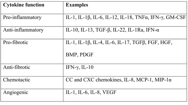
1.2.2.4. Cytokines
Cytokines are a diverse group of signaling molecules that are produced by immune system cells, most significantly by macrophages and helper T (Th) cells, but many other cell populations as well. They transmit intercellular messages in autocrine, paracrine, or endocrine fashion. They mediate innate and adaptive immune responses and induce specific cellular responses upon binding their cognate receptors in their target cells. Cytokines can be
categorized based on shared elements of their cognate receptors: some of the major cytokine groups are the chemokines, IL-1 family, IL-17 cytokines, Type I cytokines, Type II cytokines (interferons), TNF receptor family, TGF-β family 168. Growth factors may be considered a sub-family of cytokines. Cytokines can also be classed by their prototypic and most
activity169, making these functional categorizations artificial, albeit useful. For example, the anti-inflammatory cytokine IL-13 has a “main” function of reducing inflammation, but it also has important profibrotic activity (reviewed by Raja & Denton, 2015170).
Evidently, many cytokines are implicated in SSc pathology. It is beyond the scope of this thesis to discuss them all in detail. Some of the best characterized ones are summarized in Table 3 or explained in further detail in other sections of this thesis.
One cytokine that is of great interest in SSc is Interleukin 6 (IL-6). This pleiotropic cytokine is elevated in SSc skin and serum, both in early and long-standing disease171. IL-6 is a driver of chronic inflammation and endothelial cell activation. It has been shown to prevent T cell apoptosis172, one mechanism by which it contributes to chronic inflammation. It also
stimulates immunoglobulin production by activated B cells173, pointing to an additional role in autoimmunity. IL-6 can affect fibroblast proliferation and induce synthesis of ECM
components, like collagen I172. In addition, fibroblasts from SSc skin lesions constitutively produce higher amounts of IL-6 than fibroblasts from uninvolved skin or from healthy controls, giving rise to an autocrine positive feedback system172. In a 2-patient SSc study,
tocilizumab, an anti-IL-6 receptor antibody, reduced skin hardness and collagen, and improved kidney function (but not lung fibrosis)174, affirming the importance of this cytokine in SSc pathogenesis.
IL-1α is primarily a proinflammatory cytokine, whose mRNA is constitutively expressed in SSc fibroblasts175. It has autocrine activity, and induces the production of other
pro-inflammatory mediators in endothelial cells, fibroblasts and dendritic cells, such as IL-6, IL-8, IL-12, ET-1, TNF, growth factors, and adhesion molecules (reviewed by Kawaguchi176). It stimulates the proliferation and differentiation of T cells176. Interestingly, two IL1A gene
polymorphisms have been found to have associations with SSc in a Japanese population study44,177.
Table 3. Examples of cytokines classed according to their primary functions169,170,178
Cytokine function Examples
Pro-inflammatory IL-1, IL-1β, IL-6, IL-12, IL-18, TNFα, IFN-γ, GM-CSF Anti-inflammatory IL-10, IL-13, TGF-β, IL-22, IL-1Ra, IFN-α
Pro-fibrotic IL-1, IL-1β, IL-4, IL-6, IL-17, TGFβ, FGF, HGF, BMP, PDGF
Anti-fibrotic IFN-γ, IL-10
Chemotactic CC and CXC chemokines, IL-8, MCP-1, MIP-1α Angiogenic IL-1, IL-6, IL-8, VEGF
1.2.2.5. B Cells in SSc
B cells are also prominent in SSc involved tissue infiltrates, and are present in greater amounts in SSc skin as compared to normal skin179. Not surprisingly, B cells are important
players in SSc pathogenesis. Multiple B cell genes have been associated with increased susceptibility to SSc in genome-wide association studies (GWAS)180. In addition to their antibody-producing function, B cells are directly involved in antigen presentation, cytokine production, activation of T cells and dendritic cells181, thus dysfunctioning B cells contribute to inflammation and fibrosis182 via secretion of pro-inflammatory and pro-fibrotic mediators.
B cells also can stimulate pro-fibrotic activity in SSc fibroblasts by a mechanism that requires direct cell-to-cell contact and TGF-β, and that is heightened by the presence of B-cell
activating factor (BAFF)183. However, it is their role as generators of autoantibodies that has received the most attention.The high levels of IL-6 in SSc stimulate the polyclonal B cell expansion noted in SSc184, and SSc B cells appear to be chronically activated based on
increased levels of positive response regulating molecules, such as CD19185-187.
1.2.3. Autoimmunity in SSc
SSc is considered an autoimmune disease because virtually all SSc patients (more than 95%)36 harbor high titres of ANAs, and many clinical features of SSc are common with other
autoimmune systemic conditions, such as SLE, RA, MCTD, etc. Such autoantibodies exist in healthy populations as well, but at much lower levels188. A subset of SSc autoantibodies are associated very closely with particular SSc phenotypes, allowing their use as diagnostic and prognostic biomarkers. Generally speaking, there is very little overlap in ANA autoantibody profiles that are associated with limited or with diffuse SSc, or that differentiate SSc from related autoimmune rheumatic disorders (Table 1). In addition, SSc patients are known to harbor other autoantibodies that target self-antigens. Some of these have pathogenic functions with molecular mechansims that have been somewhat defined, or at least hypothesized. However, the passive transfer of antibodies has never been shown to cause development of an SSc phenotype.
The major ANAs that are specific to SSc include topisomerase I (ATA), anti-centromere (ACA), anti-Th/To, anti-RNA polymerase III (anti-RNAPIII) and anti-fibrillarin (Table 4). These can reliably be used to identify disease subtype and predict disease
progression189. ATAs (also called anti-Scl-70 antibodies) target topoisomerase I, are correlated with dcSSc (in 30-40% of dcSSc patients), and very rarely are detected in lcSSc or other systemic autoimmune rheumatic diseases (SARDs)189. Presence of ATAs points to a poorer prognosis and more severe complications. ACAs, or anti-CENP antibodies are associated with the lcSSc subset (in 80-90% of lcSSc patients, and in less than 10% of dcSSc) and although overall morbidity is better than in the ATA-associated subset, ACA-positive patients are more likely to develop and die from PAH189. ATA and ACA rarely coexist in the same patient.
The mechanisms underlying auto-antibody production in SSc are not well understood. One hypothesis to explain the presence of autoantibodies that target intracellular nuclear antigens not normally exposed to circulating B lymphocytes is that these antigens may become exposed through some form of cell damage or defective apoptosis. Some environmental factor, perhaps a viral infection, may bring about cell damage and/or apoptosis in a setting with defective apoptotic processes. Increased levels of autoantigens in the circulation due to
prolonged apoptosis or improper clearance of apoptotic material could lead to nuclear antigens being exposed to the immune system and subsequent auto-immunization, as has been reported in SLE190. Alternatively, oxidative modifications of autoantigens within apoptotic blebs191 may render them immunogenic, leading to the production of autoantibodies. Other proposed mechanisms for autoantigen-driven autoimmunity include the exposure of normally hidden epitopes after proteolytic cleavage192, or increased antigen expression in affected tissues193.
The roles or effects of autoantibodies targeting their autoantigens are also poorly elucidated. It is generally accepted that anti-endothelial cell autoantibodies (AECAs) have a role in SSc, inducing endothelial cell apoptosis in early disease. However, the target epitope and mechanism by which this occurs is not clear. Similarly, anti-fibroblast autoantibodies have
been identified, but again, the molecular events responsible for autoantibody-induced fibroblast activation remain unclear. Autoantibody-autoantigen immune complexes may be internalized by plasmacytoid dendritic cells, and subsequently stimulate endosomal TLRs and an interferon response194. More recently, autoantibodies that target cell surface receptors have been reported, which may be directly pathogenic and have agonistic or antagonistic effects leading to SSc manifestations (some of which are summarized in Table 5). For example, anti-PDGFR, anti-ETAR, and anti-AT1R autoantibodies have been shown to activate signaling
cascades initiated by their cognate autoantigens. Given that these reports are more recent, many of these functional effects are regarded as somewhat controversial.
1.2.3.1. Anti-PDGFR autoantibodies
SSc patients have been reported to harbor anti-PDGFR autoantibodies that recognize native PDGFR-α81 and induce its phosphorylation and activate down-stream signaling and profibrotic gene expression81. Strikingly, these anti-PDGFR autoantibodies were noted to be exclusive to SSc patients and present in all SSc patients of the cohort studied. Further investigations have since demonstrated stimulatory anti-PDGFR autoantibodies in patients with chronic GVHD195 as well as in SLE196, two conditions with similarities to SSc. However, others have found no significant difference between SSc patients and healthy controls, in terms of the presence of anti-PDGFR autoantibodies and that these autoantibodies do not have any PDGFR-activating properties197,198. Similarly, Classen et al. (2009)199 have reported an absence of functional
PDGFR-directed antibodies in SSc sera. Some technical experimental differences may partly explain the contradicting results. For example, Gabrielli et al.200 have pointed out that the myeloid and endothelial cells used by Classen et al.199 and Loizos et al.197, respectively, may
interact with the putative PDGFR-targeting autoantibodies differently than fibroblasts as reported by Baroni et al.81, due the presence of Fc receptors which may capture the
stimulatory autoantibodies, thus inhibiting their stimulatory activity200. These non-concordant results serve to underline that this interesting theory requires further study.
Table 4. Antinuclear antibodies relatively specific for SSc (Adapted from Chung and Utz (2004)36, Kuwana and Medsger (2017)201, and Mehra et al. (2013)189
ANA Target [main function of target] Clinical association and phenotype
Anticentromere CENP-‐A, CENP-‐B, CENP-‐C [Separation of chromosome]
Limited SSc (80-‐90% of patients); PAH,
severe peripheral vasculopathy, SRC.202,203 Anti-‐Th/To RNase P/RNase MRP
[small RNA/tRNA processing]
Limited SSc; small bowel involvement,
hypothyroidism, ILD, PAH, SRC 204-207 Anti-‐Ku Ku80 and Ku70 [DNA repair] Limited SSc; myositis overlap
Anti-‐topoisomerase-‐I (Anti-‐Scl-‐70)
DNA topo-‐isomerase I [uncoiling of DNA]
Diffuse SSc (30-‐40% of patients); severe
peripheral vasculopathy, ILD/PF, cardiac involvement. 203,208
Anti-‐RNAP I RNA polymerase I Diffuse SSc; rapidly progressive disease,
severe internal organ fibrosis, SRC. 209,210 Anti-‐RNAP III RNA polymerase III components
[small nuclear RNA transcription]
Diffuse SSc; Rapid progression, SRC,
malignancy Anti-‐PM-‐Scl Exosome complex containing PM-‐Scl-‐
100 and PM-‐Scl-‐75
[RNA processing and degradation]
Diffuse SSc; severe RP, arthritis, PF,
calcinosis, myositis. In Limited SSc associated with myositis overlap. 211,212 Anti-‐U3-‐snoRNP
(anti-‐fibrillarin)
Fibrillarin and related U3 RNA components [pre-‐ribosomal RNA processing]
Diffuse or limited SSc; cardiac
involvement, SRC, gastrointestinal involvement, ILD, PAH. 203,213-215 Anti-‐U1-‐snRNP
U1 RNA and related components [mRNA splicing]
Diffuse or limited SSc; RP, MCTD (inflam-‐
matory arthritis, myositis), PF, PAH. 27,28 Anti-‐U11/U12 RNP U11/U12 RNA and related
components [mRNA splicing]
Table 5. Examples of functional (putatively pathogenic) auto-antibodies in SSc.
Autoantibody Functional effects
Anti-endothelial cell Induce endothelial cell apoptosis; finger ischemia, PAH 65,82,216
Anti-fibroblast Stimulate fibroblasts to produce ICAM-1, IL-6, contributing to vascular injury and ECM accumulation 65
Anti-MMP-1 and anti-MMP-3
Prevent ECM degradation 217,218
Anti-PDGF Receptor Stimulate PDGFR signalling in fibroblasts, induce collagen I production, activate fibroblasts 81
M3 muscarinic receptor Stimulate receptor signalling in lower GI, inhibit muscle contraction 219 Anti-AT1R and anti-ETAR Stimulate AT1R and ETAR signaling, associated with PAH, SRC, DUs 220 Anti-fibrillin-1 Fibroblast activation, ECM accumulation, TGFβ signalling 221
1.2.4. Fibrosis in SSc
Fibrosis has long been considered the identifying feature of SSc, and a major component of SSc morbidity and mortality. Unlike most fibrotic diseases in which a single target organ is affected, SSc patients suffer from fibrosis of multiple vital organs throughout the body. With chronic fibrosis, there is a continuous accumulation of stiff connective tissue, which disrupts the affected organ's structure, thus causing its dysfunction and eventual failure. For researchers, SSc is considered to be a “model” fibrotic disease. Not surprisingly, this aspect of SSc pathology has received the majority of attention.
In the skin, fibrosis is ubiquitous, such that the terms SSc and scleroderma are often used interchangeably. Excessive amounts of various types of collagens and other ECM macromolecules are present in fibrotic SSc skin, as are myofibroblasts. Hair follicles and