Université de Montréal
Tramadol in the elderly:
Pharmacokinetic and pharmacodynamic modelling in healthy
young and elderly subjects
par Sybil Skinner-Robertson
Faculté de Pharmacie
Thèse présentée à la Faculté des études supérieures en vue de l’obtention du grade de
Philosophiæ Doctor (Ph.D.) en sciences pharmaceutiques
option pharmacologie
Résumé
Même si la douleur est très fréquente chez les personnes âgées et que ces dernières sont parmi les plus grands utilisateurs d'analgésiques, les preuves factuelles supportant les décisions médicales sont limitées. Récemment, une revue systématique des essais cliniques portant sur les douleurs aigues au bas du dos a permis de constater que les adultes de plus de 65 ans étaient systématiquement exclus des essais cliniques randomisés en dépit des incitations règlementaires à inclure de tels patients dans ces études. Les données en pharmacocinétique (PK) et pharmacodynamie (PD) concernant les analgésiques chez les patients du troisième âge, particulièrement les personnes âgées de plus de 75 ans, sont rares. Comprendre la relation pharmacocinétique-pharmacodynamique (PK/PD) des médicaments employés pour traiter les conditions qui affectent communément nos ainés est fondamentale pour un traitement optimal leur permettant de conserver une bonne qualité de vie et leur dignité et ce, tout en minimisant les effets secondaires délétères. Le tramadol est un opioïde faible communément employé chez les personnes âgées pour soulager la douleur. Pourtant, il y a peu de données sur sa relation PK/PD chez ces mêmes personnes.
Plusieurs essais cliniques visant à établir l’efficacité d’un médicament, et en particulier les analgésiques, produisent des résultats non concluants ou négatifs; les modèles expérimentaux de douleur offrent l'opportunité de comprendre la PD des analgésiques au moyen d’études de plus petite échelle qui minimisent les circonstances environnementales pouvant introduire un biais. Les analyses PK/PD par approche de population permettent d'optimiser les régimes posologiques et de concevoir des essais cliniques qui prennent en considération les connaissances acquises. Le modèle expérimental de douleur employé dans ce programme de recherche nous donne une façon d'évaluer les différences de tolérance à la douleur entre sujets jeunes et âgés de façon quantitative. L'objectif de cette thèse est de contribuer au savoir en caractérisant la relation PK/PD du tramadol et de son métabolite actif, ODM, chez les patients de 75 ans et plus, afin de déterminer s'il existe des différences reliées à l'âge.
Nous avons conduit une étude PK et PD à répartition aléatoire, contrôlée par placébo, comportant deux périodes en chassé-croisé. Treize sujets âgés de plus de 75 ans ayant une insuffisance rénale légère et 16 sujets âgés entre 18 et 40 ans ont été recrutés. Des échantillons de sang et d'urine ont été recueillis sur une durée de 48 heures post-dose. Un modèle expérimental de douleur à base de stimulation électrique a été employé pour évaluer le seuil de tolérance à la douleur (PTT), soit l'intensité maximale qu'un sujet est en mesure de tolérer et ce, employant un stimulus douloureux mais non blessant appliqué au doigt non dominant. Le PTT a été testé à des fréquences de 250 et 5 Hertz et ce, à 17 moments sur une période de 30 heures post-dose.
Une analyse PK noncompartimentale (NCA) approfondie des concentrations plasmatiques et urinaires du (+) et (-) tramadol et du (+)- et (-)-ODM de même qu'une analyse PK par approche de population du tramadol ont d’abord été exécutées. Ces analyses ont démontré que l'exposition générale au tramadol chez les patients âgés est comparable à celle des plus jeunes. Aucunes différences dans les processus d'absorption n'ont été observées. Cependant, une différence significative a été observée au niveau de la demi-vie d’élimination du tramadol chez les personnes âgées, probablement à cause d’une augmentation de sa distribution corporelle. Les différences les plus notables se situent au niveau de la PK de l'(+)-ODM, le métabolite ayant une activité opioïde. Ses concentrations plasmatiques maximales ont été observées plus tard et ont décru plus lentement chez les personnes âgées que chez les jeunes. L'exposition à l' (+)-ODM était significativement plus grande chez les sujets âgés, et tant la clairance rénale que la clairance corporelle totale étaient plus lentes. L’analyse PK populationnelle a confirmé ces observations et identifié qu'une distribution supérieure de même qu'une élimination moyenne de 50% plus longue pour le tramadol chez les sujets âgés. Il est important de souligner que, dans notre groupe de personnes âgées, l'insuffisance rénale était plus fréquente que l'insuffisance hépatique.
Par la suite, avant de procéder à l’analyse populationnelle pour établir une relation entre les concentrations de l’ODM et les seuils de tolérance à la douleur, nous avons analysé les données pharmacodynamiques sous les périodes placébo et tramadol afin de valider le nouveau modèle expérimental de douleur proposé. Nous souhaitions sélectionner le stimulus électrique (5 Hz ou 250 Hz) qui soit le plus sensible pour détecter un changement au niveau de
hla tolérance à la douleur. Tant les jeunes sujets que les plus âgés ont démontré des valeurs de base similaires pour le seuil de tolérance à la douleur et ce, aux deux fréquences sous administration active et placébo. Chez les personnes âgées, la valeur maximale du PTT était de 30% supérieure sous tramadol comparativement au placébo et ce, tant à 5 Hz que 250 Hz; toutefois, la réponse était plus variable pour la dernière fréquence. La tolérance à la douleur, telle que mesurée par la surface sous la courbe de l’effet en fonction du temps (AUEC) sur une période de 24 heures, était significativement plus élevée (au-delà de 160%) chez les personnes âgées pendant le traitement actif comparativement au placebo pour les deux fréquences de stimulation; toutefois, aucune différence significative au niveau de la tolérance n'a été observée chez les plus jeunes. Nous avons émis l’hypothèse que cette différence pouvait résulter de la plus grande exposition des sujets âgés à l' (+)-ODM. Par conséquent, une analyse PK/PD devenait nécessaire pour déterminer si ces changements au niveau du seuil de tolérance à la douleur chez les personnes âgées étaient reliés à une plus grande exposition à l'(+)-ODM.
Finalement, en utilisant des concentrations plasmatiques de (+)-ODM et les données PTT obtenues avec le stimulus de 5 Hz, nous avons conduit une analyse populationnelle exploratoire pour déterminer tout effet de l'âge sur la relation entre les concentrations plasmatiques de (+)-ODM et la tolérance à la douleur. En dépit de valeurs de base semblables pour la tolérance à la douleur, l'effet maximal possible relié au traitement était de 15% supérieur chez les sujets âgés, ce qui pourrait s’expliquer par une exposition plus élevée au métabolite actif, confirmant son mécanisme d'action opioïde. La concentration plasmatique associée à 50% de l’effet maximal n’était pas différente chez le sujet jeune et âgé, indiquant que l’âge n’est pas associé avec une plus grande sensibilité à l’ (+)-ODM.
En conclusion, ceci est le premier programme de recherche ayant étudié extensivement la PK et PD du tramadol chez les patients de 75 ans et plus. La valeur de ce programme de recherche va au-delà d'une meilleure compréhension de la PK du tramadol, en améliorant
changements survenant avec l'âge pour la PK du tramadol et de son métabolite actif chez les personnes âgées en relativement bonne santé. Ce programme contribue également au développement de modèle permettant d’effectuer davantage de recherches chez les personnes âgées puisqu’il est le premier modèle PK/PD populationnel de (+)-ODM chez les sujets de 75 ans et plus. Nos analyses démontrent que les changements reliés à l'âge dans la clairance rénale peuvent résulter en un accroissement proportionnel de l'exposition à l'ODM, et pourraient expliquer les observations faites par certains cliniciens dans la littérature qui rapportent une augmentation des effets (secondaires) à des doses équivalentes chez les personnes âgées. Ceci est d’autant plus de pertinence clinique que l'efficacité et les effets secondaires du tramadol découlant de sa nature opiacée, notamment la sédation, sont principalement reliés à l’(+)-ODM et le seraient davantage chez des patients âgés fragilisés souffrant d’une insuffisance rénale plus prononcée que celle des sujets étudiés au cours de notre recherche.
Mots-clés : Tramadol, O-desmethyltramadol, énantiomère, analyse non compartimentale, analyse populationnel pharmacocinétique, analyse populationnelle pharmacodynamique, PK/PD, seuil de tolérance de douleur, personnes âgées, Gériatrique, douleur
Abstract
Although pain is highly prevalent among the elderly and they are amongst the highest users of analgesics, research to support evidence based treatment decisions is limited. Recently a systematic review of clinical trials in low back pain found that elderly adults older than 65 were systematically excluded from randomised clinical trials despite calls to include elderly subjects in such studies. Pharmacokinetic (PK) and Pharmacodynamic (PD) data on analgesics in elderly patients, especially those older than 75 years, is sparse. Understanding the pharmacokinetic/pharmacodynamic relationship (PK/PD) of medicines used to treat conditions that commonly affect elderly people is key to treating them effectively, allowing them to live with quality of life and dignity and minimising the side effects that can interfere with this. Tramadol is a weak opioid commonly used in elderly patients for pain relief. Yet there is little data on its PK/PD in the elderly.
Many later phase clinical trials, especially in analgesics produce inconclusive or negative results; experimental pain models offer the opportunity to understand the PD of analgesics on a smaller scale and minimise confounding environmental circumstances. Population PK/PD analyses of early research data permit the optimisation of dosing regimens and of the design of phase III clinical trials by taking into account what is learned. The pain model utilised in this research program gives us a way to look at the differences in pain tolerance between young and elderly in a quantitative fashion. The objective of this thesis is to contribute to the knowledge about age-related differences in the PK/PD of tramadol and its active metabolite O-desmethyltramadol (ODM) in subjects 75 years and older in order to examine whether there are age-related differences.
We conducted a double-blind randomised, placebo-controlled, two-period crossover study including 13 elderly subjects (≥75 years) with mild renal insufficiency and 16 young (18-40 years) subjects. Blood samples and urine were collected for 48 hours post-dose. An electrically stimulated pain model (ESPM) was used to test pain tolerance threshold (PTT), the maximum intensity a subject is willing to tolerate, using a painful but non-injuring electrical stimulus applied to the non-dominant middle finger. PTT was tested at both 250 and 5 Hz at
An in depth noncompartmental analysis of the PK of (+)- and (-)-tramadol and (+)- and (-)-ODM plasma and urine concentrations as well as a population PK analysis of tramadol were performed. Maximum plasma concentrations of (+)-ODM, the active metabolite, occurred later and plasma concentrations declined more slowly in the elderly than in young subjects. These analyses showed that overall exposure to tramadol in elderly subjects is comparable to that in young subjects. No differences in absorption processes were observed. However, there was a significant difference in tramadol elimination half-life, most probably due to increased distribution in elderly subjects. The most remarkable differences were in the PK of (+)-ODM, the metabolite with opioid activity. Exposure to ODM was significantly greater in elderly subjects and both renal and overall clearance from the body were slower. The population PK analysis supported our findings and identified that a higher distribution and a 50% longer mean elimination half-life was associated with age of 75 or older. A key observation was that in our study population renal insufficiency was more prevalent in the elderly subjects than hepatic insufficiency.
Subsequently, in preparation for a population analysis of the PK and pain tolerance effect of tramadol’s active metabolite, (+)-ODM, we analysed pain tolerance data under placebo and tramadol administration to validate the exploratory experimental pain model that we used. We wanted to select the electrical stimulus (5 Hz or 250 Hz) that was most sensitive to detect changes in pain tolerance. Young and elderly subjects showed similar baseline pain tolerance at both 5 Hz and 250 Hz before administration of active and placebo, suggesting that pain tolerance is similar in either frequency. In the elderly, the peak pain tolerance was 30% greater for both 5 and 250 Hz after administration of tramadol as compared to placebo, but the response was noisier for the last frequency. The net pain tolerance over the 24 hours, as measured by area under the effect-time curve (AUEC) during active treatment was significantly higher (over 160%) compared to placebo for both 5 and 250 Hz stimulations in the elderly but no significant difference was observed in the young. We hypothesised that this difference might be due to the higher exposure of elderly subjects to ODM. And therefore, a PK/PD analysis was required to determine whether these age-related changes were due to
altered sensitivity in elderly subjects to PTT or to a greater exposure to the active (+)-ODM metabolite.
Finally utilising plasma concentrations of (+)-ODM and the PTT data from the 5 Hz stimulus, we conducted an exploratory population analysis to determine any age-related effects on the relationship between (+)-ODM concentrations and pain tolerance threshold. Although pain tolerance was similar between young and elderly subjects at baseline, there was a 15% higher maximum possible treatment-related effect that may be associated with the higher systemic exposure to ODM., the active metabolite, thereby confirming its opioid mechanism of action. The concentration at which 50% of effect was achieved was not reduced between the young and elderly, indicating that age was not associated with greater sensitivity to (+)-ODM.
In conclusion, this is the first research program to extensively report the PK and PD of tramadol in subjects 75 and older. The value of this research program goes beyond that of a better understanding of the PK of tramadol, by delineating the relative contribution of renal clearance versus overall clearance to age-related alterations in the PK of tramadol and ODM in generally healthy elderly people. This research program also contributes to the development of population models to support further research in the elderly being the first population PK/PD model developed for (+)-ODM in subjects 75 and older. Our findings show that age-related changes in renal clearance versus overall clearance can result in a proportional increase in ODM exposure, and may explain the observation of some clinicians and literature that there is increased side effects at equivalent doses in the elderly. This is potentially of clinical significance since opioid-related efficacy and side effects of tramadol, among them sedation, are primarily linked to (+)-ODM and the risk of side effects would likely be greater in frail elderly subjects with greater renal impairment than those studied in our research.
Keywords : Tramadol, O-desmethyltramadol, enantiomer, non-compartmental analysis, population pharmacokinetics, population pharmacodynamics, PK/PD, pain tolerance
Table des matières
Liste des figures ... xi
Liste des abréviations et sigles ... xii
Remerciements ... xviii
Section 1 : Introduction ... 1
Chapter 1 : Fundamental and clinical aspects of pain and aging ... 3
1.1 Anatomy and physiology of pain systems ... 3
1.1.1 Anatomy of the pain system ... 3
1.1.2 Initiation of the pain system response to noxious stimuli ... 5
1.2 Age related changes in the pain system ... 9
1.3 Age related changes in pharmacokinetics ... 10
1.4 Pharmacology of pain ... 13
1.4.1 Pain treatment in the elderly ... 13
1.4.2 Opioid mechanism of action ... 15
1.4.3 Tramadol ... 18
1.4.3.1 Tramadol mechanism of action ... 18
1.4.3.2 Tramadol Pharmacokinetics ... 22
1.5 Experimental pain models... 28
Chapter 2 : Pharmacometric modelling of analgesics ... 34
2.1 Importance of pharmacometrics in special populations... 34
2.2 Pharmacometrics ... 35 2.2.1 Noncompartmental analysis ... 36 2.2.1.1 Pharmacokinetics ... 36 2.2.1.2 Pharmacodynamics ... 39 2.2.2 Population modelling ... 41 2.2.2.1 Structural model ... 44 2.2.2.2 Metabolite kinetics ... 47 2.2.2.3 PK/PD modelling approaches ... 49 2.2.2.4 Statistical model ... 53
2.2.2.6 Addition of covariates ... 54
Chapter 3: Objectives and research hypothesis ... 61
Chapter 4: Pharmacokinetics of Tramadol and O-Desmethyltramadol Enantiomers Following Administration of Extended-Release Tablets to Elderly and Young Subjects (Manuscript 1 – Published in Drugs and Aging ) ... 64
4.1 Introduction ... 65
4.2 Manuscript ... 66
Chapter 5: Evaluation of an experimental pain model by noncompartmental analysis (Manuscript 2 – to be submitted to the Journal of Pharmacology and Experimental Therapeutics)... 100
5.1 Introduction ... 101
5.2 Manuscript ... 102
6. Population PK-PD modeling of O-desmethyltramadol in young and elderly healthy volunteers (Manuscript 3 – to be submitted to Drugs and Aging). ... 121
6.1 Introduction ... 122
6.2 Manuscript ... 123
7. General Discussion and Conclusions ... 148
7.1 General Discussion ... 148
7.2 Conclusions ... 161
Bibliographie... i
Liste des tableaux
Table 1. Relative affinity of racemic tramadol, tramadol enantiomers, O-desmethyltramadol and morphine ... 19 Table 3. Summary of pharmacokinetics of a single 100 mg oral dose of tramadol in healthy young and elderly subjects ... 24 Table 4. Pharmacokinetic parameters tramadol and ODM in healthy young subjects after oral administration of immediate release tramadol and extended release tramadol ... 28
Liste des figures
Figure 1. Ascending and descending inhibitory pain pathways ... 7
Figure 2. Neurotransmitter inhibition or enhancement of pain transmission ... 8
Figure 3. Opioid molecular mechanism of action in the spinal cord ... 17
Figure 4. Tramadol metabolism in the human liver cell ... 26
Figure 5. Electrodes and placement of electrodes for use with the Neurometer® ... 32
Figure 6. Neurometer® device and computer control ... 32
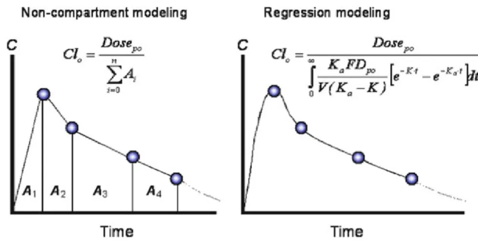
Figure 7. Comparison of NCA (left) and nonlinear regression modelling (right). ... 36
Figure 8. Illustration of positive and negative AUEC versus baseline. ... 41
Figure 9. Simple graphic representation of a one compartment structural model for a medicine with oral administration ... 45
Figure 10. Simple model of metabolite kinetics ... 48
Liste des abréviations et sigles
λz Terminal elimination rate constant Aβ Alpha-beta fibreA Alpha-delta fibre β-EP β-endorphin
Epsilon
Theta
2 Sigma squared t1/2 Half-life
Tmax Time to maximum concentration 5-HT Serotonin
AAG Alpha 1-acid glycoprotein ACH Acetylcholine
AND Adenosine
AIC Aikaike information criteria AUC Area under the curve
AUC0- Area under the plasma-concentration-time curve from 0 to infinity AUEC Area under the effect-time curve
AUMC Area under the curve for the first moment ATP Adenosine triphopshate
BIC Bayesian information criteria BLQ Below the limit of quantification BSV Between subject variability C Concentration of drug CA2+ Calcium ion
cAMP CyclicAMP CB Cannabinoids CCK Cholecystokinin
CL Clearance
CLtot Total plasma clearance Clast Last plasma concentration Cmax Maximum plasma concentration CNS Central nervous system
CPM Central pain modulation (previously DNIC) CPT Current perception threshold
CV% Percentage of coefficient of variation CYP Cytochrome P
D Deviation
DAMGO D-Ala2,N-methyl-Phe4,Gly5-ol
DA Dopamine
DF Descending facilitatory DI Descending inhibitory DH Dorsal horn
DNIC Diffuse noxious inhibitory controls (now known as CPM) DNP Descending inhibitory pathways
DP Descending pathway
DPN Descending pathways neuron DRG Dorsal root ganglia
DRT Dorsal reticular nucleus DYN Dynorphin
E Effect
ECG Electrocardiogram
EM Endomorphin
EMA European medicines agency ENK Encephalin
EPS Epsilon
F Bioavailability
FOCE First-order conditional estimation GABA Gamma-hydroxy-butyric acid GAL Galanin
GFR Glomerular filtration rate GLU Glutamate GLY Glycine GPCR G-protein-coupled receptors h Hour Hz Hertz HIST Histamine IA Intra-articular
IIV Inter-individual variability IML Intermediolateral cell column IN Interneurons
ININ Inhibitory interneurone IR Immediate release I.V. Intravenous
ka Rate constant of absorption ke Excretion rate constant kel Rate constant of elimination km Metabolism rate constant K+ Potassium ion
L litre
LRT Likelihood ratio test MC Melanocortin MIT Mean input time
MN Motoneurones
MRT Mean residence time m/s Metres per second
Mu Metabolite in urine NA Not applicable
NA Noradrenaline
NCA Non compartmental analysis NCG Nucleus cuneiformis
NGF Nerve growth factor NMDA N-methyl-D-aspartate NO Nitric oxide NP Not provided NPFF NeuropeptideFF (FLFQPQRFa) NS Nociceptive-specific NT Neurotransmitter
NTS Nucleus tractus solitaris OBSconc Observed concentration OFQ OrphaninFQ (nociceptin) OFV Objective function value
ODM O-desmethyltramadol
ORL 1 Opioid receptor-like 1
OT Oxytocin
PAF Primary afferent neuron PAG Peri acqueductal grey matter PBN Parabrachial nucleus PD Pharmacodynamic PFC Prefrontal cortex PG Prostaglandin PK Pharmacokinetics PN Projection neuron
popPK Population pharmacokinetics PPT Pain perception threshold
PTT Pain tolerance threshold
RM Raphe magnus
RUV Residual unexplained variability RVM Rostro ventromedial medulla SD Standard deviation
SP Substance P
SPID Sum of pain intensity differences S1 Primary somatosensory cortex S2 Secondary somatosensory cortex SR Sustained release
T0 Time zero
TOAD Tramadol extended release formulation intended for once daily dosing U Drug in urine
VAS Visual analogue scale Vd Volume of distribution
Vd/f Apparent volume of distribution VdSS Volume of distribution at steady state VP Vasopressin
VPC Visual predictive checks WDR Wide dynamic range neurons WRES Weighted residuals
To my Mom, who planted the dream of being a scientist when little girls weren’t encouraged to dream in that direction, who always challenged me to be curious and to never quit. To my sister who has taken up that torch. To my dear friends: Sylvie Bouchard, who gave me confidence in my ideas and intelligence and Annie Gingras, who helped me persevere and do it with a sense of humour, without their example as intelligent strong women and their encouragement and support this would not have been possible. To my husband, and children who were always there for me, encouraging me on this journey. Thank you!
Remerciements
I would like to thank my director of research, France Varin for her patience and courage in taking on an adult student. Her knowledge and experience and willingness to share it have been integral to my learning and this research.
I would like to thank my co-director, Mohamad-Samer Mouksassi, for sharing his precious time and passion and knowledge of pharmacometrics.
I would like to thank the members of my jury who have graciously given their time to evaluate my research and given their invaluable advice to improve my thesis.
I would like to thank my former work colleagues, specifically Sylvie Bouchard, Annie Gingras, David Karhu, Caroline Fradette and James Howard-Tripp for their support of me as a student and researcher.
I would like to thank my former and current lab colleagues, Chen Chun Lin, Anne Nguyen, François Gaudreault, Paul Gavra and Fady Thomas for teaching me, challenging me, supporting me and helping me to laugh.
Finally I would like to thank my husband, my children, my Mom, my sister and my dear friends who encouraged me to keep going and supported me in doing so.
Section 1 : Introduction
The proportion and absolute number of elderly people in populations around the globe are increasing because of decreased mortality in infants and young people and increased life expectancy in the elderly. It is forecasted that by 2025 life expectancy will be between 60-80 years in all regions of the world (1). Even in regions such as Africa which currently has many countries with relatively young populations, it is expected that the aging of the population, represented as a threshold of 20% elderly, will be attained at a much faster rate than in countries like France and the UK, that have currently achieved that proportion. This global demographic shift requires a better understanding and treatment of many of the health concerns that elderly persons experience, in order to ensure that individuals have the best possibility for good quality of life as they age. Furthermore, it represents a challenge for societies, particularly in countries with less economic means, to maintain health and social systems. Despite this, research on medicinal treatments used in elderly patients is lacking. A search of the Clinicaltrials.gov data base revealed that in 2010, of the 1545 clinical trials conducted in central nervous system (CNS) indications, only 1.5% included patients older than 75 years. Furthermore, less than 10% of drug delivery technology trials conducted included Pharmacokinetic (PK) assessments in the elderly (2, 3).
Older adults are at higher risk both for acute and chronic pain (4). The prevalence of pain increases up to the seventh decade of life and may be as high as 50% of persons in the community setting and 80% of persons in residential care facilities (5-7). Pain in the elderly may arise from a variety of sources, with back and neck pain and osteoarthritis being globally amongst the top ten health conditions associated with disability in populations 60 years and older. Furthermore, pain experienced by the elderly is often moderate to severe in intensity. Analyses of data from the 2008 cross-sectional, National Health and Wellness Survey in 5 European countries (France, Germany, Italy, Spain and the UK) revealed that in persons over 60 years of age reporting pain within the month prior to the survey, intensity was severe or moderate in a proportion of 24% and 63%, respectively (8). The natural adaptive response of limiting activity due to acute pain, can become maladaptive in the situation of persistent pain
motion, muscle strength and tone and increase in weight, all of which can lead to greater pain. Furthermore, the older person may also restrict social interaction, an important factor in successful aging (9, 10). The complexity of using analgesics in elderly persons cannot be underestimated, key considerations that affect PK and Pharmacodynamics (PD) include age-associated changes in body composition and function. This is particularly important in the presence of frailty and impaired cognition and must take into account the heterogeneity of the expression of these traits of aging in individuals, some of whom may remain relatively healthy into the last decades of life while others experience impairments earlier (11).
A conventional definition of “elderly” is chronological age of 65 years old or older, while individuals 65 through 74 years old are referred to as “early elderly” and those over 75 years old as late elderly (12). Others have identified that chronological age is not a reliable way to identify elderly persons at risk. Instead they propose that a phenotype of frailty is a better marker for risk in the elderly. Frailty is theoretically defined as a clinically recognizable state of increased vulnerability resulting from aging-associated decline in reserve and function across multiple physiologic systems such that the ability to cope with everyday or acute stressors is comprised (13). To meet an operational definition by Fried et al. (14) the elderly person must meet three out of five criteria: low grip strength, low energy, slowed walking speed, low physical activity, and/or unintentional weight loss. In a recent review of the definition of elderly in 20 clinical practice guidelines, Singh and Bajorek (11) found that 3 clinical guidelines define elderly based on chronological age and the remaining 17 provide no definition. They indicate that representation of ‘elderly’ in guidelines needs to be less based on chronological age or generic definitions rather they should establish a direct link between an individual patient’s characteristics and the pharmacology of their prescribed medication.
Good pain treatment in the elderly must be based on sound understanding of the circumstances of aging, including the presence of comorbidities, polypharmacy and variability in the aging process and PK and PD of medications (10). Yet PK and PD data on analgesics in elderly patients, especially those aged >75 years, are sparse (1-3, 10). Standard pain treatments must be studied to determine the impact of age related changes on PK and PD, particularly with regard to analgesic efficacy and the effect of co-morbid diseases and concomitant medications (15).
Chapter 1 : Fundamental and clinical aspects of pain and
aging
1.1 Anatomy and physiology of pain systems
The nociceptive system is a dynamic system that undergoes plastic changes and is a result of the modulation of afferent activity via peripheral and central mechanisms (12). Understanding this system requires knowledge of its physiology as well as molecular and behavioral pharmacology. Perception of and reaction to painful stimuli requires the interaction of a series of complex mechanisms: reception of noxious stimuli, transmission of information about those noxious stimuli from the periphery to central nervous system (CNS), perception and reaction in the higher centres and modulation of the pain signal.
1.1.1 Anatomy of the pain system
The cells of the pain system can be divided into four main categories: primary afferent neurons (PAF), projection neurons (PN), interneurons (IN) and neurons of the descending pathways (DPN) (Figure 1).
Primary Afferent Neurons
Primary afferent neurons (PAF) terminate in free nerve endings known as nociceptors that are found in the skin, muscles, joints and viscera. Two types of PAF are associated with these nociceptors namely Aẟ and C fibres (Figure 1). Aẟ nociceptors are responsible for the sensation of sharp, acute pain and respond to mechanical and thermal nociception and while C nociceptors are responsible for the sensation of slow burning pain from mechanical, thermal and chemical stimuli and constitute the majority of nociceptors. Aẟ nociceptors, which are larger myelinated fibres of 1-5 μm diameter, rapidly transmit nociceptive stimuli at 5-30 meters/second (m/s). Aẟ nociceptors are mainly specialized to detect dangerous mechanical and thermal stimuli and trigger a rapid response. C fibres, which are unmyelinated and smaller (0.2 to 1.5 μm) in diameter result in a slower transmission of signals (0.5-2 m/s), respond to
specialized to detect single sensations such as pinch or heat but most are polymodal. PAFs run from the peripheral site of injury primarily to the I and II laminae of the dorsal horn (DH) of the spinal cord.
Projection Neurons
Projection neurons (PN) synapse with the PAF in the DH of the spinal cord and project to the thalamus, hypothalamus, nucleus tractus solaris (NTS), parabrachial nucleus (PBN), periaqueductal grey matter (PAG) and amygdala (Figure 1). PNs can either transmit only nociceptive information or they can be non-specific receiving both nociceptive information from Aẟ and C fibres and other sensory information from Aβ fibres (sensory neurons that detect light touch) and these non-specific PN are known as wide dynamic range (WDR) neurons.
The PN decussate in the DH before ascending in the contralateral spinal tract. There are two primary tracts through which this secondary neuron may ascend:
• The spinothalamic tract is important in the localisation of pain. Secondary neurons that follow this tract synapse with a third neuron within the thalamus; this third neuron then ascends and terminates in the somatosensory cortex. • The spinoreticular tract is important in the emotional aspects of pain; it ascends
to the reticular formation of the brainstem before passing through the thalamus and hypothalamus and making many further projections into the cortex.
As the PN passes through the PAG and raphe magnus in the Rostrovental Medulla (RVM), it makes a variety of synaptic contacts that have important functions in the modulation of pain. PAF, IN and DPN interact to determine the activity of the PN (13).
Interneurons
Interneurons (IN) are located in the DH of the spinal cord and brainstem (PAG, RVM). They can act as inhibitory interneurons (ININ), also called OFF cells, or as excitatory interneurons (EXIN), acting pro-nociceptively or anti-nociceptively, respectively (Figure 1).
Neurons of the descending pathways
Neurons of the descending inhibitory pathways (DNP) can be part of the descending facilitatory pathway (DF) or the descending inhibitory (DI) pathways (Figure 1). These neurons originate in the RVM and other brainstem nuclei descending to the DH where they interact with the PAF, IN and PN as well as pre-ganglionic neurons of the sympathetic system and motorneurons (MN).
1.1.2 Initiation of the pain system response to noxious stimuli
Thermal, chemical or mechanical noxious stimuli result in the activation of mast cells close to nociceptors leading to the release of inflammatory mediators (e.g. histamine, nerve growth factor (NGF), bradykinin and prostaglandin). Binding of these inflammatory mediators to receptors such as G protein-coupled receptors (GPCR) and Tyrosine Kinase A receptors in the cell membrane of the nociceptor leads to activation of the primary afferent fibre by means of propagation of a graded action potential (14).
The signal transmitted from the nociceptor is processed within the brain. As stated earlier, the somatosensory cortex is key to the localisation of pain. However, other areas including the primary and secondary somatosensory cortex (S1 and S2), the insulae, the anterior cingulate, prefrontal cortex and thalamus are also important in pain perception and emotional and physical response.
Pain modulation is another important mechanism in the perception of and response to pain. In general, four regions of the CNS are involved in pain modulation (12):
1) Segmental signal inhibition which involves the inhibition of pain by IN in the DH of the spinal cord
2) Conditioned pain modulation (CPM (previously DNIC)) which uses heterotopic stimulation to reduce the intensity of perception of pain
3) Inhibition through the brainstem network in the PAG and RVM that modulate pain transmission through pronociceptive (ON) and antinociceptive (OFF) cells. 4) Cognitive and affective cortical centres appear to exert a top-down control in
A neuroimaging study by Hadjipavlou et al. (15) found an anatomical link between descending inhibition from higher centres of the brain, such as the prefrontal cortex (PFC) , amygdala, thalamus and hypothalamus, to the descending pain modulatory system in the PAG, the RVM and the Nucleus Cuneiformis (NCG). These higher centres may play an important role in the response to pain. The amygdala is posited to affect the uncertainty associated with pain and fear and therefore to allow humans to plan antinociceptive strategies. Opioid-induced hypoalgesia in the amygdala, PAG and RVM suggests, in turn, that these three regions are involved in planning and mediating antinociception. Descending inhibitory neurons from these higher centres project to IN and secondary neurons in the DH of the spinal cord to inhibit or enhance pain transmission. (16-22).
Figure 1. Ascending and descending inhibitory pain pathways
Aβ – Alpha-beta fibre; Cfibre – C fibre; Aδ– Alpha-delta fibre; EXIN – excitatory interneurons; DP –descending pathway; DRG – dorsal root ganglia; DRT – dorsal reticular nucleus; IML – intermediolateral cell column; IN – interneurons; ININ inhibitory interneuron; MN – motoneurons; NS – nociceptive-specific; NTS – nucleus tractus solitaries; PAF – primary afferent fibre; PAG – periaqueductal grey; PBN – parabrachial nucleus; PN – projection neurons; PreG – preganglionic; RVM – rostroventral medulla; WDR – wide dynamic range (16)
Reprinted from Prog Neurobiol.; 66(6): 355-474, 2002. Millan MJ. Descending control of pain (16) with permission of Elsevier. Whether the descending inhibitory neurons inhibit or enhance pain transmission is governed by a series of neurotransmitters, amongst them are monoamines, noradrenaline and serotonin (Figure 2) (16). Opioid receptors are highly expressed in descending modulatory pathways including RVM and PAG and activation of opioid receptors in these locations directly inhibits pain transmission in the spinal cord (13).
Figure 2. Neurotransmitter inhibition or enhancement of pain transmission
β-EP – β-endorphin; 5-HT – serotonin; ACH –acetylcholine; ADN – adenosine; CB – cannabinoids; CCK –cholecystokinin; CGRP – calcitonin gene related peptide; DA – dopamine; DRG – dorsal root ganglion; DYN – dynorphin; EM – endomorphin; ENK – encephalin; EXIN - excitatory interneuron; GABA – γ-hydroxy-butyric acid; GAL – galanin; GLU – glutamate; GLY – glycine; HIST – histamine; ININ - inhibitory interneuron; MC – melanocortin; NA – noradrenaline; NMDA – N-methyl-D-aspartate; NO – nitric oxide; NPFF – neuropeptideFF; NT-neurotransmitter ; OFQ – orphaninFQ (nociceptin); OT – oxytocin; PG – Prostaglandin; PN – projection neuron; SP – substance P; VP - vasopressin Reprinted from Prog Neurobiol.; 66(6): 355-474, 2002. Millan MJ. Descending control of pain (16) with permission of Elsevier.
1.2 Age related changes in the pain system
As referenced earlier, the prevalence of pain especially chronic pain increases from middle age onwards. Given the general trend to age related sensory decline in other sensory systems, such as the taste, auditory and visual systems, it would be surprising that there is no similar trend in the sensory capacity of the pain system (17, 18). It is generally held that the threshold for perception of painful stimuli also named presbyalgos is increased while pain tolerance threshold (PTT) is decreased in elderly subjects and patients (9, 17, 19). Research in both animal models and humans has attempted to elucidate the basis for age related changes in the perception of and response to pain.
Reviews of the preclinical literature on age deficiencies in nociception and pain behavior (18, 20) found that beginning at midlife, changes in neuroanatomy, neurochemistry and pain modulatory systems may be associated with alterations in sensitivity. The conclusion of this review was that, in rats:
Reflexive responses to painful stimuli were not changed with age; although it may take longer for older animals to undertake complex avoidance behaviors Increased sensitivity to tonic pain starting at mid-life may be the result of a
reduction in the size and number of neurons in the dorsal root ganglia and degeneration of neural inhibitory system
Relevant to our study, Hoskins et al. (21) found that, in rats, there is a loss in efficacy of spinally administered opioids and subsequent research indicated that, although the density of μ-opioid receptors was not decreased, there was a reduced affinity of [D-Ala2 ,N-methyl-Phe4,Gly5-ol]enkephalin (DAMGO), a u-opioid receptor agonist, in elderly rats as compared to young or mature rats.
Studies in humans, in general, have drawn inconsistent conclusions with regard to the purported increase in PPT and decrease in PTT in the elderly (22). In experimental studies, the modality of the painful stimulus seems to play a key role. PPT has been shown to decrease with thermally induced pain (23-26) but to increase following mechanically induced pain (27,
stimuli are less clear with one demonstrating a no change (29) and two demonstrating reduced PTT (30, 31). Perception of painful stimuli may be affected by age related effects on peripheral nociceptors. After thermal noxious stimuli, myelinated A-ẟ fibres showed reduced pain perception and longer sensory evoked potentials while both parameters remained unchanged for unmyelinated C-fibres. This apparent discrepancy is possibly due to reduced density and function of myelinated fibres, including structural modification and reduced conduction velocity with age (32, 33). Tseng et al. (34) found a reduction in the sensory areas of the brain activated and the magnitude of the activation in the elderly using functional magnetic resonance imaging after noxious thermal stimulation.
Changes in pain modulation mechanisms affect both opioid and non-opioid mechanisms and may play an important role in differences seen in pain tolerance. Evidence has been demonstrated for age related reduced pain-modulatory capacity with regard to central pain modulation (CPM )(35, 36). This research suggested that CPM effects resulted in a higher tolerance to heterotopic cold pain in young subjects and pain ratings associated with the cold stimulus were higher in elderly subjects. Thus, in the elderly increased sensitivity of WDR neurons to noxious stimulation resulting from deterioration of CPM mechanisms could result in a net increase in perceived pain. Clinically, this could explain the lower pain tolerance seen in elderly patients and the increasing prevalence of chronic pain conditions. Moreover, there appears to be differences in neuroplasticity in elderly persons: temporal summation occurring more readily, resulting in heightened sensitivity and heightened risk of the occurrence of chronic pain with age (27).
1.3 Age related changes in pharmacokinetics
The pharmacokinetics of many drugs are altered in the elderly (37). Alterations in organ function, body composition, concomitant medications and the higher risk of co-morbid diseases all play a part in these differences (38). These changes can affect absorption, distribution, metabolism and elimination. Pharmacokinetics of medicines in the elderly, particularly with older analgesics is not well documented (1-3).
Absorption
For orally administered medications, increases in the gastric emptying time and decrease in peristalsis can result in slower transit through the gastrointestinal system altering the time during which the system is exposed to the drug and can absorb it. Gastric pH is increased, decreasing gastric dissolution of basic medications and decreasing absorption of acidic medications. On the other hand, higher content of mucosal connective tissue and reduced mesenteric blood flow along with atrophy of the macro and microvilli result in reduced ability of the system to absorb medications. As a result, many medications have altered bioavailability in the elderly (37).
Distribution
Differences in body composition, rate of blood flow and changes in binding of medications to plasma proteins, fatty tissue and other biologic matter which can lead to medications being distributed differently in the bodies of healthy elderly persons. Many of the co-morbid diseases for which elderly have greater risk can further affect distribution.
Elderly persons have a lower lean body mass and higher ratio of fatty tissues. With increasing age total body fat increases from 18% to 48% in females and from 18% to 36% in males. The amount of extracellular fluid remains unchanged but its proportion in the body increases with age along with a decrease in intracellular fluid which is a reflection of decreasing cell mass. All of these can have the effect of medications having a different volume of distribution than in younger subjects, since less or more of the medication may be retained in the circulatory or central compartment. For example for lipophilic drugs, volume of distribution (Vd) is increased.
Blood flow is reduced with age. Cardiac output declines roughly 1% yearly after the age of 25 and regional blood flow shows a similar yearly decline in flow to the brain (-0.35 to -0.5%), heart (-0.5%), liver (-0.3 to -1.5%), and kidneys (-1.1 to -1.9%). Corresponding changes in the ability of medications to distribute to less vascularized compartments such as fatty tissue and peripheral tissues will follow.
Plasma proteins remain roughly the same with age with the exception of plasma albumin in the frail elderly subject which can affect the Vd of highly bound acidic drugs with varying clinical significance; a greater free fraction of albumin bound drugs carries the potential for greater efficacy and toxicities. Alpha 1-acid glycoprotein (AAG) is increased in acute illness and chronic inflammatory diseases decreasing the free fraction of basic drugs such as propranolol (and tramadol) resulting in the potential for reduced efficacy in the elderly (39).
Metabolism
Hepatic metabolism or clearance of medications is related to the ability of the liver to biotransform medications to more easily eliminated metabolites and the ability of the cardiovascular system to present the drug to the liver where enzymes capable of metabolizing the drug are present. Hepatic clearance is the result of liver blood flow and hepatic extraction ratio. Depending on the ratio of hepatic clearance of a drug to the hepatic blood flow, extraction is generally classified as high (>0.7), intermediate (0.3-0.7) or low (<0.3) and represents the fraction of drug removed during one pass through the liver. Age related decrease in hepatic blood flow can reduce hepatic clearance of medications with a high hepatic extraction ratio and, in turn, increase bioavailability.
Several low extraction or capacity limited drugs metabolised by Phase I reactions have shown a significant reduction in clearance in the elderly. Cytochrome P 450 (CYP) enzymes are important in Phase I metabolic reactions of many medicines. CYP2D6, found in the liver and brain and CYP3A4 in the liver and gut, are important in the metabolism of opioid analgesics. CYP3A4 is responsible for metabolism of almost 50% of medicines as such medicines that utilise this metabolic pathway have a high potential for interaction with other medicines metabolised by CYP3A4, an important consideration in elderly patients who often take many medications. It has been shown in several studies that CYP2D6 remains unchanged with age while in some studies CYP3A4 has been shown to be reduced while others show no change (40-42). Studies have shown that monoamine oxidase activity is maintained with ageing (41, 43, 44). Conjugative metabolism is not generally affected by aging (45, 46).
Excretion
Renal clearance is reduced in the elderly and may have a variety of causes including reduced renal blood flow, reduced active tubular transport, loss of functional nephrons or all of these (37). Both glomerular filtration and maximum tubular secretion decline by approximately 0.6 percent per year after 25 years of age. Drugs having a high fraction excreted unchanged in urine will be mostly affected. However, it is difficult to distinguish the relative contribution of hepatic clearance and renal clearance to overall clearance, as both are susceptible to age related changes.
Elimination
Often half-life is prolonged in the elderly. In absence of intravenous drug administration in both young and elderly subjects, it is almost impossible to determine whether the net effect on half-life is related to alteration in total body distribution or clearance.
1.4 Pharmacology of pain
1.4.1 Pain treatment in the elderly
Pain is highly prevalent in the elderly, recent observational studies have shown that elderly patients are systematically undertreated (47-49). The selection of appropriate analgesics in elderly requires careful consideration of a variety of factors such as age related changes in body composition, co-morbid medical conditions and polypharmacy which can lead to heterogeneity in analgesic effect and side effects. The picture can be further complicated by the potential presence of frailty which is not necessarily tied to chronological age or impaired cognition.
A cross-sectional study of 21 380 nursing home residents aged 65 and older in nursing homes in 10 U.S. states found that the most common treatments for persistent pain were acetaminophen (37.2%), propoxyphene (18.2%), hydrocodone (6.8%) and tramadol (5.4%) (49). The 2008 consensus statement on opioid use for severe chronic pain in the elderly, focused their review on buprenorphine, fentanyl, hydromorphone, methadone, morphine and
cancer related and non-cancer related pain conditions. Non-cancer related pain includes conditions such as low back pain, osteoarthritis and neuropathic pain.
Acetaminophen is a widely used analgesic and the drug of choice for mild to moderate pain on its own or in combination with stronger analgesics such as opioids (50, 51). It is frequently used to treat mild to moderate osteoarthritis and other painful conditions that affect the elderly. Although a recent meta-analysis of 137 studies comprising 33 243 participants found that acetaminophen was least likely amongst diclofenac, ibuprofen, naproxen, celecoxib, intra-articular (IA) corticosteroids and IA hyaluronic acid to be efficacious (52). There may be some effect of age on the PK of acetaminophen in healthy elders with reported results being variable (45, 53-55). Decreases in volume of distribution have been observed with increasing age (55). Frailty in older persons does seem to be associated with reduction in total clearance of acetaminophen in the elderly (45, 54-56), suggesting that in healthy older people intrinsic oxidative metabolism may be intact while in frail elderly it may be compromised (10). This is of particular concern with regard to unintentional overdose of acetaminophen, liver disease or use with alcohol amongst other factors putting the frail elderly patient at higher risk for formation N-acetyl-p-benzoquinoneimine and hepatic centrilobular necrosis (10). Recently, the safety of recommended doses of acetaminophen in the elderly are being questioned particularly in the frail elderly. Elderly people may have a worse benefit/risk ratio. Risk factors can include polymedication, glutathione depletion, organ insufficiency, malnutrition, dehydration and fragility (10, 57, 58).
Commonly used pain relievers such as ibuprofen and naproxen are non-selective cyclo-oxygenase inhibitors and carry a significant risk of cardiovascular events including death, gastrointestinal bleeding and kidney dysfunction and are used with extreme caution or not at all in the elderly. Oral nonselective and selective NSAIDs are rarely used in elderly patients due to the potential cardiovascular risks (10, 50-52). These side effects are generally not associated with use of tramadol and other opioids, making them an option for older patients with chronic pain (50).
When pain worsens, traditional opioids such as morphine, oxycodone, fentanyl, or buprenorphine may be added. There is debate about the value of tramadol and tapentadol, as these drugs having opioid and non-opioid mechanisms of action exhibit side effects and potential for drug interactions leading to serotonin syndrome. However, there is a general agreement that making use of multiple mechanisms of action (referred to as multimodal analgesia) along with non-pharmaceutical approaches provide better relief (51).
Treatment of moderate to severe pain in elderly patients is an important aspect of their care and opioids are considered an important tool (2, 50, 51, 59) with well-known efficacy but also potential for harm. Opioid related harm is significantly related to increasing age including risks of respiratory depression and falls and fractures (60). There are a variety of opioid options for treating elderly patients in pain, including morphine, codeine oxycodone, hydromorphone, fentanyl, tramadol, methadone, buprenorphine and tapentadol (59). Important considerations in the choice of an opioid in elderly especially frail elderly patients are the patient’s renal function and the route of excretion of the opioid chosen, renally excreted opioids may accumulate in elderly people with impaired renal function (10, 59). Furthermore, since elderly patients are often taking many medications, understanding the potential for drug-drug interactions with opioids especially those metabolised by CYP enzymes such as tramadol and codeine is important. The choice of which opioid to use should be considered in the context of the characteristics of the individual patient, such as presence of complex comorbidities, psychosocial considerations, co-medications and careful management of side effects is a key consideration (59).
1.4.2 Opioid mechanism of action
Modulation of pain perception and response involves critical endogenous opioid systems and these systems are a major target of analgesic strategies (61, 62). Opioids are implicated in many molecular/cellular responses related to pain and affect including behaviours related to analgesia, reward, depression and anxiety. Opioid receptors are expressed in a variety of locations throughout the pain system including in afferent nociceptive neurons, the spinal cord and the descending modulatory pathways. In primary
inflammation (63, 64). In the spinal cord and in pain modulating descending pathways, they directly inhibit interneurons, which in turn inhibit spinal cord transmission (62, 65). Descending pain control centres tend to have high concentrations of opioid receptors and endogenous opioids (66). There are four opioid receptor subtypes: μ, ẟ, k and opioid receptor-like 1 (ORL1) receptors. The μ receptor is the most ubiquitous opioid receptor in the spinal cord and is the main modulator of the pain system (67). In the spinal cord, 70% of opioid receptors are located pre-synaptically where they inhibit calcium influx by enhancing outward movement of potassium or inhibit adenylate cyclase conversion of adenosine triphosphate (ATP) to cyclic AMP (cAMP) and therefore, preventing the release of Substance P and CGRP (Figure 3). Post-synaptical opioid receptor activation results in inhibition of potassium ion efflux which, in turn, decreases neuron excitability. Opioids mainly excite the prefrontal cortex, hypothalamus, amygdala and cingulate gyrus resulting in an indirect excitation of neurons in the PAG, as well as also directly exciting PAG neurons projection to the RVM where they will affect ON and OFF cells by inhibiting opioid receptor bearing ON cells. They also inhibit GABAergic inputs to OFF cells leading to inhibition of the transmission of nociception (Figure 3).
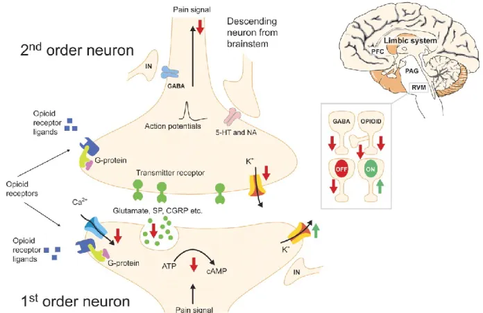
Figure 3. Opioid molecular mechanism of action in the spinal cord
5-HT – serotonin; ATP – adenosine triphosphate; CA2+ – calcium ion; cAMP –cyclic adenosine monophosphate; CGRP – calcitonin gene related peptide; GABA –
γ
-hydroxy-butyric acid; K+ - potassium ion; IN – interneuron; NA – noradrenaline; PFC – prefrontal cortex; PAG – periaqueductal grey matter; RVM – rostroventral medulla; SP – substance P (62). Reprinted from Olesen AE, Andresen T, Staahl C, Drewes AM. Human experimental pain models for assessing the therapeutic efficacy of analgesic drugs. Pharmacol Rev. 2012;64(3):722-79 with permission of Aspet journals.Opioid receptors are 7-transmembrane spanning proteins that, following activation by an agonist, couple to inhibitory G-proteins (Gα and Gβγ subunits), then dissociate from one another and subsequently act on various intracellular effector pathways (61).
Opioids are currently the most efficacious analgesics for moderate to severe pain (68), yet their clinical utility continues to be limited by a compromise between efficacy and side effects, particularly in the elderly. The most common side effects of opioids can be divided into peripheral (constipation, urinary retention, hives, bronchospasm) and central effects (nausea, sedation, respiratory depression, hypotension, myosis, cough suppression) (61).
1.4.3 Tramadol
1.4.3.1 Tramadol mechanism of action
First synthesised in 1962, tramadol hydrochloride is a centrally acting analgesic which is structurally related to morphine and codeine (69). It is a racemic 1:1 mixture of (+)-tramadol and (-)-tramadol and is metabolised to (+/-)-O-desmethyltramadol (ODM; also known as M1) and (+/-)-N-desmethyltramadol as well as a number of other metabolites (70). The racemate, its enantiomers and the ODM metabolite, are all implicated in the production of anti-nociception through both non-opioid and opioid mechanisms (71). In-vitro and in-vivo studies have shown enantioselective pharmacology, pharmacokinetics and metabolism. These studies also reported that each enantiomer contributes to the analgesic effect of tramadol via different mechanisms of action and each enantiomer contributes synergistically to the drug effect (72, 73).
Tramadol acts as an opioid agonist by selectively binding to receptors in the spinal cord and brain (74, 75), although with much less affinity than codeine (1/10) and morphine (1/6000). The parent compound, tramadol, binds weakly to μ-opioid receptors; however, the (+)-ODM metabolite has 200 times the affinity of the parent drug. As a result, the opioid action of tramadol is thought to be primarily linked to the (+)-ODM metabolite (76, 77). Table 1 presents the affinities of tramadol, O-desmethyltramadol and their enantiomers as well as that of morphine for the μ-opioid receptor and serotonin (5-HT-2C) transporters.
Table 1. Relative affinity of racemic tramadol, tramadol enantiomers, O-desmethyltramadol and morphine Ki (µM) μ –opioid receptor Serotonin (5-HT 2C) transporter NE transporter (+/-)-tramadol (78) 2.4 0.78 0.90
(+)-tramadol (78) Not reported 2.51 0.53
(-)-tramadol (78) Not reported 0.43 2.35
(+/-)-O-desmethyltramadol (79) 0.0054 Not reported Not reported (+)- O-desmethyltramadol (79) 0.0034 Not reported Not reported (+/-)-O-desmethyltramadol (79) 0.24 Not reported Not reported
Morphine 0.0012 No effect No effect
The non-opioid mechanism of tramadol has been elucidated through studies that demonstrated a lack of reversibility of the analgesic effect by naloxone, lack of any naloxone induced withdrawal symptoms, production of mydriasis rather than miosis and reduction of analgesic effect with co-administration with non-opioid antagonists (71, 73, 80). In a study examining the actions of (+)-tramadol, (-)-tramadol and (+)-O-desmethyltramadol and (-)-O-desmethyltramadol on electrically evoked norepinephrine efflux and re-uptake in rat coeruleus brain slices, mean norepinephrine efflux was significantly (p < 0.01) increased by racemic tramadol (66%; SEM: 10%) and its (+)- enantiomer (57%; SEM: 10%) and (-)-enantiomer (64%; SEM: 13%). Norepinephrine re-uptake was blocked only by (-)-tramadol (p < 0.01), which increased the re-uptake half-time to 499% (SEM 63%) of pre-drug values. At the test drug concentrations, O-desmethyltramadol was inactive with regard to norepinephrine efflux or re-uptake (81). In a study of the actions of racemic tramadol, (+)-tramadol, (-)-tramadol and O-desmethyltramadol on electrically evoked serotonin efflux and uptake in rat dorsal raphe nuclei in the RVM, racemic tramadol and the (+)-tramadol enantiomer significantly blocked
(+)-tramadol: p < 0.05) while O-desmethyltramadol and the (-)- enantiomer were inactive at the concentrations used in the study. (82) The non-opioid mechanism of action of tramadol involves activation of descending noradrenergic and serotonergic pathways (71). The (+)-tramadol enantiomer preferentially inhibits serotonin reuptake and enhances serotonin release while (-)-tramadol preferentially inhibits norepinephrine reuptake and enhances stimulation evoked norepinephrine release; (-)-ODM inhibits monoamine uptake (73, 83).
Several human studies using experimental pain models have demonstrated greater analgesic effect of tramadol compared to placebo (80, 84-86). Although designed to demonstrate efficacy versus placebo or other treatments, they do provide some information about onset and duration of analgesic effect which generally appears to occur within 2 hours of administration and to last until the end of the dosing interval (6 hours for immediate release (IR) formulations and 12 h for sustained release (SR) formulations this is limited by sampling frequency and lack of detailed presentation of onset and offset information (Table 1). In a study by Sarbu et al. (87), 47 patients with acute low back pain were administered a single 200 mg extended release tramadol tablet (intended for once daily administration). The patients indicated the time of onset of pain relief using the stopwatch method. Ratings of pain intensity and pain relief and pharmacokinetic samples were taken prior to dosing, at the onset of pain relief and 3 and 6 hours postdose. No rescue medication was permitted until the end of the study (6-hour postdose). Adverse events were monitored throughout the study. Onset of perceptible pain relief was achieved within 1 hour for the majority of patients and at plasma levels, suggesting a therapeutic threshold between 50 and 100 ng/mL.
Table 2. Summary of results from selected experimental pain models of tramadol Desmeules et al.
n =10a Hummel et al. n = 20b Hogger et al. n = 12c Thurauf et al.n = 20d Study design Randomised,
double-blind, placebo controlled, 4-way crossover Randomised, double-blind, placebo controlled, 3-way crossover Randomised, double-blind, 6-way crossover Randomised, double-blind, controlled, 3-way crossover. Experimental
Pain Model Transcutaneous electrical stimulation of the sural nerve
Tonic pain: dry airstream delivered to right nostril
Phasic pain: CO2 stream applied to nasal mucosa
Electrical stimulation of the tooth pulp of central incisors
Phasic and tonic pain: CO2 stream applied to nasal mucosa alternately in nasal cavity Measurement Electromyographic response measured on the ipsilateral biceps femoris
Pain threshold using a Pain Numerical Rating Scale Chemo-sensory event related potentials (phasic stimuli) Pain VAS Somato-sensory Evoked potentials 8-point categorical pain intensity scale Chemo-sensory event related potentials Pain VAS Treatment Tramadol 100 mg, tramadol + yohimbine, tramadol + yohibine+naloxone, placebo Tramadol IR 100 mg Tramadol SR 100 mg, Tramadol SR 150 mg, placebo 50 mg tramadol, 50 mg tildine + 4 mg naloxone, bromofenac 25, 50 and 75 mg Tramadol 100 mg SR, Tramadol 200 mg SR, placebo Route of Administration
Oral oral Oral oral
Onset of
analgesia (h) Not reported <2 h Not reported < 2 h Duration of
Analgesia
6h 12h* Not reported > 12 h
Time to peak
effect 3.7 h Not reported Not reported 6 h
Result Both subjective (PNRS) and objective (nociceptive reflex/ RIII) pain threshold were increased
VAS and amplitudes of evoked potentials decreased, latencies of evoked potentials and EEG frequency spectrum unchanged
No parameters were
affected Decreased: VAS to tonic pain, amplitude of evoked potentials,, Unchanged: VAS to CO2 stimulation unchanged, latencies of evoked potentials unchanged a n=10 healthy young male volunteers mean age of 25.6 ± 4.5;b n = 20 healthy young volunteers (13 male and 7 female) mean age 27.8 years (range 23-41 years); c n = 12 healthy young volunteers (6 male and 6 female) mean age 25 ± 3.5 year; d n = 20 healthy young volunteers (10 male and 10 female) mean age 26.10 years (22-32
Studies of the analgesic effect of tramadol after use of a percutaneous electrical stimulation and cold pressor experimental pain models in CYP2D6 poor and extensive metabolisers have demonstrated greater analgesic effect among extensive metabolisers than among poor metabolisers, although poor metabolisers still achieved analgesia, possibly as a result of the non-opioid mechanisms of action of the parent compound (88, 89).
The efficacy of tramadol has been demonstrated in studies including elderly subjects up to 80 years of age in a variety of conditions including osteoarthritis, neuropathic pain, acute and chronic low back pain, post-operative pain and dental pain (90-97). It is indicated for moderate to severe pain at doses between 100-400 mg and requires titration to minimize side effects and achieve optimal efficacy.
1.4.3.2 Tramadol Pharmacokinetics
Tramadol and ODM have been shown in humans to have stereoselective pharmacokinetics and metabolism (98). Steady state concentrations of (+)-tramadol were found to be approximately 30% higher than (-)-tramadol and (+)-tramadol half-life was approximately 1 hour slower. Serum concentrations of (-)-ODM were found to also be approximately 30% higher than (+)-ODM in 12 healthy young (18-22 years of age) male subjects administered as a single oral 100 mg dose of tramadol sustained release tablets twice daily for 11 days. Of note the volunteers were not screened for CYP2D6 status. It is not expected that these differences are clinically significant (98). A population pharmacokinetic (popPK) analysis of two studies: one in 12 healthy young male volunteers and a second in 24 healthy young (22-26 years of age) volunteers (12 males and 12 females) administered intravenous (I.V.) and oral tramadol found similar results and furthermore that the enantioselectivity appears to be administration route dependent (99).
After I.V. administration, tramadol has an initial distribution phase with half-life of 6 minutes, which consists of a faster distribution into tissues of the central compartment consisting of blood and highly perfused tissues (e.g., kidney, liver) and a slower distribution phase with a half-life of 1.7 hours for equilibrium between tissues of the peripheral compartment and the blood (72, 100) A bioavailability study comparing 10 healthy young male subjects administered intravenous and oral tramadol as a 100 mg single dose found a