Skin Tensile Properties in Patients Treated for Acromegaly
Catherine Braham a Daniela Betea b Claudine Piérard-Franchimont a Albert Beckers b Gérald E Piérard a
Departments of (a) Dermatopathology and (b) Endocrinology, University Medical Center of Liège, Belgium
Correspondence : Prof G.E. Piérard, MD, PhD Department of Dermatopathology,
CHU Sart Tilman B-4000 Liège (Belgium)
Tel +32-43662408, Fax +32-43662976 Email Gerald.Pierard@ulg.ac.be
Abstract
Background : Somatotropic effects are described in the skin. Indeed, acromegaly is in part clinically recognized by cutaneous coarsening. The actual changes in tensile properties associated with the cutaneous manifestations are largely unknown. Objectives : To study the relationships between the skin tensile properties and the severity of acromegaly as assessed by serum levels of growth hormone (GH) and insulin-like growth factor-1 (IGF-1). Patients and method : Assessments were made in 13 patients with acromegaly treated by somatostatin agonists combined or not with surgery. A total of 39 age-and sex-matched healthy subjects served as controls. Skin tensile properties were measured on the forearm and nape of the neck using a computerized suction device. Results : Significant differences were yielded between the skin tensile properties in patients and normal subjects. The highest IGF-1 values in the patientsʼ medical records were positively correlated with both skin distensibility and biologic elasticity. The most recent IGF-1 serum levels were negatively correlated with the visco-elastic ratio. No correlations were yielded between any of the biomechanical parameters and GH levels, disease duration and treatment dosages, respectively. Conclusion : Skin in acromegaly appears to be functionally more redundant and elastic than normal skin. The biomechanical changes appear quite different from those observed in other diseases with collagen deposition such as diabetes mellitus and scleroderma.
Key Words : Acromegaly. Dermis. Mechanical properties. Growth hormone. Insulin-like growth factor-1.
The overall tensile properties of skin largely depend on the composition and architecture of the dermis [1]. As a consequence, many connective tissue disorders are in part characterized by typical changes in the tensile characteristics of the skin [1-3]. Several non-invasive methods are available to assess such features [4].
In acromegaly, skin is enlarged, acquires a doughy texture and typically presents disproportionate skinfolds [5, 6]. Skin coarsening is primarily due to an increase in the amount of collagen and glycosaminoglycans [7-9].Objectivation of the functional consequences of this organomegaly has not yet been studied thoroughly. Reports have shown that injections of growth hormone (GH) in adults may result in increased skin thickness [10] and in unusual viscoelastic changes in the skin [11]. In fact, there is ample evidence that fibroblasts and other cells of the skin respond to GH and insulin-like growth factor 1 (IGF-1) [12-22]. These findings suggest that epidermal, adnexal and dermal cell populations can be direct and indirect tarrgets for GH [22]. Indeed, transgenic mice overexpressing GH show skin overgrowth with increased dermal thickness, significant dermal fibrosis and replacement of subcutaneous adipose tissue by fibrous tissue [23]. By contrast, GH and IGF-1 deficiencies show effects on the skin by decreasing its thickness and weakening its mechanical properties [24, 25].
The aim of the present study was to assess the in vivo tensile properties of the skin in patients treated for acromegaly, and to compare it with serum levels of GH and IGF-1.
Patients and methods
Thirteen patients with acromegaly were enrolled in the study (Table I). The M:F sex ratio was 1.6. All but one patients were treated with lanreotide (Somatuline®,
Ipsen) or octreotide (Table I). Serum levels of GH (GH IRMA C.T., BC 1012, Biocode, Belgium) and IGF-1 (IGF-1 IRMA, BC 1010, Biocode, Belgium) were retrieved from the medical files. The highest GH and IGF-1 values in the medical records of each patient were considered to be representative of the climax severity of the disease. The serum levels of these biomolecules recorded at the time of the study were also retained to assess the current status of the disease. A control group was formed by 39 age- and sex-matched healthy subjects (3 normal matched volunteers for each patient with acromegaly).
The tensile properties of the skin were evaluated using a computerized suction device (Cutometer® SEM 474, C + K Electronic, Cologne, Germany) equipped with a hand-held probe having a central suction head of 4 mm in diameter. The time/strain mode was used with three cycles of 5-s traction under negative pressure of 500 mbar separated by 5-s relaxation periods (Fig. 1). The skin tensile parameters considered in this study have been previously described in detail [4, 26-30]. Each load cycle of suction to the skin results in an immediate elastic distension (ED), recorded at 0.15 s, followed by a delayed visoelastic distension, ending with the measurement of the maximum distension (MD) of the skin obtained after a 5-s traction. When the traction is discontinued, the skin tends to return to its initial position. During that phase, two parameters are recorded, namely the immediate elastic retraction (ER) after 0.1 s of relaxation and the resilient distension (RD) after a 5-s relaxation time. These measures were taken during the first and third load cycles. The differential distension (DD) was measured as the difference in MD between the third and the first cycles. In addition,, certain biologically relevant ratios of these parameters were calculated (Table II).
Measurements were taken from the left mid volar forearm and the nape of the neck. The values were expressed as means (M) and SD with calculation of the coefficient of variation (CV =102 SD. M -1). Differences between body sites were tested for their statistical significance using the paired Student t-test. Regression analysis models were applied to evaluate the relationships between the biomechanical values and hormonal levels. The best model, i.e. linear, logarithmic, exponential or power, was chosen on the basis of the highest coefficient of correlation (r). A p value lower than 0.05 was considered significant.
Results
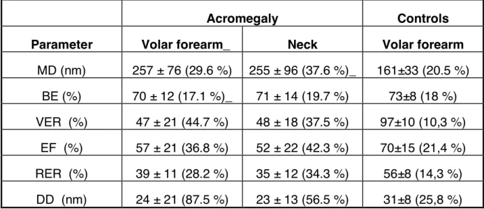
For each biomechanical parameter, interindividual variability was quite large among patients with acromegaly (Table III). In contrast, intraindividual differences between the two body test sites were very low. In absence of any significant regional differences (Table III), the values obtained from the volar forearm and the nape of the neck were averaged for further search of correlations with hormonal values. Compared to matched normal subjects, patients with acromegaly showed a significant increase in MD (p < 0.01) combined with significant reductions in VER (p < 0.05), EF (p < 0.01) and RER (p < 0.05).
From all the correlations tested between each of the GH and IGF-1 levels, disease duration, somatostatin agonist dosages and skin tensile parameters, only three reached significance (Fig. 2a, b, 3). MD and BE increased in parallel with the values of maximum IGF-1 (linear correlation, r = 0.60 and 0.64, respectively, p < 0.01) . In contrast, VER values were negatively correlated with the most recent IGF-1 serum levels (logarithmic correlation, r = -0.54, p < 0.05).
There is ample evidence that skin is under the influence of many neuroendocrine influences [22]. Previous studies indicated that the precise assessments of the mechanical properties of this organ reveal quite specific changes in hormone-related connective tissue disorders including menopause, diabetes and GH deficiency [1, 11, 25, 28-33]. In addition, such assessments reveal some correlations between the severity in skin and bone alterations [30]. Thus, determining the mechanical properties of skin may also represent a non-invasive tool for predicting internal manifestations of some connective tissue disorders.
It is recognized that the suction method parameters do not lend themselves to quick simple explanation. In the present test design, the 4-mm probe aperture allowed the suction force to be transmitted to the first 1 to 1.5 mm of the dermis. This limitation permits indeed a comparison between the intrinsic properties of normal skin and those of a presumably thicker dermis in acromegaly. Hence, the well recognized effect of tissue thickness upon the biomechanical properties of skin [1, 25, 34] is minimized. Our study indicates that the skin tensile properties are markedly altered in patients treated for acromegaly. This was indeed not a surprise because there is ample clinical clue for such changes. In the overall assessment, skin in acromegaly exhibited increased distensibility with more subtle changes in its viscoelastic aspects. The interindividual variations in skin tensile properties were larger in the patients than in the healthy subjects.
The present data distinguish the most recent and the maximum GH and IGF-1 imbalances in acromegaly. No significant correlation was yielded between GH serum levels and the skin tensile properties. In contrast, distinct correlations were found between each of the IGF-1 levels and values of some biomechanical parameters.
This may be explained by the quite stable intra-individual IGF-1 values in contrast with the variable amplitude and frequency in GH pulses [12, 35]. There was evidence that both maximum skin distensibility (MD) and biologic elasticity (BE) increased with the disease severity as assessed by the highest recorded IGF-1 values. Such a finding is at variance with other skin conditions including diabetes and scleroderma which are also associated with deposits of collagen [27-29]. In these disorders, MD is typically decreased and elasticity changes are variable. Indeed, the functional consequences of increased collagen content in the dermis may be quite different according to the agencement of the fibrils and bundles. It is noteworthy that such structures are stretched and tightly bound each other in scleroderma, resulting in increased skin stiffness. Skin in acromegaly shows a different microanatomy and lacks these tensile characteristics. It appears to be more enlarged, redundant and doughy rather than tight and stretched. Hormonal changes in acromegaly induce imbalanced soft tissue enlargement with increased dermal thickness [12]. This feature cannot explain the MD increase, but rather would be suspected to be responsible for the reverse. The present data suggest that the skin envelope is more increased in area than in thickness. Thus, skin increases in size at a greater extent than needed to simply cover the volume of the underlying bones, muscles and subcutaneous tissues.
The short term effects of hormonal disturbance in acromegaly appear different from those related to the climax severity of the disease. Viscosity of skin (VER) decreased proportionally to the increase in the last IGF-1 recorded value. Such a negative correlation was relatively weak and the scatterplot showed considerable spread. In addition, this aspect is at variance with previous findings made in GH
abusers among adult body builders [11]. In this latter group of individuals, the musculature was indeed impressive and much more developed than in patients with acromegaly. Hence, the skin tension imposed by the volume of muscles was likely different in this distinct group of individuals. In addition, fluid retention was likely present in the dermis. A direct effect of somatostatin agonists on the tensile properties of skin cannot be dismissed from the present observations. However, no correlation was found in this study between the biochemical parameters and the mean drug dosages and the total amount of lanreotide and octreotide administered to the patients.
In conclusion, IGF-1 levels appear to control, at least in part, the tensile properties of skin. The excess in IGF-1 may yield distinct short-term and long-term functional consequences on the skin physiology. The predictive value of the presently described biomechanical alterations for the severity of internal manifestations remains to be settled.
References
1. Piérard GE and the EEMCO group. EEMCO guidance to the in vivo assessment of tensile functional properties of the skin. Part 1: Relevance to the structures and ageing of the skin and subcutaneous tissues. Skin Pharmacol Appl Skin Physiol 1999;12: 352-362.
2. Marks R. Mechanical properties of the skin. In Goldsmith LA (ed) Biomechemistry and Physiology of the Skin. New York, Oxford University Press 1983; pp 1237-1254.
3. Piérard GE, Henry F. Essai de classement catégoriel des propriétés biomécaniques de la peau. Evaluations par la méthode de succion. Nouv Dermatol 1995;14:630-636.
4. Rodrigues L and the EEMCO group. EEMCO guidance to the in vivo assessement of tensile functional properties of the skin. Part 2 : Instrumentation and test modes. Skin Pharmacol Appl Skin Physiol 2001; 14: 52-61.
5. Ezzat S, Forster MJ, Berchtold P, Redelmeier DA, Boerlin V, Harris AG. Acromegaly. Clinical and biochemical features in 500 patients. Medicine 1994;73:233-240.
6. Thiboutot DM. Dermatological manifestations of endocrine disorders. J Clin Endocrinol Metab 1995;80:3082-3087.
7. Gabrilove JL, Schwartz A, Churg J. Effect of hormones on the skin in endocrinologic diseases. J Clin Endocrinol Metab 1962;22:688-692.
8. Matsuoka LY, Wortsman J, Kupchella Ce, Eng A, Dietrich JE. Histochemical characterization of the cutaneous involvement of acromegaly. Arch Intern Med 1982;142:1820-1823.
9. Jorgensen PH, Andreassen TT, Jorgensen KD. Growth hormone influences collagen deposition and mechanical strength of intact rat skin. Acta Endocrinol 1989;120:767-772.
10. Rudman D, Feller AG, Nagraj HS, Gergans GA, Lalitha PY, Goldberg AF, Schlenker RA, Cohn L, Rudman IW, Mattson DE. Effects of growth hormone in men over 60 year old. N. Engl J Med 1990;323:1-6.
11. Piérard-Franchimont C, Henry F, Crielaard JM, Piérard GE. Mechanical properties of skin in recombinant human growth factor abusers among adult body builders. Dermatology 1996;192: 389-392.
12. Barkan AL, Beitins IZ, Kelch RP. Plasma insulin-like growth factor-1/somatomedin-C in acromegaly : correlation with the degree of growth hormone secretion. J Clin Endocrinol Metab 1988;67:69-73.
13. Lobie PE, Breipohl W, Lincoln DT, Garcia-Aragon J, Waters MJ. Localization of the growth hormone receptor/binding protein in skin. J Endocrinol 1990;126:467-472.
14. Granot I, Halevy O, Hurwitz SA et al. Growth hormone and insulin-like growth factor I regulate collagen gene expression and extracellular collagen in cultures of avian skin fibroblasts. Mol Cell Endorinol 1991;80:1-9.
15. Jorgensenr O. Human growth hormone replacement therapy : Pharmacological and clinical aspects. Endocr Rev 1991; 121: 189-207.
16. Lobie PE, Garcia-Aragon J, Wang BS, Baumbach WR, Waters MJ. Cellular localization of the growth hormone binding protein in the rat. Endocrinology 1992;130:3057-3065.
17. Oakes SR, Haynes KM, Waters MJ, Herington AC, Werther GA. Demonstration and localization of growth hormone receptor in human skin and skin fibroblast. J Clin Endocrinol Metab 1992;75;1368-1373.
18. Tavakkol A, Elder JT, Griffiths CEM, Cooper KD, Talwar H, Fischer GJ, Keane KM, Foltin SK, Voorhees JJ. Expression of growth hormone receptor insulin-like growth factor (IGF-1) and IGF-1 receptor mRNA and proteins in human skin. J Invest Dermatol 1992;99:343-349.
19. Batch JA, Mercuri FA, Edmondson SR, Werther GA. Localization of messenger ribonucleic acid for insulin-like growth factor-binding proteins in human skin by in situ hybridization. J Clin Endocrinol Metab 1994;79:1444-1449.
20. Deplewski D, Rosenfield RL. Growth hormone and insulin-like growth factors have different effects on sebaceous cell growth and differentiation. Endocrinology 1999;140:4089-4094.
21. Edmondson SR, Russo VC, McFarlane AC, Wraight CJ, Werther GA. Interactions between growth hormone, insulin-like growth factor I, and basic growth factor in melanocyte growth. J Clin Endocrinol Metab 1999;84:1638-1644.
22. Slominski A, Wortsman J. Neuroendocrinology of the skin. Endocr Rev 2000;21:457-487.
23. Wanke R, Milz Z, Rieger N, Ogiolda L, Renner-Muller I, Brem G, Hermanns W, Wolf E. Overgrowth of skin in growth hormone transgenic mice depends on the presence of male gonads. J Invest Dermatol 1999;113:967-971.
24. Gilhar A, Ish-Shalom S, Pillar T, Etzioni a, Silbermann M. Effect of anti-insulin-like growth factor I on epidermal proliferation of human skin transplanted onto nude mice treated with growth hromone. Endocrinology 1994;234:229-232.
25. Conte F, Diridollou S, Joret B, Turlier V, Charveron M, Gall Y, Rochiccioli P, Bieth E, Tauber M. Evaluation of cutaneous modifications in seventy-seven growth hormone-deficient children. Horm Res 2000;134: 229-232.
26. Henry F, Goffin V, Piérard-Franchimont C, Piérard GE. Mechanical properties of skin in Ehlers-Danlos syndrome, types I, II and III. Ped Dermatol 1996;13: 464-467.
27. Nikkels-Tassoudji N, Henry F, Piérard-Franchimont C, Piérard GE. Computerized evaluation of skin stiffening in scleroderma. Eur J Clin Invest 1996;26:457-460.
28. Nikkels-Tassoudji N, Henry F, Letawe C, Piérard-Franchimont C, Lefebvre P, Piérard GE. Mechanical properties of the diabetic waxy skin. Dermatology 1996;192:19-22.
29. Piérard-Franchimont C, Nikkels-Tassoudji N , Lefebvre P, Piérard GE. Subclinical skin stiffening in adults suffering from type 1 diabetes mellitus. A comparison with Raynaud's syndrome. J Med Eng Technol 1998;22:206-210.
30. Piérard GE, Piérard-Franchimont C, Vanderplaetsen S, Franchimont N, Gaspard U, Malaise M. Relationships between bone mass density and tensile properties of skin in women. Eur J Clin Invest 2001;31:731-735.
31. Piérard GE, Letawe C, Dowlati A, Piérard-Franchimont C. Effect of hormone replacement therapy for menopause on the mechanical properties of skin. Journal of the American Geriatrics Society 1995;43: 662-665.
32. Callens A, Vaillant L, Lecomte P, Berson M, Gall Y, Lorette G. Does hormonal skin aging exist ? A study of the influence of different hormone therapy regimens on the skin of post-menopausal women using non-invasive measurement techniques. Dermatology 1996;193:289-294.
33. Piérard-Franchimont C, Cornil C, Dehavay J, Letot B, Deleixhe-mauhin F, Pierard GE. Climacteric skin ageing of the face. A prospective longitudinal intent-to-treat trial on the effect of oral hormone replacement therapy. Maturitas, 199;32:87-93.
34. Diridollou S, Black D, Lagarde JM, Gall Y, Berson M, Vabre V, Patat F, Vaillant L . Sex and site-dependent variations in the thickness and the mechanical properties of human skin in vivo. Int J Cosmet Sci 2000;22:421-435.
35. Finkelstein JW, Boyar RM, Roffwarg HP, Kream J, Hellman L. Age-related change in the twenty-four-hour spontaneous secretion of growth hormone. J Clin Endocrinol Metab 1972;35:665-670.
Table I - Demographic data of the patients with acromegaly
Patient Gender Age (year)
Time elapsed since diagnosis (year)
Surgery Somatostatin agonist treatment Duration (years) Current mean dose
month Disease status 1 M 54 17 + none healed 2 M 65 5 0 Lanreotide 5 y, 90 mg statilization 3 M 65 1 + Lanreotide 0.5 y, 0 mg healed 4 M 38 1 + Lanreotide 0.5 y, 0 mg healed
5 M 34 5 + Lanreotide 3 y, 120 mg moderate improvement
6 M 49 11 + Lanreotide 11 y, 120 mg stabilization 7 M 73 5 0 Lanreotide 5 y, 90 mg stabilization 8 M 51 13 + Octreotide 11 y, 60 mg stabilization 9 F 71 5 0 Lanreotide 5 y, 120 mg stabilization 10 F 43 17 + Lanreotide 3 y, 120 mg stabilization 11 F 67 13 + Lanreotide 13 y, 60 mg stabilization 12 F 56 9 + Lanreotide 4 y, 90 mg stabilization 13 F 31 6 0 Lanreotide 6 y, 120 mg Stabilization
Table II Rheologic ratios (%) quantifying the skin tensile strength
Extension phase during skin elevation
Viscoelastic ratio, VER = 102 (MD-ED)ED-1 Recovery phase during skin relaxation
Biologic elasticity, BE = 102 (Md-RD)MD-1
Relative elastic recovery, RER = 102 (MD-ER)MD-1 Elastic function, EF = 102 (MD-ER) ED-1
Table III : Mean ± SD and coefficient of variation (CV, %) of the biomechanical parameters evaluated on the volar forearm and neck
Acromegaly Controls
Parameter Volar forearm_ Neck Volar forearm MD (nm) 257 ± 76 (29.6 %) 255 ± 96 (37.6 %)_ 161±33 (20.5 %) BE (%) 70 ± 12 (17.1 %)_ 71 ± 14 (19.7 %) 73±8 (18 %) VER (%) 47 ± 21 (44.7 %) 48 ± 18 (37.5 %) 97±10 (10,3 %) EF (%) 57 ± 21 (36.8 %) 52 ± 22 (42.3 %) 70±15 (21,4 %) RER (%) 39 ± 11 (28.2 %) 35 ± 12 (34.3 %) 56±8 (14,3 %) DD (nm) 24 ± 21 (87.5 %) 23 ± 13 (56.5 %) 31±8 (25,8 %)
Figure captions
Fig 1 Deformation of skin in response to three cycles 5s- suction separated by 5-s relaxation. See text for the definition of the biomechanical parameters.
Fig 2 Scatterplot of (a) MD values µm and (b) BE values (%) according to the maximum IGF-1 levels (ng/ml) in the patientsʼ history. Positive linear relationships are yielded (MD : r = 0.60, p < 0.01; BE : r = 0.64, p < 0.01).
Fig 3 Scatterplot of VER values (%) according to the most recent IGF-1 levels (ng/ml) in the patientʼs history. A negative linear relationship is yielded (r = -0.50, p < 0.05).

![Rôles et mécanismes d'action du récepteur AT[indice inférieur 2] de l'angiotensine II d'un modèle cellulaire neuronal au tissu prostatique humain l'importance du contexte](data:image/gif;base64,R0lGODlhAQABAIAAAP///wAAACH5BAEAAAAALAAAAAABAAEAAAICRAEAOw==)